Review

Rice Lesion Mimic Mutants (LMM): The Current Understanding of Genetic Mutations in the Failure of ROS Scavenging during Lesion Formation

1
Department of Biotechnology, Institute of Biotechnology, Yeungnam University, Gyeongsan 38541, Gyeongbuk, Korea
2
Department of Forestry, North Eastern Regional Institute of Science and Technology, Nirjuli 791109, Arunachal Pradesh, India
3
Department of Genetic Engineering and Biotechnology, University of Rajshahi, Rajshahi 6205, Bangladesh
*
Authors to whom correspondence should be addressed.
Submission received: 1 June 2021 / Revised: 26 July 2021 / Accepted: 30 July 2021 / Published: 4 August 2021
(This article belongs to the Section Plant Genetics, Genomics and Biotechnology)

Abstract

:
Rice lesion mimic mutants (LMMs) form spontaneous lesions on the leaves during vegetative growth without pathogenic infections. The rice LMM group includes various mutants, including spotted leaf mutants, brown leaf mutants, white-stripe leaf mutants, and other lesion-phenotypic mutants. These LMM mutants exhibit a common phenotype of lesions on the leaves linked to chloroplast destruction caused by the eruption of reactive oxygen species (ROS) in the photosynthesis process. This process instigates the hypersensitive response (HR) and programmed cell death (PCD), resulting in lesion formation. The reasons for lesion formation have been studied extensively in terms of genetics and molecular biology to understand the pathogen and stress responses. In rice, the lesion phenotypes of most rice LMMs are inherited according to the Mendelian principles of inheritance, which remain in the subsequent generations. These rice LMM genetic traits have highly developed innate self-defense mechanisms. Thus, although rice LMM plants have undesirable agronomic traits, the genetic principles of LMM phenotypes can be used to obtain high grain yields by deciphering the efficiency of photosynthesis, disease resistance, and environmental stress responses. From these ailing rice LMM plants, rice geneticists have discovered novel proteins and physiological causes of ROS in photosynthesis and defense mechanisms. This review discusses recent studies on rice LMMs for the Mendelian inheritances, molecular genetic mapping, and the genetic definition of each mutant gene.

Graphical Abstract

1. Introduction

Rice (Oryza sativa L.) is a staple food for more than one-third of the world’s population. High-yield rice is needed to meet the demand for the rapidly increasing global population, which is proposed by the United Nations to reach 9.3 billion and 10.1 billion in 2050 and 2100, respectively On the other hand, rice diseases are constantly posing a major threat to meeting the essential foods demand for the global population. For instance, the rice blast disease caused by the fungus Magnaporthe oryzae results in a major loss of net rice production [1]. Plants respond to pathogen attacks and develop complex signaling and defense mechanisms to protect themselves [2,3].
One of the most efficient and immediate resistance reactions is the hypersensitive response (HR), which is characterized by the rapid death of plant cells directly in contact with, or close to the pathogens [4,5,6]. Plants regulate the signaling pathways triggered by HR that leads to cellular programmed cell death (PCD), including in response to pathogen infections. On the other hand, some lesions of PCD appear spontaneously on the leaf surfaces in the absence of a pathogenic infection. These phenotypes are similar to pathogen inducible phenotypes and are called lesion mimic mutants (LMMs) [7,8] (Figure 1).
The phenotypic mutants of LMMs are commonly found in most plant species, including Arabidopsis sp. [9,10,11,12], rice [13,14,15,16,17,18,19,20,21,22,23,24,25,26], maize [7,26,27,28,29], barley [30,31,32,33], soybean [34], wheat [35], potato [36], and cotton [37]. The LMMs of rice have been well documented in genetic research, including spotted leaf mutants, brown leaf mutants, white-stripe leaf mutants, and other lesion phenotypic mutants (Table 1). On the other hand, as most LMM plants have a significantly lower grain yield, genetic studies of LMMs have been conducted to understand their negative effects on agronomic traits. The genetic principles of LMMs can be used to understand the molecular network governing plant defense, immunity, and vegetative growth [15]. Moreover, the achievements of molecular genetic studies for LMMs contribute to the utilization of crop breeding.
This paper describes the genetic and molecular functions of mutants with lesion mimic phenotypes in rice and their involvement in other cellular activities. In addition, this paper proposes the application of precious rice gene resources to be used in breeding programs to develop disease resistance and higher yield in rice.

2. Inheritance and Genetic Definition of Rice LMMs

In rice genetics, the spotted leaf (spl) trait of rice LMM has been well documented [38,39] (Figure 1). These spl genetic traits, including spl1, spl3 spl4, spl5, spl6, spl7, and spl11, show a typical recessive genetic inheritance [13,14,18,19,20,21,22,39], whereas the SPL18 gene is found as the dominant inheritance [22]. Infrequently, white lesion leaf mutants, the white-stripe leaves and panicles 1 (wspl) and the white-lesion mimic leaf1 (wlml1) are determined by recessive alleles [40,41]. In addition, light-induced rice LMMs, such as the light-induced lesion mimic mutant 1 (LIL1), are governed by a semi-dominant allelic gene [17]. Therefore, most rice LMM traits are frequently inherited as recessive genetic linked phenotypes.
As the mutant traits reflect the function of the genes, the genetic definition of the rice LMMs has been studied intensively in plant molecular genetics. In particular, the complete DNA sequencing of Oryza sativa performed by the International Rice Genome Sequencing Project (IRGSP) essentially helped to decipher the molecular genetics of rice [42]. Moreover, the defined genes of rice LMM traits have been made available to the public since the integration of Rice Annotation Project Data Bases (RAP-DB) in the IRGSP and the MSU Rice Genome Annotation Project [43,44] (Table 1). Science communities, specifically plant geneticists, have been praised for their contributions to building the respective rice genome databases.
Rice lesion mutants have been well documented in genetics and physiology [45,46]. Recently, numbers of the genes caused by the rice LMM phenotypes have been identified using the map-based gene cloning technique in terms of forwarding genetic analysis (Table 1). These genetic analyses are quite accurate because the map-based gene cloning is achieved by simple-sequence repeat (SSR) markers assisting in gene mapping by polymerase chain reaction (PCR) methods based on the Mendelian genetic segregation [47].
In the current molecular genetics of rice LMM research, a candidate locus of the phenotypic trait is mapped and cloned for the corresponding gene and then used for genetic complementation analysis; as a result, the genetic definitions of rice phenotypic LMM mutant traits have been defined accurately. Moreover, the Mendel’s dominant and recessive genetic laws are well defined based on the achievements of the molecular genetics of rice LMM studies to date. Therefore, many phenotypic traits of rice LMMs following the Mendelian segregation have been elucidated from their main causal DNA sequences as genes [48,49,50,51]. Table 1 summarizes the characteristics of the cloned rice LLM genes with their respective phenotypes.
Table 1. Identified LMM genes with their gene products, phenotypes, and names in rice.
Gene MutantsGene Locus
RAP ID,
MSU ID
RAP-DB Gene SymbolsCGSNL
Gene
Symbols
CGSNL Gene NameOryzabase Gene Symbol SynonymsOryzabase
Gene Name Synonyms
Gene Product (Protein)ProgramMed Cell Death (PCD) PhenotypeTrait Class and Mechanisms InvolveReference
bbs1Os03g0364400
LOC_Os03g24930
OsBBS1, OsRLCK109BBS1BILATERAL BLADE SENESCENCE 1OsRLCK109, RLCK109, OsBBS1, OSBBS1/OsRLCK109Receptor-like cytoplasmic kinase 109, bilateral blade senescence 1Receptor-like cytoplasmic kinaseDark brown lesions in leaves;
Regulates cell death and defense responses
Disease resistance, Receptor-like cytoplasmic kinase 109 regulates cell death[16,50]
cea62Os02g0110200,
LOC_Os02g02000.1
HPL3, OsHPL3, CYP74B2HPL3HYDROPEROXIDE LYASE 3OsHPL3, CYP74B2, OsCYP74B2 Fatty acid 13-hydroperoxide lyaseBrown lesion spot over the entire leaf surfaceDisease resistance, insect resistance, viral disease resistance; Due to constitutive induction of JA signaling[51,52]
csfl6Os08g0160500
LOC_Os08g06380.1
CSLF6, OsCslF6, OsCSLF6CSLF6CELLULOSE SYNTHASE LIKE F6OsCslF6, OsCSLF6, OsCLD3, CLD3 MLG (mixed-linkage glucan) synthaseSpontaneous, discrete, necrotic lesions in flag and old leavesDue to a decrease in mixed-linkage glucan contents[53]
lil1Os07g0488400
LOC_Os07g30510
LIL1LIL1LIGHT-INDUCED LESION MIMIC MUTANT 1 Light-induced lesion mimic mutant 1Cysteine-rich receptor-like kinase (CRK),Light-induced, small, rust-red lesions on the leaf; accumulation of ROS; PCDLMM, Disease resistance; Putative cysteine-rich receptor-like kinase (CRK) casual light-induced LMM[17]
llm1Os04g0610800
LOC_Os04g52130.1
RLIN1 RICE LESION INITIATION 1CPO, CPOX, LLM1, HEMFCoproporphyrinogen III oxidase, leaf lesion mimic mutantCoproporphyrinogen III oxidaseShowed programmed cell death and accumulated ROSLMM, Stress tolerance; Chloroplast damage
in lesion formation in rice
[54]
spl3Os03g0160100OsSPL3, LCRN1SPL3, OsEDR1 ACDR1SPOTTED LEAF3 (SPL3) OsEDR1, OsACDR1, EDR1Tolerance and resis- tance, lesi- on mimic, Mitogen-activated protein kinase kinase kinase (MAPKKK),lesion spots on leaves at the late tiller- ing stagecaused direct- ly by excess amounts of H2O2 accumulation[55]
lmsOs02g0639000LMS, OsLMSLMSLESION MIMIC AND SENESCENCEOsLMS Double-stranded RNA binding domain (dsRBD) containing proteinReddish-brown lesions on leaves and rapid senescence after floweringFailure of regulating the stress responses[56]
lsd1Os08g0159500
LOC_Os03g43840.1
OsLOL1, OsLSD1 OsLSD1, OsLOL1, OsLSD1.1, LSD1.1, LSD1Zinc finger protein LSD1,
LESION SIMULATING DISEASE1.1,
C2C2-type zinc finger proteinRegulates PCD and callus differentiationNegative regulator of PCD and hypersensitive response (HR)[57]
mloOs06g0486300OsMLO2MLO_POWDERY-MILDEW-RESISTANCE GENE O_OsMLO6, OsMLO1, OsMlo1, Mlo, OsMlo-1, Mlo1, Powdery-mildew-resistance gene O6,MLO-like protein 1Modulating defense responses and cell deathInvolved in modulation of pathogen defense and cell death. Ca2+-dependent calmodulin-binding[58]
SlOs12g0268000
LOC_Os01g50520.1
SEKIGUCHI LESIONOsSLSEKIGUCHI LESIONOsSL, spl1, CYP71P1, OsLLM1Sekiguchi lesion, cytochrome P450 71P1, tryptamine 5-hydroxylase, large lesion mimic 1Cytochrome P450 monooxygenaseSpotted lesions on leavesDisease resistance, Cytochrome P450 monooxygenase function[18,59]
spl4Os06g0130000
LOC_Os06g03940
LMR, SPL4LRD6-6LESION RESEMBLING DISEASE 6-6LMR, LRD6-6Lesion resembling disease 6-6 (LRD6-6)AAA-type ATPase,
Plant spastin
Lesion formation and also affects leaf senescence in riceLMM, disease resistance, endosomes-mediated vesicular trafficking[60,61,62,63]
spl5Os07g0203700
LOC_Os02g04950.1
SPL5SPOTTED LEAF 5spl5-1Spotted leaf5Putative splicing factor 3b subunit 3 (SF3b3)
CPSF A subunit region
Small reddish-brown spots scattering over the whole surface of leavesLMM, splicing factor regulates gene expression, [49,64]
spl7Os05g0530400
LOC_Os04g48030.1
SPL7, SPOTTED LEAF 7spl7, HSFA4DSpotted lea
, spotted leaf-7, heat stress transcription factor Spl7
Heat stress transcription factorSmall, reddish-brown lesions over the whole surface of leavesLMM, Balancing ROS during biotic and abiotic stress[14,25]
spl11Os12g0570000
LOC_Os01g60860.1
SPL11SPOTTED LEAF 11spl11, spl11*, OsPUB11, PUB11Spotted leaf11;
plant U-box-containing protein 11
Protein with E3 ubiquitin ligase activity; Armadillo-like helical domain-containing proteinChlorosis and spotted lesions on leaves in LD conditions
Red spots distribute on leaf
LMM, disease resistance, E3 ligase negatively regulates PCD[45]
spl18Os10g0195600 SPL18SPOTTED LEAF 18Spl18, OsAT1Spotted leaf 18Acyltransferase proteinFormation of necrotic lesion on the leaf. OsAT1 transferase proteinLMM, disease resistance, Hypersensitive reaction in tobacco[22]
spl28Os01g0703600
LOC_Os01g25110
SPL28SPL28SPOTTED LEAF 28 Clathrin-associated adaptor protein complex 1 medium subunit mu1 (AP1M1),Spotted lesions on leaves and early senescenceDisease resistance, regulation of vesicular trafficking[48]
spl30Os12g0566300
LOC_Os12g37870
OsACL-A2, SPL30ACLA2ATP-CITRATE LYASE A2SPL30, OsACL-A2, ACL-A2Spotted leaf 30, ATP-citrate lyase A2Subunit A of the heteromeric ATP-citrate lyaseNegative regulation of cell death, disease resistanceLMM accumulates ROS and degrades nuclear deoxyribonucleic acids[65]
spl32Os07g0658400
LOC_Os07g46460.1
OsGLU, OsFd-GOGAT, Fd-GOGATABC1ABNORMAL CYTOKININ RESPONSE 1OsABC1, SPL32Ferredoxin glutamate synthase-spotted leaf 32Ferredoxin-dependent glutamate synthaseThe spl32 plants displayed early leaf senescenceLMM, decreased chlorophyll, upregulate superoxide dismutase[66]
spl33Os01g0116600
LOC_Os01g02720
SPL33SPL33SPOTTED LEAF 33LMM5.1, SPL33/LMM5.1Spotted leaf 33, lesion mimic mutant 5.1Eukaryotic translation elongation factor 1 alpha (eEF1A)-like proteinNegative regulation of cell death, Defense response; death and early leaf senescenceLMM, consisting of a nonfunctional zinc finger domain and three functional EF-Tu domains[67]
spl35Os03g0205000
LOC_Os03g10750
OsSPL35 SPOTTED LEAF 35 Spotted leaf 35CUE (coupling of ubiquitin conjugation to ER degradation) domain-containing proteinInduce cell death resulting lesion mimic mutant and enhanced disease resistance to fungal and bacterial pathogensLMM, Decreased chlorophyll content, accumulation of H2O2, and upregulated defense-related genes [68]
spl40Os05g0312000SPL40SPL40SPOTTED-LEAF 40OsMed5_1, Med5_1Mediator 5_1, spotted-leaf 40 Cell death around the lesion and burst of ROSDisease resistance; Bacterial blight resistance[69]
wsp1Os04g0601800
LOC_Os05g0482400
WSP1WSP1WHITE-STRIPE LEAVES AND PANICLES 1OsWSP1, OsMORF2b, MORF2bWhite-stripe leaves and panicles 1, multiple organellar RNA editing factor 2bMultiple organellar RNA editing factor (MORF) family proteinBrown spots and white lesion mimic spots on the tip and leavesStress tolerance; brown spots and white lesion mimic spots on the tip of the leaves[70]
CGSNL: The Committee on Gene Symbolization, Nomenclature, and Linkage [71]; Oryzabases: (https://shigen.nig.ac.jp/rice/oryzabase/, last accessed on 3 August 2021); RAP: The Rice Annotation Project Database (RAP-DB), (https://rapdb.dna.affrc.go.jp/, last accessed on 3 August 2021); MSU: The Michigan State University Rice Genome Annotation Project, (http://rice.plantbiology.msu.edu/, last accessed on 3 August 2021).
The rice Sekiguchi lesion (SL) mutant was identified on the Os12g0268000 locus as involved in cell death and disease resistance [18,72]. The SL gene encoded a tryptamine 5-hydroxylase of cytochrome P450 monooxygenase family protein that catalyzes the conversion of tryptamine to serotonin [73]. The SL protein functions as NADPH-dependent tryptamine 5-hydroxylase to remove oxygen. The SL phenotype showed an increase light-dependent tryptophan decarboxylase and monoamine oxidase activities upon infection with Magnaporthe grisea [73].
The SL gene expression was induced by the chitin elicitor and by infection with a rice blast disease pathogen, M. grisea [59,73]. In the SL mutant studies, the endogenous plant serotonin-induced defense gene expression plays a vital role in the plant’s innate immunity. Tian et al. reported that a loss of the functional mutant in the SL allele of Minghui 86 resulted in higher levels of defense hormones, such as salicylic acid and jasmonic acid, and suggested to enhance the pathogen triggered immunity responses [74].
The rice spotted leaf 3 (spl3) mutant was induced by gamma rays and manifested as lesion spots on leaves at the late-tillering stage while completely covering the old leaves [55] (Figure 1). Wang et al. [55] demonstrated with genetic experiments that the cell death of the spl3 mutant was caused directly by excess amounts of the H2O2 accumulation by comparison with that of the normal leaves in wild-type rice. Interestingly, they also reported that the spl3 mutant was unresponsive to abscisic acid (ABA) with delayed leaf senescence and a loss of catalase activity. This resulted in a failure to scavenge the ROS, specifically H2O2, causing lesion formation on spl3 leaves [55]. Furthermore, the spl3 mutant locus was mapped on the Os03g0160100 and encoded the Mitogen-Activated Protein Kinase Kinase Kinase 1 (OsMAPKKK1) [55]. Thus, the spl3 mutant can be considered as a genetic model to explain the molecular mechanism for plant stress-signaling hormones, including the MAPK cascades in ethylene signaling, ABA-signaling, and resistance to stress and pathogens in rice.
The rice spotted leaf 4 (spl4) mutant is specifically linked to large reddish-brown spots scattering on leaves [75] (Figure 1). Of note, 40 years after the spl4 genetic mutant was identified and named the gene LESION RESEMBLING DISEASE 6-6 (LRD6-6), the DNA locus of the gene was found to be the Os06g0130000 locus [62]. Song et al. [62] reported that the gamma ray-induced spl4-1 mutant was caused by a single base substitution in the Os06g0130000 DNA segment, resulting in the production of a nonfunctional ATP-dependent microtubule-interacting-and-transport (MIT) spastin protein. Song et al. [62] also suggested that the spl4-1 mutant inhibit the polarization of cortical MTs in internode caused by damage in the spastin protein function, which results in a shortened internode; hence, the spl4-1 mutant allele exhibited semi-dwarfism.
The rice spotted leaf 5 (spl5) mutant displayed reddish-brown lesion spots on leaves from the seedling stage followed by proliferation to the complete leaf surface with maturation (Figure 1). The gene of the spl5 mutant was cloned as the Os07g0203700 locus, which encodes a splicing factor 3b subunit 3 (SF3b3) and involved in splicing premature RNAs [49,64]. Chen et al. proposed that the SPL5 protein regulates the RNA splicing mediated in the negative regulation of cell death for pathogen resistance [49]. Moreover, a frame shift mutant of the spl5 gene was noted in the premature termination of its transcription. Thus, the mutant lost regulation of RNA splicing, which results in programmed cell death (PCD) to develop lesions on leaves [49].
Yamanouchi et al. first identified the rice spotted leaf 7 (spl7) mutant, which encodes a heat stress transcription factor and regulates cell death, and a defense mechanism in rice [14]. Of note, the rice spl7 mutant was located on the Os05g0530400 locus encoding a heat stress transcription factor and regulates the resistance and defense mechanism in the spl7 mutant [14].
The rice spotted leaf 11 (spl11) mutant, cloned on the Os12g0570000 locus encodes the SPL11 protein, shows red spots distributed on the leaves, and confers the non-race-specific resistance to blast and bacterial blight [14]. The wild type of SPL11 protein contains both a U-box domain and an armadillo (ARM) repeat domain [45]. Interestingly, the proteins with U-box and ARM domains were examined as the key components in apoptosis by ubiquitination and protein–protein interactions in yeast and mammalian systems, respectively [76]. In plants, ubiquitination-mediated protein degradation is an important process required in the photomorphogenesis and regulation of hormone signaling [77]. The spl11 mutant was produced by the single nucleotide substitution of the wild-type SPL11 protein, resulting in nonfunctional protein for the E3 ubiquitin ligase activity for ubiquitination. Therefore, the lesion spots of the spl11 mutant leaves were triggered by cellular homeostasis failure and an interruption of negative regulation of PCD for defense activation [45]. It is an essential hypothesis in that the E3 ubiquitin ligase activity mediates the ubiquitination system to control the PCD and defense in plants [45].
The rice spotted leaf 18 (Spl18) mutant identified on the Os10g0195600 locus encodes an acyltransferase protein by the T-DNA gene tagging [22]. The Spl18 showed a dominant phenotype with resistance against disease. Furthermore, plants overexpressing the acyltransferase protein of the Spl18 gene also showed resistance to bacterial blast disease.
The rice novel spotted-leaf 28 (spl28) mutant was identified by the treatment with N-methyl-N-nitrosourea (MNU) and showed a typical rice spotted-leaf phenotype with small lesions in progressing vegetative development [48]. In particular, the MNU-induced spl28 mutant showed a gain of pathogen resistance against the rice blast and bacterial blight. The gene for the spl28 allele was designated at the RAP locus Os01g0703600 and encoded a clathrin-associated adaptor protein complex 1 medium subunit μ1 (AP1M1), which is involved in the post-Golgi trafficking pathway [48]. The clathrin protein performs critical roles in forming clathrin-coated vesicles (CCV) in the cytoplasm for intracellular trafficking as a type of cargo at the cell membrane, trans-Golgi network, and endosomal compartments for multiple membrane traffic pathways. Qiao et al. reported that the spl28 mutant is due to a dysfunction of CCV formation, which results in the failure of vesicular trafficking and, hence, causes the formation of a hypersensitive response (HR) [48]. This is an important finding in that hypersensitivity to ROS is involved in the destruction of vascular transport systems and may be resistant to pathogens.
The rice spotted-leaf 30 (spl30) mutant, a phenotypic mutant caused by a single recessive gene, exhibits red-brown lesions in response to the light and temperature [78]. Ruan et al. confirmed that the SPL30 gene located at the LOC_Os12g37870 encodes the ATP-citrate lyases A2 (OsACL-A2) protein [65]. Specifically, the rice spl30-1 mutant was caused by the defective function of the OsACL-A2 protein. Overall, the rice spl30 gene is positioned at the Os12g0566300 locus of RAP [65]. The cell death spots of the rice spl30-1 mutant were identified as a homozygous recessive allele caused by the OsACL-A2 enzyme dysfunction, which results in the significant accumulation of ROS and, hence, accelerates the degradation of nuclear deoxyribonucleic acids [65].
The rice spotted leaf 32 (spl32) mutant, caused by a recessive allele, showed small lesion spots on the leaves from the seedling stage and covered longitudinal lesion strips and leaf veins with the development process. The normal SPL32 gene was cloned on the Os07g0658400 locus encoding a ferredoxin-dependent glutamate synthase (Fd-GOGAT) (Table 1) [66]. Therefore, the necrosis lesion of the rice spl32 mutant resulted from the null function of the Fd-GOGAT protein by a single base mutation in the open reading frame (ORF) of the Fd-GOGAT gene [66].
The rice spotted leaf 33 (spl33) mutant showed lesion spots on the leaves caused by ROS accumulation and the PCD-mediated cell death resulting in early leaf senescence. The spl33 gene was located at the Os01g0116600 locus and noted for the production of eukaryotic translation elongation factor 1 alpha (eEF1A)-like protein composed of a nonfunctional zinc finger domain and three functional EF-Tu domains [67]. Based on the expression analysis of the wild-type SPL33 gene, Wang et al. proposed that an eEF1A-like protein mediates the PCD and provides resistance against pathogens [67].
The rice spotted leaf 40 (spl40) mutant showed lesion formation at the leaf tips during the seedling stage, which progressively dispersed over the whole leaf surface at the tillering stage depending on the light influx and demonstrated enhance bacterial blight resistance in rice [69]. The gene of spl40 was defined as the Os05g0312000, but it has not been characterized in rice completely.
The light-induced lesion mimic mutant (LIL1) of rice exhibited the light-induced, red-colored lesions on the leaf blades during the development stages. The lesion formation of the LIL1 mutant was carried out by a semi-dominant allele based on genetic analysis [17]. Recently, the LIL1 gene was identified on the Os07g0488400 locus and encodes a putative cysteine-rich receptor-like kinase (CRK), predicted based on a Map-based cloning technique [17]. Further research showed that the LIL1 mutant phenotype was caused by a base substitution mutation in the fourth exon of the LOC_Os07g30510 (MSU ID) [17].
The rice bilateral blade senescence 1 (bbs1) showed early leaf senescence after the ethyl methane sulfonate (EMS) treatment. The leaves of the phenotype bbs1 were distinguished by the bilateral blade margins with the withered and yellow-colored and governed by a single recessive nuclear gene [50]. The normal OsBBS1 gene locus was cloned as the LOC_Os03g24930 (MSU) and the Os03g0364400, which encoded a receptor-like cytoplasmic kinase 109 (RLCK109) predicted by the map-based gene cloning method [50]. The mutant phenotype bbs1 was caused by guanine (G) insertion mutation in the ORF, resulting in a frameshift mutation [50].
A lesion mimic mutant lmm24 was isolated from ZhongHui8015 (ZH8015) rice after ethyl methane sulfonate (EMS) treatment [16]. The lmm24 exhibited spontaneous cell death from the seedling stage to the yellow mature stage and enhanced resistance to rice blast fungus M. oryzae [16]. Zhang reported that the lmm24 was caused by a deletion mutation of the LOC_Os03g24930 (MSU) sequences for the RLCK109. Therefore, both the llm24 and bbs1 mutants were elucidated to be the result of mutations in the same gene encoding RLCK109 protein. Furthermore, both independent findings showed the receptor-like cytoplasmic kinase 109 (RLCK109) involvement in the immune signaling pathway for the defense response [16].
The rice white stripe leaf/panicle (wsp1) mutant showed green and white sectors distributed along the major veins in the leaves by a deficiency in chloroplast development [70]. The WSP1 gene was located on the Os04g51280 locus and encoded a putative product of the multiple organelle RNA editing factor 2b (MORF) proteins. The white striped leaf phenotype of the wsp1 mutant was caused by a nonfunctional MORF protein to regulate inaccuracies in RNA editing in chloroplast development. Zhang et al. reported that the WSP1 factor is essentially required for chloroplast development in rice [70], contributed to understand the chloroplast development and function.
The rice spotted leaf sheath (sles) mutant showed lesion mimic spots on the leaf sheath with early senescence and was controlled by a single recessive nuclear gene [79]. Lee et al. reported that the sles mutant phenotype varied from the rice spotted leaf mutants [79]. The lesion spots of the sles mutant appeared on the leaf sheath at the two-leaf stage and later expanded to cover the entire leaf sheath rather than on the surface of leaf. Genetic mapping showed that the sles mutant was located at the LOC_Os07g25680, which encodes a kinase domain-containing protein of the Raf MAPKKK family.
In the sles mutant, ROS scavenging genes and pathogenesis-related (PR) genes were upregulated significantly. The discovery of the sles mutant reaffirmed that Mitogen-Activated Protein Kinase Kinase Kinase (MAPKKK) is involved in the regulation of ROS homeostasis. Therefore, the failure of the MAPK cascade functions in the sles mutants results in unbalanced ROS homeostasis, leading to cell death and accelerated senescence by ROS accumulation in the leaf and sheath [79]. Thus, understanding the genetic mechanism of the sles mutation linked with ROS homeostatic regulation may further provide clues on the pathogenic resistance of rice.
In molecular genetic studies of rice LMM mutations, the lesion formations in LMM rice were caused by the abnormal ROS production in the cell physiology or dysfunctional proteins for ROS scavenging systems [69]. Based on the Mendel’s inheritance, most LMMs were caused by recessive alleles; hence, several LMMs were mapped for the DNA locus and proteins in rice genomes (Table 1). Therefore, the interpretation of genetic mechanisms in rice LMMs can be used to define the PCD and resistance to diseases.

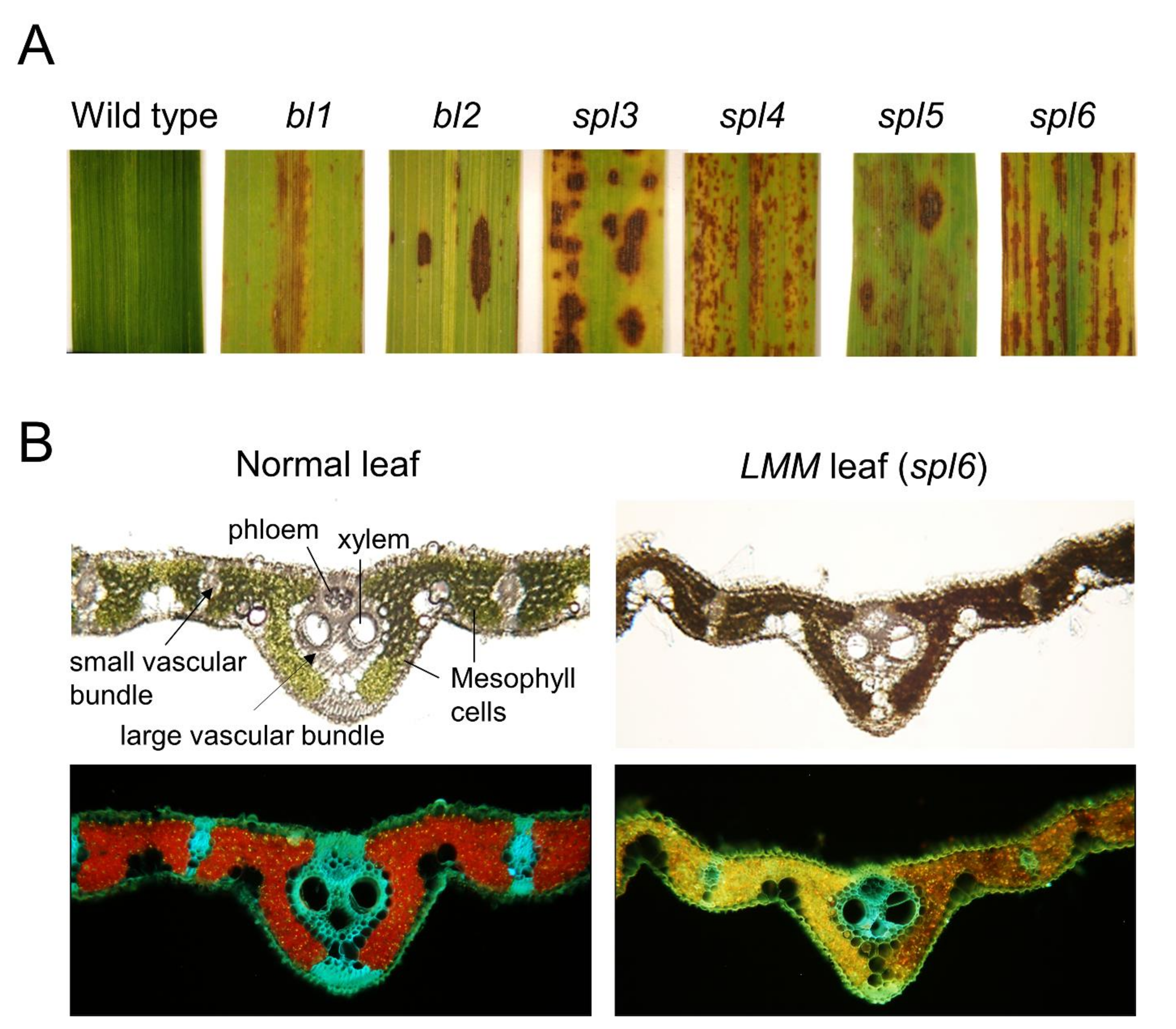
3. Development of Lesion Formation in Rice LMMs

Rice and maize showed similar types of lesion formation and are modulated during the development process [19,27]. Although no spots were reported in newly formed young leaves, the metabolically active flag leaves showed severe lesions [19,21,27]. In most cases, initially, lesions first appeared as specks near the tip of the leaf and then eventually, completely covered the leaf. Finally, the leaf surfaces exhibited yellow, brown, red, and black spots of dead necrotic cells [21] (Figure 1).
Many studies reported that lesion formation accelerated the severity of spot formation in rice leaves under high temperature and high light intensity [16,24,78]. Depending on the developmental differences of the lesion formations, the LMM phenotypes could be categorized into whole life lesion mimics, vegetative initiation lesion mimics, and reproductive initiation lesion mimics [7,8,26,28,80,81]. There are significant differences in the patterns of lesion formation of the LMM genes, particularly the spot color, size, and intensity (Figure 1). Most lesion colors of LMMs typically present yellow, brown, and red spots. However, the white-stripe leaves and panicles 1 (wsp1) mutant showed white-stripe leaves and panicles in rice [70].
Recently, a novel white-spotted leaf mutant was also identified in a japonica-type Korean cultivar that showed both brown spots and white lesion spots on the tip of the leaves from the vegetative stage [40]. In the spl5 mutant, the spot intensity was less than the others, whereas bl1, bl2, spl3, spl4, and spl6 produced a dense intensity of spots (Figure 1). Thus, various spot sizes, colors, and patterns in LMMs indicate the involvement of different genes. For example, the spot number and spot intensity were inversely proportional, where a few spots were noted with large spot size as found in bl2 and spl4 (Figure 1).
The formation of spots on most rice spl mutants of LMMs was controlled by the developmental pattern, as it was formed at a certain stage of plant age. Once spots are developed on all the leaves, they mostly remain in all the developmental stages. Interestingly, the flag leaves are affected severely by the lesion spots resulting in earlier senescence. In most spl mutants, no visible spots existed on the leaf at the seedling stage; however, spots were appeared as tiny spots at the tillering stage (45 days) and developed into severe phenotype at the milk stage (60 days) under natural field conditions between 30 to 35 °C [21].
Lesion formation can spread throughout the generations where the lesions initiate sparsely, and once formed, they can expand quickly over the entire leaf blade. Most of the spots were formed throughout the minor leaf veins and rarely on the central vein. Finally, lesions were spread all over the leaf surface that caused earlier leaf senescence and the death of the localized cells (Figure 1). Except for leaf blades, no other organs were affected by premature cell death because no visible spots existed, indicated that the photosynthetic organ produce the spots due to the accumulation of high ROS levels during photosynthesis and results in lesion formation by cell death.
In addition, the leaf portions of the spl6 plants protected from sunlight showed resistance to the development of lesions, it indicated the influence of light via photosynthesis in the induction of the spot formation on the mutant leaves. Similarly, researchers reported that fewer lesions were formed on leaves covered with aluminum foil [82,83]. To note, the covered area on the leaf either did not develop any spots or fewer lesions were formed by comparison to that of the non-covered area [72]. Interestingly, the lesions development was noted only under white light, while no lesion formation and reduced lesion formation were documented on the leaves exposed to either blue or red light [26,84]. Hence, the lesion formation of LMMs may be linked directly to the light reactions of the photosynthesis process.
Some patterns of lesion spot formation in rice LMMs are classified as progressive types during the developmental stages, including spl3, spl4, and spl6 mutants [14,19]. Typically, these mutants are arranged in groups of longitudinal lines along the leaf blade as dense spots towards the leaf apex rather than the leaf base (Figure 1). Noticeably, rice spl mutants produced more than two times number of spots with an increase in temperature, which was similar to Lls1 and slm1 mutants in maize [20,82,83].
In the spl6 mutant leaves, the highest levels of H2O2 accumulation were detected, which caused the initiation of severe cell death at a plant age of 45 days, suggesting the toxic accumulation of H2O2 in plants to initiate the cell death [85]. Such a fatal process was observed, particularly when the developing lesions of spl6 were closely dissected and the thylakoid membranes in the chloroplast of the spotted leaves were damaged severely (Figure 2).
This is because the lesion-formed cells failed to remove excessive amounts of ROS with the progression of photosynthesis. Moreover, the light intensity and temperature, which enhance the photosynthesis rate, were also marked as critical environmental factors that trigger the phenotypic expression of lesions [9,11,12,14,20]. For example, the rice spl7 mutant showed a variable lesion density under various temperature conditions, where the leaves showed a reduction in lesion density under low temperatures [14]. Whereas the spl6 mutant exhibited an increase in lesion spot formation corresponding to high light intensity [20].
Furthermore, Yamanouchi et al. proposed that the lesion formation of rice spl7 mutants was caused by the dysfunction of a heat stress transcription factor (HSF) [14]. As reviewed, different phenotypic expression pathways of the LMMs may exist specifically that depend on the genotypes of LMMs (Table 1). On the other hand, due to genetic differences, the spl6 gene is located on chromosome 1, and the spl7 gene is on chromosome 5; thus, the spl6 mutant can be caused by a functional protein other than HSF of the spl7 mutant. Moreover, the brown leaf (bl) mutant and spl mutants showed varied lesion patterns on the leaves. Hence, the LMMs may differ in each gene mutation; however, the lesion formation in similar phenotypes occurred due to the failure of scavenging excess ROS by various genes.

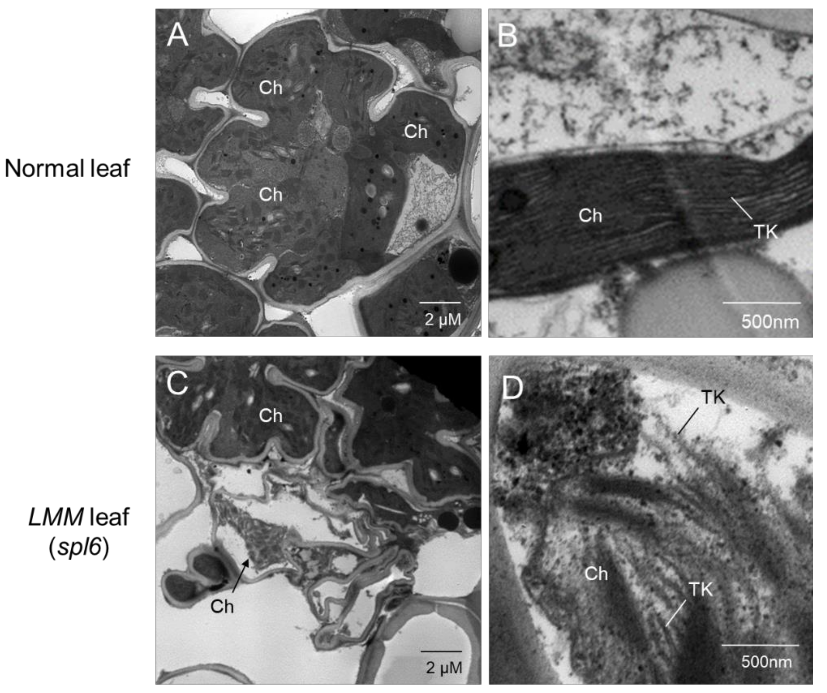
4. Chloroplast Damage from Disrupted Photosynthetic Apparatus in LMMs

During lesion formation in the LMMs, the chloroplast is the primary target to be damaged, which affects the photosynthetic machinery. The rice LMM traits cannot control the level of photosynthesis, resulting in plant death. Chlorophyll is a photosynthetic pigment that is positively correlated with the photosynthetic rate [86]. The chlorophyll content usually increases with increasing plant age.
On the other hand, the chlorophyll content decreased slightly in the rice later during senescence [87]. In the presence of light, chloroplasts in plant cells are the major source of ROS and have been shown to control the PCD in response to reactive oxygen species [88]. However, no significant differences in the chlorophyll content were noted in the later stages of wild type plants, while substantial differences were observed in the lesion spots of the spl6 mutant, predicted as a result of damage in the lamella and thylakoid membrane of chloroplasts (Figure 1 and Figure 2) [20].
The genetic basis of chlorophyll is quite complex, and many metabolic steps and genes are involved [87,89]. The RuBisCO is abundant mostly in the normal chloroplasts, indicated a strong correlation of spl mutant with reduction in RuBisCO at a severe cell death stage [19]. In the spl6 mutants, the induction of ROS, which causes reduced transcription level and cell death, is marked for a decrease in chloroplast production and reduced photosynthesis.
As depicted in Figure 2, TEM images show heavily damaged chloroplasts or an absence of mesophyll chloroplasts in the spl6 mutant by comparison to wild-type. Damaged thylakoid membranes in the mesophyll chloroplasts were also observed in the non-spotted area of the spl6 mutant, and no chloroplasts were observed in the mesophyll cells from the spotted area of the spl6 mutant. Chloroplast damage was caused by an oxidative burst, which results in excessive deposition of substances, such as plastoglobules and callose, in chloroplasts [20,24]. This suggests that overexpression of the ROS is linked to lesion formation and cell death in the LMMs, facilitating the mechanisms underlying protection against stress conditions.
Similar to the spl6, the degradation and changes in the chloroplast structure were also observed in the spl36 [24]. Moreover, the rice spl26 mutant was observed to disturb the photosynthetic capacity by a reduction in chlorophyll content accompanied by an increase in the membrane ion leakage rate and malonaldehyde level [90]. As another example, the spl33 mutant impaired the downregulation of photosynthesis-related genes and the up-regulation of senescence-associated genes.
In addition, it also showed a loss in chlorophyll and a breakdown of chloroplasts [67]. The rice llm1I mutant showed chloroplasts damaged by a malfunction of the cysteine-rich receptor-like kinase [55]. In summary, the genetic analysis of rice LMM traits showed that an uncontrolled photosynthesis rate resulted in the formation of lesions, which causes phytotoxicity in the plant.

5. Chlorophyll Content and Lesion Severity

Defects in the chlorophyll catabolism can also cause cell death in plants. A disruption of two essential enzymes, i.e., pheophorbide a oxygenase and red chlorophyll catabolite reductase, are involved in the degradation of chlorophyll, which generates spontaneous lesion in the accelerated cell death 1 (acd1) and accelerated cell death 2 (acd2) mutants [91,92,93]. Also, Arabidopsis acd1 was characterized by the accumulation of pheophorbide a (pheide a) due to the disruption of the ACD1/LLS1/PHEIDE A OYGENASE (PAO1) gene [7,93]. Of note, the primary function of the pheophorbide a oxygenase is to convert the pheide into the red chlorophyll catabolite (RCC) [94,95]. However, excessive accumulation of the photoreactive pheide in the chloroplast induces cell death in a light-dependent manner [96]. In addition, the ACD2 gene encodes an RCC reductase, which catalyzes the subsequent reaction and converts the RCC into the primary fluorescent chlorophyll catabolite [92]. Thus, the spontaneous cell death observed in the acd2 mutant was correlated with the accumulation of the RCC and singlet oxygen (1O2) formation [95].
Moreover, the ACD2 may bind to a porphyrin-related molecule in the mitochondria to prevent the PCD [97]. Interestingly, during pathogen infection, the RCC reductase as product of the ACD2 gene dynamically localizes between the chloroplast and mitochondria, resulting in protection against mitochondrial oxidative bursts [98]. Many LMMs are affected by chlorophyll biosynthesis or degradation; it is unclear whether a change in the accumulation of photoreactive chlorophyll precursors or degradation product contributes to the PCD. Chloroplasts play a key role as a sensor of light stress that delays the photosynthetic electron transport and regulates the ROS production.
Among the ROS, singlet oxygen (1O2) specie activates a signaling pathway in chloroplasts that is regulated by the two plastid-localized proteins, EXECUTER 1 and 2 [99]. The EXECUTER (EX) proteins exhibit protective role in the wild-type plant under high light stress conditions [100]. Therefore, the EXECUTER 1 activation assists to halt the stress by the release of singlet oxygen.
The ROS is generated in the chloroplast from various pathways that further triggers an HR and initiate the appearance of light-dependent LMM phenotypes owing to the failure of the plant machinery to dissolve excess excitation energy (EEE) [101,102,103]. This is highlighted by the light-dependent LMMs, including lsd1, acd1, and acd2, which showed defects in the EEE dissipation or chlorophyll catabolism caused by photooxidative damage and the formation of the ROS [92,94,104,105]. In addition, the light-induced bl2 and spl6 mutants progressed the destruction of thylakoids in chloroplasts, resulting in lesion formation (Figure 2) [21,85]. Hence, collected evidences suggest that the lesion formation of light-induced LMMs may be caused by genetic defects in the EEE distributions machinery of the chloroplast.

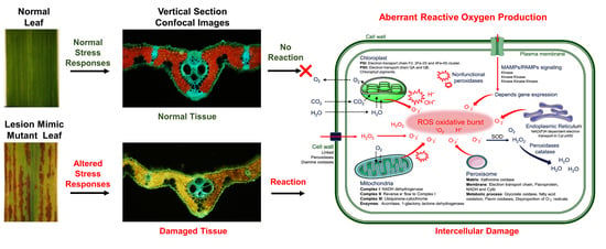
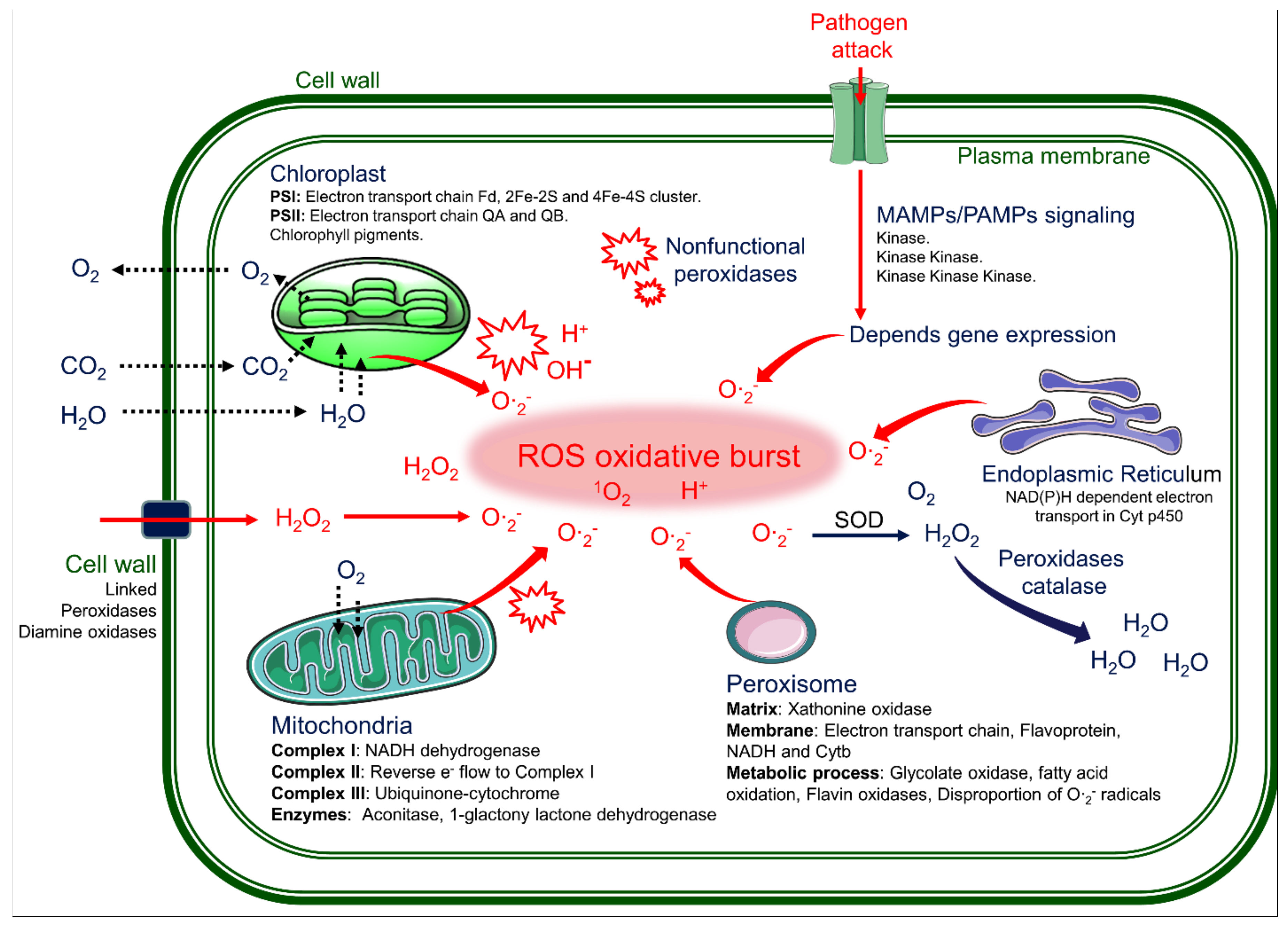
6. Reactive Oxygen Species Cause Lesions by in the PCD of the LMMs

The extensive formation of lesions in rice LMMs are induced by abiotic stresses, such as water, temperature, and light, and biotic stresses, such as reactive oxygen species (ROS) and pathogen infection [19,21,82,106,107]. In particular, the ROS can partially reduce or activate the derivatives of oxygen, such as superoxide radicals (O∙2-), hydroxyl radicals (OH∙), singlet oxygen (1O2), and hydrogen peroxide (H2O2). The ROS are always formed by the inevitable leakage of electrons to O2 from the electron transport activities of chloroplast, mitochondria, and plasma membranes or as a byproduct of various metabolic pathways localized in different cellular compartments [108]. Therefore, these ROS molecules are highly reactive and toxic and lead to the oxidative destruction of cells. A large amount of the ROS leads to the destruction of several subcellular organelles, such as chloroplasts, mitochondria, the plasma membrane, peroxisomes, apoplasts, the endoplasmic reticulum, and the cell wall in rice LMMs (Figure 3 and Figure 4).
Most of the lesion formation processes in LMM plants are rapid and cause the spontaneous death of localized cells at the location of stress. Physiological evidence in LMM studies concluded that a sudden increase in the ROS production in the mesophyll cells of a spotted leaf is a causal agent for cell death. For example, high accumulations of the H2O2 and superoxide (O2) in the lesion area of spotted leaves have been identified in spotted leaf mutants [8,10,17,55,85].
During rapid cell death caused by lesion formation in leaves, several toxic substances have been found to delay the electron transport chains (ETC) and to block antioxidant enzymes, leading to the excessive accumulation of the ROS. This accumulated ROS finally results in oxidative damage, lipid peroxidation, membrane leakage, and localized cell death [21,109] (Figure 2 and Figure 3). The destruction of the thylakoid membrane has been shown in images of chloroplasts in mesophyll cells in situ (Figure 2) [20,21]. Therefore, the ROS eruption is the main cause of lesion spot initiation.
The normal amount of the ROS in cells has been implicated as secondary messengers in intracellular signaling cascades that mediate several plant responses in plant cells, including stomatal closure at low or moderate concentrations [110], programmed cell death [111], root gravitropism [112], and the acquisition of tolerance to both abiotic and biotic stresses [113]. In addition, the low concentration of the ROS served as a messenger to induce the ascorbic acid production, which constitutes an essential substance in the network of antioxidants. In the spl6 mutant, the ascorbic acid contents in lesion-developed leaves showed gradual increment with an increase in the spot severity (Figure 4) [20].
The occurrence of the ROS is inevitable due to plant photosynthesis. On the other hand, the ROS elimination in plant cells is achieved by the ROS scavenging system, which is mediated by various enzymatic defense systems, including superoxide dismutase (SOD), catalase (CAT), and peroxidases (POX) [114]. The SOD catalyzes the superoxide into oxygen and H2O2 [115]. Also the CAT plays a role in the catalysis of the H2O2 in cells [116], while the peroxidases (POD) catalyze the oxidation of phenolic compounds and reduce the H2O2 production [101].
Under high ROS levels, the H2O2 is generated endogenously by the metabolic pathway and induces the PCD in plant cells [117]. The H2O2 is reduced by the protein thioredoxin peroxidase (TPX) through the intracellular redox signaling pathway [118,119]. On the other hand, the reduced level of the TPX in the LMMs failed to eliminate the H2O2, leading to the PCD and consecutive lesions in the spotted leaves of the spl1 and spl6 mutants [20,59,120].
Therefore, ROS accumulation in LMM plants may cause the extensive lesions formation mediated by PCD, resulting in a broad spectrum tolerance to the stress responses and pathogens [121,122]. Additionally, the ROS accumulation in LMMs also affects plant growth and differentiation.

7. Role of the MAP-Kinases Pathway in the PCD of the LMMs

In pathogen attack, the PCD is processed rapidly to protect plants, and several protein kinases play important roles in regulating the stress signal transduction pathways (Figure 3). During biotic and abiotic stresses, several specific kinase-signaling pathways should be considered to initiate an oxidative burst in the LMMs. This evidence was reviewed in several LMMs with a typical phenotype of the PCD without a pathogen attack. Besides, the mitogen-activated protein kinase (MAPK) cascade plays an important function during external environmental stimuli, thereby performing the transduction of extracellular signals to intracellular targets [123].
Recently, the MAPKKK cascade was also discovered to cause lesion spot formation in rice [55]. The protein of the spotted leaf sheath (sles) gene was also discovered to contain Raf MAPKKK [79]. Thus, the nonfunctional Raf MAPKKK of the sles mutant may be associated with loss of the ROS homeostatic regulation, which results in the oxidative burst for lesion formation in leaves. The MAPK cascade contains many other kinases, including MAPKs, MAPKKs (MAPK kinases), and MAPKK kinases (MAPKKKs) [124,125,126]. Plants have more MAPKKKs than MAPKs and MAPKKs, which leads to complicated and fickle regulatory MAPK cascades. On the other hand, some MAPKKKs have been characterized and are involved in several biological processes, including biotic [127,128] and abiotic stress responses [129,130], innate immunity [131], and the defense and stress responses [121]. Conclusively, the MAPK cascade function is critical to modulate the ROS homeostasis in plants; hence, it needs to be investigated for genetic mechanisms linked with oxidative bursts in the LMMs.

8. Role of Sphingolipids in LMMs

Sphingolipids are ubiquitous in a wide variety of organisms, including eukaryotes and prokaryotes. Ceramides, derivatives of sphingolipids, are components of the cell membrane and participate in various cellular signaling processes. Examples include regulating differentiation, proliferation, and programmed cell death (PCD) [132]. In plant-microbial interactions, pathogen-specific sphingolipids were found to induce the Ca2+ signaling, MAPK, and ROS production in rice [133].
Genetic evidence has shown the ceramide functions in plant PCD. For example, the ACD5 (ACCELERATED CELL DEATH5) and ACD11 encoded genes for the ceramide kinase and sphingosine transfer proteins, respectively, are involved in the sphingolipid metabolism [134]. The acd5 mutant helped to reduce the ceramide kinase activity and displayed an enhanced cell death after bacterial pathogen infections. The reduction of ceramide kinase resulted in the accumulation of precursor molecules, likely ceramide or sphinganine [135].
Therefore, the homeostatic balance between ceramides/sphingolipids and phosphorylated derivatives is important for lesion formation and cell death in plants. Another study showed that the acd11 lesion mimics a mutant of A. thaliana that exhibits an autoimmune phenotype, such as constitutive defense responses and death without pathogen infections [117]. ACD11 encoded a putative sphingosine transfer protein; however, the precise role during these processes is still unknown.
On the other hand, the defeat of ceramide kinase in the acd5 mutant of Arabidopsis sp. lost defenses to the ceramide accumulation and mitochondrial H2O2 bursts [136]. The physiological effects of the PCD mediated by the sphingolipid metabolism found in Arabidopsis LMMs are similar to rice LMMs; however, the underlying mechanism related to ceramide functions is not well known in rice LMMs.

9. Resistant to Pathogen Infection in the LMMs

Upon pathogen attack, the ROS is induced immediately to kill the infected cells and served as a signal to activate the defense response [137]. The ROS studies with findings in LMMs showed that low ROS concentrations acted as a second messenger in several plant responses, including stomatal closure, root gravitropism, seed germination, programmed cell death, lignin biosynthesis, hypersensitivity responses, and osmotic stress (Figure 4). Upon stress induction, the accumulation of salicylic acid was observed, which leads to an increase of the endogenous level of H2O2, which could then serve as a secondary messenger to induce the PCD.
This ROS-mediated lesion formation of the LMMs may induce the systematic resistant system against pathogens. To avoid oxidative damage from the ROS, the antioxidant enzymes, including SOD, CAT, APX (Ascorbate peroxidases), and GST (glutathione-s-transferase), help to remove excess amounts of the ROS. Enzymatic defenses include superoxide dismutase, which converts the superoxide radical to the H2O2 while the catalases and peroxidases, which trigger the conversion of H2O2 to water and oxygen under normal conditions and can handle the oxidative load [138]. In this way, most LMMs may show enhanced resistance to rice blast and bacterial blight [13,17,22,23,24,48,62,69,106,138,139,140,141]. Moreover, the ROS accumulation due to the loss-of-function of the ROS scavenging systems in the LMMs affect the respiratory burst oxidase homolog (Rbohs)-mediated signaling in plant growth, differentiation, and reactions to biotic and abiotic stresses [141,142,143]. Thus, it is predicted that some of the rice Rboh gene families in LMMs may contribute to resistance against pathogens.
SPL35 was shown to enhance the disease resistance to fungal and bacterial pathogens [68]. Furthermore, the bbs1 (lmm24) mutant showed up-regulation of defense response genes and resisted the rice blast fungus Magnaporthe oryzae [16]. For example, the excessive accumulation of the H2O2 in the leaves of the spl5 mutant induced the HR and increased the resistance to pathogens [49,55]. Thus, the up-regulation of the APX and GST are hypothesized to act as scavenger of the ROS in the spl5 mutant. However, experimental observations showed insufficient production of the APX and GST in the spl5 mutant to detoxify the overproduction of the ROS during plant growth.
In rice, failure of the vesicular trafficking system is involved in cell death. Rice AAA-ATPase1 plays a role in the association with salicylic acid-regulation for disease resistance and defense responses against the blast fungus M. oryzae [63]. In another study, the lrd6-6 mutant showed multivesicular bodies (MVBs)/endosomes-mediated vesicular trafficking, which might play important roles in plant immunity and cell death [61]. Similarly, the spl28 mutant failed to exhibit normal vesicular trafficking resulting in spotted leaves and early senescence. On the other hand, the LMMs can act as a negative regulator of cell death with enhanced resistance to rice blast and bacterial blight [60].
Recent studies reported that rice LMMs would be helpful in resistant breeding because most of them showed spontaneous activation of defense responsive genes in pathogen attacks [15]. Manigbas et al. reported that UV radiations could induce an increase in the H2O2 production, which could inflict injury to crops affecting growth and productivity [142]. Therefore, some rice LMM plants were selected as the rice breeding lines related to abiotic stresses, such as UV and H2O2 resistance.
In particular, catalase (CAT) in the spl6 and bl2 mutants was upregulated during lesion development, resulting in enhanced ROS scavenging in damaged cells, thereby, lowering the H2O2 levels [20,142]. For example, the SL gene of the Sekiguchi lesion (sl) mutant encoding a P450 monooxygenase protein-producing serotonin showed pathogen defense responses for rice blast disease [59,73,74]. Therefore, most LMMs have resistance to pathogenic infections depending on their genes, which are useful in genetic resources breeding disease-resistant plants.

10. Conclusions and Future Prospects

The plant lesion mimic mutants (LMMs) show aberrant regulation of cell death without a pathogenic infection. Genetic analysis of the rice spotted leaf genes of the LMMs showed that a recessive gene governs most LMMs. This study reviewed rice LMMs regarding the morpho-physiological features, biochemical functions, subcellular localization, and protein interactions. Most of the rice LMMs failed to scavenge the excess ROS and excitation energy generated during photosynthesis. Therefore, the ROS burst resulted in the formation of lesion spots mediated by the PCD. Some rice LMMs exhibited enhanced disease resistance via the consecutive production of the ROS, which can provide insight into the mechanisms underlying the HR, PCD, and immunity.
The lesion phenotypes and the genetic mode of rice LLMs are also involved in the chlorophyll metabolisms, stress tolerance, and ROS detoxification pathways. Consequently, more intensive research will be needed to obtain further insights, identify the target molecules, explore more mutants, and obtain genetic definitions of the corresponding genes.
The necrotic lesion spots caused by the ROS burst induce cell death for both the pathogens and plant cells to prevent the spread of invading pathogens throughout the plant body. Therefore, the rice LMM genetic traits are commonly elucidated to have highly developed self-defense mechanisms. Moreover, the identified LMM genes exhibiting the molecular genetic mechanisms of lesion formation will be useful for breeding rice with disease resistance and environmental stress tolerance as well as to achieve a high yield with improved photosynthesis efficiency.

Author Contributions

Conceptualization, S.G.K.; Corresponding and original draft preparation, S.G.K., K.E.L., M.S., M.N.M., and P.K.; Supervision, S.G.K.; funding acquisition, S.G.K. All authors have read and agreed to the published version of the manuscript.

Funding

The preparation of this paper was funded by the Ministry of Education (NRF2013R1A1A2013248 and NRF-2017R1D1A3B03035886) of South Korea.

Institutional Review Board Statement

Not applicable.

Informed Consent Statement

Not applicable.

Data Availability Statement

The data of this article is within the text.

Acknowledgments

We are grateful to thanks for Shiv Bharadwaj, Dep. of Biotechnology, Yeungnam University, Korea, for editing the manuscript. This research was supported by Basic Science Research Program through the National Research Foundation of Korea (NRF) funded by the Ministry of Education (NRF-2017R1D1A3B03035886).

Conflicts of Interest

The authors declare no conflict of interest.

References

  1. Skamnioti, P.; Gurr, S.J. Against the grain: Safeguarding rice from rice blast disease. Trends Biotechnol. 2009, 27, 141–150. [Google Scholar] [CrossRef] [PubMed] [Green Version]
  2. Greenberg, J.T.; Yao, N. The role and regulation of programmed cell death in plant–pathogen interactions. Cell. Microbiol. 2004, 6, 201–211. [Google Scholar] [CrossRef] [PubMed]
  3. Andreasson, E.; Ellis, B. Convergence and specificity in the Arabidopsis MAPK nexus. Trends Plant Sci. 2010, 15, 106–113. [Google Scholar] [CrossRef] [PubMed]
  4. Greenberg, J.T. Programmed cell death: A way of life for plants. Proc. Natl. Acad. Sci. USA 1996, 93, 12094–12097. [Google Scholar] [CrossRef] [Green Version]
  5. Laloi, C.; Apel, K.; Danon, A. Reactive oxygen signalling: The latest news. Curr. Opin. Plant Biol. 2004, 7, 323–328. [Google Scholar] [CrossRef] [PubMed]
  6. Valandro, F.; Menguer, P.K.; Cabreira-Cagliari, C.; Margis-Pinheiro, M.; Cagliari, A. Programmed cell death (PCD) control in plants: New insights from the Arabidopsis thaliana deathosome. Plant Sci. 2020, 299, 110603. [Google Scholar] [CrossRef] [PubMed]
  7. Gray, J.; Close, P.S.; Briggs, S.P.; Johal, G.S. A novel suppressor of cell death in plants encoded by the Lls1 gene of maize. Cell 1997, 89, 25–31. [Google Scholar] [CrossRef] [Green Version]
  8. Lorrain, S.; Vailleau, F.; Balagué, C.; Roby, D. Lesion mimic mutants: Keys for deciphering cell death and defense pathways in plants? Trends Plant Sci. 2003, 8, 263–271. [Google Scholar] [CrossRef]
  9. Dietrich, R.A.; Delaney, T.P.; Uknes, S.J.; Ward, E.R.; Ryals, J.A.; Dangl, J.L. Arabidopsis mutants simulating disease resistance response. Cell 1994, 77, 565–577. [Google Scholar] [CrossRef]
  10. Jabs, T.; Dietrich, R.A.; Dangl, J.L. Initiation of runaway cell death in an Arabidopsis mutant by extracellular superoxide. Science 1996, 273, 1853–1856. [Google Scholar] [CrossRef]
  11. Noutoshi, Y.; Kuromori, T.; Wada, T.; Hirayama, T.; Kamiya, A.; Imura, Y.; Yasuda, M.; Nakashita, H.; Shirasu, K.; Shinozaki, K. Loss of necrotic spotted lesions 1 associates with cell death and defense responses in Arabidopsis thaliana. Plant Mol. Biol. 2006, 62, 29–42. [Google Scholar] [CrossRef] [PubMed]
  12. Bouchez, O.; Huard, C.; Lorrain, S.; Roby, D.; Balagué, C. Ethylene is one of the key elements for cell death and defense response control in the Arabidopsis lesion mimic mutant vad1. Plant Physiol. 2007, 145, 465–477. [Google Scholar] [CrossRef] [PubMed] [Green Version]
  13. Yin, Z.; Chen, J.; Zeng, L.; Goh, M.; Leung, H.; Khush, G.S.; Wang, G.-L. Characterizing rice lesion mimic mutants and identifying a mutant with broad-spectrum resistance to rice blast and bacterial blight. Mol. Plant Microbe Interact. 2000, 13, 869–876. [Google Scholar] [CrossRef] [PubMed] [Green Version]
  14. Yamanouchi, U.; Yano, M.; Lin, H.; Ashikari, M.; Yamada, K. A rice spotted leaf gene, Spl7, encodes a heat stress transcription factor protein. Proc. Natl. Acad. Sci. USA 2002, 99, 7530–7535. [Google Scholar] [CrossRef] [PubMed] [Green Version]
  15. Xiaobo, Z.H.U.; Mu, Z.E.; Mawsheng, C.; Xuewei, C.; Jing, W. Deciphering rice lesion mimic mutants to understand molecular network governing plant immunity and growth. Rice Sci. 2020, 27, 278–288. [Google Scholar] [CrossRef]
  16. Zhang, Y.; Liu, Q.; Zhang, Y.; Chen, Y.; Yu, N.; Cao, Y.; Zhan, X.; Cheng, S.; Cao, L. LMM24 encodes receptor-like cytoplasmic kinase 109, which regulates cell death and defense responses in rice. Int. J. Mol. Sci. 2019, 20, 3243. [Google Scholar] [CrossRef] [Green Version]
  17. Zhou, Q.; Zhang, Z.; Liu, T.; Gao, B.; Xiong, X. Identification and map-based cloning of the light-induced lesion mimic mutant 1 (LIL1) gene in rice. Front. Plant Sci. 2017, 8, 2122. [Google Scholar] [CrossRef] [Green Version]
  18. Liu, G.; Wang, L.; Zhou, Z.; Leung, H.; Wang, G.-L.; He, C. Physical mapping of a rice lesion mimic gene, Spl1, to a 70-kb segment of rice chromosome 12. Mol. Genet. Genom. 2004, 272, 108–115. [Google Scholar] [CrossRef]
  19. Matin, M.N.; Suh, H.S.; Kang, S.G. Characterization of phenotypes of rice lesion resembling disease mutants. Korean J. Genet. 2006, 28, 221–228. [Google Scholar]
  20. Matin, M.N.; Saief, S.A.; Rahman, M.M.; Lee, D.H.; Kang, H.; Lee, D.S.; Kang, S.G. Comparative phenotypic and physiological characteristics of spotted leaf 6 (spl6) and brown leaf spot2 (bl2) lesion mimic mutants (LMM) in rice. Mol. Cells 2010, 30, 533–543. [Google Scholar] [CrossRef]
  21. Kang, S.G.; Matin, M.N.; Bae, H.; Natarajan, S. Proteome analysis and characterization of phenotypes of lesion mimic mutant spotted leaf 6 in rice. Proteomics 2007, 7, 2447–2458. [Google Scholar] [CrossRef]
  22. Mori, M.; Tomita, C.; Sugimoto, K.; Hasegawa, M.; Hayashi, N.; Dubouzet, J.G.; Ochiai, H.; Sekimoto, H.; Hirochika, H.; Kikuchi, S. Isolation and molecular characterization of a Spotted leaf 18 mutant by modified activation-tagging in rice. Plant Mol. Biol. 2007, 63, 847–860. [Google Scholar] [CrossRef] [PubMed]
  23. Wang, J.; Ye, B.; Yin, J.; Yuan, C.; Zhou, X.; Li, W.; He, M.; Wang, J.; Chen, W.; Qin, P. Characterization and fine mapping of a light-dependent leaf lesion mimic mutant 1 in rice. Plant Physiol. Biochem. 2015, 97, 44–51. [Google Scholar] [CrossRef]
  24. Rao, Y.; Jiao, R.; Wu, X.; Wang, S.; Ye, H.; Li, S.; Hu, J.; Lin, H.; Ren, D.; Wang, Y. SPL36 encodes a receptor-like protein kinase precursor and regulates programmed cell death and defense responses in rice. Res. Sq. 2020. [Google Scholar] [CrossRef]
  25. Hoang, T.V.; Vo, K.T.X.; Rahman, M.M.; Choi, S.-H.; Jeon, J.-S. Heat stress transcription factor OsSPL7 plays a critical role in reactive oxygen species balance and stress responses in rice. Plant Sci. 2019, 289, 110273. [Google Scholar] [CrossRef] [PubMed]
  26. Hoisington, D.A.; Neuffer, M.G.; Walbot, V. Disease lesion mimics in maize: I. Effect of genetic background, temperature, developmental age, and wounding on necrotic spot formation with Les1. Dev. Biol. 1982, 93, 381–388. [Google Scholar] [CrossRef]
  27. Simmons, C.; Hantke, S.; Grant, S.; Johal, G.S.; Briggs, S.P. The maize lethal leaf spot 1 mutant has elevated resistance to fungal infection at the leaf epidermis. Mol. Plant Microbe Interact. 1998, 11, 1110–1118. [Google Scholar] [CrossRef] [Green Version]
  28. Hu, G.; Yalpani, N.; Briggs, S.P.; Johal, G.S. A porphyrin pathway impairment is responsible for the phenotype of a dominant disease lesion mimic mutant of maize. Plant Cell 1998, 10, 1095–1105. [Google Scholar] [CrossRef]
  29. Moeder, W.; Yoshioka, K. Lesion mimic mutants: A classical, yet still fundamental approach to study programmed cell death. Plant Signal. Behav. 2008, 3, 764–767. [Google Scholar] [CrossRef] [PubMed] [Green Version]
  30. Wolter, M.; Hollricher, K.; Salamini, F.; Schulze-Lefert, P. The mlo resistance alleles to powdery mildew infection in barley trigger a developmentally controlled defence mimic phenotype. Mol. Gen. Genet. MGG 1993, 239, 122–128. [Google Scholar] [CrossRef]
  31. Büschges, R.; Hollricher, K.; Panstruga, R.; Simons, G.; Wolter, M.; Frijters, A.; Van Daelen, R.; van der Lee, T.; Diergaarde, P.; Groenendijk, J. The barley Mlo gene: A novel control element of plant pathogen resistance. Cell 1997, 88, 695–705. [Google Scholar] [CrossRef] [Green Version]
  32. Rostoks, N.; Schmierer, D.; Mudie, S.; Drader, T.; Brueggeman, R.; Caldwell, D.G.; Waugh, R.; Kleinhofs, A. Barley necrotic locus nec1 encodes the cyclic nucleotide-gated ion channel 4 homologous to the Arabidopsis HLM1. Mol. Genet. Genom. 2006, 275, 159–168. [Google Scholar] [CrossRef] [PubMed]
  33. Keisa, A.; Kanberga-Silina, K.; Nakurte, I.; Kunga, L.; Rostoks, N. Differential disease resistance response in the barley necrotic mutant nec1. BMC Plant Biol. 2011, 11, 1–10. [Google Scholar] [CrossRef] [Green Version]
  34. Al Amin, G.M.; Kong, K.; Sharmin, R.A.; Kong, J.; Bhat, J.A.; Zhao, T. Characterization and rapid gene-mapping of leaf lesion mimic phenotype of spl-1 mutant in soybean (Glycine max (L.) Merr.). Int. J. Mol. Sci. 2019, 20, 2193. [Google Scholar] [CrossRef] [Green Version]
  35. Sugie, A.; Murai, K.; Takumi, S. Alteration of respiration capacity and transcript accumulation level of alternative oxidase genes in necrosis lines of common wheat. Genes Genet. Syst. 2007, 82, 231–239. [Google Scholar] [CrossRef] [PubMed] [Green Version]
  36. Abad, M.S.; Hakimi, S.M.; Kaniewski, W.K.; Rommens, C.M.T.; Shulaev, V.; Lam, E.; Shah, D.M. Characterization of acquired resistance in lesion-mimic transgenic potato expressing bacterio-opsin. Mol. Plant Microbe Interact. 1997, 10, 635–645. [Google Scholar] [CrossRef]
  37. Kumar, V.; Parkhi, V.; Joshi, S.G.; Christensen, S.; Jayaprakasha, G.K.; Patil, B.S.; Kolomiets, M.V.; Rathore, K.S. A novel, conditional, lesion mimic phenotype in cotton cotyledons due to the expression of an endochitinase gene from Trichoderma virens. Plant Sci. 2012, 183, 86–95. [Google Scholar] [CrossRef]
  38. Iwata, N.; Omura, T. Linkage analysis by reciprocal translocation method in rice plants (Oryza sativa L.): I. Linkage groups corresponding to the chromosome 1, 2, 3 and 4. Jpn. J. Breed. 1971, 21, 19–28. [Google Scholar] [CrossRef] [Green Version]
  39. Iwata, N.; Satoh, H.; Omura, T. Relationship between the twelve chromosomes and the linkage groups (studies on the trisomics in rice plants)(Oryza sativa L.). V. Ikushugaku Zasshi 1984, 34, 314–321. [Google Scholar] [CrossRef] [Green Version]
  40. Kim, B.; Lee, H.; Jin, Z.; Lee, D.; Koh, H.-J. Characterization and genetic mapping of white-spotted leaf (wspl) mutant in rice. Plant Breed. Biotechnol. 2019, 7, 340–349. [Google Scholar] [CrossRef]
  41. Chen, Z.; Chen, T.; Sathe, A.P.; He, Y.; Zhang, X.; Wu, J. Identification of a novel semi-dominant spotted-leaf mutant with enhanced resistance to Xanthomonas oryzae pv. oryzae in rice. Int. J. Mol. Sci. 2018, 19, 3766. [Google Scholar] [CrossRef] [PubMed] [Green Version]
  42. Sasaki, T.; Burr, B. International rice genome sequencing project: The effort to completely sequence the rice genome. Curr. Opin. Plant Biol. 2000, 3, 138–142. [Google Scholar] [CrossRef]
  43. Ohyanagi, H.; Tanaka, T.; Sakai, H.; Shigemoto, Y.; Yamaguchi, K.; Habara, T.; Fujii, Y.; Antonio, B.A.; Nagamura, Y.; Imanishi, T.; et al. The rice annotation project database (RAP-DB): Hub for Oryza sativa ssp. japonica genome information. Nucleic Acids Res. 2006, 34, 741–744. [Google Scholar] [CrossRef] [Green Version]
  44. Kawahara, Y.; de la Bastide, M.; Hamilton, J.P.; Kanamori, H.; McCombie, W.R.; Ouyang, S.; Schwartz, D.C.; Tanaka, T.; Wu, J.; Zhou, S. Improvement of the Oryza sativa Nipponbare reference genome using next generation sequence and optical map data. Rice 2013, 6, 1–10. [Google Scholar] [CrossRef] [PubMed] [Green Version]
  45. Zeng, L.-R.; Qu, S.; Bordeos, A.; Yang, C.; Baraoidan, M.; Yan, H.; Xie, Q.; Nahm, B.H.; Leung, H.; Wang, G.-L. Spotted leaf11, a negative regulator of plant cell death and defense, encodes a U-box/armadillo repeat protein endowed with E3 ubiquitin ligase activity. Plant Cell 2004, 16, 2795–2808. [Google Scholar] [CrossRef] [Green Version]
  46. Vega-Sánchez, M.E.; Zeng, L.; Chen, S.; Leung, H.; Wang, G.-L. SPIN1, a K homology domain protein negatively regulated and ubiquitinated by the E3 ubiquitin ligase SPL11, is involved in flowering time control in rice. Plant Cell 2008, 20, 1456–1469. [Google Scholar] [CrossRef] [Green Version]
  47. McCouch, S.R.; Teytelman, L.; Xu, Y.; Lobos, K.B.; Clare, K.; Walton, M.; Fu, B.; Maghirang, R.; Li, Z.; Xing, Y. Development and mapping of 2240 new SSR markers for rice (Oryza sativa L.)(supplement). DNA Res. 2002, 9, 257–279. [Google Scholar] [CrossRef] [Green Version]
  48. Qiao, Y.; Jiang, W.; Lee, J.; Park, B.; Choi, M.; Piao, R.; Woo, M.; Roh, J.; Han, L.; Paek, N. SPL28 encodes a clathrin-associated adaptor protein complex 1, medium subunit μ1 (AP1M1) and is responsible for spotted leaf and early senescence in rice (Oryza sativa). New Phytol. 2010, 185, 258–274. [Google Scholar] [CrossRef]
  49. Chen, X.; Hao, L.; Pan, J.; Zheng, X.; Jiang, G.; Jin, Y.; Gu, Z.; Qian, Q.; Zhai, W.; Ma, B. SPL5, a cell death and defense-related gene, encodes a putative splicing factor 3b subunit 3 (SF3b3) in rice. Mol. Breed. 2012, 30, 939–949. [Google Scholar] [CrossRef]
  50. Zeng, D.-D.; Yang, C.-C.; Qin, R.; Alamin, M.; Yue, E.-K.; Jin, X.-L.; Shi, C.-H. A guanine insert in OsBBS1 leads to early leaf senescence and salt stress sensitivity in rice (Oryza sativa L.). Plant Cell Rep. 2018, 37, 933–946. [Google Scholar] [CrossRef] [PubMed]
  51. Liu, X.; Li, F.; Tang, J.; Wang, W.; Zhang, F.; Wang, G.; Chu, J.; Yan, C.; Wang, T.; Chu, C. Activation of the jasmonic acid pathway by depletion of the hydroperoxide lyase OsHPL3 reveals crosstalk between the HPL and AOS branches of the oxylipin pathway in rice. PLoS ONE 2012, 7, e50089. [Google Scholar] [CrossRef]
  52. Tong, X.; Qi, J.; Zhu, X.; Mao, B.; Zeng, L.; Wang, B.; Li, Q.; Zhou, G.; Xu, X.; Lou, Y. The rice hydroperoxide lyase OsHPL3 functions in defense responses by modulating the oxylipin pathway. Plant J. 2012, 71, 763–775. [Google Scholar] [CrossRef]
  53. Vega-Sánchez, M.E.; Verhertbruggen, Y.; Christensen, U.; Chen, X.; Sharma, V.; Varanasi, P.; Jobling, S.A.; Talbot, M.; White, R.G.; Joo, M. Loss of cellulose synthase-like F6 function affects mixed-linkage glucan deposition, cell wall mechanical properties, and defense responses in vegetative tissues of rice. Plant Physiol. 2012, 159, 56–69. [Google Scholar] [CrossRef] [PubMed] [Green Version]
  54. Sun, C.; Liu, L.; Tang, J.; Lin, A.; Zhang, F.; Fang, J.; Zhang, G.; Chu, C. RLIN1, encoding a putative coproporphyrinogen III oxidase, is involved in lesion initiation in rice. J. Genet. Genom. 2011, 38, 29–37. [Google Scholar] [CrossRef] [PubMed]
  55. Wang, S.-H.; Lim, J.-H.; Kim, S.-S.; Cho, S.-H.; Yoo, S.-C.; Koh, H.-J.; Sakuraba, Y.; Paek, N.-C. Mutation of SPOTTED LEAF3 (SPL3) impairs abscisic acid-responsive signalling and delays leaf senescence in rice. J. Exp. Bot. 2015, 66, 7045–7059. [Google Scholar] [CrossRef] [PubMed] [Green Version]
  56. Undan, J.R.; Tamiru, M.; Abe, A.; Yoshida, K.; Kosugi, S.; Takagi, H.; Yoshida, K.; Kanzaki, H.; Saitoh, H.; Fekih, R. Mutation in OsLMS, a gene encoding a protein with two double-stranded RNA binding motifs, causes lesion mimic phenotype and early senescence in rice (Oryza sativa L.). Genes Genet. Syst. 2012, 87, 169–179. [Google Scholar] [CrossRef] [Green Version]
  57. Wang, L.; Pei, Z.; Tian, Y.; He, C. OsLSD1, a rice zinc finger protein, regulates programmed cell death and callus differentiation. Mol. Plant Microbe Interact. 2005, 18, 375–384. [Google Scholar] [CrossRef] [PubMed] [Green Version]
  58. Liu, Q.; Zhu, H. Molecular evolution of the MLO gene family in Oryza sativa and their functional divergence. Gene 2008, 409, 1–10. [Google Scholar] [CrossRef]
  59. Ueno, M.; Shibata, H.; Kihara, J.; Honda, Y.; Arase, S. Increased tryptophan decarboxylase and monoamine oxidase activities induce Sekiguchi lesion formation in rice infected with Magnaporthe grisea. Plant J. 2003, 36, 215–228. [Google Scholar] [CrossRef] [PubMed]
  60. Fekih, R.; Tamiru, M.; Kanzaki, H.; Abe, A.; Yoshida, K.; Kanzaki, E.; Saitoh, H.; Takagi, H.; Natsume, S.; Undan, J.R. The rice (Oryza sativa L.) LESION MIMIC RESEMBLING, which encodes an AAA-type ATPase, is implicated in defense response. Mol. Genet. Genomics 2015, 290, 611–622. [Google Scholar] [CrossRef]
  61. Zhu, X.; Yin, J.; Liang, S.; Liang, R.; Zhou, X.; Chen, Z.; Zhao, W.; Wang, J.; Li, W.; He, M. The multivesicular bodies (MVBs)-localized AAA ATPase LRD6-6 inhibits immunity and cell death likely through regulating MVBs-mediated vesicular trafficking in rice. PLoS Genet. 2016, 12, e1006311. [Google Scholar] [CrossRef] [Green Version]
  62. Song, G.; Kwon, C.-T.; Kim, S.-H.; Shim, Y.; Lim, C.; Koh, H.-J.; An, G.; Kang, K.; Paek, N.-C. The rice SPOTTED LEAF4 (SPL4) encodes a plant spastin that inhibits ROS accumulation in leaf development and functions in leaf senescence. Front. Plant Sci. 2019, 9, 1925. [Google Scholar] [CrossRef]
  63. Liu, X.; Inoue, H.; Tang, X.; Tan, Y.; Xu, X.; Wang, C.; Jiang, C.-J. Rice OsAAA-ATPase1 is induced during blast infection in a salicylic acid-dependent manner, and promotes blast fungus resistance. Int. J. Mol. Sci. 2020, 21, 1443. [Google Scholar] [CrossRef] [Green Version]
  64. Ge, C.; Zhi-guo, E.; Pan, J.; Jiang, H.; Zhang, X.; Zeng, D.; Dong, G.; Hu, J.; Xue, D. Map-based cloning of a spotted-leaf mutant gene OsSL5 in Japonica rice. Plant Growth Regul. 2015, 75, 595–603. [Google Scholar] [CrossRef]
  65. Ruan, B.; Hua, Z.; Zhao, J.; Zhang, B.; Ren, D.; Liu, C.; Yang, S.; Zhang, A.; Jiang, H.; Yu, H. Os ACL-A2 negatively regulates cell death and disease resistance in rice. Plant Biotechnol. J. 2019, 17, 1344–1356. [Google Scholar] [CrossRef] [PubMed]
  66. Sun, L.; Wang, Y.; Liu, L.; Wang, C.; Gan, T.; Zhang, Z.; Wang, Y.; Wang, D.; Niu, M.; Long, W. Isolation and characterization of a spotted leaf 32 mutant with early leaf senescence and enhanced defense response in rice. Sci. Rep. 2017, 7, 1–13. [Google Scholar] [CrossRef] [Green Version]
  67. Wang, S.; Lei, C.; Wang, J.; Ma, J.; Tang, S.; Wang, C.; Zhao, K.; Tian, P.; Zhang, H.; Qi, C. SPL33, encoding an eEF1A-like protein, negatively regulates cell death and defense responses in rice. J. Exp. Bot. 2017, 68, 899–913. [Google Scholar] [CrossRef]
  68. Ma, J.; Wang, Y.; Ma, X.; Meng, L.; Jing, R.; Wang, F.; Wang, S.; Cheng, Z.; Zhang, X.; Jiang, L. Disruption of gene SPL 35, encoding a novel CUE domain-containing protein, leads to cell death and enhanced disease response in rice. Plant Biotechnol. J. 2019, 17, 1679–1693. [Google Scholar] [CrossRef] [PubMed] [Green Version]
  69. Sathe, A.P.; Su, X.; Chen, Z.; Chen, T.; Wei, X.; Tang, S.; Zhang, X.; Wu, J. Identification and characterization of a spotted-leaf mutant spl40 with enhanced bacterial blight resistance in rice. Rice 2019, 12, 1–15. [Google Scholar] [CrossRef]
  70. Zhang, Z.; Cui, X.; Wang, Y.; Wu, J.; Gu, X.; Lu, T. The RNA editing factor WSP1 is essential for chloroplast development in rice. Mol. Plant 2017, 10, 86–98. [Google Scholar] [CrossRef] [Green Version]
  71. McCouch, S.R. Gene nomenclature system for rice. Rice 2008, 1, 72–84. [Google Scholar] [CrossRef] [Green Version]
  72. Eun, L.; Ahmed, S.; Kang, S.G. Phenotype characteristics of spotted leaf 1 mutant in rice. Res. J. Biotechnol. 2012, 7, 9–15. [Google Scholar]
  73. Fujiwara, T.; Maisonneuve, S.; Isshiki, M.; Mizutani, M.; Chen, L.; Wong, H.L.; Kawasaki, T.; Shimamoto, K. Sekiguchi lesion gene encodes a cytochrome P450 monooxygenase that catalyzes conversion of tryptamine to serotonin in rice. J. Biol. Chem. 2010, 285, 11308–11313. [Google Scholar] [CrossRef] [PubMed] [Green Version]
  74. Tian, D.; Yang, F.; Niu, Y.; Lin, Y.; Chen, Z.; Li, G.; Luo, Q.; Wang, F.; Wang, M. Loss function of SL (sekiguchi lesion) in the rice cultivar Minghui 86 leads to enhanced resistance to (hemi) biotrophic pathogens. BMC Plant Biol. 2020, 20, 1–14. [Google Scholar] [CrossRef] [PubMed]
  75. Iwata, N.; Omura, T.; Satoh, H. Linkage studies in rice (Oryza sativa L.): On some mutants for physiological leaf spots. J. Fac. Agric. Kyushu Univ. 1978, 22, 243–251. [Google Scholar]
  76. Lee, J.C.; Peter, M.E. Regulation of apoptosis by ubiquitination. Immunol. Rev. 2003, 193, 39–47. [Google Scholar] [CrossRef] [PubMed]
  77. Sullivan, J.A.; Shirasu, K.; Deng, X.W. The diverse roles of ubiquitin and the 26S proteasome in the life of plants. Nat. Rev. Genet. 2003, 4, 948–958. [Google Scholar] [CrossRef] [PubMed]
  78. Huang, Q.; Shi, Y.; Yang, Y.; Feng, B.; Wei, Y.; Chen, J.; Baraoidan, M.; Leung, H.; Wu, J. Characterization and genetic analysis of a light-and temperature-sensitive spotted-leaf mutant in rice. J. Integr. Plant Biol. 2011, 53, 671–681. [Google Scholar] [CrossRef] [PubMed]
  79. Lee, D.; Lee, G.; Kim, B.; Jang, S.; Lee, Y.; Yu, Y.; Seo, J.; Kim, S.; Lee, Y.-H.; Lee, J. Identification of a spotted leaf sheath gene involved in early senescence and defense response in rice. Front. Plant Sci. 2018, 9, 1274. [Google Scholar] [CrossRef] [Green Version]
  80. Johal, G.S.; Hulbert, S.H.; Briggs, S.P. Disease lesion mimics of maize: A model for cell death in plants. Bioessays 1995, 17, 685–692. [Google Scholar] [CrossRef]
  81. Dangl, J.L.; Dietrich, R.A.; Richberg, M.H. Death don’t have no mercy: Cell death programs in plant-microbe interactions. Plant Cell 1996, 8, 1793. [Google Scholar] [CrossRef] [PubMed]
  82. Gray, J.; Janick-Buckner, D.; Buckner, B.; Close, P.S.; Johal, G.S. Light-dependent death of maize lls1 cells is mediated by mature chloroplasts. Plant Physiol. 2002, 130, 1894–1907. [Google Scholar] [CrossRef] [Green Version]
  83. Penning, B.W.; Johal, G.S.; McMullen, M.D. A major suppressor of cell death, slm1, modifies the expression of the maize (Zea mays L.) lesion mimic mutation les23. Genome 2004, 47, 961–969. [Google Scholar] [CrossRef] [PubMed]
  84. Arase, S.; Fukuyama, R.; Honda, Y. Light-dependent induction of sekiguchi lesion formation by bipolaris oryzae in Rice cv. Sekiguchi-asahi. J. Phytopathol. 2000, 148, 193–196. [Google Scholar] [CrossRef]
  85. Matin, M.N.; Pandeya, D.; Baek, K.-H.; Lee, D.S.; Lee, J.-H.; Kang, H.; Kang, S.G. Phenotypic and genotypic analysis of rice lesion mimic mutants. Plant Pathol J. 2010, 26, 159–169. [Google Scholar] [CrossRef] [Green Version]
  86. Davis, M.S.; Forman, A.; Fajer, J. Ligated chlorophyll cation radicals: Their function in photosystem II of plant photosynthesis. Proc. Natl. Acad. Sci. USA 1979, 76, 4170–4174. [Google Scholar] [CrossRef] [PubMed] [Green Version]
  87. Wang, F.; Wang, G.; Li, X.; Huang, J.; Zheng, J. Heredity, physiology and mapping of a chlorophyll content gene of rice (Oryza sativa L.). J. Plant Physiol. 2008, 165, 324–330. [Google Scholar] [CrossRef] [PubMed]
  88. Lee, J.; Nam, J.; Park, H.C.; Na, G.; Miura, K.; Jin, J.B.; Yoo, C.Y.; Baek, D.; Kim, D.H.; Jeong, J.C. Salicylic acid-mediated innate immunity in Arabidopsis is regulated by SIZ1 SUMO E3 ligase. Plant J. 2007, 49, 79–90. [Google Scholar] [CrossRef]
  89. Suzuki, J.Y.; Bollivar, D.W.; Bauer, C.E. Genetic analysis of chlorophyll biosynthesis. Annu. Rev. Genet. 1997, 31, 61–89. [Google Scholar] [CrossRef] [Green Version]
  90. Ting, C.; Zheng, C.; Sathe, A.P.; Zhihong, Z.; Liangjian, L.I.; Huihui, S.; Shaoqing, T.; Xiaobo, Z.; Jianli, W.U. Characterization of a novel gain-of-function spotted-leaf mutant with enhanced disease resistance in rice. Rice Sci. 2019, 26, 372–383. [Google Scholar] [CrossRef]
  91. Greenberg, J.T.; Ausubel, F.M. Arabidopsis mutants compromised for the control of cellular damage during pathogenesis and aging. Plant J. 1993, 4, 327–341. [Google Scholar] [CrossRef] [PubMed]
  92. Mach, J.M.; Castillo, A.R.; Hoogstraten, R.; Greenberg, J.T. The Arabidopsis-accelerated cell death gene ACD2 encodes red chlorophyll catabolite reductase and suppresses the spread of disease symptoms. Proc. Natl. Acad. Sci. USA 2001, 98, 771–776. [Google Scholar] [CrossRef]
  93. Tanaka, R.; Hirashima, M.; Satoh, S.; Tanaka, A. The Arabidopsis-accelerated cell death gene ACD1 is involved in oxygenation of pheophorbide a: Inhibition of the pheophorbide a oxygenase activity does not lead to the “stay-green” phenotype in Arabidopsis. Plant Cell Physiol. 2003, 44, 1266–1274. [Google Scholar] [CrossRef] [PubMed] [Green Version]
  94. Pružinská, A.; Tanner, G.; Anders, I.; Roca, M.; Hörtensteiner, S. Chlorophyll breakdown: Pheophorbide a oxygenase is a Rieske-type iron–sulfur protein, encoded by the accelerated cell death 1 gene. Proc. Natl. Acad. Sci. USA 2003, 100, 15259–15264. [Google Scholar] [CrossRef] [Green Version]
  95. Pružinská, A.; Anders, I.; Aubry, S.; Schenk, N.; Tapernoux-Lüthi, E.; Müller, T.; Kräutler, B.; Hörtensteiner, S. In vivo participation of red chlorophyll catabolite reductase in chlorophyll breakdown. Plant Cell 2007, 19, 369–387. [Google Scholar] [CrossRef] [Green Version]
  96. Hirashima, M.; Tanaka, R.; Tanaka, A. Light-independent cell death induced by accumulation of pheophorbide a in Arabidopsis thaliana. Plant Cell Physiol. 2009, 50, 719–729. [Google Scholar] [CrossRef] [Green Version]
  97. Yao, N.; Greenberg, J.T. Arabidopsis ACCELERATED CELL DEATH2 modulates programmed cell death. Plant Cell 2006, 18, 397–411. [Google Scholar] [CrossRef] [PubMed] [Green Version]
  98. Pattanayak, G.K.; Venkataramani, S.; Hortensteiner, S.; Kunz, L.; Christ, B.; Moulin, M.; Smith, A.G.; Okamoto, Y.; Tamiaki, H.; Sugishima, M. Accelerated cell death 2 suppresses mitochondrial oxidative bursts and modulates cell death in Arabidopsis. Plant J. 2012, 69, 589–600. [Google Scholar] [CrossRef] [Green Version]
  99. Wagner, D.; Przybyla, D.; op den Camp, R.; Kim, C.; Landgraf, F.; Lee, K.P.; Würsch, M.; Laloi, C.; Nater, M.; Hideg, E. The genetic basis of singlet oxygen–induced stress responses of Arabidopsis thaliana. Science 2004, 306, 1183–1185. [Google Scholar] [CrossRef]
  100. Kim, C.; Meskauskiene, R.; Zhang, S.; Lee, K.P.; Ashok, M.L.; Blajecka, K.; Herrfurth, C.; Feussner, I.; Apel, K. Chloroplasts of Arabidopsis are the source and a primary target of a plant-specific programmed cell death signaling pathway. Plant Cell 2012, 24, 3026–3039. [Google Scholar] [CrossRef] [Green Version]
  101. Asada, K. The water-water cycle in chloroplasts: Scavenging of active oxygens and dissipation of excess photons. Annu. Rev. Plant Biol. 1999, 50, 601–639. [Google Scholar] [CrossRef]
  102. Karpinski, S.; Reynolds, H.; Karpinska, B.; Wingsle, G.; Creissen, G.; Mullineaux, P. Systemic signaling and acclimation in response to excess excitation energy in Arabidopsis. Science 1999, 284, 654–657. [Google Scholar] [CrossRef] [PubMed]
  103. Fryer, M.J.; Ball, L.; Oxborough, K.; Karpinski, S.; Mullineaux, P.M.; Baker, N.R. Control of ascorbate peroxidase 2 expression by hydrogen peroxide and leaf water status during excess light stress reveals a functional organisation of Arabidopsis leaves. Plant J. 2003, 33, 691–705. [Google Scholar] [CrossRef]
  104. Mateo, A.; Mühlenbock, P.; Rustérucci, C.; Chang, C.C.-C.; Miszalski, Z.; Karpinska, B.; Parker, J.E.; Mullineaux, P.M.; Karpinski, S. LESION SIMULATING DISEASE 1 is required for acclimation to conditions that promote excess excitation energy. Plant Physiol. 2004, 136, 2818–2830. [Google Scholar] [CrossRef] [PubMed] [Green Version]
  105. Wituszyńska, W.; Szechyńska-Hebda, M.; Sobczak, M.; Rusaczonek, A.; Kozłowska-Makulska, A.; Witoń, D.; Karpiński, S. Lesion simulating disease 1 and enhanced disease susceptibility 1 differentially regulate UV-C-induced photooxidative stress signalling and programmed cell death in Arabidopsis thaliana. Plant Cell Environ. 2015, 38, 315–330. [Google Scholar] [CrossRef]
  106. Takahashi, A.; Kawasaki, T.; Henmi, K.; Shii, K.; Kodama, O.; Satoh, H.; Shimamoto, K. Lesion mimic mutants of rice with alterations in early signaling events of defense. Plant J. 1999, 17, 535–545. [Google Scholar] [CrossRef] [PubMed]
  107. Balagué, C.; Lin, B.; Alcon, C.; Flottes, G.; Malmström, S.; Köhler, C.; Neuhaus, G.; Pelletier, G.; Gaymard, F.; Roby, D. HLM1, an essential signaling component in the hypersensitive response, is a member of the cyclic nucleotide–gated channel ion channel family. Plant Cell 2003, 15, 365–379. [Google Scholar] [CrossRef] [Green Version]
  108. Sharma, P.; Jha, A.B.; Dubey, R.S.; Pessarakli, M. Reactive oxygen species, oxidative damage, and antioxidative defense mechanism in plants under stressful conditions. J. Bot. 2012, 2012. [Google Scholar] [CrossRef] [Green Version]
  109. Schutzendubel, A.; Polle, A. Plant responses to abiotic stresses: Heavy metal-induced oxidative stress and protection by mycorrhization. J. Exp. Bot. 2002, 53, 1351–1365. [Google Scholar] [CrossRef] [PubMed]
  110. Kwak, J.M.; Mori, I.C.; Pei, Z.; Leonhardt, N.; Torres, M.A.; Dangl, J.L.; Bloom, R.E.; Bodde, S.; Jones, J.D.G.; Schroeder, J.I. NADPH oxidase AtrbohD and AtrbohF genes function in ROS-dependent ABA signaling in Arabidopsis. EMBO J. 2003, 22, 2623–2633. [Google Scholar] [CrossRef] [PubMed]
  111. Mittler, R. Oxidative stress, antioxidants and stress tolerance. Trends Plant Sci. 2002, 7, 405–410. [Google Scholar] [CrossRef]
  112. Joo, J.H.; Bae, Y.S.; Lee, J.S. Role of auxin-induced reactive oxygen species in root gravitropism. Plant Physiol. 2001, 126, 1055–1060. [Google Scholar] [CrossRef] [Green Version]
  113. Torres, M.A.; Dangl, J.L.; Jones, J.D.G. Arabidopsis gp91phox homologues AtrbohD and AtrbohF are required for accumulation of reactive oxygen intermediates in the plant defense response. Proc. Natl. Acad. Sci. USA 2002, 99, 517–522. [Google Scholar] [CrossRef] [PubMed] [Green Version]
  114. Mittler, R.; Vanderauwera, S.; Gollery, M.; Van Breusegem, F. Reactive oxygen gene network of plants. Trends Plant Sci. 2004, 9, 490–498. [Google Scholar] [CrossRef] [PubMed]
  115. Raychaudhuri, S.S.; Deng, X.W. The role of superoxide dismutase in combating oxidative stress in higher plants. Bot. Rev. 2000, 66, 89–98. [Google Scholar] [CrossRef]
  116. Scandalios, J.G. The rise of ROS. Trends Biochem. Sci. 2002, 27, 483–486. [Google Scholar] [CrossRef]
  117. Brodersen, P.; Petersen, M.; Pike, H.M.; Olszak, B.; Skov, S.; Ødum, N.; Jørgensen, L.B.; Brown, R.E.; Mundy, J. Knockout of Arabidopsis accelerated-cell-death11 encoding a sphingosine transfer protein causes activation of programmed cell death and defense. Genes Dev. 2002, 16, 490–502. [Google Scholar] [CrossRef] [PubMed] [Green Version]
  118. Schröder, E.; Ponting, C.P. Evidence that peroxiredoxins are novel members of the thioredoxin fold superfamily. Protein Sci. 1998, 7, 2465–2468. [Google Scholar] [CrossRef] [PubMed] [Green Version]
  119. Hiraga, S.; Sasaki, K.; Ito, H.; Ohashi, Y.; Matsui, H. A large family of class III plant peroxidases. Plant Cell Physiol. 2001, 42, 462–468. [Google Scholar] [CrossRef] [Green Version]
  120. Huang, L.; Sun, Q.; Qin, F.; Li, C.; Zhao, Y.; Zhou, D.-X. Down-regulation of a SILENT INFORMATION REGULATOR2-related histone deacetylase gene, OsSRT1, induces DNA fragmentation and cell death in rice. Plant Physiol. 2007, 144, 1508–1519. [Google Scholar] [CrossRef] [Green Version]
  121. Groom, Q.J.; Torres, M.A.; Fordham-Skelton, A.P.; Hammond-Kosack, K.E.; Robinson, N.J.; Jones, J.D.G. rbohA, a rice homologue of the mammalian gp91phox respiratory burst oxidase gene. Plant J. 1996, 10, 515–522. [Google Scholar] [CrossRef] [PubMed]
  122. Levine, A.; Tenhaken, R.; Dixon, R.; Lamb, C. H2O2 from the oxidative burst orchestrates the plant hypersensitive disease resistance response. Cell 1994, 79, 583–593. [Google Scholar] [CrossRef]
  123. Widmann, C.; Gibson, S.; Jarpe, M.B.; Johnson, G.L. Mitogen-activated protein kinase: Conservation of a three-kinase module from yeast to human. Physiol. Rev. 1999, 79, 143–180. [Google Scholar] [CrossRef] [PubMed]
  124. Schaeffer, H.J.; Weber, M.J. Mitogen-activated protein kinases: Specific messages from ubiquitous messengers. Mol. Cell. Biol. 1999, 19, 2435–2444. [Google Scholar] [CrossRef] [Green Version]
  125. Hamel, L.-P.; Nicole, M.-C.; Sritubtim, S.; Morency, M.-J.; Ellis, M.; Ehlting, J.; Beaudoin, N.; Barbazuk, B.; Klessig, D.; Lee, J. Ancient signals: Comparative genomics of plant MAPK and MAPKK gene families. Trends Plant Sci. 2006, 11, 192–198. [Google Scholar] [CrossRef]
  126. Rao, K.P.; Richa, T.; Kumar, K.; Raghuram, B.; Sinha, A.K. In silico analysis reveals 75 members of mitogen-activated protein kinase kinase kinase gene family in rice. DNA Res. 2010, 17, 139–153. [Google Scholar] [CrossRef] [PubMed] [Green Version]
  127. Kieber, J.J.; Rothenberg, M.; Roman, G.; Feldmann, K.A.; Ecker, J.R. CTR1, a negative regulator of the ethylene response pathway in Arabidopsis, encodes a member of the raf family of protein kinases. Cell 1993, 72, 427–441. [Google Scholar] [CrossRef]
  128. Frye, C.A.; Tang, D.; Innes, R.W. Negative regulation of defense responses in plants by a conserved MAPKK kinase. Proc. Natl. Acad. Sci. USA 2001, 98, 373–378. [Google Scholar] [CrossRef] [PubMed]
  129. Gao, L.; Xiang, C.-B. The genetic locus At1g73660 encodes a putative MAPKKK and negatively regulates salt tolerance in Arabidopsis. Plant Mol. Biol. 2008, 67, 125–134. [Google Scholar] [CrossRef]
  130. Huang, Y.; Li, C.Y.; Qi, Y.; Park, S.; Gibson, S.I. SIS 8, a putative mitogen-activated protein kinase kinase kinase, regulates sugar-resistant seedling development in Arabidopsis. Plant J. 2014, 77, 577–588. [Google Scholar] [CrossRef] [Green Version]
  131. Asai, T.; Tena, G.; Plotnikova, J.; Willmann, M.R.; Chiu, W.-L.; Gomez-Gomez, L.; Boller, T.; Ausubel, F.M.; Sheen, J. MAP kinase signalling cascade in Arabidopsis innate immunity. Nature 2002, 415, 977–983. [Google Scholar] [CrossRef]
  132. Taha, T.A.; Mullen, T.D.; Obeid, L.M. A house divided: Ceramide, sphingosine, and sphingosine-1-phosphate in programmed cell death. Biochim. Biophys. Acta (BBA) Biomembr. 2006, 1758, 2027–2036. [Google Scholar] [CrossRef] [Green Version]
  133. Kurusu, T.; Hamada, H.; Sugiyama, Y.; Yagala, T.; Kadota, Y.; Furuichi, T.; Hayashi, T.; Umemura, K.; Komatsu, S.; Miyao, A. Negative feedback regulation of microbe-associated molecular pattern-induced cytosolic Ca2+ transients by protein phosphorylation. J. Plant Res. 2011, 124, 415–424. [Google Scholar] [CrossRef] [PubMed]
  134. Liang, H.; Yao, N.; Song, J.T.; Luo, S.; Lu, H.; Greenberg, J.T. Ceramides modulate programmed cell death in plants. Genes Dev. 2003, 17, 2636–2641. [Google Scholar] [CrossRef] [Green Version]
  135. Greenberg, J.T.; Silverman, F.P.; Liang, H. Uncoupling salicylic acid-dependent cell death and defense-related responses from disease resistance in the Arabidopsis mutant acd5. Genetics 2000, 156, 341–350. [Google Scholar] [CrossRef] [PubMed]
  136. Bi, F.-C.; Liu, Z.; Wu, J.-X.; Liang, H.; Xi, X.-L.; Fang, C.; Sun, T.-J.; Yin, J.; Dai, G.-Y.; Rong, C. Loss of ceramide kinase in Arabidopsis impairs defenses and promotes ceramide accumulation and mitochondrial H2O2 bursts. Plant Cell 2014, 26, 3449–3467. [Google Scholar] [CrossRef] [Green Version]
  137. Shigeoka, S.; Ishikawa, T.; Tamoi, M.; Miyagawa, Y.; Takeda, T.; Yabuta, Y.; Yoshimura, K. Regulation and function of ascorbate peroxidase isoenzymes. J. Exp. Bot. 2002, 53, 1305–1319. [Google Scholar] [CrossRef]
  138. Walker, M.A.; Mckersie, B.D. Role of the ascorbate-glutathione antioxidant system in chilling resistance of tomato. J. Plant Physiol. 1993, 141, 234–239. [Google Scholar] [CrossRef]
  139. Wu, C.; Bordeos, A.; Madamba, M.R.S.; Baraoidan, M.; Ramos, M.; Wang, G.; Leach, J.E.; Leung, H. Rice lesion mimic mutants with enhanced resistance to diseases. Mol. Genet. Genom. 2008, 279, 605–619. [Google Scholar] [CrossRef]
  140. Tu, R.; Wang, H.; Liu, Q.; Wang, D.; Zhou, X.; Xu, P.; Zhang, Y.; Wu, W.; Chen, D.; Cao, L. Characterization and genetic analysis of the oshpl3 rice lesion mimic mutant showing spontaneous cell death and enhanced bacterial blight resistance. Plant Physiol. Biochem. 2020, 154, 94–104. [Google Scholar] [CrossRef] [PubMed]
  141. Mizobuchi, R.; Hirabayashi, H.; Kaji, R.; Nishizawa, Y.; Yoshimura, A.; Satoh, H.; Ogawa, T.; Okamoto, M. Isolation and characterization of rice lesion-mimic mutants with enhanced resistance to rice blast and bacterial blight. Plant Sci. 2002, 163, 345–353. [Google Scholar] [CrossRef]
  142. Manigbas, N.L.; Park, D.; Park, S.; Kim, S.; Hwang, W.; Wang, H.; Kang, S.; Kang, H.; Yi, G. Rapid evaluation protocol for hydrogen peroxide and ultraviolet radiation resistance using lesion mimic mutant rice (Oryza sativa L.). Philipp. J. Crop. Sci. 2011, 36, 19–27. [Google Scholar]
  143. Sasidharan, R.; Schippers, J.H.M.; Schmidt, R.R. Redox and low-oxygen stress: Signal integration and interplay. Plant Physiol. 2021, 86, 66–78. [Google Scholar] [CrossRef]
Figure 1. Phenotypes of spot formation on the leaf blade of LMM mutants; bl1, bl2, spl3, spl4, spl5, and spl6. (A) Leaf shape and spot patterns. In each LMM mutant, the left leaves show symptoms of lesion spots. Apparent differences regarding the structure, color, arrangement, and severity of spots at the mature stage are identified. In spl6, small spots joined together to form longitudinal lines, leaving non-spotted areas on the midrib. (B) Mid-leaf cross-section of wild type and spl6 mutant under fluorescence optical microscopy observed under UV light at 488 nm. The clear red color of chloroplasts in mesophyll cells was observed as active photosynthesis in the wild-type leaf. The brown and yellow color of mesophyll cells exhibit degraded chloroplasts, resulting in inactive photosynthesis in the mesophyll cells of spl6 leaves (LMM).
Plants 10 01598 g001
Figure 2. Electron microscopy images of spl6 and normal wild-type rice leaves, (A,B) Dense chloroplasts of normal wild type and (C,D) degraded chloroplasts (Ch) in the spl6 LMM mutant. Normal leaves of wild type showed that chloroplasts were well-developed in mesophyll cells with a firm structure of thylakoids (TK), but typical LMM leaves (spl6) showed the progression of chloroplast degradation in some mesophyll cells with broken lamella and thylakoids (arrows).
Plants 10 01598 g002
Figure 3. Schematic diagram for the sites of the ROS production from enzymatic and non-enzymatic complexes during an “oxidative burst” under biotic and abiotic stress. Also, as a result of the defense mechanism of pathogen infections, an “oxidative burst” occurs, which is associated with microbe/pathogen-associated molecular patterns (MAMPs/PAMPs) for identification of attacking pathogen. Oxidative burst mechanisms in the cellular response are different in innate genetic LMM phenotypes and pathogen attack; however, the results are similar. SOD, Superoxide dismutase.
Plants 10 01598 g003
Figure 4. Summary on the role of the ROS at low and high concentrations for the plant and pathogen. The ROS at low concentrations acted as a second messengers in several plant responses, including stomatal closure, root gravitropism, seed germination, programmed cell death, lignin biosynthesis, the hypersensitivity response, and osmotic stress in plant cells for defense. However, high concentrations of the ROS induce cell death through oxidative damage to the lipids, proteins, and DNA in plant cells and pathogens.
Plants 10 01598 g004
Publisher’s Note: MDPI stays neutral with regard to jurisdictional claims in published maps and institutional affiliations.

