Article

Evolutionary History and Functional Diversification of the JmjC Domain-Containing Histone Demethylase Gene Family in Plants

1
Zhengzhou Research Base, State Key Laboratory of Cotton Biology, School of Agricultural Sciences, Zhengzhou University, Zhengzhou 450001, China
2
State Key Laboratory of Cotton Biology, Institute of Cotton Research, Chinese Academy of Agricultural Sciences, Anyang 455000, China
3
Collaborative Innovation Center of Henan Grain Crops, Agronomy College, Henan Agricultural University, Zhengzhou 450002, China
*
Authors to whom correspondence should be addressed.
These authors contributed equally to this work.
Submission received: 16 March 2022 / Revised: 1 April 2022 / Accepted: 8 April 2022 / Published: 12 April 2022
(This article belongs to the Topic Plant Breeding, Genetics and Genomics)

Abstract

:
Histone demethylases containing JumonjiC (JmjC) domains regulate gene transcription and chromatin structure by changing the methylation status of lysine residues and play an important role in plant growth and development. In this study, a total of 332 JmjC family genes were identified from 21 different plant species. The evolutionary analysis results showed that the JmjC gene was detected in each species, that is, the gene has already appeared in algae. The phylogenetic analysis showed that the KDM3/JHDM2 subfamily genes may have appeared when plants transitioned from water to land, but were lost in lycophytes (Selaginella moellendorffii). During the evolutionary process, some subfamily genes may have been lost in individual species. According to the analysis of the conserved domains, all of the plant JmjC genes contained a typical JmjC domain, which was highly conserved during plant evolution. The analysis of cis-acting elements showed that the promoter region of the JmjC gene was rich in phytohormones and biotic and abiotic stress-related elements. The transcriptome data analysis and protein interaction analyses showed that JmjC genes play an important role in plant growth and development. The results clarified the evolutionary history of JmjC family genes in plants and lay the foundation for the analysis of the biological functions of JmjC family genes.

1. Introduction

Epigenetics is a discipline that studies on the changes in gene expression, which can be heritable changes in without changes in the DNA sequence [1,2]. Gene expression is regulated by epigenetics, including DNA methylation, histone modification, and chromatin remodeling. Non-coding RNA plays a key role in the regulation of chromatin structure and dynamic gene expression [3,4,5]. Histone lysine methylation plays an important role in the epigenetic regulation of gene expression in multicellular eukaryotes (including plants) [6]. Histone methylation mainly occurs at the N-terminal lysine (K) and arginine (R) sites of histones H3 and H4, including K4, K9, K27, K36, K79 of H3; K20 of H4; and R2, R17, R26 of H3, R3 of H4. The methyl group on S-adenosylmethionine is transferred to the guanidine group at the end of target protein lysine residue by histone methyl transferase (HMT). Lysine can undergo three types of methylation, namely, monomethylation (Kme1), dimethylation (Kme2), or trimethylation (Kme3) modification, while arginine can only undergo monomethylation (Rme1) modification, symmetric dimethylation (Rme2s) modification, or asymmetric dimethylation (Rme2a) modification [7]. The methylation of histone lysine has a variety of effects, not only affecting the electrostatic charge of the modified residues, but also improving the hydrophobicity and changing the intramolecular or intermolecular interactions [8]. In addition, histone lysine methylation modification at different sites has different effects on transcriptional regulation. The methylation of histones H3K4, H3K36, and H3K79 is mainly concentrated in the promoter region of active transcription, so it is related to the transcription activation of genes; the methylation of H3K9 can be used as a heterochromatin marker and inhibit gene transcription. The methylation of H3K27 is associated with gene silencing [9].
According to the different modes of action, the two histone lysine demethylases that play an important role in the demethylation of lysine residues in organisms are divided into two categories: the first is histone lysine Acid-specific demethylase 1 (LSD1), which can reverse the monomethylation or dimethylation of lysine residues through flavin adenine dinucleoside FAD (Flavin adenine dinucleotide), but does not act on trimethylated lysine [10]. The second histone lysine demethylase, JHDM, is a histone demethylase containing Jumonji C (JmjC) domain, and its function depends on ferrous ion (Fe [II]) and α-Ketoglutarate (α-KG) as prosthetic groups, and has demethylation activity on lysine modified by one-two-trimethylation through hydroxylation [11]. An increasing amount of evidence shows that the JmjC protein is an important histone lysine demethylase family, which plays an important role in maintaining stable histone methylation in the body [10,11].
The JmjC gene family has been comprehensively identified in several plant species, including Zea mays [12], Glycine max [13], and Oryza sativa [14]. Twenty-one JMJ gene family members have been identified in Arabidopsis; these proteins are involved in RNA silencing, DNA methylation, and the regulation of brassinosteroid (BR) signaling pathways. They have important functions such as influencing flowering, biological rhythms, and bud regeneration [15]. At present, according to sequence similarity and catalytic specificity, Arabidopsis JmjC genes can be divided into five categories: KDM4/JHDM3 (AtJMJ11-13), KDM5/JARID (AtJMJ14-19), JMJD6 (AtJMJ21/22), KDM3/JHDM2 (AtJMJ24-29), and the JmjC domain-only (AtJMJ20 and AtJMJ30-32) subfamily [11,15,16]. Members of different subfamily acting on different substrates can cause varying degrees of impact. Specifically, the KDM5/JARID subfamily members can remove the methylation of H3K4me1/2/3 [17], the KDM4/JHDM3 subfamily can remove the methylation of H3K9me2/3 and H3K36me2/3, while the KDM3/JHDM2 subfamily can demethylate H3K9me1 and H3K9me2 [18]. JMJD6 subfamily proteins have H3K27me2/3 demethylation activity [11,19], and JmjC domain-only subfamily proteins can remove the methylation of H3K27me3 [20].
JmjC-like proteins participate in the regulation of numerous gene expression and chromatin activities through histone demethylation and interaction with other chromatin modifications, and involve biological processes such as development, metabolism, and environmental response [21,22,23]. Early Flowering 6 (ELF6/AtJMJ11) and Relative of Early Flowering 6 (REF6/AtJMJ12) in Arabidopsis both belong to the KDM4/JHDM3 subfamily, and have histone H3K9me2/3 and H3K27me2/3 demethylase activities, but the two have completely opposite functions in controlling flowering time [24]. AtJMJ15 and AtJMJ18 belong to the KDM5/JARID subfamily, which can bind FLOWERING LOCUS C (FLC) chromatin. The overexpression of AtJMJ15 and AtJMJ18 reduces the level of H3K4 methylation in FLC chromatin and inhibits FLC expression, thereby promoting the expression of FLOWERING LOCUS T (FT) in accompanying cells, stimulating flowering, and leading to a significant early flowering phenotype [25,26]. The zinc finger protein SOMNUS (SOM) is an important seed germination inhibitor [27]. JMJ22 is a member of the JMJD6 subfamily, and its expression with JMJ20 can be directly inhibited by SOM to control seed germination [28]. Rice JMJ706 and Arabidopsis IBM1/JMJ25 belong to the KDM3/JHDM2 subfamily and have been shown to remove H3K9 methylation [29,30]. Although JMJ24 belongs to the same subfamily as IBM1/JMJ25, JMJ24 may have the function of counteracting IBM1/JMJ25 in gene regulation [31]. AtJMJ30 is a member of the JmjC domain-only subfamily. It is a component of the plant biological clock and participates in the regulation of the circadian rhythm, mainly by participating in the regulation of the length of the circadian rhythm [32]. In addition, the JmjC genes also play a vital role in plant response to stress. In Arabidopsis, compared with wild-type, plants overexpressing AtJMJ15 shows stronger salt tolerance, while mutants with loss of function are more sensitive to salt stress [33]. In the rice jmj704 deletion mutant, the level of H3K4me2/3 is significantly increased. JMJ704 suppresses the negative regulation of rice plants’ defense by reducing the activation marker H3K4me2/3 on it [34]. Overexpression of OsJMJ705 reduces the methylation level of H3K27me2/3, which results in preferential activation of H3K27me3-labeled biotic stress response genes, and enhances rice plants’ resistance to bacterial leaf blight (Xanthomonas oryzae pv. oryzae) [35]. In rice, JMJ706 can specifically remove the demethylation of histone H3K9me2 and participate in the response to salt, drought, ABA, and paraquat stress; the response to paraquat stress is more obvious. The loss-of-function mutant plants of JMJ706 have enhanced resistance to paraquat and hydrogen peroxide [36].
However, the evolution of the histone demethylase gene containing the JmjC domain in plants is still unclear and requires further investigation. In this study, JmjC genes in various green plants were identified, and an evolutionary analysis of the whole genome was performed. We focused on the identification of JmjC genes in different species, the evolutionary relationship, and an analysis of conserved domains and promoter elements. The results of this study provide a theoretical basis for the evolution and biological functions of the JmjC genes in plants.

2. Results

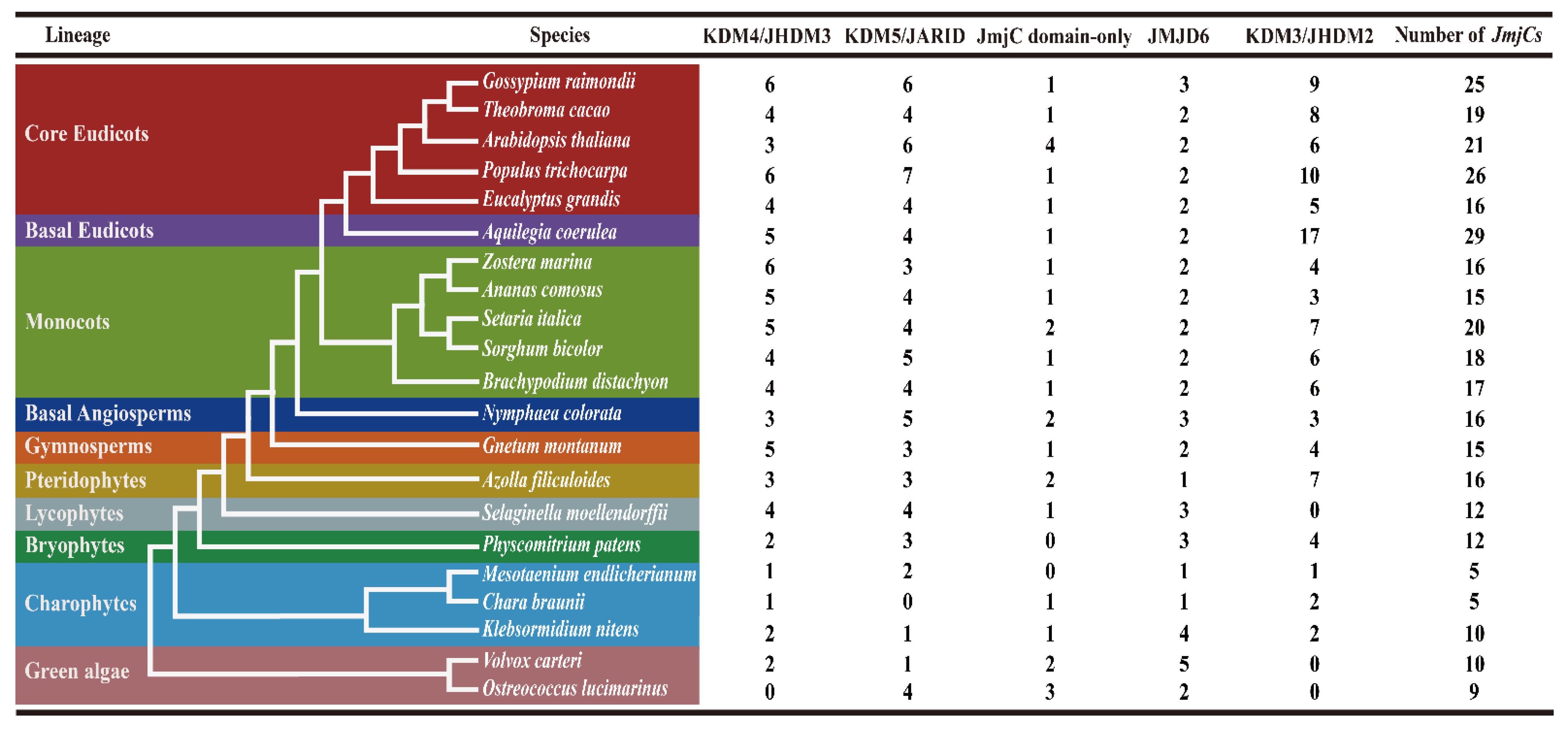
2.1. Identification of JmjC Genes in Green Plants

In order to comprehensively understand the JmjC genes in green plants, 21 plant species, which have published genome-wide data, were used to screen the JmjC genes. A total of 332 JmjC family genes were identified, and each sequence contained a conserved domain of JmjC (Table S1). These 21 species represent 10 plant evolutionary lineages, including green algae (Ostreococcus lucimarinus and Volvox carteri), charophytes (Mesotaenium endlicherianum, Chara braunii, and Klebsormidium nitens), bryophytes (Physcomitrium patens), lycophytes (Selaginella moellendorffii), pteridophytes (Azolla filiculoides), gymnosperms (Gnetum montanum), basal angiosperms (Nymphaea colorata), monocots (Brachypodium distachyon, Sorghum bicolor, Setaria italica, Ananas comosus, and Zostera marina), basal eudicots (Aquilegia coerulea), and core eudicots (Gossypium raimondii, Theobroma cacao, Arabidopsis thaliana, Populus trichocarpa, and Eucalyptus grandis) (Figure 1). The JmjC gene family has already appeared in green algae, with 9 and 10 JmjC genes in O. lucimarinus and V. carteri, respectively. The copy number of JmjC genes varied widely in other plants, ranging from 5 in C. braunii and M. endlicherianum to 29 in A. coerulea. Specifically, 10 JmjC genes were found in K. nitens, 12 in P. patens and S. moellendorffii, 15 in G. montanum and A. comosus, while there were 16 in A. filiculoides, N. colorata, Z. marina, and E. grandis, 17 in B. distachyon, 18 in S. bicolor, 19 in T. cacao, 20 in S. italica, 21 in A. thaliana, 25 in G. raimondii [37], and 26 in P. trichocarpa, which indicated that JmjC family members had expanded largely in the process of evolution.

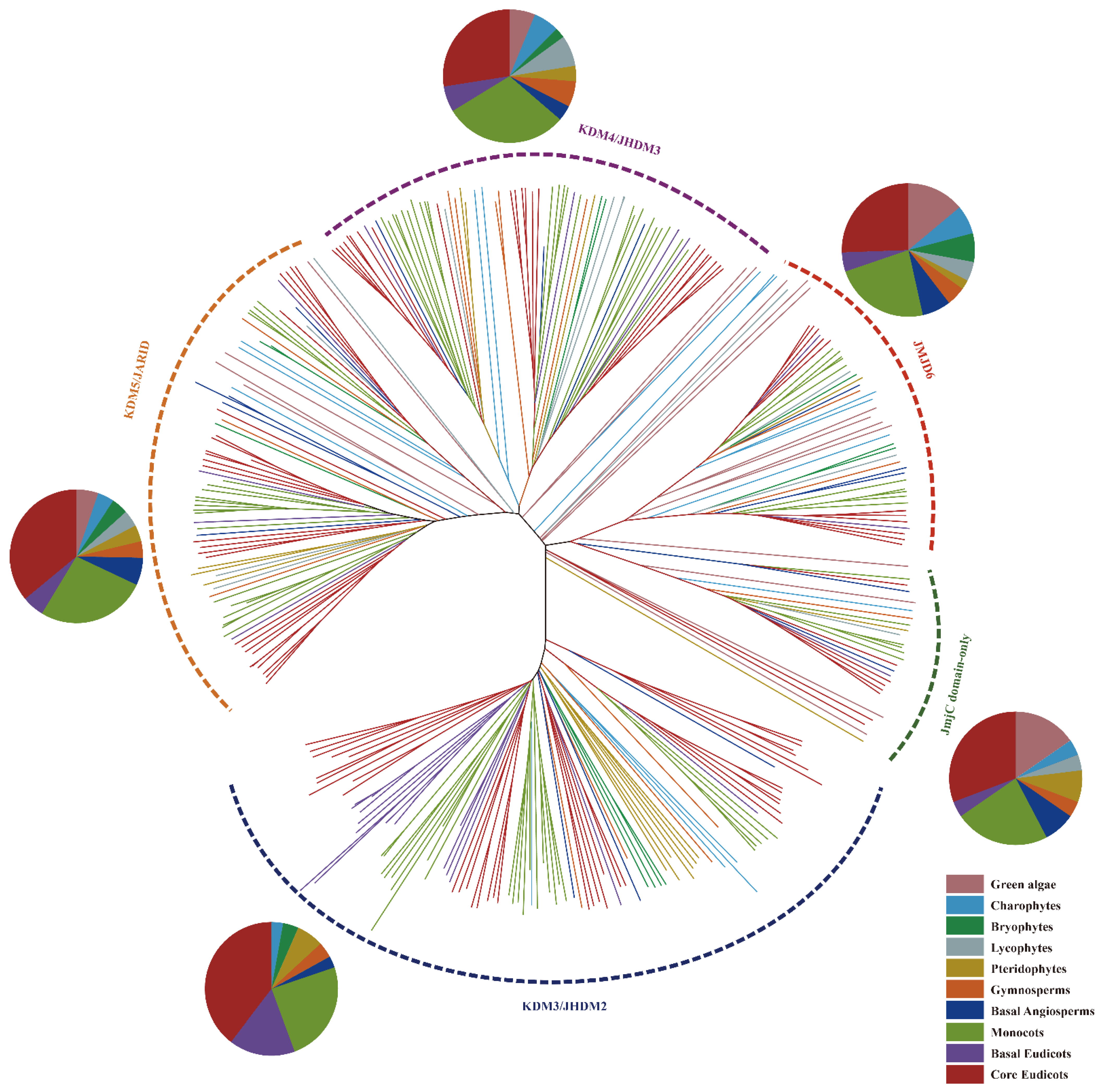
2.2. Phylogenetic Evolution Analysis of Plant JmjC Genes

In order to further study the evolutionary relationship between JmjC proteins in green plants, a multiple sequence alignment was performed on the full-length amino acid sequences of 332 JmjCs, and the neighbor-joining (NJ) phylogenetic tree was constructed using MEGA 7.0 software. As shown in Figure 2, all JmjC genes formed five branches, namely, KDM4/JHDM3, KDM5/JARID, JmjC domain-only, JMJD6, and KDM3/JHDM2 (Table S2). From the pie chart analysis, it can be clearly seen that most of the JmjC genes in all subfamilies were present in eudicotyledonous and monocotyledonous plants (Figure S1). Moreover, there were differences in the number of JmjC genes among different subfamilies. For example, the KDM3/JHDM2 subfamily had the largest number of JmjC family members, a total of 104, but no members of green algae of lycophytes belonged to the subfamily. Additionally, the number of KDM3/JHDM2 subfamily members varied the most, ranged from 0 to 17 (A. coerulea). Additionally, the number of JmjC genes in the JmjC domain-only subfamily was the least, only 28 in total, and the majority contained four genes in A. thaliana. In chara (M. endlicherianum) and bryophytes (P. patens), no members of the JmjC domain-only subfamily were found, and there were three genes in green algae (O. lucimarinus); two genes in V. carteri, A. filiculoides, N. colorata, and S. italica; and only one JmjC gene in the other plants. Except for the KDM3/JHDM2 and JmjC domain-only branches, the remaining branches contained the JmjC genes of all the lineages analyzed. There were a total of 75 JmjC genes in the KDM4/JHDM3 branch, and there were similar numbers of genes in eudicotyledons and monocots, 23 and 24 JmjC genes, respectively, but no genes from O. lucimarinus belonged to this family. There were a total of 77 JmjC genes in the KDM5/JARID branch; among them, no genes were found in C. braunii, and 27 and 20 genes were found in eudicotyledons and monocots, respectively. There were 48 JmjC genes in the JMJD6 branch (Table S3).

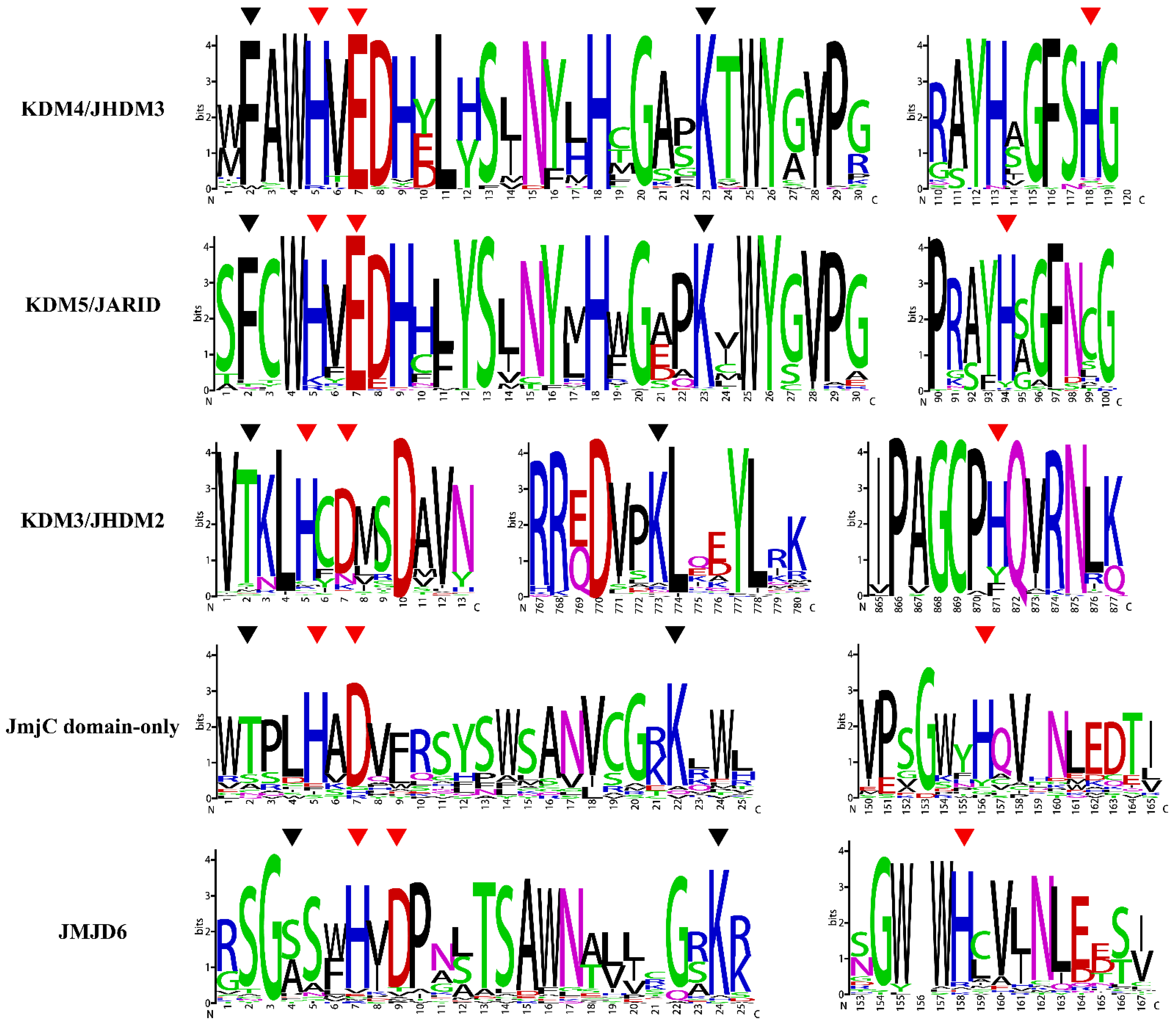
2.3. Analysis of Conserved Amino Acid Residues of JmjC

Jumonji C (JmjC) domain demethylates histones through an oxidative mechanism that requires ferrous ion (Fe [II]) and α-ketoglutarate (α-KG) as cofactors in addition to the hydroxylation of proteins [38]. The three residues that bind to Fe (II) (His 188, Glu/Asp 190, and His 276), and two residues that bind to α-KG (Thr/Phe 185 and Lys 206) are highly conserved and play an important role in the function of enzyme activity. To further assess the conservation of the JmjC domain, a multiple sequence alignment of JmjC domains in 21 plants species of different subfamilies was performed, and the results were displayed as sequence logos. As shown in Figure 3, the conserved residues varied within the subfamily. The subfamilies KDM4/JHDM3 and KDM5/JARID contained the original sites, namely, His (H), Glu (E), and His (H) and Phe (F) and Lys (K), respectively. The residues of subfamilies KDM3/JHDM2 and JmjC domain-only changed to His (H), Asp (D) and His (H) and Thr (T) and Lys (K), respectively. In addition, the first residue of the JMJD6 subfamily was replaced by Ser (S)/Ala (A), and the remaining four sites were conserved. Among the five subfamilies, the JmjC domain-only subfamily had the largest variation in amino acids, followed by the JMJD6 subfamily. Compared with the other four subfamilies, the amino acid residues of the KDM3/JHDM2 subfamily were farther apart. The conservation of the interaction site was related to the demethylase activity of the JmjC gene, suggesting that functional differentiation of the JmjC gene may have occurred in the process of evolution.

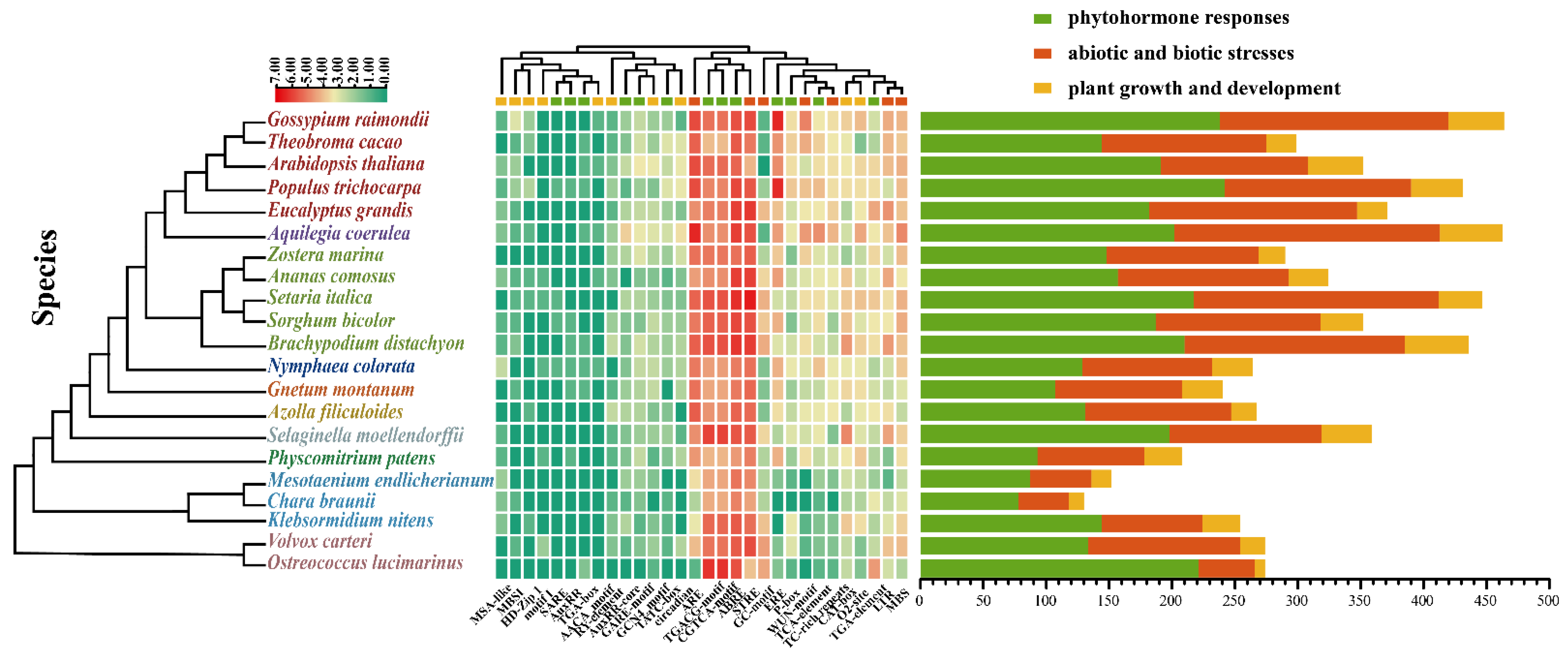
2.4. Analysis of Cis-Acting Elements in the Promoter Regions of JmjC Genes

Recent research has shown that JmjC genes have biological functions in plant growth and development, hormone response, and abiotic stress [39]. Therefore, we analyzed the cis-acting elements of the 2000 bp upstream regions from the transcription start site (TSS) of all the selected JmjC genes to explore their potential function. Finally, three types of cis-acting elements related to phytohormone response, abiotic and biotic stress response, and plant growth and development were detected (Figure 4 and Table S4). The results revealed six types of phytohormone-responsive regulatory elements that were identified in the JmjC promoter regions, including abscisic acid (ABA)-responsive elements (ABRE), gibberellin (GA)-responsive elements (GARE-motif, TATC-box, and P-box), methyl jasmonate (MeJA)-responsive elements (TGACG-motif and CGTCA-motif), salicylic acid (SA)-responsive elements (TCA-element and SARE), auxin (IAA)-responsive elements (TGA-element, AuxRR-core, TGA-box, and AuxRR), and ethylene-responsive elements (ERE). Among them, the elements associated with ABA and MeJA were abundant in all the species, which are major signaling molecules affecting plant growth and stress responses [40]. Furthermore, seven types of stress-response elements, namely, LTR, MBS, ARE, GC-motif, WUN-motif, TC-rich repeats, and STRE, which are responsible for low-temperature stress response, drought stress response, anaerobic induction, enhancing anoxic specific inducibility, wound stress response, defense stress response, and stress response, respectively, were identified. The most common elements that related to abiotic and biotic stress response were ARE and STRE. The cis-acting element related to plant growth and development mainly included the cellular development and metabolism regulation categories. To be specific, CAT-box, GCN4_motif, MSA-like, RY-element, AACA_motif, motif I, and HD-Zip 1 are associated with cell differentiation and tissue development. While circadian, O2-site, and MBSI are associated with circadian control, zein metabolism regulation, and flavonoid biosynthesis gene regulation, respectively. These analyses showed that there were numerous different cis-acting elements in the promoter of JmjC genes, suggesting that they might not only participate in the transduction of hormone signals, but also in the response to external pressure.

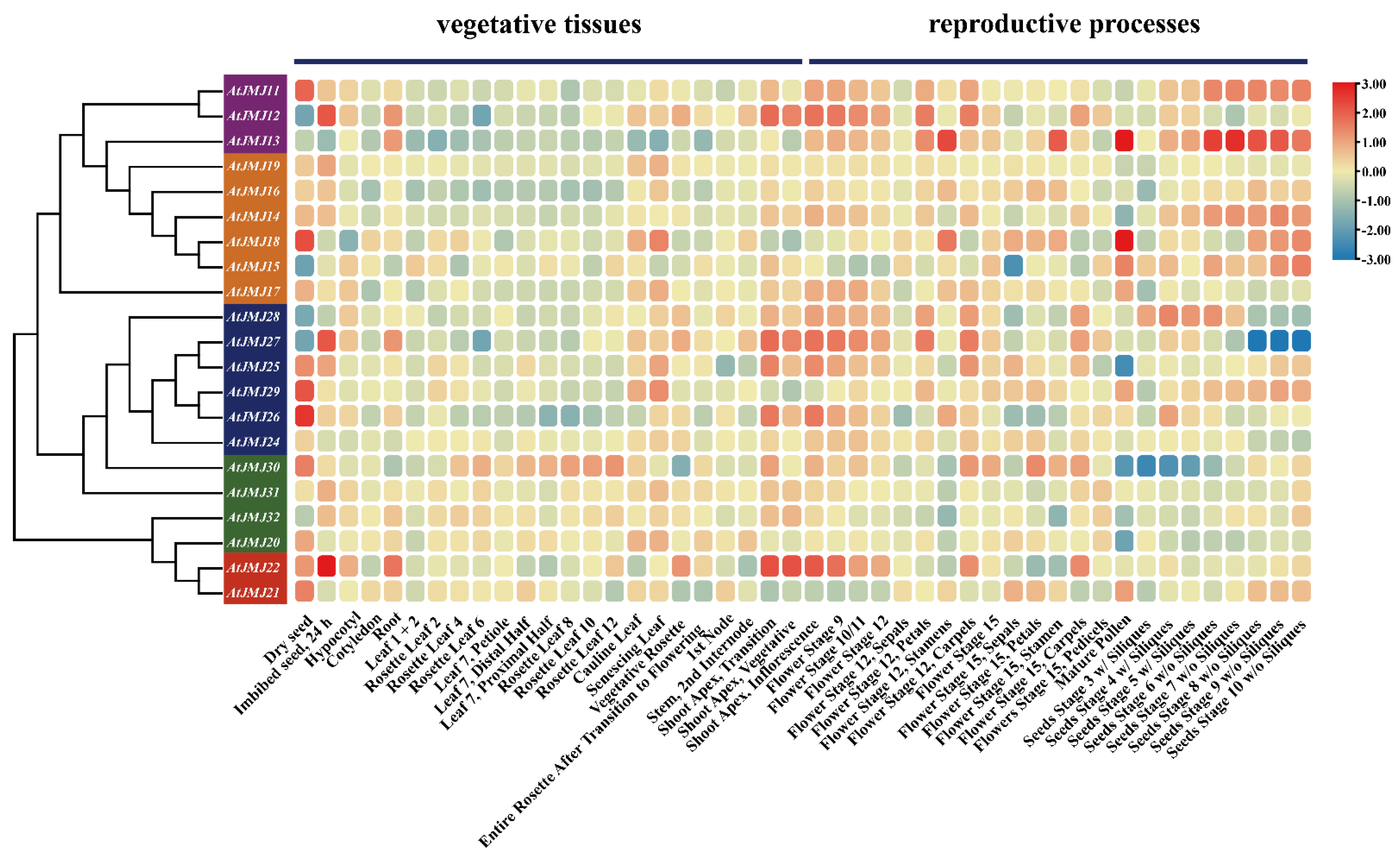
2.5. Expression Profiles of JmjC Genes in Different Tissues

To investigate the role of plant JmjC genes in different stages of tissue development, RNA-seq data were used to study the expression profile of JmjC genes in model plant A. thaliana. The transcriptome data of A. thaliana revealed that JmjC genes had different expression patterns in different tissues (Figure 5), suggesting that these genes had different functions in various developmental stages. In 23 vegetative tissues, most AtJMJs had high expression in dry seed, cauline leaf, senescing leaf, and shoot apex. AtJMJ12, AtJMJ22, and AtJMJ27 were highly expressed in seed imbibed for 24 h. Moreover, most AtJMJs had higher expression levels in tissues during the reproductive processes than that in vegetative tissues. In particular, AtJMJ13 was highly expressed only during the reproductive stage. The expression levels of AtJMJs in the shoot apex; inflorescence; and in flowering stages 9, 10/11, and 12 were relatively high. Some genes were expressed significantly only at certain stages, for example, AtJMJ14 was highly expressed only in the seed stages, and AtJMJ15 only in the mature pollen and seed stages. Additionally, there were individual genes, such as AtJMJ16, that had low expression levels in all the tissues selected. The same subfamily may have different gene expression patterns: AtJMJ22 was highly expressed in various tissues, while AtJMJ21 only in dry seed.

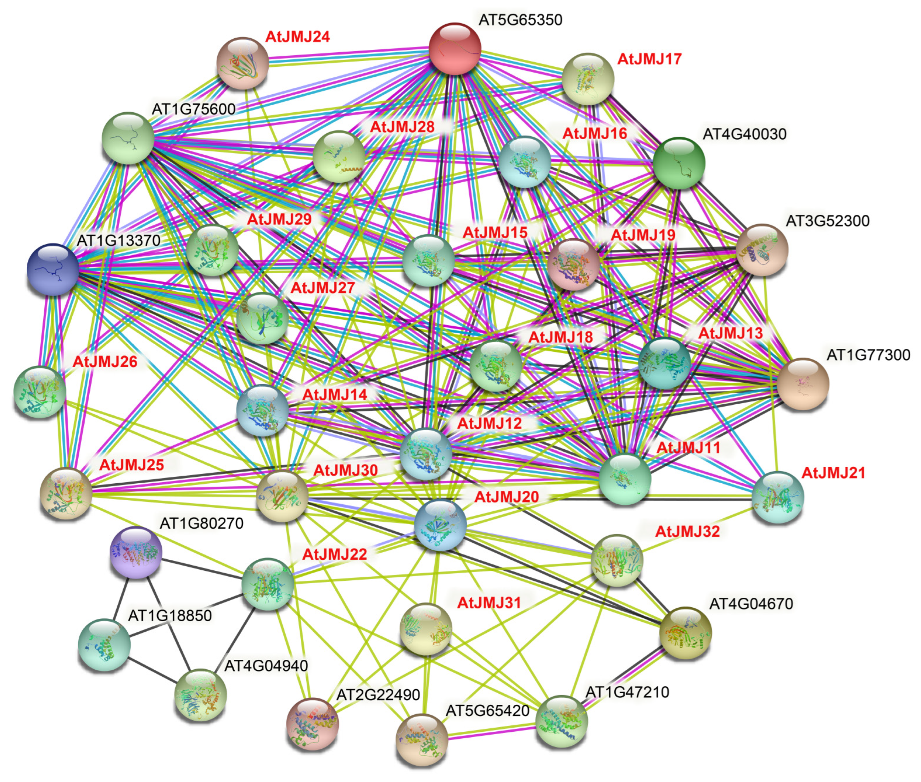
2.6. Interaction of the JmjC Protein in Plants

To further detect the potential roles of JmjC family members, we investigated the interaction of the JmjC protein in A. thaliana using the STRING database (Figure 6). The results showed that the interaction network of the JmjC protein exhibited complex functional relationships. For instance, the majority of A. thaliana JmjC proteins interacted with the histone superfamily proteins (AT5G65350.1/HTR11, AT5G10980.1/HTR4, AT1G75600.1/AT1G75600, AT1G13370.1/AT1G13370), and AT5G19840/AtJMJ31 interacted with cyclins (AT5G65420.3/CYCD4;1, AT2G22490.2/CYCD2;1. AT1G47210.2/CYCA3;2). Moreover, AtJMJ11/AT5G04240, AtJMJ12/AT3G48430 and AtJMJ13/AT5G46910 interacted with each other, and they all interacted with methyltransferase-associated proteins (AT1G77300.1/EFS), suggesting that the JmjC protein is involved in many biological processes through protein interactions.

3. Discussion

The plant JmjC gene family is an important class of histone lysine demethylation enzymes, which have important effects on plant growth and development. In this study, a total of 332 JmjC genes were identified in 21 green plants, and each sequence contained a JmjC conserved domain. According to the analysis of species evolution (Figure 1), the JmjC genes existed in all the analyzed plants, and the copy number of the JmjC genes varied widely, ranging from 5 in C. braunii and M. endlicherianum to 29 in A. coerulea, indicating that the JmjC family genes presented a wide range of changes in the course of plant evolution. The phylogenetic relationship of plant JmjCs indicated (Figure 2) that among the five subfamilies, the KDM3/JHDM2 subfamily appeared in charophytes first, and no JmjC genes of this subfamily were found in S. moellendorffii. Additionally, no JmjC domain-only subfamily members were found in either M. endlicherianum or P. patens, no KDM4/JHDM3 members were found in O. lucimarinus, and no KDM5/JARID members were found in C. braunii, which indicated that the JmjC genes of this subfamily in these plants were lost during evolution. Only the JMJD6 subfamily of the JMJC genes were retained in all the plant species. The five amino acid residues within the cofactor binding sites predicted by the JmjC domain were conserved and are important for the enzyme activity of the JmjC protein [11,15]. Among them, three residues bind to the Fe (II) cofactor, while α-ketoglutarate (α-KG) requires another two residues [15]. The five amino acid residues were highly conserved in the KDM4/JHDM3, KDM5/JARID, KDM3/JHDM2, and JmjC domain-only subfamilies. However, the JMJD6 subfamily lost a key α-KG binding residue (F/T) and was replaced by S/A (Figure 3). Mutations in amino acids and changes in the distance between residues may lead to changes in plants’ JmjC protein function.
The promoter is a DNA sequence that is located in the upstream region of the 5′ end of the structural gene, which can activate RNA polymerase, making it accurately bind to the template DNA and have specificity for transcription initiation [41]. The specificity of gene expression depends on cis-regulatory elements and their association with the JmjC genes [42]. Therefore, 2000 bp sequences upstream of the 5′ end of all the JmjC genes were extracted, and the cis-acting elements in the promoter regions were predicted using the PlantCRAE online tool (Figure 4). The results showed that many cis-acting elements in response to plant hormones, biotic and abiotic stresses, and plant growth and development were included. ABRE is an ABA-responsive cis-acting element. Under the condition of water shortage, the ABA content in plant leaves increases and causes stomatal closure, so it can be used as an anti-transpiration agent. In addition, drought, cold, high temperatures, salinization, and waterlogging stress can rapidly increase ABA in plants and enhance stress resistance. For example, ABA can induce the re-synthesis of certain enzymes and increase the cold, flood, and salt resistance of plants [41,42,43]. Methyl jasmonate is involved in the germination, rooting, flowering, and senescence of plants, which is also thought to be a defense mechanism that positively responds to most biotic and abiotic stresses in plants [44]. Additionally, there were amounts of cis-elements related to anaerobic induction (ARE) and stress response (STRE). The cis-acting elements associated with the cellular development and metabolism regulation categories were common in the promoter regions in all the species. Therefore, it was speculated that the JmjC family genes play an important role in plant growth and stress response.
The potential role of the JmjC family members was further explored by predicting Arabidopsis transcriptome data and protein interaction networks (Figure 5 and Figure 6). Most AtJMJs had higher expression levels in tissues during the reproductive processes than that in vegetative tissues, suggesting that they may play a more important role in the reproductive stage. Meanwhile, the protein interaction prediction indicated that AtJMJ13/At5G46910 interacted with the AtJMJ11/At5G04240 and AtJMJ12/At3G48430 proteins, and both of them interacted with methyltransferase-related proteins. Additionally, the mutant of AtJMJ11/At5G04240 leads to an early flowering phenotype in Arabidopsis [45]. The overexpression of ATJMJ12/AT3G48430 shows a pleiotropic phenotype of early flowering, short stature, and various degrees. Furthermore, the analysis of the cellular and molecular levels has found that the overexpression of AtJMJ12/At3G48430 results in a decrease in the cells of the leaf surface [46]. In addition, studies have shown that AtJMJ13/AT5G46910 is an Arabidopsis H3K27Me3 demethylase and a temperature photoperiod-dependent inhibitory factor for flowering. The Arabidopsis jmj13 mutant blooms early under short-day and high-temperature conditions [47]. Therefore, Arabidopsis JmjC proteins play a major role in plant growth and development.

4. Materials and Methods

4.1. Genome-Wide Identification of JmjC Family Genes in Plants

In this study, the genome-wide data of 21 plant species were analyzed, and 21 A. thaliana JmjC genes were downloaded from TAIR (https://www.arabidopsis.org/ (accesses on 18 August 2021)) [48]. The sources of genomic data for other species are listed in Table S5. To identify JmjC family genes in the selected species, BlastP and tBlastN searches [49] were applied to search the genome data with JmjC protein sequences in A. thaliana as queries. All the identified candidates were submitted to the Pfam [50] and SMART [51] databases, and genes encoding proteins containing a conserved protein domain (PF02373) were identified as JmjC genes.

4.2. Evolutionary Analysis

The ClustalW [52] program was used for the multiple sequence alignment of all the JmjC protein sequences in 21 plant species with the default parameters. Then, a phylogenetic tree was constructed using the MEGA 7.0 [53] with the neighbor-joining (NJ) method. The reliability was assessed with the p-distance model and 1000 bootstrap replications. Excel was used to count the number of genes contained in each JmjC subfamily in different species, and Origin 8.0 software [54] was used to make a pie chart.

4.3. Analysis of Conserved Amino Acid Residues of JmjC

Multiple sequence alignments were performed for each subfamily of JmjC protein sequences using the ClustalX 2.1 [55] software with the default parameters to obtain the conserved regions. Sequence logos for the conserved domains in JmjCs were generated using the WebLogo (http://weblogo.berkeley.edu/logo.cgi (accesses on 4 February 2022)) online tool [56].

4.4. Analysis of Cis-Acting Elements in the Promoter Regions of JmjC Genes

The 2000 bp upstream regions from the transcription start sites (TSS) of all the JmjC genes were obtained from the Phytozome database [57] and genome files, and then the PlantCARE (http://bioinformatics.psb.ugent.be/webtools/plantcare/html (accesses on 4 February 2022)) online tool [58] was used to search for cis-acting elements. The results were classified and counted [59], and finally visualized using the TBtools software [60].

4.5. Expression Pattern of Plant JmjC Genes in Different Tissues

Open expression data of various tissues in A. thaliana were acquired from the Bio-Analytic Resource for Plant Biology public database (http://bar.utoronto.ca/ (accesses on 4 February 2022)) [61], based on JmjC IDs of A. thaliana, including 23 vegetative tissues and 23 tissues during reproductive processes. A heatmap generated by the TBtools software [60] was used to display the relative expression levels.

4.6. Prediction of JmjC Protein Interactions

The prediction of Arabidopsis JmjC protein interactions was built using the STRING database (https://cn.string-db.org/ (accessed on 10 February 2022)) [62], with the confidence parameter set at a threshold of 0.40 and the number of interactions set to 5; the other parameters were as default.

5. Conclusions

In this study, a total of 332 JmjC genes were identified from 21 different plants. The evolutionary analysis suggested that JmjC genes had already appeared in algae, and the conserved domain analysis showed that each sequence contained a typical JmjC conserved domain, indicating that it was strongly conserved in plant evolution. The phylogenetic analysis demonstrated that the genes of the KDM3/JHDM2 subfamily might have emerged during the transition from water to land. Additionally, the analysis of the cis-acting elements showed that the JmjC genes were rich in cis-acting elements in response to plant hormones and biotic and abiotic stresses. The transcriptome data and protein interaction analysis indicated that JmjC genes play an important role in plant growth and development.

Supplementary Materials

The following supporting information can be downloaded at: https://0-www-mdpi-com.brum.beds.ac.uk/article/10.3390/plants11081041/s1, Refs [63,64,65,66,67,68,69,70,71,72,73,74,75,76,77,78,79,80,81], Table S1: JmjC gene IDs in 21 species; Table S2: Species selection and subfamily distribution in the plant JmjC gene phylogenetic tree; Table S3: JmjC gene family distribution; Table S4: List of identified cis-elements in the putative promoter region of JmjC genes; Table S5: The genome data sources for the species used in this study; Figure S1: JmjC gene family classification.

Author Contributions

Methodology, S.M. and Z.Z.; software, W.L.; formal analysis, S.M., Z.Z. and W.L.; investigation, S.M., Z.Z., Y.L., W.H. and J.Z.; resources, Y.Z., X.Y., J.Z. and X.L.; data curation, J.Z. and Q.D.; writing—original draft preparation, S.M., Z.Z. and W.L.; writing—review and editing, Y.L., D.Y. and X.M.; funding acquisition, W.L. and X.M. All authors have read and agreed to the published version of the manuscript.

Funding

This work was supported by the National Natural Science Foundation of China (32072114), the Natural Science Foundation of Henan (202300410550), the State Key Laboratory of Cotton Biology Open Fund (CB2021A13), the Innovation Project of the Chinese Academy of Agricultural Sciences (CAAS-ASTIP-ICR-KP-2021-01), and the Xinjiang Tianshan Talents Program (2021).

Data Availability Statement

The data presented in this study are available in the Supplementary Materials.

Conflicts of Interest

The authors declare no conflict of interest.

References

  1. Egger, G.; Liang, G.; Aparicio, A.; Jones, P.A. Epigenetics in human disease and prospects for epigenetic therapy. Nature 2004, 429, 457–463. [Google Scholar] [CrossRef] [PubMed]
  2. Wei, J.W.; Huang, K.; Yang, C.; Kang, C.S. Non-coding RNAs as regulators in epigenetics (Review). Oncol. Rep. 2017, 37, 3–9. [Google Scholar] [CrossRef] [PubMed] [Green Version]
  3. Alvarez-Venegas, R.; Abdallat, A.A.; Guo, M.; Alfano, J.R.; Avramova, Z. Epigenetic control of a transcription factor at the cross section of two antagonistic pathways. Epigenetics 2007, 2, 106–113. [Google Scholar] [CrossRef] [PubMed] [Green Version]
  4. Cairns, B.R. The logic of chromatin architecture and remodelling at promoters. Nature 2009, 461, 193–198. [Google Scholar] [CrossRef] [PubMed]
  5. Allis, C.D.; Jenuwein, T. The molecular hallmarks of epigenetic control. Nat. Rev. Genet. 2016, 17, 487–500. [Google Scholar] [CrossRef]
  6. Xiao, J.; Lee, U.S.; Wagner, D. Tug of war: Adding and removing histone lysine methylation in Arabidopsis. Curr. Opin. Plant Biol. 2016, 34, 41–53. [Google Scholar] [CrossRef] [Green Version]
  7. Liu, C.; Lu, F.; Cui, X.; Cao, X. Histone methylation in higher plants. Annu. Rev. Plant Biol. 2010, 61, 395–420. [Google Scholar] [CrossRef]
  8. Kouzarides, T. Chromatin modifications and their function. Cell 2007, 128, 693–705. [Google Scholar] [CrossRef] [Green Version]
  9. Mosammaparast, N.; Shi, Y. Reversal of histone methylation: Biochemical and molecular mechanisms of histone demethylases. Annu. Rev. Bioch. 2010, 79, 155–179. [Google Scholar] [CrossRef]
  10. Shi, Y.; Lan, F.; Matson, C.; Mulligan, P.; Whetstine, J.R.; Cole, P.A.; Casero, R.A.; Shi, Y. Histone demethylation mediated by the nuclear amine oxidase homolog LSD1. Cell 2004, 119, 941–953. [Google Scholar] [CrossRef] [Green Version]
  11. Klose, R.J.; Kallin, E.M.; Zhang, Y. JmjC-domain-containing proteins and histone demethylation. Nat. Rev. Genet. 2006, 7, 715–727. [Google Scholar] [CrossRef] [PubMed]
  12. Qian, Y.; Chen, C.; Jiang, L.; Zhang, J.; Ren, Q. Genome-wide identification, classification and expression analysis of the JmjC domain-containing histone demethylase gene family in maize. BMC Genom. 2019, 20, 256. [Google Scholar] [CrossRef] [PubMed]
  13. Han, Y.; Li, X.; Cheng, L.; Liu, Y.; Wang, H.; Ke, D.; Yuan, H.; Zhang, L.; Wang, L. Genome-wide analysis of soybean JmjC domain-containing proteins suggests evolutionary conservation following whole-genome duplication. Front. Plant Sci. 2016, 7, 1800. [Google Scholar] [CrossRef] [PubMed] [Green Version]
  14. Chowrasia, S.; Panda, A.K.; Rawal, H.C.; Kaur, H.; Mondal, T.K. Identification of jumonjiC domain containing gene family among the Oryza species and their expression analysis in FL478, a salt tolerant rice genotype. Plant Physiol. Biochem. 2018, 130, 43–53. [Google Scholar] [CrossRef]
  15. Lu, F.; Li, G.; Cui, X.; Liu, C.; Wang, X.J.; Cao, X. Comparative analysis of JmjC domain-containing proteins reveals the potential histone demethylases in Arabidopsis and rice. J. Integr. Plant Biol. 2008, 50, 886–896. [Google Scholar] [CrossRef]
  16. Luo, M.; Hung, F.-Y.; Yang, S.; Liu, X.; Wu, K. Histone lysine demethylases and their functions in plants. Plant Mol. Biol. Rep. 2013, 32, 558–565. [Google Scholar] [CrossRef]
  17. Tahiliani, M.; Mei, P.; Fang, R.; Leonor, T.; Rutenberg, M.; Shimizu, F.; Li, J.; Rao, A.; Shi, Y. The histone H3K4 demethylase SMCX links REST target genes to X-linked mental retardation. Nature 2007, 447, 601–605. [Google Scholar] [CrossRef]
  18. Yamane, K.; Toumazou, C.; Tsukada, Y.; Erdjument-Bromage, H.; Tempst, P.; Wong, J.; Zhang, Y. JHDM2A, a JmjC-containing H3K9 demethylase, facilitates transcription activation by androgen receptor. Cell 2006, 125, 483–495. [Google Scholar] [CrossRef] [Green Version]
  19. Allis, C.D.; Berger, S.L.; Cote, J.; Dent, S.; Jenuwien, T.; Kouzarides, T.; Pillus, L.; Reinberg, D.; Shi, Y.; Shiekhattar, R.; et al. New nomenclature for chromatin-modifying enzymes. Cell 2007, 131, 633–636. [Google Scholar] [CrossRef] [Green Version]
  20. Jones, M.A.; Morohashi, K.; Grotewold, E.; Harmer, S.L. Arabidopsis JMJD5/JMJ30 acts independently of LUX ARRHYTHMO within the plant circadian clock to enable temperature compensation. Fron. Plant Sci. 2019, 10, 57. [Google Scholar] [CrossRef] [Green Version]
  21. Jenuwein, T.; Allis, C.D. Translating the histone code. Science 2001, 293, 1074–1080. [Google Scholar] [CrossRef] [PubMed] [Green Version]
  22. Chen, X.; Hu, Y.; Zhou, D.X. Epigenetic gene regulation by plant Jumonji group of histone demethylase. Biochim. Biophys. Acta 2011, 1809, 421–426. [Google Scholar] [CrossRef] [PubMed]
  23. Kooistra, S.M.; Helin, K. Molecular mechanisms and potential functions of histone demethylases. Nat. Rev. Mol. Cell Biol. 2012, 13, 297–311. [Google Scholar] [CrossRef] [PubMed]
  24. Noh, B.; Lee, S.H.; Kim, H.J.; Yi, G.; Shin, E.A.; Lee, M.; Jung, K.J.; Doyle, M.R.; Amasino, R.M.; Noh, Y.S. Divergent roles of a pair of homologous jumonji/zinc-finger-class transcription factor proteins in the regulation of Arabidopsis flowering time. Plant Cell 2004, 16, 2601–2613. [Google Scholar] [CrossRef] [Green Version]
  25. Yang, H.; Han, Z.; Cao, Y.; Fan, D.; Li, H.; Mo, H.; Feng, Y.; Liu, L.; Wang, Z.; Yue, Y.; et al. A companion cell-dominant and developmentally regulated H3K4 demethylase controls flowering time in Arabidopsis via the repression of FLC expression. PLoS Genet. 2012, 8, e1002664. [Google Scholar] [CrossRef]
  26. Yang, H.; Mo, H.; Fan, D.; Cao, Y.; Cui, S.; Ma, L. Overexpression of a histone H3K4 demethylase, JMJ15, accelerates flowering time in Arabidopsis. Plant Cell Rep. 2012, 31, 1297–1308. [Google Scholar] [CrossRef]
  27. Park, J.; Lee, N.; Kim, W.; Lim, S.; Choi, G. ABI3 and PIL5 collaboratively activate the expression of SOMNUS by directly binding to its promoter in imbibed Arabidopsis seeds. Plant Cell 2011, 23, 1404–1415. [Google Scholar] [CrossRef] [Green Version]
  28. Cho, J.N.; Ryu, J.Y.; Jeong, Y.M.; Park, J.; Song, J.J.; Amasino, R.M.; Noh, B.; Noh, Y.S. Control of seed germination by light-induced histone arginine demethylation activity. Dev. Cell 2012, 22, 736–748. [Google Scholar] [CrossRef] [Green Version]
  29. Saze, H.; Shiraishi, A.; Miura, A.; Kakutani, T. Control of genic DNA methylation by a jmjC domain-containing protein in Arabidopsis thaliana. Science 2008, 319, 462–465. [Google Scholar] [CrossRef]
  30. Sun, Q.; Zhou, D.X. Rice jmjC domain-containing gene JMJ706 encodes H3K9 demethylase required for floral organ development. Proc. Nat. Acad. Sci. USA 2008, 105, 13679–13684. [Google Scholar] [CrossRef] [Green Version]
  31. Audonnet, L.; Shen, Y.; Zhou, D.X. JMJ24 antagonizes histone H3K9 demethylase IBM1/JMJ25 function and interacts with RNAi pathways for gene silencing. Gene Expr. Patterns 2017, 25–26, 1–7. [Google Scholar] [CrossRef]
  32. Lu, S.X.; Knowles, S.M.; Webb, C.J.; Celaya, R.B.; Cha, C.; Siu, J.P.; Tobin, E.M. The Jumonji C domain-containing protein JMJ30 regulates period length in the Arabidopsis circadian clock. Plant Physiol. 2011, 155, 906–915. [Google Scholar] [CrossRef] [PubMed] [Green Version]
  33. Shen, Y.; Conde, E.S.N.; Audonnet, L.; Servet, C.; Wei, W.; Zhou, D.X. Over-expression of histone H3K4 demethylase gene JMJ15 enhances salt tolerance in Arabidopsis. Front. Plant Sci. 2014, 5, 290. [Google Scholar] [CrossRef] [PubMed] [Green Version]
  34. Hou, Y.; Wang, L.; Wang, L.; Liu, L.; Li, L.; Sun, L.; Rao, Q.; Zhang, J.; Huang, S. JMJ704 positively regulates rice defense response against Xanthomonas oryzae pv. oryzae infection via reducing H3K4me2/3 associated with negative disease resistance regulators. BMC Plant Biol. 2015, 15, 286. [Google Scholar] [CrossRef] [PubMed] [Green Version]
  35. Li, T.; Chen, X.; Zhong, X.; Zhao, Y.; Liu, X.; Zhou, S.; Cheng, S.; Zhou, D.X. Jumonji C domain protein JMJ705-mediated removal of histone H3 lysine 27 trimethylation is involved in defense-related gene activation in rice. Plant Cell 2013, 25, 4725–4736. [Google Scholar] [CrossRef] [PubMed] [Green Version]
  36. Liu, H.; Guo, S.; Xu, Y.; Li, C.; Zhang, Z.; Zhang, D.; Xu, S.; Zhang, C.; Chong, K. OsmiR396d-regulated OsGRFs function in floral organogenesis in rice through binding to their targets OsJMJ706 and OsCR4. Plant Physiol. 2014, 165, 160–174. [Google Scholar] [CrossRef] [PubMed] [Green Version]
  37. Zhang, J.; Feng, J.; Liu, W.; Ren, Z.; Zhao, J.; Pei, X.; Liu, Y.; Yang, D.; Ma, X. Characterization and stress response of the JmjC domain-containing histone demethylase gene family in the allotetraploid cotton species Gossypium hirsutum. Plants 2020, 9, 1617. [Google Scholar] [CrossRef]
  38. Sun, Z.; Wang, X.; Qiao, K.; Fan, S.; Ma, Q. Genome-wide analysis of JMJ-C histone demethylase family involved in salt-tolerance in Gossypium hirsutum L. Plant Physiol. Biochem. 2021, 158, 420–433. [Google Scholar] [CrossRef]
  39. Wang, Z.; Liu, D.; Xu, B.; Tian, R.; Zuo, Y. Modular arrangements of sequence motifs determine the functional diversity of KDM proteins. Brief. Bioinform. 2021, 22, bbaa215. [Google Scholar] [CrossRef]
  40. Yu, T.F.; Zhao, W.Y.; Fu, J.D.; Liu, Y.W.; Chen, M.; Zhou, Y.B.; Ma, Y.Z.; Xu, Z.S.; Xi, Y.J. Genome-Wide Analysis of CDPK Family in Foxtail Millet and Determination of SiCDPK24 Functions in Drought Stress. Front. Plant Sci. 2018, 9, 651. [Google Scholar] [CrossRef]
  41. Hernandez-Garcia, C.M.; Finer, J.J. Identification and validation of promoters and cis-acting regulatory elements. Plant Sci. 2014, 217–218, 109–119. [Google Scholar] [CrossRef] [PubMed] [Green Version]
  42. Zhou, D.X. Regulatory mechanism of plant gene transcription by GT-elements and GT-factors. Trends Plant Sci. 1999, 4, 210–214. [Google Scholar] [CrossRef]
  43. Zhang, J.Y.; Huang, S.N.; Wang, G.; Xuan, J.P.; Guo, Z.R. Overexpression of Actinidia deliciosa pyruvate decarboxylase 1 gene enhances waterlogging stress in transgenic Arabidopsis thaliana. Plant Physiol. Biochem. 2016, 106, 244–252. [Google Scholar] [CrossRef]
  44. Wasternack, C.; Hause, B. Jasmonates and octadecanoids: Signals in plant stress responses and development. Prog. Nucleic Acid Res. Mol. Biol. 2002, 72, 165–221. [Google Scholar]
  45. Yu, X.; Li, L.; Li, L.; Guo, M.; Chory, J.; Yin, Y. Modulation of brassinosteroid-regulated gene expression by Jumonji domain-containing proteins ELF6 and REF6 in Arabidopsis. Proc. Nat. Acad. Sci. USA 2008, 105, 7618–7623. [Google Scholar] [CrossRef] [PubMed] [Green Version]
  46. Lu, F.; Cui, X.; Zhang, S.; Jenuwein, T.; Cao, X. Arabidopsis REF6 is a histone H3 lysine 27 demethylase. Nat. Genet. 2011, 43, 715–719. [Google Scholar] [CrossRef] [PubMed]
  47. Zheng, S.; Hu, H.; Ren, H.; Yang, Z.; Qiu, Q.; Qi, W.; Liu, X.; Chen, X.; Cui, X.; Li, S.; et al. The Arabidopsis H3K27me3 demethylase JUMONJI 13 is a temperature and photoperiod dependent flowering repressor. Nat. Commun. 2019, 10, 1303. [Google Scholar] [CrossRef] [Green Version]
  48. Zhao, W.; Shafiq, S.; Berr, A.; Shen, W.H. Genome-wide gene expression profiling to investigate molecular phenotypes of Arabidopsis mutants deprived in distinct histone methyltransferases and demethylases. Genom. Data 2015, 4, 143–145. [Google Scholar] [CrossRef]
  49. Zhang, Z.; Liu, W.; Ma, Z.; Zhu, W.; Jia, L. Transcriptional characterization and response to defense elicitors of mevalonate pathway genes in cotton (Gossypium arboreum L.). PeerJ 2019, 7, e8123. [Google Scholar] [CrossRef] [Green Version]
  50. El-Gebali, S.; Mistry, J.; Bateman, A.; Eddy, S.R.; Luciani, A.; Potter, S.C.; Qureshi, M.; Richardson, L.J.; Salazar, G.A.; Smart, A.; et al. The Pfam protein families database in 2019. Nucleic Acids Res. 2019, 47, D427–D432. [Google Scholar] [CrossRef]
  51. Letunic, I.; Copley, R.R.; Schmidt, S.; Ciccarelli, F.D.; Doerks, T.; Schultz, J.; Ponting, C.P.; Bork, P. SMART 4.0: Towards genomic data integration. Nucleic Acids Res. 2004, 32, D142–D144. [Google Scholar] [CrossRef] [PubMed] [Green Version]
  52. Li, K.B. ClustalW-MPI: ClustalW analysis using distributed and parallel computing. Bioinformatics 2003, 19, 1585–1586. [Google Scholar] [CrossRef] [PubMed] [Green Version]
  53. Kumar, S.; Stecher, G.; Tamura, K. MEGA7: Molecular evolutionary genetics analysis version 7.0 for bigger datasets. Mol. Biol. Evol. 2016, 33, 1870–1874. [Google Scholar] [CrossRef] [PubMed] [Green Version]
  54. Liu, W.; Zhang, Z.; Zhu, W.; Ren, Z.; Jia, L.; Li, W.; Ma, Z. Evolutionary conservation and divergence of genes encoding 3-Hydroxy-3-methylglutaryl coenzyme A synthase in the allotetraploid cotton species Gossypium hirsutum. Cells 2019, 8, 412. [Google Scholar] [CrossRef] [PubMed] [Green Version]
  55. Larkin, M.A.; Blackshields, G.; Brown, N.P.; Chenna, R.; McGettigan, P.A.; McWilliam, H.; Valentin, F.; Wallace, I.M.; Wilm, A.; Lopez, R.; et al. Clustal W and Clustal X version 2.0. Bioinformatics 2007, 23, 2947–2948. [Google Scholar] [CrossRef] [PubMed] [Green Version]
  56. Crooks, G.E.; Hon, G.; Chandonia, J.M.; Brenner, S.E. WebLogo: A sequence logo generator. Genome Res. 2004, 14, 1188–1190. [Google Scholar] [CrossRef] [PubMed] [Green Version]
  57. Goodstein, D.M.; Shu, S.; Howson, R.; Neupane, R.; Hayes, R.D.; Fazo, J.; Mitros, T.; Dirks, W.; Hellsten, U.; Putnam, N.; et al. Phytozome: A comparative platform for green plant genomics. Nucleic Acids Res. 2012, 40, D1178–D1186. [Google Scholar] [CrossRef]
  58. Lescot, M.; Déhais, P.; Thijs, G.; Marchal, K.; Moreau, Y.; Van de Peer, Y.; Rouzé, P.; Rombauts, S. PlantCARE, a database of plant cis-acting regulatory elements and a portal to tools for in silico analysis of promoter sequences. Nucleic Acids Res. 2002, 30, 325–327. [Google Scholar] [CrossRef]
  59. Li, W.; Sun, K.; Ren, Z.; Song, C.; Pei, X.; Liu, Y.; Wang, Z.; He, K.; Zhang, F.; Zhou, X.; et al. Molecular evolution and stress and phytohormone responsiveness of SUT genes in Gossypium hirsutum. Front Genet. 2018, 9, 494. [Google Scholar] [CrossRef]
  60. Chen, C.; Chen, H.; Zhang, Y.; Thomas, H.R.; Frank, M.H.; He, Y.; Xia, R. TBtools: An integrative toolkit developed for interactive analyses of big biological data. Mol. Plant 2020, 13, 1194–1202. [Google Scholar] [CrossRef]
  61. Winter, D.; Vinegar, B.; Nahal, H.; Ammar, R.; Wilson, G.V.; Provart, N.J. An “Electronic Fluorescent Pictograph” browser for exploring and analyzing large-scale biological data sets. PLoS ONE 2007, 2, e718. [Google Scholar] [CrossRef]
  62. Szklarczyk, D.; Morris, J.H.; Cook, H.; Kuhn, M.; Wyder, S.; Simonovic, M.; Santos, A.; Doncheva, N.T.; Roth, A.; Bork, P.; et al. The STRING database in 2017: Quality-controlled protein-protein association networks, made broadly accessible. Nucleic Acids Res. 2017, 45, D362–D368. [Google Scholar] [CrossRef] [PubMed]
  63. Palenik, B.; Grimwood, J.; Aerts, A.; Rouzé, P.; Salamov, A.; Putnam, N.; Dupont, C.; Jorgensen, R.; Derelle, E.; Rombauts, S.; et al. The tiny eukaryote Ostreococcus provides genomic insights into the paradox of plankton speciation. Proc. Natl. Acad. Sci. USA 2007, 104, 7705–7710. [Google Scholar] [CrossRef] [PubMed] [Green Version]
  64. Prochnik, S.E.; Umen, J.; Nedelcu, A.M.; Hallmann, A.; Miller, S.M.; Nishii, I.; Ferris, P.; Kuo, A.; Mitros, T.; Fritz-Laylin, L.K.; et al. Genomic analysis of organismal complexity in the multicellular green alga Volvox carteri. Science 2010, 329, 223–226. [Google Scholar] [CrossRef] [PubMed] [Green Version]
  65. Cheng, S.; Xian, W.; Fu, Y.; Marin, B.; Keller, J.; Wu, T.; Sun, W.; Li, X.; Xu, Y.; Zhang, Y.; et al. Genomes of subaerial zygnematophyceae provide insights into land plant evolution. Cell 2019, 179, 1057–1067.e14. [Google Scholar] [CrossRef]
  66. Nishiyama, T.; Sakayama, H.; de Vries, J.; Buschmann, H.; Saint-Marcoux, D.; Ullrich, K.K.; Haas, F.B.; Vanderstraeten, L.; Becker, D.; Lang, D.; et al. The chara genome: Secondary complexity and implications for plant terrestrialization. Cell 2018, 174, 448–464.e424. [Google Scholar] [CrossRef] [Green Version]
  67. Hori, K.; Maruyama, F.; Fujisawa, T.; Togashi, T.; Yamamoto, N.; Seo, M.; Sato, S.; Yamada, T.; Mori, H.; Tajima, N.; et al. Klebsormidium flaccidum genome reveals primary factors for plant terrestrial adaptation. Nat. Commun. 2014, 5, 3978. [Google Scholar] [CrossRef]
  68. Lang, D.; Ullrich, K.K.; Murat, F.; Fuchs, J.; Jenkins, J.; Haas, F.B.; Piednoel, M.; Gundlach, H.; Van Bel, M.; Meyberg, R.; et al. The Physcomitrella patens chromosome-scale assembly reveals moss genome structure and evolution. Plant J. 2018, 93, 515–533. [Google Scholar] [CrossRef] [Green Version]
  69. Banks, J.A.; Nishiyama, T.; Hasebe, M.; Bowman, J.L.; Gribskov, M.; de Pamphilis, C.; Albert, V.A.; Aono, N.; Aoyama, T.; Ambrose, B.A.; et al. The Selaginella genome identifies genetic changes associated with the evolution of vascular plants. Science 2011, 332, 960–963. [Google Scholar] [CrossRef] [Green Version]
  70. Li, F.W.; Brouwer, P.; Carretero-Paulet, L.; Cheng, S.; de Vries, J.; Delaux, P.M.; Eily, A.; Koppers, N.; Kuo, L.Y.; Li, Z.; et al. Fern genomes elucidate land plant evolution and cyanobacterial symbioses. Nat. Plants 2018, 4, 460–472. [Google Scholar] [CrossRef] [Green Version]
  71. Wan, T.; Liu, Z.M.; Li, L.F.; Leitch, A.R.; Leitch, I.J.; Lohaus, R.; Liu, Z.J.; Xin, H.P.; Gong, Y.B.; Liu, Y.; et al. A genome for gnetophytes and early evolution of seed plants. Nat. Plants 2018, 4, 82–89. [Google Scholar] [CrossRef] [PubMed]
  72. Zhang, L.; Chen, F.; Zhang, X.; Li, Z.; Zhao, Y.; Lohaus, R.; Chang, X.; Dong, W.; Ho, S.Y.W.; Liu, X.; et al. The water lily genome and the early evolution of flowering plants. Nature 2020, 577, 79–84. [Google Scholar] [CrossRef] [PubMed] [Green Version]
  73. International Brachypodium Initiative. Genome sequencing and analysis of the model grass Brachypodium distachyon. Nature 2010, 463, 763–768. [Google Scholar] [CrossRef] [PubMed]
  74. McCormick, R.F.; Truong, S.K.; Sreedasyam, A.; Jenkins, J.; Shu, S.; Sims, D.; Kennedy, M.; Amirebrahimi, M.; Weers, B.D.; McKinley, B.; et al. The Sorghum bicolor reference genome: Improved assembly, gene annotations, a transcriptome atlas, and signatures of genome organization. Plant J. 2018, 93, 338–354. [Google Scholar] [CrossRef] [Green Version]
  75. Bennetzen, J.L.; Schmutz, J.; Wang, H.; Percifield, R.; Hawkins, J.; Pontaroli, A.C.; Estep, M.; Feng, L.; Vaughn, J.N.; Grimwood, J.; et al. Reference genome sequence of the model plant Setaria. Nat. Biotechnol. 2012, 30, 555–561. [Google Scholar] [CrossRef] [PubMed] [Green Version]
  76. Ming, R.; VanBuren, R.; Wai, C.M.; Tang, H.; Schatz, M.C.; Bowers, J.E.; Lyons, E.; Wang, M.L.; Chen, J.; Biggers, E.; et al. The pineapple genome and the evolution of CAM photosynthesis. Nat. Genet. 2015, 47, 1435–1442. [Google Scholar] [CrossRef] [Green Version]
  77. Olsen, J.L.; Rouzé, P.; Verhelst, B.; Lin, Y.C.; Bayer, T.; Collen, J.; Dattolo, E.; De Paoli, E.; Dittami, S.; Maumus, F.; et al. The genome of the seagrass Zostera marina reveals angiosperm adaptation to the sea. Nature 2016, 530, 331–335. [Google Scholar] [CrossRef] [Green Version]
  78. Filiault, D.L.; Ballerini, E.S.; Mandáková, T.; Aköz, G.; Derieg, N.J.; Schmutz, J.; Jenkins, J.; Grimwood, J.; Shu, S.; Hayes, R.D.; et al. The Aquilegia genome provides insight into adaptive radiation and reveals an extraordinarily polymorphic chromosome with a unique history. Elife 2018, 7, e36426. [Google Scholar] [CrossRef]
  79. Tuskan, G.A.; Difazio, S.; Jansson, S.; Bohlmann, J.; Grigoriev, I.; Hellsten, U.; Putnam, N.; Ralph, S.; Rombauts, S.; Salamov, A.; et al. The genome of black cottonwood, Populus trichocarpa (Torr. & Gray). Science 2006, 313, 1596–1604. [Google Scholar]
  80. Argout, X.; Martin, G.; Droc, G.; Fouet, O.; Labadie, K.; Rivals, E.; Aury, J.M.; Lanaud, C. The cacao Criollo genome v2.0: An improved version of the genome for genetic and functional genomic studies. BMC Genom. 2017, 18, 730. [Google Scholar] [CrossRef]
  81. Myburg, A.A.; Grattapaglia, D.; Tuskan, G.A.; Hellsten, U.; Hayes, R.D.; Grimwood, J.; Jenkins, J.; Lindquist, E.; Tice, H.; Bauer, D.; et al. The genome of Eucalyptus grandis. Nature 2014, 510, 356–362. [Google Scholar] [CrossRef] [PubMed] [Green Version]
Figure 1. Phylogenetic relationships among 21 species. Different background colors in the phylogenetic tree show different pedigrees. The right side shows the number of JmjC genes in each species of the different subfamilies and the total number of JmjC genes found in the genome of each species.
Plants 11 01041 g001
Figure 2. Phylogenetic tree of JmjC proteins in 21 plant species. JmjCs are divided into five subfamilies, which are represented by arcs of different colors. The lines of different colors in the evolutionary tree represent the JmjCs of 10 different lineages. The color of the pie chart in the figure corresponds to the color of the JmjCs in the evolutionary tree.
Plants 11 01041 g002
Figure 3. Conserved amino acid residues of JmjC protein in plants. The height of each letter in the figure indicates the degree of conservation of amino acid residues. The number on the abscissa indicates the relative position of the amino acid in the motif. The three conserved catalytic residues of Fe (II) (His 188, Glu/Asp 190, and His 276) are marked with red triangles. The two conserved catalytic residues of α-KG (Ther/Phe 185 and Lys 206) are marked with black triangles.
Plants 11 01041 g003
Figure 4. Analysis of promoter cis-elements of JmjC genes in plants. The left side of the heatmap is the evolutionary history of 21 species. Green, red, and yellow boxes represent cis-elements related to phytohormone responses, biotic and abiotic stresses, and plant growth and development, respectively. The histogram indicates the number of cis-acting elements of different types in each species.
Plants 11 01041 g004
Figure 5. Expression profiles of the JmjC genes in different tissues and developmental stages of A. thaliana. On the left is the phylogenetic tree of Arabidopsis JmjCs.
Plants 11 01041 g005
Figure 6. Functional network assembly of the A. thaliana JmjC proteins. Red letters are the A. thaliana protein. Light blue and purple lines represent known interactions determined by the database and experiments, respectively. Green, red, and blue lines represent predicted interactions from gene proximity, fusion, and symbiosis, respectively. Light green, black, and gray lines indicate other interactions from text mining, co-expression, and protein homology, respectively. Empty nodes: proteins of unknown three-dimensional structure. Filled nodes: some three-dimensional structures are known or predicted.
Plants 11 01041 g006
Publisher’s Note: MDPI stays neutral with regard to jurisdictional claims in published maps and institutional affiliations.

