
Biological Activities of the Fruit Essential Oil, Fruit, and Root Extracts of Ferula drudeana Korovin, the Putative Anatolian Ecotype of the Silphion Plant †
 , ,
, ,  and
and
Abstract
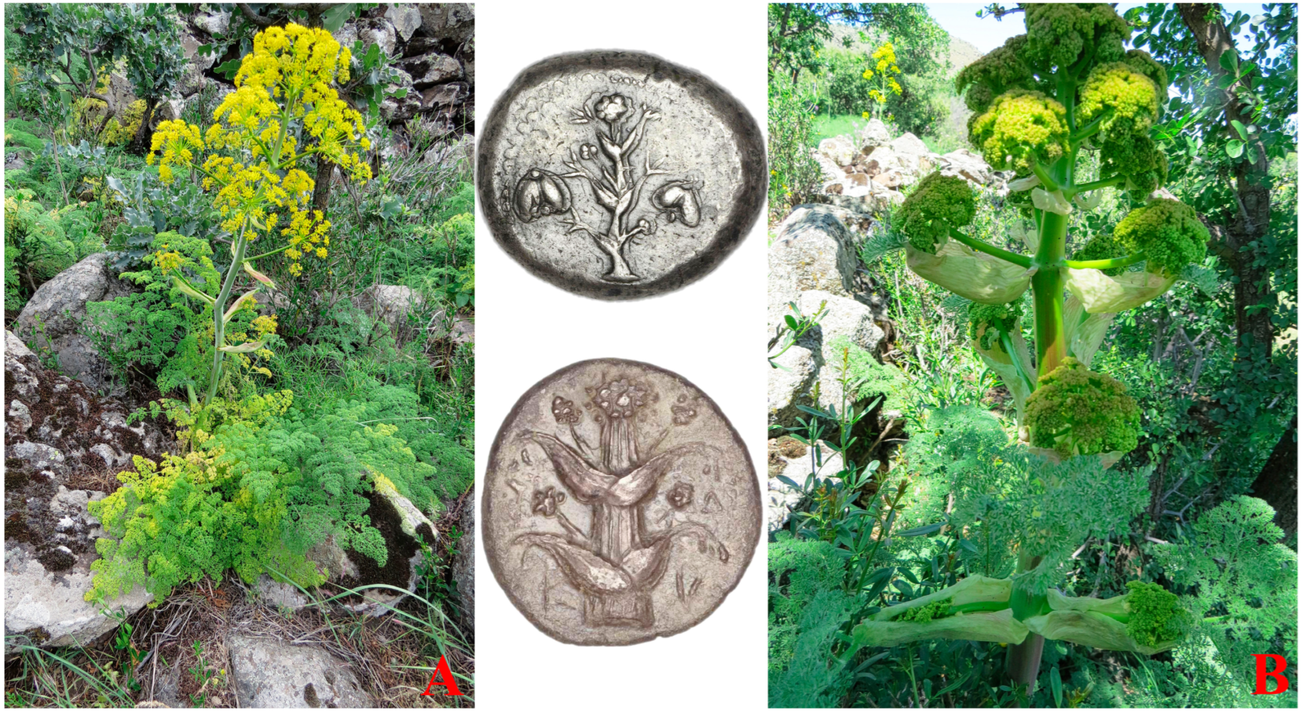
:1. Introduction
2. Results and Discussion
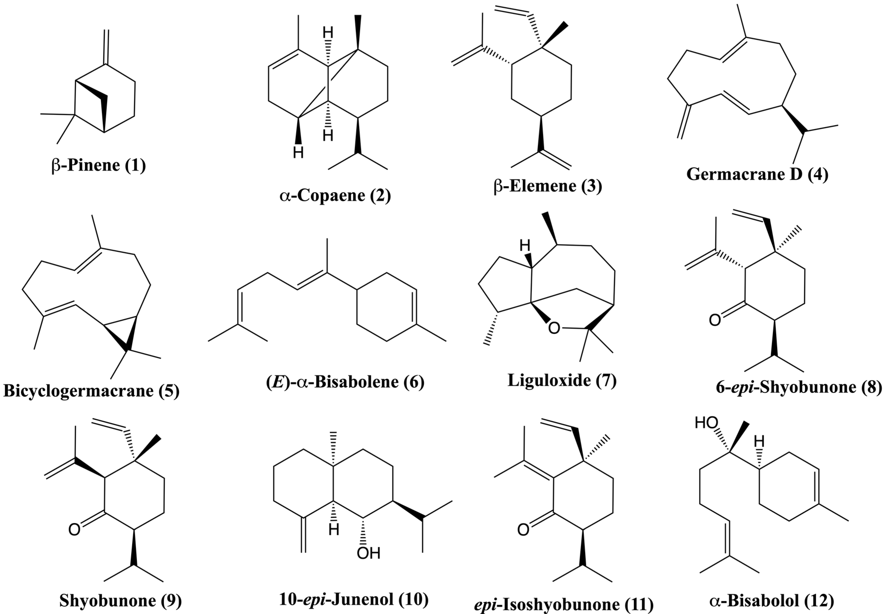
2.1. Volatile Composition of the Fruit Essential Oil of Ferula drudeana
2.2. Antimicrobial Testing of the Fruit Essential Oil of Ferula drudeana
2.3. Antioxidant Activity Determination
2.3.1. Qualitative TLC Spot Testing Evaluation of the Antioxidant Activities of the Fruit Essential Oil, Fruit, and Root Extracts of Ferula drudeana
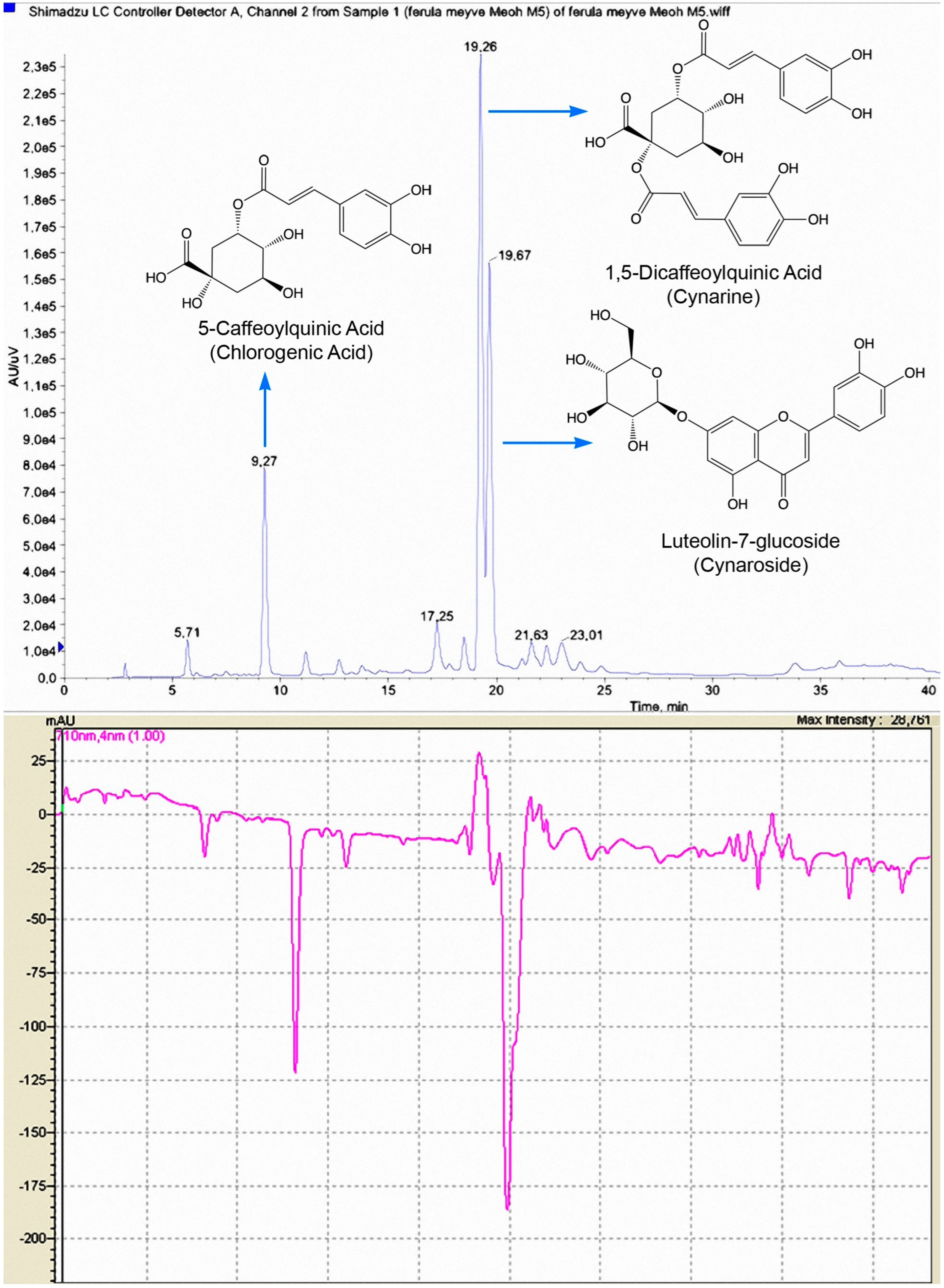
2.3.2. Online HPLC–ABTS•+ Identification of Major Antioxidant Compounds of the Methanolic Extracts of Ferula drudeana
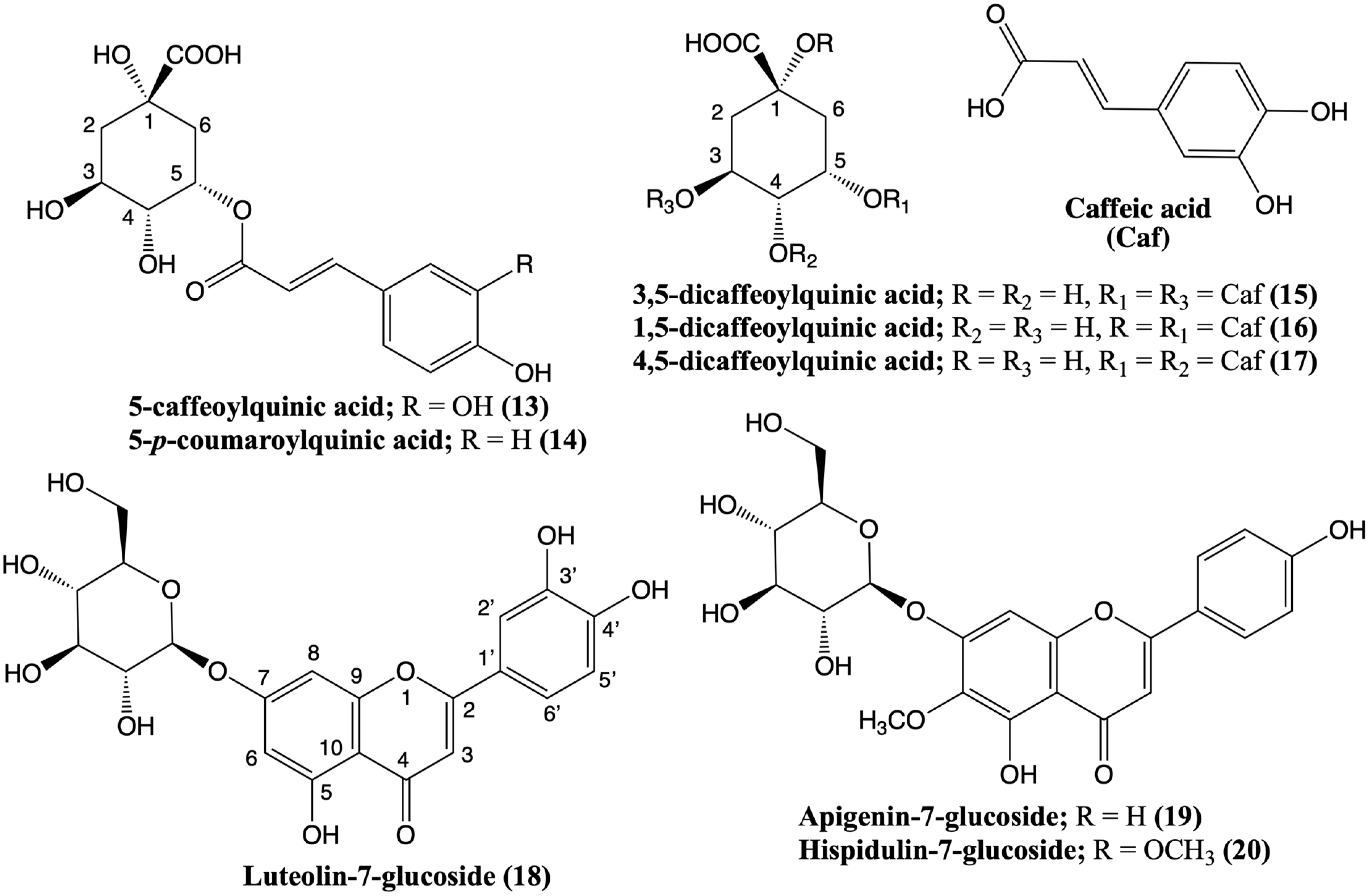
2.3.3. HPLC–MS/MS Analysis of the Methanolic Extract of the Fruits of Ferula drudeana
2.3.4. Determination of the Antioxidant Potential of the Methanolic Extracts of Ferula drudeana by DPPH and ABTS Free Radical Scavenging Activity Assessment
2.4. Biological Activities of the Ferula drudeana Metabolites
3. Materials and Methods
3.1. General Experimental Procedures
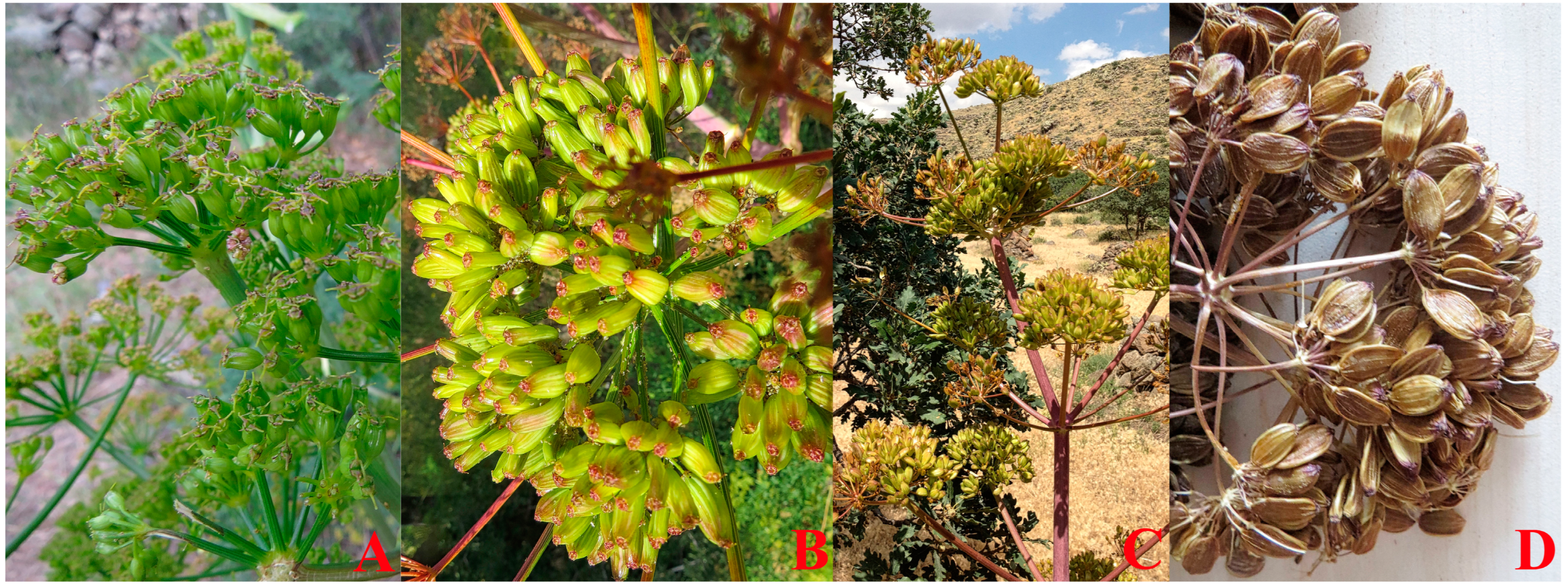
3.2. Material
3.3. Extraction
3.4. Gas Chromatography–Gas Chromatography/Mass Spectrometry Analyses of Ferula drudeana Essential Oil
3.5. Tested Microorganisms and Standard Antimicrobial Agents
3.6. Antimicrobial Activity
3.7. Antioxidant Activity
3.7.1. DPPH• TLC Spot Testing
3.7.2. HPLC–ABTS•+ Derivatization
3.7.3. LC–MS/MS Analysis
3.7.4. Determination of the Antioxidant Potential of Methanolic Extracts
DPPH Radical Scavenging Activity
ABTS Radical Scavenging Activity
4. Conclusions
Supplementary Materials
Author Contributions
Funding
Institutional Review Board Statement
Informed Consent Statement
Data Availability Statement
Acknowledgments
Conflicts of Interest
References
- POWO. Plants of the World Online. Facilitated by the Royal Botanic Gardens, Kew. Published on the Internet. Available online: https://powo.science.kew.org/taxon/30105171-2#publications (accessed on 31 December 2022).
- Peşmen, H. Ferula, Flora of Turkey and the East Aegean Islands; Edinburgh University Press: Edinburgh, UK, 1972; Volume 4, pp. 440–453. [Google Scholar]
- Pimenov, M.; Leonov, M. The Asian Umbelliferae biodiversity database (ASIUM) with particular reference to South-West Asian taxa. Turk. J. Bot. 2004, 28, 139–145. Available online: https://journals.tubitak.gov.tr/botany/vol28/iss1/13 (accessed on 7 February 2023).
- Akalın, E.; Tuncay, H.O.; Olcay, B.; Miski, M. A new Ferula (Apiaceae) species from Southwest Anatolia: Ferula pisidica Akalın & Miski. Plants 2020, 9, 740. [Google Scholar] [CrossRef]
- Mohammadhosseini, M.; Venditti, A.; Sarker, S.D.; Nahar, L.; Akbarzadeh, A. The genus Ferula: Ethnobotany, phytochemistry and bioactivities—A review. Ind. Crop. Prod. 2019, 129, 350–394. [Google Scholar] [CrossRef]
- Gunther, R.T. The Greek Herbal of Dioscorides; Hafner Publishing Company: London, UK; New York, NY, USA, 1968. [Google Scholar]
- Korovin, E.P. Generis Ferula (Tourn.) L. monographia Illustrata; Academiae Scientiarum UzRSS: Tashkent, Uzbekistan, 1947. [Google Scholar]
- Miski, M. Next chapter in the legend of silphion: Preliminary morphological, chemical, biological and pharmacological evaluations, initial conservation studies, and reassessment of the regional extinction event. Plants 2021, 10, 102. [Google Scholar] [CrossRef]
- Bostock, J.; Riley, H.T. Book XXII, The Properties of Plants and Fruits, Chapter 49; Laser: Thirty-nine remedies. In Pliny the Elder, Natural History; Collected Works of Pliny the Elder; Delphi Classics, Hastings; Delphi Publishing Ltd.: East Sussex, UK, 2015. [Google Scholar]
- Hort, A.F. Book VI of Under-Shrubs [3.2–3.4]. In Theophrastus, Enquiry into Plants; Collected Works of Theophrastus; Delphi Classics, Hastings; Delphi Publishing Ltd.: East Sussex, UK, 2019. [Google Scholar]
- French, D.H. Ethnobotany of the Umbelliferae. In The Biology and Chemistry of the Umbelliferae; Academic Press Inc.: London, UK, 1971; pp. 385–412. [Google Scholar]
- Totelin, L.M.V. Hippocratic Recipes, Oral and Written Transmission of Pharmacological Knowledge in Fifth- and Fourth-Century Greece; Brill: Leiden, The Netherlands, 2009; pp. 158–161. [Google Scholar]
- Süzgeç-Selçuk, S.; Özek, G.; Meriçli, A.H.; Baser, K.H.C.; Haliloglu, Y.; Özek, T. Chemical and biological diversity of the leaf and rhizome volatiles of Acorus calamus L. from Turkey. J. Essent. Oil Bear. Plants 2017, 20, 646–661. [Google Scholar] [CrossRef]
- Babushok, V.I.; Linstrom, P.J.; Zenkevich, I.G. Retention indices for frequently reported compounds of plant essential oils. J. Phys. Chem. Ref. Data 2011, 40, 043101-1–043101-47. [Google Scholar] [CrossRef]
- Demirci, F.; Demirci, B.; Gurbuz, I.; Yeşilada, E.; Başer, K.H.C. Characterization and biological activity of Achillea teretifolia Willd. and A. nobilis L. subsp. neilreichii (Kerner) Formanek Essential Oils. Turk. J. Biol. 2009, 33, 129–136. [Google Scholar] [CrossRef]
- Demirci, B.; Koltuksuz Yasdikcıoglu, G.; Baser, K.H.C. Sesquiterpene hydrocarbons of the essential oil of Actinolema macrolema Boiss. Turk. J. Chem. 2013, 37, 917–926. [Google Scholar] [CrossRef]
- Ozek, G.; Chidibayeva, A.; Ametov, A.; Nurmahanova, A.; Ozek, T. Chemical composition of flower volatiles and seeds fatty acids of Rosa iliensis Chrshan, an endemic species from Kazakhstan. Rec. Nat. Prod. 2022, 16, 225–235. [Google Scholar] [CrossRef]
- Merle, H.; Verdeguer, M.; Blázquez, M.A.; Boira, H. Chemical composition of the essential oils from Eriocephalus africanus L. var. africanus populations growing in Spain. Flav. Fragr. J. 2007, 22, 461–464. [Google Scholar] [CrossRef]
- 10-Epijunenol (Compound). Available online: https://pubchem.ncbi.nlm.nih.gov/compound/573015#section=Experimental-Properties (accessed on 31 December 2022).
- Baser, K.H.C.; Demirci, B.; Sagiroglu, M.; Duman, H. Essential oil of Ferula species of Turkey. In Proceedings of the 38th International Symposium on Essential Oils, Graz, Austria, 9–12 September 2007. [Google Scholar]
- Martins, R.M.; Xavier-Júnior, F.H.; Barros, M.R.; Menezes, T.M.; de Assis, C.R.; de Melo, A.C.G.; Veras, B.O.; Ferraz, V.P.; Filho, A.A.M.; Yogui, G.T.; et al. Impact on cholinesterase-inhibition and in silico investigations of sesquiterpenoids from Amazonian Siparuna guianensis Aubl. Spectrochim. Acta A Mol. Biomol. Spectrosc. 2021, 252, 119511. [Google Scholar] [CrossRef]
- Rossi, P.G.; Bao, L.; Luciani, A.; Panighi, J.; Desjobert, J.M.; Costa, J.; Casanova, J.; Bolla, J.-M.; Berti, L. (E)-Methylisoeugenol and elemicin: Antibacterial components of Daucus carota L. essential oil against Campylobacter jejuni. J. Agric. Food Chem. 2007, 55, 7332–7336. [Google Scholar] [CrossRef]
- Yamamura, S.; Iguchi, A.; Nishiyama, A.; Niwa, M.; Koyama, H.; Hirata, Y. Sesquiterpenes from Acorus calamus L. Tetrahedron 1971, 27, 5419–5431. [Google Scholar] [CrossRef]
- Garneau, F.X.; Collin, G.; Gagnon, H.; Bélanger, A.; Lavoie, S.; Savard, N.; Pichette, A. Aromas from Quebec. I. Composition of the essential oil of the rhizomes of Acorus calamus L. J. Essent. Oil Res. 2008, 20, 250–254. [Google Scholar] [CrossRef]
- Znati, M.; Jabrane, A.; Hajlaoui, H.; Harzallah-Skhiri, F.; Bouajila, J.; Casanova, J.; Jannet, H.B. Chemical composition and in vitro evaluation of antimicrobial and anti-acetylcholinesterase properties of the flower oil of Ferula lutea. Nat. Prod. Comm. 2012, 7, 947–950. [Google Scholar] [CrossRef]
- Zellagui, A.; Gherraf, N.; Hegazy, M.; Akkal, S.; Rhouati, S.; Dendougui, H. Phytochemical investigation and antimicrobial activity of crude extract of the roots of Ferula vesceritensis. Chem. Nat. Compd. 2012, 48, 891–892. [Google Scholar] [CrossRef]
- Pavlović, I.; Petrović, S.; Radenković, M.; Milenković, M.; Couladis, M.; Branković, S.; Drobac, M.P.; Niketić, M. Composition, antimicrobial, antiradical and spasmolytic activity of Ferula heuffelii Griseb. ex Heuffel (Apiaceae) essential oil. Food Chem. 2012, 130, 310–315. [Google Scholar] [CrossRef]
- Özek, G.; Özek, T.; Işcan, G.; Başer, K.H.C.; Duran, A.; Hamzaoglu, E. Composition and antimicrobial activity of the oils of Ferula szowitsiana DC. from Turkey. J. Essent. Oil Res. 2008, 20, 186–190. [Google Scholar] [CrossRef]
- Maggi, F.; Cecchini, C.; Cresci, A.; Coman, M.M.; Tirillini, B.; Sagratini, G.; Papa, F. Chemical composition and antimicrobial activity of the essential oil from Ferula glauca L. (F. communis L. subsp. glauca) growing in Marche (central Italy). Fitoterapia 2009, 80, 68–72. [Google Scholar] [CrossRef]
- Kavoosi, G.; Rowshan, V. Chemical composition, antioxidant and antimicrobial activities of essential oil obtained from Ferula assa-foetida oleo-gum-resin: Effect of collection time. Food Chem. 2013, 138, 2180–2187. [Google Scholar] [CrossRef]
- Iranshahi, M.; Hassanzadeh-Khayat, M.; Bazzaz, B.S.F.; Sabeti, Z.; Enayati, F. High content of polysulphides in the volatile oil of Ferula latisecta Rech. F. et Aell. fruits and antimicrobial activity of the oil. J. Essent. Oil Res. 2008, 20, 183–185. [Google Scholar] [CrossRef]
- Ibraheim, Z.Z.; Abdel-Mageed, W.M.; Dai, H.; Guo, H.; Zhang, L.; Jaspars, M. Antimicrobial antioxidant daucane sesquiterpenes from Ferula hermonis Boiss. Phytother. Res. 2012, 26, 579–586. [Google Scholar] [CrossRef]
- Asili, J.; Sahebkar, A.; Bazzaz, B.S.F.; Sharifi, S.; Iranshahi, M. Identification of essential oil components of Ferula badrakema fruits by GC-MS and 13C-NMR methods and evaluation of its antimicrobial activity. J. Essent. Oil-Bear. Plants 2009, 12, 7–15. [Google Scholar] [CrossRef]
- Al-Yahya, M.A.; Muhammad, I.; Mirza, H.H.; El-Feraly, F.S. Antibacterial constituents from the rhizomes of Ferula communis. Phytother. Res. Int. J. Devoted Pharmacol. Toxicol. Eval. Nat. Prod. Deriv. 1998, 12, 335–339. [Google Scholar] [CrossRef]
- Samaranayake, Y.H.; Samaranayake, L. Candida krusei: Biology, epidemiology, pathogenicity and clinical manifestations of an emerging pathogen. J. Med. Microbiol. 1994, 41, 295–310. [Google Scholar] [CrossRef] [PubMed]
- Kumar, S.; Sandhir, R.; Ojha, S. Evaluation of antioxidant activity and total phenol in different varieties of Lantana camara leaves. BMC Res. Notes 2014, 7, 560. [Google Scholar] [CrossRef] [PubMed]
- Sathuvan, M.; Vignesh, A.; Thangam, R.; Palani, P.; Rengasamy, R.; Murugesan, K. In vitro antioxidant and anticancer potential of bark of Costus pictus D. Don. Asian Pac. J. Trop. Biomed. 2012, 2, S741–S749. [Google Scholar] [CrossRef]
- He, W.; Liu, X.; Xu, H.; Gong, Y.; Yuan, F.; Gao, Y. On-line HPLC-ABTS screening and HPLC-DAD-MS/MS identification of free radical scavengers in Gardenia (Gardenia jasminoides Ellis) fruit extracts. Food Chem. 2010, 123, 521–528. [Google Scholar] [CrossRef]
- Gouveia, S.; Goncalves, J.; Castilho, P.C. Characterization of phenolic compounds and antioxidant activity of ethanolic extracts from flowers of Andryala glandulosa ssp varia (Lowe ex DC.) R.Fern., an endemic species of Macaronesia region. Ind. Crop. Prod. 2013, 42, 573–582. [Google Scholar] [CrossRef]
- Clifford, M.N.; Johnston, K.L.; Knight, S.; Kuhnert, N. Hierarchical scheme for LC-MS n identification of chlorogenic acids. J. Agric. Food Chem. 2003, 51, 2900–2911. [Google Scholar] [CrossRef] [PubMed]
- Clifford, M.N.; Knight, S.; Kuhnert, N. Discriminating between the six isomers of dicaffeoylquinic acid by LC-MS. J. Agric. Food Chem. 2005, 53, 3821–3832. [Google Scholar] [CrossRef]
- Kontogianni, V.G.; Tomic, G.; Nikolic, I.; Nerantzaki, A.A.; Sayyad, N.; Stosic-Grujicic, S.; Stojanovic, I.; Gerothanassis, I.P.; Tzakos, A.G. Phytochemical profile of Rosmarinus officinalis and Salvia officinalis extracts and correlation to their antioxidant and anti-proliferative activity. Food Chem. 2013, 136, 120–129. [Google Scholar] [CrossRef]
- Topdas, E.F.; Sengul, M.; Taghizadehghalehjoughi, A.; Hacimuftuoglu, A. Neuroprotective potential and antioxidant activity of various solvent extracts and essential oil of Ferula orientalis L. J. Essent. Oil-Bear Plants 2020, 23, 121–138. [Google Scholar] [CrossRef]
- Romo Vaquero, M.; Yáñez-Gascón, M.J.; Garcia Villalba, R.; Larrosa, M.; Fromentin, E.; Ibarra, A.; Roller, M.; Tomas-Barberan, F.; de Gea, J.C.E.; García-Conesa, M.T. Inhibition of gastric lipase as a mechanism for body weight and plasma lipids reduction in Zucker rats fed a rosemary extract rich in carnosic acid. PLoS ONE 2012, 7, e39773. [Google Scholar] [CrossRef]
- Kızıltaş, H.; Gören, A.; Bingol, Z.; Alwasel, S.; Gülçin, İ. Anticholinergic, antidiabetic and antioxidant activities of Ferula orientalis L. determination of its polyphenol contents by LC-HRMS. Rec. Nat. Prod. 2021, 15, 513–528. [Google Scholar] [CrossRef]
- Rahali, F.Z.; Lamine, M.; Rebey, I.B.; Wannes, W.A.; Hammami, M.; Selmi, S.; Mliki, A.; Sellami, I.H. Biochemical characterization of fennel (Ferula communis L.) different parts through their essential oils, fatty acids and phenolics. Acta Sci. Pol. Hortorum Cultus 2021, 20, 3–14. [Google Scholar] [CrossRef]
- Rahali, F.Z.; Kefi, S.; Bettaieb Rebey, I.; Hamdaoui, G.; Tabart, J.; Kevers, C.; Franck, T.; Mouithys-Mickalad, A.; Hamrouni Sellami, I. Phytochemical composition and antioxidant activities of different aerial parts extracts of Ferula communis L. Plant Biosyst. Int. J. Deal. All Asp. Plant Biol. 2019, 153, 213–221. [Google Scholar] [CrossRef]
- Pavlović, I.; Radenković, M.; Branković, S.; Milenković, M.T.; Niketić, M.; Ušjak, L.; Petrović, S. Spasmolytic, gastroprotective and antioxidant activities of dry methanol extract of Ferula heuffelii underground parts. Chem. Biodivers. 2022, 19, e202200047. [Google Scholar] [CrossRef] [PubMed]
- Kahraman, C.; Baysal, I.; Çankaya, I.; Goger, F.; Kirimer, N.; Akdemir, Z. Acetylcholinesterase inhibitory activities and LC-MS analysis of the antioxidant Ferula caspica M. Bieb. and F. halophila Pesmen extracts. J. Res. Pharm. 2019, 23, 543–551. [Google Scholar] [CrossRef]
- Chen, X.; Liu, Q. Luteolin glycosides as taxonomic markers in Ferula and related genera. Biochem. Syst. Ecol. 1989, 17, 309–310. [Google Scholar] [CrossRef]
- Rivas da Silva, A.C.; Lopes, P.M.; Barros de Azevedo, M.M.; Costa, D.C.; Alviano, C.S.; Alviano, D.S. Biological activities of α-pinene and β-pinene enantiomers. Molecules 2012, 17, 6305–6316. [Google Scholar] [CrossRef] [PubMed] [Green Version]
- İşcan, G. Antibacterial and anticandidal activities of common essential oil constituents. Rec. Nat. Prod. 2017, 11, 374–388. [Google Scholar]
- de Macedo Andrade, A.C.; Rosalen, P.L.; Freires, I.A.; Scotti, L.; Scotti, M.T.; Aquino, S.G.; de Castro, R.D. Antifungal activity, mode of action, docking prediction and anti-biofilm effects of (+)-beta-pinene enantiomers against Candida spp. Curr. Top. Med. Chem. 2018, 18, 2481–2490. [Google Scholar] [CrossRef]
- Chowhan, N.; Singh, H.P.; Batish, D.R.; Kaur, S.; Ahuja, N.; Kohli, R.K. β-Pinene inhibited germination and early growth involves membrane peroxidation. Protoplasma 2013, 250, 691–700. [Google Scholar] [CrossRef]
- Guzmán-Gutiérrez, S.L.; Bonilla-Jaime, H.; Gómez-Cansino, R.; Reyes-Chilpa, R. Linalool and β-pinene exert their antidepressant-like activity through the monoaminergic pathway. Life Sci. 2015, 128, 24–29. [Google Scholar] [CrossRef] [PubMed]
- Machado, T.Q.; Felisberto, J.R.S.; Guimarães, E.F.; Queiroz, G.A.D.; Fonseca, A.C.C.D.; Ramos, Y.J.; Marques, A.M.; Moreira, D.L.; Robbs, B.K. Apoptotic effect of β-pinene on oral squamous cell carcinoma as one of the major compounds from essential oil of medicinal plant Piper rivinoides Kunth. Nat. Prod. Res. 2015, 36, 1636–1640. [Google Scholar] [CrossRef]
- Salehi, B.; Upadhyay, S.; Erdogan Orhan, I.; Jugran, A.K.; Jayaweera, S.L.D.; Dias, D.A.; Sharopov, F.; Taheri, Y.; Martins, N.; Baghalpour, N.; et al. Therapeutic potential of α-and β-pinene: A miracle gift of nature. Biomolecules 2019, 9, 738. [Google Scholar] [CrossRef]
- Felipe, C.F.B.; Albuquerque, A.M.S.; de Pontes, J.L.X.; de Melo, J.Í.V.; Rodrigues, T.C.M.L.; de Sousa, A.M.P.; Monteiro, A.B.; da Silva Ribeiro, A.E.; Lopes, J.P.; de Menezes, I.R.A.; et al. Comparative study of alpha-and beta-pinene effect on PTZ-induced convulsions in mice. Fundam. Clin. Pharmacol. 2019, 33, 181–190. [Google Scholar] [CrossRef]
- Türkez, H.; Çelik, K.; Toğar, B. Effects of copaene, a tricyclic sesquiterpene, on human lymphocytes cells in vitro. Cytotechnology 2014, 66, 597–603. [Google Scholar] [CrossRef]
- Turkez, H.; Togar, B.; Tatar, A.; Geyikoglu, F.; Hacımuftuoglu, A. Cytotoxic and cytogenetic effects of α-copaene on rat neuron and N2a neuroblastoma cell lines. Biologia 2014, 69, 936–942. [Google Scholar] [CrossRef]
- Nishida, R.; Shelly, T.E.; Whittier, T.S.; Kaneshiro, K.Y. α-Copaene, a potential rendezvous cue for the Mediterranean fruit fly, Ceratitis capitata. J. Chem. Ecol. 2000, 26, 87–100. [Google Scholar] [CrossRef]
- Chavan, M.J.; Wakte, P.S.; Shinde, D.B. Analgesic and anti-inflammatory activities of the sesquiterpene fraction from Annona reticulata L. bark. Nat. Prod. Res. 2012, 26, 1515–1518. [Google Scholar] [CrossRef]
- Dong, R.; Chen, X.; Wu, T.; Liu, G.J. Elemene for the treatment of lung cancer. Cochrane Database Syst. Rev. 2007, CD005054. [Google Scholar] [CrossRef]
- Li, Q.Q.; Lee, R.X.; Liang, H.; Zhong, Y. Anticancer activity of β-elemene and its synthetic analogs in human malignant brain tumor cells. Anticancer Res. 2013, 33, 65–76. [Google Scholar]
- Guan, C.; Liu, W.; Yue, Y.; Jin, H.; Wang, X.; Wang, X.J. Inhibitory effect of β-elemene on human breast cancer cells. Int. J. Clin. Exp. Pathol. 2014, 7, 3948. [Google Scholar]
- Wang, J.; Xu, C.; Chen, Y.; Shao, L.; Li, T.; Fan, X.; Yu, L.; Zhang, R.; Chen, B.; Chen, H.; et al. β-elemene enhances the antitumor activity of erlotinib by inducing apoptosis through AMPK and MAPK pathways in TKI-resistant H1975 lung cancer cells. J. Cancer 2021, 12, 2285–2294. Available online: https://www.jcancer.org/v12p2285.htm (accessed on 7 February 2023). [CrossRef]
- Chen, X.; Huang, C.; Li, K.; Liu, J.; Zheng, Y.; Feng, Y.; Kai, G.Y. Recent advances in biosynthesis and pharmacology of β-elemene. Phytochem. Rev. 2022, 1–18. [Google Scholar] [CrossRef]
- Zhai, B.; Zeng, Y.; Zeng, Z.; Zhang, N.; Li, C.; Zeng, Y.; Yu, Y.; Wang, S.; Chen, X.; Sui, X.; et al. Drug delivery systems for elemene, its main active ingredient β-elemene, and its derivatives in cancer therapy. Int. J. Nanomed. 2018, 13, 6279. [Google Scholar] [CrossRef] [PubMed]
- Yuan, R.; Zhang, D.; Yang, J.; Wu, Z.; Luo, C.; Han, L.; Yang, F.; Lin, J.; Yang, M. Review of aromatherapy essential oils and their mechanism of action against migraines. J. Ethnopharmacol. 2021, 265, 113326. [Google Scholar] [CrossRef]
- Casiglia, S.; Bruno, M.; Bramucci, M.; Quassinti, L.; Lupidi, G.; Fiorini, D.; Maggi, F. Kundmannia sicula (L.) DC: A rich source of germacrene D. J. Essent. Oil Res. 2017, 29, 437–442. [Google Scholar] [CrossRef]
- Røstelien, T.; Borg-Karlson, A.K.; Fäldt, J.; Jacobsson, U.; Mustaparta, H. The plant sesquiterpene germacrene D specifically activates a major type of antennal receptor neuron of the tobacco budworm moth Heliothis virescens. Chem. Senses 2000, 25, 141–148. [Google Scholar] [CrossRef] [PubMed]
- Zamora, C.M.P.; Torres, C.A.; Nufiez, M.B. Antimicrobial activity and chemical composition of essential oils from Verbenaceae species growing in South America. Molecules 2018, 23, 544. [Google Scholar] [CrossRef]
- Govindarajan, M.; Benelli, G. Eco-friendly larvicides from Indian plants: Effectiveness of lavandulyl acetate and bicyclogermacrene on malaria, dengue and Japanese encephalitis mosquito vectors. Ecotoxicol. Environ. Saf. 2016, 133, 395–402. [Google Scholar] [CrossRef]
- Xu, Y.; Wu, B.; Cao, X.; Zhang, B.; Chen, K. Citrus CmTPS1 is associated with formation of sesquiterpene bicyclogermacrene. Sci. Hortic. 2017, 226, 133–140. [Google Scholar] [CrossRef]
- Kazemi, M.; Rostami, H. Chemical composition, antimicrobial and antioxidant activities of the essential oil of Psammogeton canescens. Nat. Prod. Res. 2015, 29, 277–280. [Google Scholar] [CrossRef]
- Yeo, S.K.; Ali, A.Y.; Hayward, O.A.; Turnham, D.; Jackson, T.; Bowen, I.D.; Clarkson, R. β-Bisabolene, a sesquiterpene from the essential oil extract of opoponax (Commiphora guidottii), exhibits cytotoxicity in breast cancer cell lines. Phytother. Res. 2016, 30, 418–425. [Google Scholar] [CrossRef]
- de Almeida Júnior, J.S.; da Silva, É.B.S.; Moraes, T.M.P.; Kasper, A.A.M.; Sartoratto, A.; Baratto, L.C.; de Oliveira, E.C.P.; Oliveira, E.; Barata, L.E.S.; Minervino, A.H.H.; et al. Anti-Inflammatory potential of the oleoresin from the Amazonian tree Copaifera reticulata with an unusual chemical composition in rats. Vet. Sci. 2021, 8, 320. [Google Scholar] [CrossRef]
- He, X.; Zhang, L.; Chen, J.; Sui, J.; Yi, G.; Wu, J.; Ma, Y. Correlation between chemical composition and antifungal activity of Clausena lansium essential oil against Candida spp. Molecules 2019, 24, 1394. [Google Scholar] [CrossRef] [PubMed]
- Chen, H.P.; Yang, K.; Zheng, L.S.; You, C.X.; Cai, Q.; Wang, C.F. Repellant and insecticidal activities of shyobunone and isoshyobunone derived from the essential oil of Acorus calamus rhizomes. Pharmacogn. Mag. 2015, 11, 675–681. Available online: https://www.phcog.com/text.asp?2015/11/44/675/165543 (accessed on 7 February 2023).
- Hindu, M.; Bhavana Harendra, H.; Jain, A.S.; Srinivasa, C.; Chandrappa, K.G.; Shreevatsa, B.; Pradeep, S.; Dharmashekara, C.; Kollur, S.P.; Deepak, T.S.; et al. Computational approach assessing the antibacterial activity of Acorus calamus against Helicobacter pylori. Lett. Appl. NanoBioSci. 2022, 12, 91. [Google Scholar] [CrossRef]
- Ramazani, E.; Akaberi, M.; Emami, S.A.; Tayarani-Najaran, Z. Pharmacological and biological effects of alpha-bisabolol: An updated review of the molecular mechanisms. Life Sci. 2022, 21, 120728. [Google Scholar] [CrossRef] [PubMed]
- Kamatou, G.P.; Viljoen, A.M. A review of the application and pharmacological properties of α-bisabolol and α-bisabolol-rich oils. JAOCS 2010, 87, 1–7. [Google Scholar] [CrossRef]
- Rigo, A.; Ferrarini, I.; Lorenzetto, E.; Darra, E.; Liparulo, I.; Bergamini, C.; Sissa, C.; Cavalieri, E.; Vinante, F. BID and the α-bisabolol-triggered cell death program: Converging on mitochondria and lysosomes. Cell Death Dis. 2019, 10, 1–13. [Google Scholar] [CrossRef]
- Rocha, N.F.M.; de Oliveira, G.V.; de Araújo, F.Y.R.; Rios, E.R.V.; Carvalho, A.M.R.; Vasconcelos, L.F.; Macedo, D.S.; Soares, P.M.G.; de Sousa, D.P.; de Sousa, F.C.F. (−)-α-Bisabolol-induced gastroprotection is associated with reduction in lipid peroxidation, superoxide dismutase activity and neutrophil migration. Eur. J. Pharm. Sci. 2011, 44, 455–461. [Google Scholar] [CrossRef]
- Sampaio, T.L.; de Menezes, R.R.P.P.B.; da Costa, M.F.B.; Meneses, G.C.; Arrieta, M.C.V.; Filho, A.J.M.C.; de Morais, G.B.; Libório, A.B.; Alves, R.S.; Evangelista, J.S.A.M.; et al. Nephroprotective effects of (−)-α-bisabolol against ischemic-reperfusion acute kidney injury. Phytomedicine 2016, 23, 1843–1852. [Google Scholar] [CrossRef] [PubMed]
- Muñoz-Pérez, V.M.; Ortiz, M.I.; Ponce-Monter, H.A.; Monter-Pérez, V.; Barragán-Ramírez, G. Anti-inflammatory and utero-relaxant effect of α-bisabolol on the pregnant human uterus. Korean J. Physiol. Pharmacol. 2018, 22, 391–398. [Google Scholar] [CrossRef] [PubMed]
- Hajaji, S.; Sifaoui, I.; López-Arencibia, A.; Reyes-Batlle, M.; Jiménez, I.A.; Bazzocchi, I.L.; Piñero, J.E. Leishmanicidal activity of α-bisabolol from Tunisian chamomile essential oil. Parasitol. Res. 2018, 117, 2855–2867. [Google Scholar] [CrossRef]
- Seki, T.; Kokuryo, T.; Yokoyama, Y.; Suzuki, H.; Itatsu, K.; Nakagawa, A.; Mizatuni, T.; Miyake, T.; Uno, M.; Yamauchi, K.; et al. Antitumor effects of α-bisabolol against pancreatic cancer. Cancer Sci. 2011, 102, 2199–2205. [Google Scholar] [CrossRef]
- Zhu, X.; Zhang, H.; Lo, R. Phenolic compounds from the leaf extract of artichoke (Cynara scolymus L.) and their antimicrobial activities. J. Agric. Food Chem. 2004, 52, 7272–7278. [Google Scholar] [CrossRef]
- Adzet, T.; Camarasa, J.; Laguna, J.C. Hepatoprotective activity of polyphenolic compounds from Cynara scolymus against CCl4 toxicity in isolated rat hepatocytes. J. Nat. Prod. 1987, 50, 612–617. [Google Scholar] [CrossRef]
- Hakkou, Z.; Maciuk, A.; Leblais, V.; Bounani, N.E.; Mekhfi, H.; Bnouham, M.; Aziz, M.; Ziyyat, A.; Rauf, A.; Hadda, T.B.; et al. Antihypertensive and vasodilator effects of methanolic extract of Inula viscosa: Biological evaluation and POM analysis of cynarin, chlorogenic acid as potential hypertensive. Biomed. Pharmacother. 2017, 93, 62–69. [Google Scholar] [CrossRef]
- Kirchhoff, R.; Beckers, C.; Kirchhoff, G.M.; Trinczek-Gärtner, H.; Petrowicz, O.; Reimann, H.J. Increase in choleresis by means of artichoke extract. Phytomedicine 1994, 1, 107–115. [Google Scholar] [CrossRef] [PubMed]
- Li, X.; Li, K.; Xie, H.; Xie, Y.; Li, Y.; Zhao, X.; Jiang, X.; Chen, D. Antioxidant and cytoprotective effects of the Di-O-Caffeoylquinic acid family: The mechanism, structure–activity relationship, and conformational effect. Molecules 2018, 23, 222. [Google Scholar] [CrossRef]
- Kim, D.B.; Unenkhuu, B.; Kim, G.J.; Kim, S.W.; Kim, H.S. Cynarin attenuates LPS-induced endothelial inflammation via upregulation of the negative regulator MKP-3. Animal Cells Syst. 2022, 26, 119–128. [Google Scholar] [CrossRef]
- Wu, C.; Chen, S.; Liu, Y.; Kong, B.; Yan, W.; Jiang, T.; Tian, H.; Liu, Z.; Shi, Q.; Wang, Y.; et al. Cynarin suppresses gouty arthritis induced by monosodium urate crystals. Bioengineered 2022, 13, 11782–11793. [Google Scholar] [CrossRef] [PubMed]
- Chen, Y.; Geng, S.; Liu, B. Three common caffeoylquinic acids as potential hypoglycemic nutraceuticals: Evaluation of α-glucosidase inhibitory activity and glucose consumption in HepG2 cells. J. Food Biochem. 2020, 44, 13361. [Google Scholar] [CrossRef] [PubMed]
- Etemadi-Tajbakhsh, N.; Faramarzi, M.A.; Delnavazi, M.R. 1, 5-dicaffeoylquinic acid, an α-glucosidase inhibitor from the root of Dorema ammoniacum D. Don. Res. Pharm. Sci. 2020, 15, 429–436. [Google Scholar] [CrossRef]
- Murlanova, K.; Cohen, N.; Pinkus, A.; Vinnikova, L.; Pletnikov, M.; Kirby, M.; Gorelick, J.; Drori, E.; Pinhasov, A. Antidepressant-like effects of a chlorogenic acid- and cynarine-enriched fraction from Dittrichia viscosa root extract. Sci. Rep. 2022, 12, 3647. [Google Scholar] [CrossRef]
- Mamat, N.; Lu, X.Y.; Kabas, M.; Aisa, H.A. Potential anti-vitiligo properties of cynarine extracted from Vernonia anthelmintica (L.) Willd. Int. J. Mol. Med. 2018, 42, 2665–2675. [Google Scholar] [CrossRef]
- Slanina, J.; Táborská, E.; Bochořáková, H.; Slaninova, I.; Humpa, O.; Robinson, W.E.; Schram, K.H. New and facile method of preparation of the anti-HIV-1 agent, 1,3-dicaffeoylquinic acid. Tetrahedron Lett. 2001, 42, 3383–3385. [Google Scholar] [CrossRef]
- Corona, A.; Fanunza, E.; Salata, C.; Morwitzer, M.J.; Distinto, S.; Zinzula, L.; Sanna, C.; Frau, A.; Daino, G.L.; Quartu, M.; et al. Cynarin blocks Ebola virus replication by counteracting VP35 inhibition of interferon-beta production. Antiviral Res. 2022, 198, 105251. [Google Scholar] [CrossRef]
- Liao, H.J.; Tzen, J.T.C. The potential role of phenolic acids from Salvia miltiorrhiza and Cynara scolymus and their derivatives as JAK inhibitors: An in silico study. Int. J. Mol. Sci. 2022, 23, 4033. [Google Scholar] [CrossRef] [PubMed]
- Mileo, A.M.; Di Venere, D.; Linsalata, V.; Fraioli, R.; Miccadei, S. Artichoke polyphenols induce apoptosis and decrease the invasive potential of the human breast cancer cell line MDA-MB231. J. Cell Physiol. 2012, 227, 3301–3309. [Google Scholar] [CrossRef] [PubMed]
- Hwang, S.J.; Kim, Y.-W.; Park, Y.; Lee, H.J.; Kim, K.-W. Anti-inflammatory effects of chlorogenic acid in lipopolysaccharide stimulated RAW 264.7 cells. Inflamm. Res. 2014, 63, 81–90. [Google Scholar] [CrossRef] [PubMed]
- Jin, S.; Chang, C.; Zhang, L.; Liu, Y.; Huang, X.; Chen, Z. Chlorogenic acid improves late diabetes through adiponectin receptor signaling pathways in db/db mice. PLoS ONE 2015, 10, e0120842. [Google Scholar] [CrossRef] [Green Version]
- Wu, D.; Bao, C.; Li, L.; Fu, M.; Wang, D.; Xie, J.; Gong, X. Chlorogenic acid protects against cholestatic liver injury in rats. J. Pharm. Sci. 2015, 129, 177–182. [Google Scholar] [CrossRef]
- Kwon, S.-H.; Lee, H.-K.; Kim, J.-A.; Hong, S.-I.; Kim, H.-C.; Jo, T.-H.; Park, Y.-I.; Lee, C.-K.; Kim, Y.-B.; Lee, S.-Y.; et al. Neuroprotective effects of chlorogenic acid on scopolamine-induced amnesia via anti-acetylcholinesterase and anti-oxidative activities in mice. Eur. J. Pharmacol. 2010, 649, 210–217. [Google Scholar] [CrossRef]
- Ding, Y.; Cao, Z.; Cao, L.; Ding, G.; Wang, Z.; Xiao, W. Antiviral activity of chlorogenic acid against influenza A (H1N1/H3N2) virus and its inhibition of neuraminidase. Sci. Rep. 2017, 7, 45723. [Google Scholar] [CrossRef]
- Ong, K.W.; Hsu, A.; Tan, B.K.H. Anti-diabetic and anti-lipidemic effects of chlorogenic acid are mediated by ampk activation. Biochem. Pharmacol. 2013, 85, 1341–1351. [Google Scholar] [CrossRef]
- Yan, Y.; Liu, N.; Hou, N.; Dong, L.; Li, J. Chlorogenic acid inhibits hepatocellular carcinoma in vitro and in vivo. J. Nutr. Biochem. 2017, 46, 68–73. [Google Scholar] [CrossRef]
- Bouayed, J.; Rammal, H.; Dicko, A.; Younos, C.; Soulimani, R. Chlorogenic acid, a polyphenol from Prunus domestica (Mirabelle), with coupled anxiolytic and antioxidant effects. J. Neurol. Sci. 2007, 262, 77–84. [Google Scholar] [CrossRef] [PubMed]
- Bagdas, D.; Cinkilic, N.; Ozboluk, H.Y.; Ozyigit, M.O.; Gurun, M.S. Antihyperalgesic activity of chlorogenic acid in experimental neuropathic pain. J. Nat. Med. 2012, 67, 698–704. [Google Scholar] [CrossRef] [PubMed]
- Mubarak, A.; Bondonno, C.P.; Liu, A.H.; Considine, M.J.; Rich, L.; Mas, E.; Croft, K.D.; Hodgson, J.M. Acute effects of chlorogenic acid on nitric oxide status, endothelial function, and blood pressure in healthy volunteers: A randomized trial. J. Agric. Food Chem. 2012, 60, 9130–9136. [Google Scholar] [CrossRef]
- Heitman, E.; Ingram, D.K. Cognitive and neuroprotective effects of chlorogenic acid. Nutr. Neurosci. 2017, 20, 32–39. [Google Scholar] [CrossRef] [PubMed]
- Ma, Y.; Gao, M.; Liu, D. Chlorogenic acid improves high fat diet-induced hepatic steatosis and insulin resistance in mice. Pharm. Res. 2015, 32, 1200–1209. [Google Scholar] [CrossRef]
- Wang, Z.; Lam, K.-L.; Hu, J.; Ge, S.; Zhou, A.; Zheng, B.; Zeng, S.; Lin, S. Chlorogenic acid alleviates obesity and modulates gut microbiota in high-fat-fed mice. Food Sci. Nutr. 2019, 7, 579–588. [Google Scholar] [CrossRef]
- Zeng, J.; Zhang, D.; Wan, X.; Bai, Y.; Yuan, C.; Wang, T.; Yuan, D.; Zhang, C.; Liu, C. Chlorogenic acid suppresses miR-155 and ameliorates ulcerative colitis through NF-kB/NLRP3 inflammasome pathway. Mol. Nutr. Food Res. 2020, 64, 2000452. [Google Scholar] [CrossRef]
- Xue, N.; Zhou, Q.; Ji, M.; Jin, J.; Lai, F.; Chen, J.; Zhang, M.; Jia, J.; Yang, H.; Zhang, J.; et al. Chlorogenic acid inhibits glioblastoma growth through repolarizating macrophage from M2 to M1 phenotype. Sci. Rep. 2017, 7, 39011. [Google Scholar] [CrossRef]
- Changizi, Z.; Moslehi, A.; Rohani, A.H.; Eidi, A. Chlorogenic acid induces 4T1 breast cancer tumor’s apoptosis via p53, Bax, Bcl-2, and caspase-3 signaling pathways in BALB/c mice. J. Biochem. Mol. Toxicol. 2020, 35, e22642. [Google Scholar] [CrossRef]
- Jin, U.-H.; Lee, J.-Y.; Kang, S.-K.; Kim, J.-K.; Park, W.-H.; Kim, J.-G.; Moon, S.-K.; Kim, C.-H. A phenolic compound, 5-caffeoylquinic acid (chlorogenic acid), is a new type and strong matrix metalloproteinase-9 inhibitor: Isolation and identification from methanol extract of Euonymus alatus. Life Sci. 2005, 77, 2760–2769. [Google Scholar] [CrossRef]
- Mamat, N.; Dou, J.; Lu, X.; Eblimit, A.; Akber, A.H. Isochlorogenic acid A promotes melanin synthesis in B16 cell through the β-catenin signal pathway. Acta Biochim. Biophys. Sin. 2017, 49, 800–807. [Google Scholar] [CrossRef] [PubMed]
- Roh, K.-B.; Jang, Y.; Cho, E.; Park, D.; Kweon, D.-H.; Jung, E. Chlorogenic acid isomers isolated from Artemisia lavandulaefolia exhibit anti-Rosacea effects in vitro. Biomedicines 2022, 10, 463. [Google Scholar] [CrossRef]
- Moradi-Afrapoli, F.; Saremi, G.; Nasseri, S.; Emami, S.A.; Mojarrab, M. Isolation of two isochlorogenic acid isomers from phenolic rich fraction of Artemisia turanica Krasch. Iranian J. Pharm. Res. 2020, 19, 59–66. [Google Scholar] [CrossRef]
- Gebhardt, R. Choleretic and anticholestatic activities of flavonoids of artichoke (Cynara scolymus L. subsp. scolymus (L.) Hayek). Acta Hort. 2005, 681, 429–435. [Google Scholar] [CrossRef]
- Olennikov, D.N.; Chirikova, N.K.; Vennos, C. Chemical composition, antioxidant and anticholinesterase activities of Gentianella azurea from Russian Federation. Nat. Prod. Commun. 2017, 12, 55–56. [Google Scholar] [CrossRef] [Green Version]
- Dong, W.; Chen, D.; Chen, Z.; Sun, H.; Xu, Z. Antioxidant capacity differences between the major flavonoids in cherry (Prunus pseudocerasus) in vitro and in vivo models. LWT 2021, 141, 110938. [Google Scholar] [CrossRef]
- Yu, H.; Li, J.; Hu, X.; Feng, J.; Wang, H.; Xiong, F. Protective effects of cynaroside on oxidative stress in retinal pigment epithelial cells. J. Biochem. Mol. Toxicol. 2019, 33, e22352. [Google Scholar] [CrossRef]
- Zou, Y.; Zhang, M.; Zhang, T.; Wu, J.; Wang, J.; Liu, K.; Zhan, N. Antioxidant and anti-inflammatory activities of cynaroside from Elsholtiza bodinieri. Nat. Prod. Commun. 2018, 13, 1501–1504. [Google Scholar] [CrossRef]
- Mogana, R.; Adhikari, A.; Tzar, M.N.; Ramliza, R.; Viart, C. Antibacterial activities of the extracts, fractions and isolated compounds from Canarium patentinervium Miq. against bacterial clinical isolates. BMC Complement. Med. Ther. 2020, 20, 55. [Google Scholar] [CrossRef] [PubMed]
- Odontoya, G.; Hoult, J.R.S.; Houghton, P.J. Structure-activity relationship for anti-inflammatory effect of luteolin and its derived glycosides. Phytother. Res. 2005, 19, 782–786. [Google Scholar] [CrossRef]
- Lee, S.A.; Park, B.R.; Moon, S.M.; Kim, J.-S.; Kim, D.K.; Kim, C.S. Cynaroside protects human periodontal ligament cells from lipopolysaccharide-induced damage and inflammation through suppression of NF-κB activation. Arch. Oral Biol. 2020, 120, 104944. [Google Scholar] [CrossRef] [PubMed]
- Szekalska, M.; Sosnowska, K.; Tomczykowa, M.; Winnicka, K.; Kasacka, I.; Tomczyk, M. In vivo anti-inflammatory and anti-allergic activities of cynaroside evaluated by using hydrogel formulations. Biomed. Pharmacother. 2020, 121, 109681. [Google Scholar] [CrossRef] [PubMed]
- Chung, H.S. Inhibition of monoamine oxidase by a flavone and its glycoside from Ixeris dentata Nakai. Nutraceuticals Food 2003, 8, 141–144. [Google Scholar] [CrossRef]
- Brown, J.E.; Rice-Evans, C.A. Luteolin-rich artichoke extract protects low density lipoprotein from oxidation in vitro. Free Rad. Res. 1998, 29, 247–255. [Google Scholar] [CrossRef]
- Van, L.V.; Pham, E.C.; Nguyen, C.V.; Duong, N.T.N.; Thi, V.L.T.; Truong, T.N. In vitro and in vivo antidiabetic activity, isolation of flavonoids, and in silico molecular docking of stem extract of Merremia tridentata (L.). Biomed. Pharmacother. 2022, 146, 112611. [Google Scholar] [CrossRef]
- Borges, P.H.O.; Pedreiro, S.; Baptista, S.J.; Geraldes, C.F.G.C.; Batista, M.T.; Maria, M.C.; Silva, M.M.C.; Figueirinha, A. Inhibition of α-glucosidase by flavonoids of Cymbopogon citratus (DC) Stapf. J. Ethnopharmacol. 2021, 280, 114470. [Google Scholar] [CrossRef]
- Zang, Y.; Igarashi, K.; Li, Y.L. Anti-diabetic effects of luteolin and luteolin-7-O-glucoside on KK-Ay mice. Biosci. Biotechnol. Biochem. 2016, 80, 1580–1586. [Google Scholar] [CrossRef] [PubMed]
- Kim, J.H.; Son, Y.K.; Kim, G.H.; Hwang, K.H. Xanthoangelol and 4-hydroxyderricin are the major active principles of the inhibitory activities against monoamine oxidases on Angelica keiskei K. Biomol. Ther. 2013, 21, 234–240. [Google Scholar] [CrossRef]
- Goodarzi, S.; Tabatabaei, M.J.; Jafari, R.M.; Shemirani, F.; Tavakolib, S.; Mansur, M.; Tofighi, Z. Cuminum cyminum fruits as source of luteolin-7-O-glucoside, potent cytotoxic flavonoid against breast cancer cell lines. Nat. Prod. Res. 2020, 34, 1602–1606. [Google Scholar] [CrossRef] [PubMed]
- Seelinger, G.; Merfort, I.; Wolfle, U.; Schempp, C.M. Anti-carcinogenic effects of the flavonoid luteolin. Molecules 2008, 13, 2628–2651. [Google Scholar] [CrossRef]
- Velmurugan, B.K.; Lin, J.T.; Mahalakshmi, B.; Chuang, Y.C.; Lin, C.C.; Lo, Y.S.; Hsieh, M.J.; Chen, M.K. Luteolin-7-O-glucoside inhibits oral cancer cell migration and invasion by regulating matrix metalloproteinase-2 expression and extracellular signal-regulated kinase pathway. Biomolecules 2020, 10, 502–518. [Google Scholar] [CrossRef]
- Damasceno, L.M.O.; Silva, A.L.N.; dos Santos, R.F.; Feitosa, T.A.; Viana, L.G.F.; de Oliveira-Júnior, R.G.; Silva, M.G.; Larissa, A.; Rolim, L.A.; Camila, S.; et al. Cytotoxic activity of chemical constituents and essential oil from the leaves of Leonotis nepetifolia (Lamiaceae). Rev. Virtual Quim. 2019, 11, 517–528. [Google Scholar] [CrossRef]
- Yang, R.; Han, S.; Clayton, J.; Haghighatian, M.; Tsai, C.-C.; Yao, Y.; Li, P.; Shen, J.; Zhou, Q. A proof-of concept inhibitor of endothelial lipase suppresses triple-negative breast cancer cells by hijacking the mitochondrial function. Cancers 2022, 14, 3763. [Google Scholar] [CrossRef]
- Ji, J.; Wang, Z.; Sun, W.; Li, Z.; Cai, H.; Zhao, E.; Cui, H. Effects of cynaroside on cell proliferation, apoptosis, migration and invasion though the MET/AKT/mTOR axis in gastric cancer. Int. J. Mol. Sci. 2021, 22, 12125. [Google Scholar] [CrossRef]
- Žemlička, L.; Fodran, P.; Lukeš, V.; Vagánek, A.; Slováková, M.; Staško, A.; Dubaj, T.; Liptaj, T.; Karabín, M.; Birošová, L. Physicochemical and biological properties of luteolin-7-O-β-d-glucoside (cynaroside) isolated from Anthriscus sylvestris (L.) Hoffm. Monatsh. Chem. 2014, 145, 1307–1318. [Google Scholar] [CrossRef]
- Lee, S.A.; Park, B.R.; Moon, S.M.; Hong, J.H.; Kim, D.K.; Kim, C.S. Chondroprotective effect of cynaroside in IL-1 β-induced primary rat chondrocytes and organ explants via NF-κB and MAPK signaling inhibition. Oxid. Med. Cell Longev. 2020, 2020, 9358080. [Google Scholar] [CrossRef] [PubMed]
- Wang, L.; Ma, X.; Wang, J.; Li, C. In vitro inhibitory effects of cynaroside on human liver cytochrome P450 enzymes. Pharmacology 2019, 104, 296–302. [Google Scholar] [CrossRef] [PubMed]
- Mendel, M.; Chłopecka, M.; Latek, U.; Karlik, W.; Tomczykowa, M.; Straw, J.; Tomczyk, M. Evaluation of the effects of Bidens tripartita extracts and their main constituents on intestinal motility—An ex vivo study. J. Ethnopharmacol. 2020, 259, 112982. [Google Scholar] [CrossRef]
- Feng, J.H.; Dong, X.W.; Yu, H.L.; Shen, W.; Lv, X.-Y.; Wang, R.; Cheng, X.-X.; Xiong, F.; Hu, X.-L.; Wang, H. Cynaroside protects the blue light-induced retinal degeneration through alleviating apoptosis and inducing autophagy in vitro and in vivo. Phytomedicine 2021, 88, 153604. [Google Scholar] [CrossRef]
- Rigano, D.; Formisano, C.; Basile, A.; Lavitola, A.; Senatore, F.; Rosselli, S.; Bruno, M. Antibacterial activity of flavonoids and phenylpropanoids from Marrubium globosum ssp. libanoticum. Phytother. Res. 2007, 21, 395–397. [Google Scholar] [CrossRef]
- Moussaoui, F.; Zellagui, A.; Segueni, N.; Touil, A.; Rhouati, S. Flavonoid Constituents from Algerian Launaea resedifolia (O.K.) and their antimicrobial activity. Rec. Nat. Prod. 2010, 4, 91–95. [Google Scholar]
- Villa-Rodriguez, J.A.; Kerimi, A.; Abranko, L.; Tumova, S.; Ford, L.; Blackburn, R.S.; Rayner, C.; Williamson, G. Acute metabolic actions of the major polyphenols in chamomile: An in vitro mechanistic study on their potential to attenuate postprandial hyperglycaemia. Sci. Rep. 2018, 8, 5471. [Google Scholar] [CrossRef] [PubMed]
- Smiljkovic, M.; Stanisavljevic, D.; Stojkovic, D.; Petrovic, I.; Vicentic, J.M.; Popovic, J.; Grdadolnik, S.G.; Markovic, D.; Sankovic-Babice, S.; Glamoclija, J. Apigenin-7-O-glucoside versus apigenin: Insight into the modes of anticandidal and cytotoxic actions. EXCLI J. 2017, 16, 795–807. [Google Scholar] [CrossRef] [PubMed]
- Minda, D.; Avram, S.; Pavel, I.Z.; Kis, B.; Ghitu, A.; Zupkó, I.; Dehelean, C.; Buda, V.; Diaconeasa, Z.; Scurtu, A.; et al. An in vitro evaluation of apigenin and apigenin-7-O-glucoside against hela human cervical cancer cell line. Rev. Chim. 2020, 71, 140–144. [Google Scholar] [CrossRef]
- Liu, M.M.; Ma, R.H.; Ni, Z.J.; Thakur, K.; Cespedes-Acuña, C.L.; Jiang, L.; Wei, Z.J. Apigenin 7-O-glucoside promotes cell apoptosis through the PTEN/PI3K/AKT pathway and inhibits cell migration in cervical cancer Hela cells. Food Chem. Toxicol. 2020, 146, 111843. [Google Scholar] [CrossRef] [PubMed]
- Weng, X.C.; Wang, W. Antioxidant activity of compounds isolated from Salvia plebeia. Food Chem. 2000, 71, 489–493. [Google Scholar] [CrossRef]
- Ai-li, J.; Chang-hai, W. Antioxidant properties of natural components from Salvia plebeia on oxidative stability of ascidian oil. Process Biochem. 2006, 41, 1111–1116. [Google Scholar] [CrossRef]
- Gülcemal, D.; Alankus-Çalıskan, Ö.; Karaalp, C.; Örs, A.U.; Ballar, P.; Bedir, E. Phenolic Glycosides with antiproteasomal activity from Centaurea urvillei DC. subsp. urvillei. Carbohydr. Res. 2010, 345, 2529–2533. [Google Scholar] [CrossRef]
- Genc, Y.; Guragac Dereli, F.T.; Saracoglu, I.; Kupeli Akkol, E. The inhibitory effects of isolated constituents from Plantago major subsp. major L. on collagenase, elastase and hyaluronidase enzymes: Potential wound healer. Saudi Pharm. J. 2020, 28, 101–106. [Google Scholar] [CrossRef]
- Clinical and Laboratory Standards Institute. Methods for Dilution Antimicrobial Susceptibility Tests for Bacteria That Grow Aerobically; Approved Standard, 2nd ed.; CLSI Document M7-A7; Clinical and Laboratory Standards Institute: Wayne, PA, USA, 2006; ISBN 1-56238-587-9. [Google Scholar]
- NCCLS. Reference Method for Broth Dilution Antifungal Susceptibility Testing of Yeasts; Approved Standard, 2nd ed.; NCCLS Document M27-A2; NCCLS: Wayne, PA, USA, 2002; ISBN 1-56238-469-4. [Google Scholar]
- Koleva, I.I.; Niederländer, H.A.; van Beek, T.A. Application of ABTS radical cation for selective on-line detection of radical scavengers in HPLC eluates. Anal. Chem. 2001, 73, 3373–3381. [Google Scholar] [CrossRef]
- Brand-Williams, W.; Cuvelier, M.; Berset, C. Use of a free radical method to evaluate antioxidant activity. LWT Food Sci. Technol. 1995, 28, 25–30. [Google Scholar] [CrossRef]
- Bader, A.; Martini, F.; Schinella, G.R.; Rios, J.L.; Prieto, J.M. Modulation of Cox-1, 5-, 12-and 15-Lox by Popular Herbal Remedies Used in Southern Italy Against Psoriasis and Other Skin Diseases. Phytother Res. 2015, 29, 108–113. [Google Scholar] [CrossRef] [PubMed]
- Re, R.; Pellegrini, N.; Proteggente, A.; Pannala, A.; Yang, M.; Rice-Evans, C. Antioxidant activity applying an improved ABTS radical cation decolorization assay. Free Radic. Biol. Med. 1999, 26, 1231–1237. [Google Scholar] [CrossRef] [PubMed]


| No. | RRI a | RRI b | Molecular Formula/MW | Compound Name | Peak Area (%) | IM |
|---|---|---|---|---|---|---|
| 1 | 1032 | 1032 c, 1008–1039 d | C10H16/136 | α-Pinene | 0.2 | tR, MS |
| 2 | 1118 | 1118 c, 1085–1130 d | C10H16/136 | β-Pinene (1) | 5.8 | tR, MS |
| 3 | 1132 | 1132 c | C10H16/136 | Sabinene | tr | tR, MS |
| 4 | 1174 | 1174 c | C10H16/136 | Myrcene | tr | tR, MS |
| 5 | 1299 | 1299 e | C10H20O2/172 | 2-Methylbutyl isovalerate | tr | MS |
| 6 | 1491 | 1495 c | C15H24/204 | Bicycloelemene | 0.1 | MS |
| 7 | 1492 | 1445–1549 d | C15H24/204 | Cyclosativene | 0.4 | MS |
| 8 | 1497 | 1497 c | C15H24/204 | α-Copaene (2) | 0.5 | MS |
| 9 | 1550 | 1559 d, 1534–1580 d | C15H24/204 | cis-α-Bergamotene | 0.2 | MS |
| 10 | 1597 | 1583–1668 d | C15H24/204 | α-Guaiene | 0.3 | MS |
| 11 | 1600 | 1565–1608 d | C15H24/204 | β-Elemene (3) | 0.6 | MS |
| 12 | 1612 | 1612 c | C15H24/204 | β-Caryophyllene | 0.2 | tR, MS |
| 13 | 1621 | - | -/204 | Unknown 1 | 1.8 | MS |
| 14 | 1668 | 1669 e | C15H24/204 | (Z)-β-Farnesene | 0.2 | MS |
| 15 | 1669 | 1668 c, 1627–1668 d | C15H24/204 | Sesquisabinene | 0.3 | tR, MS |
| 16 | 1671 | 1643–1684 d | C15H24/204 | (E)-β-Farnesene | 0.1 | MS |
| 17 | 1687 | 1687 c | C15H24/204 | α-Humulene | 0.1 | tR, MS |
| 18 | 1719 | 1718 f | C15H24/204 | γ-Guaiene | 0.2 | MS |
| 19 | 1726 | 1726 c | C15H24/204 | Germacrene D (4) | 1.5 | MS |
| 20 | 1755 | 1755 c | C15H24/204 | Bicyclogermacrene (5) | 2.7 | tR, MS |
| 21 | 1772 | 1773 c | C15H24/204 | δ-Cadinene | 0.2 | tR, MS |
| 22 | 1784 | 1773–1786 d | C15H24/204 | (E)-α-Bisabolene (6) | 1.0 | MS |
| 23 | 1804 | 1812 g, 1808 h | C15H26O/222 | Liguloxide (7) | 1.6 | MS |
| 24 | 1868 | 1861 c | C15H24O/220 | 6-epi-Shyobunone (8) | 12.6 | MS |
| 25 | 1900 | 1893 c | C15H24O/220 | Isoshyobunone | tr | MS |
| 26 | 1916 | 1903 c | C15H24O/220 | Shyobunone (9) | 44.2 | MS |
| 27 | 1977 | 2028 k, 2052 k | C15H26O/222 | 10-epi-Junenol (10) | 5.8 | MS |
| 28 | 2053 | 2044 c | C15H24O/220 | epi-Isoshyobunone (11) | 9.8 | MS |
| 29 | 2084 | - | -/236 | Unknown 2 | 2.2 | MS |
| 30 | 2092 | - | -/220 | Unknown 3 | 1.1 | MS |
| 31 | 2232 | 2178–2234 d | C15H26O/222 | α-Bisabolol (12) | 0.5 | tR, MS |
| Monoterpene Hydrocarbons | 6.0 | |||||
| Sesquiterpene Hydrocarbons | 8.6 | |||||
| Oxygenated Sesquiterpenes | 74.5 | |||||
| Others | 5.1 | |||||
| Total % | 94.2 |
| Microorganisms | EOF | F1 | F2 | F3 | R1 | R2 | R3 | S1 | S2 |
|---|---|---|---|---|---|---|---|---|---|
| Escherichia coli | 2000 | 1250 | 1250 | 2500 | 1250 | 1800 | 1800 | 3.9 | 1 |
| Pseudomonas aeruginosa | 2000 | 625 | 1250 | 2500 | 1250 | 900 | 900 | 62.5 | 15.6 |
| Salmonella typhimurium | 500 | 1250 | 1250 | 2500 | 625 | 900 | 900 | 3.9 | 1 |
| Bacillus cereus | 1000 | 2500 | 625 | 1250 | 1250 | 3600 | 450 | 7.8 | 1 |
| Bacillus subtilis | 1000 | 1250 | 1250 | 2500 | 1250 | 450 | 900 | 1.9 | 1 |
| Serratia marcescens | 1000 | 625 | 625 | 1250 | 1250 | 450 | 900 | 15.6 | 15.6 |
| Staphylococcus epidermidis | 2000 | 2500 | 625 | 625 | 312 | 1800 | 900 | 3.9 | 1 |
| E. coli O157:H7 | 2000 | 1250 | 1250 | 2500 | 625 | 900 | 1800 | 3.9 | 1 |
| Microorganisms | EOF | F1 | F2 | F3 | R1 | R2 | R3 | S1 | S2 |
|---|---|---|---|---|---|---|---|---|---|
| Candida albicans * | 250 | 312 | 1250 | 1250 | 156 | 450 | 900 | 0.05 | 0.1 |
| Candida utilis | 500 | 39 | 78 | 312 | 19.5 | 112 | 225 | 1.6 | 0.05 |
| Candida tropicalis | 2000 | 625 | 312 | 1250 | 310 | 900 | 450 | 0.2 | 0.2 |
| Candida krusei | 500 | 39 | 312 | 625 | 9.75 | 450 | 900 | 1.6 | 0.2 |
| Candida albicans | 2000 | 312 | 1250 | 1250 | 156 | 450 | 450 | 0.1 | 0.2 |
| Candida glabrata | 2000 | 156 | 625 | 156 | 78 | 225 | 450 | 3.2 | 0.2 |
| No | Rt (min) | [M-H]− (m/z) | Fragment Ion (m/z) | Identification | Reference |
|---|---|---|---|---|---|
| 1 | 9.3 | 353 | 353 (16), 191 (100), 179 (5) | 5-Caffeoylquinic acid (13) | [40,41] |
| 2 | 11.4 | 293 | 293 (100), 131 (20) | Unknown 4 | |
| 3 | 12.8 | 337 | 337 (16), 191 (100) | 5-p-Coumaroylquinic acid (14) | [40] |
| 4 | 17.4 | 515 | 191 (31), 179 (31), 173 (46), 135 (100) | Unknown 5 | |
| 5 | 18.7 | 515 | 515 (43), 353 (43) 191 (100) 179 (38) | 3,5-Dicaffeoylquinic acid (15) | [40] |
| 6 | 19.4 | 515 | 515 (23), 353 (35), 335 (5), 191 (100), 179 (12), 161 (9) | 1,5-Dicaffeoylquinic acid (16) | [41] |
| 7 | 19.7 | 447 | 447 (73), 285 (100) | Luteolin-7-glucoside (18) | [42] |
| 8 | 21.8 | 515 | 515 (17), 353 (17), 191 (39), 179 (44), 173 (100) | 4,5-Dicaffeoylquinic acid (17) | [41] |
| 9 | 23.1 | 431 | 431 (84), 268 (100) | Apigenin-7-glucoside (19) | [43] |
| 10 | 23.9 | 461 | 461 (100), 446 (32), 313 (11), 298 (42), 283 (37), 255 (68) | Hispidulin-7-glucoside (20) | [44] |
| Extracts/Compounds | DPPH [IC50, mg/mL] 2 | TEAC 1 [mM] 1 mg/mL | TEAC [mM] 0.1 mg/mL |
|---|---|---|---|
| Fruit Methanolic Extract of F. drudeana | 0.087 ± 0.011 | 0.41 ± 0.07 | na 3 |
| Root Methanolic Extract of F. drudeana | 0.189 ± 0.048 | 0.25 ± 0.09 | na |
| Chlorogenic acid 4 | 0.013 ± 0.001 | 2.94 ± 0.29 | 0.16 ± 0.07 |
| Luteolin-7-glucoside 4 | 0.0073 ± 0.0005 | 3.02 ± 0.05 | 1.03 ± 0.02 |
| Gallic acid 5 | 0.002 ± 0.0001 | 3.22 ± 0.02 | 3.24 ± 0.01 |
| BHT 5 | 0.042 ± 0.008 | 3.16 ± 0.04 | 0.37 ± 0.05 |
| Ascorbic acid 5 | 0.006 ± 0.001 | 3.24 ± 0.05 | 0.73 ± 0.07 |
| Secondary Metabolite | Biological Activities |
|---|---|
| β-Pinene (1) | Antibacterial, anticandidal [51,52], antibiofilm [53], phytotoxic [54], antidepressant-like activity [55], cytotoxic [56], gastroprotective [57], anticonvulsant [58] |
| α-Copaene (2) | Cytotoxic, antioxidant, antigenotoxic [59,60], insect attractant [61], analgesic and anti-inflammatory [62] |
| β-Elemene (3) | Antitumor, anticancer activity [63,64,65,66,67,68], antimigraine [69] |
| Germacrene D (4) | Cytotoxic, antioxidant, insecticidal [70], insect attractant [71], antibacterial [72] |
| Bicyclogermacrene (5) | Larvicidal [73], radical scavenger [74] |
| (E)-α-Bisabolene (6) | Antioxidant [75], cytotoxic [76], anti-inflammatory [77], antifungal [78] |
| Shyobunone (8) | Insecticidal, repellent activity [79], neuroprotective, cholinesterase inhibitor, anti-Alzheimer [21], antibacterial (against Helicobacter pylori) [80] |
| 6-epi-shyobunone (9) | Cholinesterase inhibitor, anti-Alzheimer, neuroprotective [21] |
| Epi-isoshyobunone (11) | Insecticidal, repellent activity [79], cholinesterase inhibitor, anti-Alzheimer, neuroprotective [21] |
| α-Bisabolol (12) | Anti-inflammatory, analgesic [81], antioxidant, anti-infective [82], cytotoxic [83], gastroprotective [84], nephroprotective [85], uterorelaxant [86], antileishmanial [87], antitumoral [88] |
| Cynarine (16) (1,5-Dicaffeoylquinic acid) | Antimicrobial [89], hepatoprotective [90], antihypertensive, vasodilator [91], choleretic [92], antioxidant [93], anti-inflammatory [94,95], antidiabetic [96,97], antidepressant [98], antivitiligo [99], anti-HIV-1 [100], Ebola virus inhibitor [101], Janus kinase (JAK) inhibitor [102] |
| Chlorogenic acid (13) (5-Caffeoylquinic acid) | Antimicrobial [89], hepatoprotective [90], antihypertensive, vasodilator [91], antitumor [103], anti-inflammatory [104], improves late diabetes [105], protects against cholestatic liver injury [106], neuroprotective [107], antiviral activity against influenza A (H1N1/H3N2) virus [108], antidiabetic and antilipidemic [109], inhibits hepatocellular carcinoma [110], anxiolytic and antioxidant [111], antihyperalgesic [112], cardioprotective [113], neuroprotective and cognitive improvement [114], improves hepatic steatosis and insulin resistance [115], alleviates obesity and modulates gut microbiota [116], ameliorates ulcerative colitis [117], inhibits glioblastoma growth [118], induces 4T1 breast cancer tumor’s apoptosis [119], strong matrix metalloproteinase-9 inhibitor [120] |
| 3,5-Dicaffeoylquinic acid (15) (Isochlorogenic acid A) | Promotes melanin synthesis [121], antirosacea [122], antioxidant [93,123] |
| 4,5-Dicaffeoylquinic acid (17) (Isochlorogenic acid C) | Antirosacea [122], antioxidant [93,123] |
| Cynaroside (18) (Luteolin-7-glucoside) | Choleretic and anticholestatic [124], antioxidant [125,126,127,128], anticholinesterase [125], antibacterial against multidrug-resistant clinical isolate strains [129], anti-inflammatory [128,130,131,132], antiallergic [132], inhibitor of monoamine oxidase B [133], inhibitor of low-density lipoprotein oxidation [134], antidiabetic [135,136,137], antidepressant [138], cytotoxic, anticancer [139,140,141,142,143,144], antimicrobial [89,145], antimutagenic [145], hepatoprotective [90], chondroprotective [146], CYP1A2 inhibitor [147], intestinal motility [148], retinal protective [149] |
| Apigenin-7-glucoside (19) | Antibacterial [150], antibacterial and antifungal [151], inhibition of α-amylase activity [152], anticandidal [153], cytotoxic, anticancer [142,153,154,155] |
| Homoplantaginin (20) (Hispidulin- 7-glucoside) | Antioxidant [156,157], antiproteasomal [158], collagenase, elastase and hyaluronidase enzyme inhibitory [159] |
Disclaimer/Publisher’s Note: The statements, opinions and data contained in all publications are solely those of the individual author(s) and contributor(s) and not of MDPI and/or the editor(s). MDPI and/or the editor(s) disclaim responsibility for any injury to people or property resulting from any ideas, methods, instructions or products referred to in the content. |
© 2023 by the authors. Licensee MDPI, Basel, Switzerland. This article is an open access article distributed under the terms and conditions of the Creative Commons Attribution (CC BY) license (https://creativecommons.org/licenses/by/4.0/).
Share and Cite
Tosun, F.; Göger, F.; İşcan, G.; Kürkçüoğlu, M.; Kuran, F.K.; Miski, M. Biological Activities of the Fruit Essential Oil, Fruit, and Root Extracts of Ferula drudeana Korovin, the Putative Anatolian Ecotype of the Silphion Plant. Plants 2023, 12, 830. https://0-doi-org.brum.beds.ac.uk/10.3390/plants12040830
Tosun F, Göger F, İşcan G, Kürkçüoğlu M, Kuran FK, Miski M. Biological Activities of the Fruit Essential Oil, Fruit, and Root Extracts of Ferula drudeana Korovin, the Putative Anatolian Ecotype of the Silphion Plant. Plants. 2023; 12(4):830. https://0-doi-org.brum.beds.ac.uk/10.3390/plants12040830
Chicago/Turabian StyleTosun, Fatma, Fatih Göger, Gökalp İşcan, Mine Kürkçüoğlu, Fadıl Kaan Kuran, and Mahmut Miski. 2023. "Biological Activities of the Fruit Essential Oil, Fruit, and Root Extracts of Ferula drudeana Korovin, the Putative Anatolian Ecotype of the Silphion Plant" Plants 12, no. 4: 830. https://0-doi-org.brum.beds.ac.uk/10.3390/plants12040830