Article

Antifungal Activity and Effect of Plant-Associated Bacteria on Phenolic Synthesis of Quercus robur L.

1
Education and Research Institute of Forestry and Landscape-Park Management, National University of Life and Environmental Sciences of Ukraine, 03041 Kyiv, Ukraine
2
Institute for Evolutionary Ecology NAS of Ukraine, 37 Lebedeva Str., 03143 Kiev, Ukraine
3
Forestry Department, Weihenstephan-Triesdorf University of Applied Sciences, Germany, Hans-Carl-von-Carlowitz-Platz 3, 85354 Freising, Germany
4
Danylo Zabolotny Institute of Microbiology and Virology National Academy of Sciences of Ukraine, 154 Zabolotnogo Str., 03143 Kyiv, Ukraine
5
State Enterprise “State Centre of Agricultural Products Certification and Examination”, Janusha Korchaka Str. 9/12, 03143 Kyiv, Ukraine
*
Author to whom correspondence should be addressed.
Submission received: 3 February 2023 / Revised: 9 March 2023 / Accepted: 10 March 2023 / Published: 17 March 2023
(This article belongs to the Special Issue Plant Growth Promoting Bacteria)

Abstract

:
Europe’s forests, particularly in Ukraine, are highly vulnerable to climate change. The maintenance and improvement of forest health are high-priority issues, and various stakeholders have shown an interest in understanding and utilizing ecological interactions between trees and their associated microorganisms. Endophyte microbes can influence the health of trees either by directly interacting with the damaging agents or modulating host responses to infection. In the framework of this work, ten morphotypes of endophytic bacteria from the tissues of unripe acorns of Quercus robur L. were isolated. Based on the results of the sequenced 16S rRNA genes, four species of endophytic bacteria were identified: Bacillus amyloliquefaciens, Bacillus subtilis, Delftia acidovorans, and Lelliottia amnigena. Determining the activity of pectolytic enzymes showed that the isolates B. subtilis and B. amyloliquefaciens could not cause maceration of plant tissues. Screening for these isolates revealed their fungistatic effect against phytopathogenic micromycetes, namely Fusarium tricinctum, Botrytis cinerea, and Sclerotinia sclerotiorum. Inoculation of B. subtilis, B. amyloliquefaciens, and their complex in oak leaves, in contrast to phytopathogenic bacteria, contributed to the complete restoration of the epidermis at the sites of damage. The phytopathogenic bacteria Pectobacterium and Pseudomonas caused a 2.0 and 2.2 times increase in polyphenol concentration in the plants, respectively, while the ratio of antioxidant activity to total phenolic content decreased. Inoculation of Bacillus amyloliquefaciens and Bacillus subtilis isolates into oak leaf tissue were accompanied by a decrease in the total pool of phenolic compounds. The ratio of antioxidant activity to total phenolic content increased. This indicates a qualitative improvement in the overall balance of the oak leaf antioxidant system induced by potential PGPB. Thus, endophytic bacteria of the genus Bacillus isolated from the internal tissues of unripe oak acorns have the ability of growth biocontrol and spread of phytopathogens, indicating their promise for use as biopesticides.

1. Introduction

Plants are constantly exposed to stress factors of biotic and abiotic nature. As a result of climate change, changes in zonal vegetation types and a significant increase in areas with unfavourable conditions for the growth of woody plants are expected by the end of the XXI century. Under such conditions, a substantial decrease in the productivity of forest-forming species, a weakening of their resistance to pests and diseases, and a gradual loss of reproductive capacity and the possibility of natural regeneration of forests is predicted [1,2,3]. Due to global challenges, the development of biological compensatory mechanisms that support the sustainability of forest ecosystems is particularly relevant.
One of the components of the plant resistance system under extreme conditions consists of endophytic microorganisms, which together with epiphytes form associated microbiomes [4]. Recently, much data have been obtained confirming positive effects of plant-associated bacteria on plants [5,6,7,8]. Their unique role in maintaining the viability of the plant organism is demonstrated by the fact that endophytes are found in the tissues of almost all plant organs: roots, shoots, flowers, fruits, and seeds [6,7,8,9]. However, the interaction mechanisms between plants and endophytic microorganisms are quite complex and depend on many factors [10,11].
Nowadays, special attention has been paid to studies of taxonomic groups of endophytes, potential ways of colonization of plant tissues and organs [12,13], physiological processes responsible for the formation of the acquired resistance of plants [14,15,16], and plant-microbial interactions and expression of related genes [17,18,19,20]. Some species of endophytic bacteria are capable of fixing atmospheric nitrogen [21,22,23,24]. They synthesize biologically active substances and plant growth regulators [25,26,27,28,29]. Endophytes participate in protection of plants from infections [30] and show antagonistic activity against pathogens [31,32]. It is known that bacteria of the genera Rhizobium, Bacillus, Pseudomonas, Pantoea, Paenibacillus, Burkholderia, Achromobacter, Azospirillum, Microbacterium, Methylobacterium, Variovorax, and Enterobacter can increase plants’ resistance to abiotic stresses [33,34].
Among the microorganisms associated with plants, a specific group called Plant Growth Promotion Bacteria (PGPB) deserves particular attention. These bacteria occupy ecological niches in the phyllosphere, rhizosphere, and endosphere of the plant organism and they are in symbiotic interaction with them. The plant endosphere, compared to other groups of PGPB, represents a specific and insufficiently studied niche where only about 2% of the list of plant species inhabited by endophytic microorganisms has been studied [17,35,36,37].
In the context of conservation and restoration of forests, it is extremely important to study the importance of endophytic bacteria for unifier plants that can significantly transform the environment. One of the main forest-forming species in forest ecosystems of Europe is the common oak (Quercus robur L.). The oak phyllosphere is extremely diverse [38,39]. To maintain a stable symbiosis, endophytes produce biologically active substances. They increase plant resistance to abiotic and biotic stresses [40,41,42,43]. Endophytic bacteria often have long-term ecological interactions with host plants, including symbiosis, mutualism, and commensalism (Table 1).
Isolated from the endo- and rhizosphere of the root system of Quercus spp. bacteria of the genera Luteibacter, Pseudomonas, and Arthrobacter showed that in the complex of cell-associated hydrolytic enzymes [44], the vast majority of their total activity belongs to strains of the genus Luteibacter. Cellulolytic and hemicellulolytic enzymes are quite common among strains of the genera Luteibacter and Pseudomonas and are rarely found in Arthrobacter [44]. Cell-associated enzymes of bacteria of the root system of Quercus spp. are an essential factor in the transformation of organic matter [45], affecting the cycles of C, N, and P, and significantly impact the functionality of forest ecosystems [46,47,48,49]. Endophytes show chemotaxis in relation to root exudates of host plants [50,51,52,53]. This is particularly important for middle-aged plants and perennial trees, which have a stabilizing effect on forest ecosystems as a whole [54,55]. Study of the features of Quercus petraea (Matt.) Liebl colonization by endophytes and their further influence on the plants’ growth and development in alpine forests was carried out [56].
Table 1. Study of the endophytic mycobiota of Quercus spp.
RegionSpeciesEcological NicheAuthors
Central, Western, Southern, and Northern EuropeQ. petraeaendophytes of leaves, shoots
and branches of different ages
Halmschlager et al., 1993, Fort et al., 2021 [17]
Q. robur Griffith et Boddy, 1990, Petrini et Fisher, 1990, Kowalski et Kehr 1996, Gennaro et al., 2003, Ragazzi et al., 2003, Gonthier et al., 2006, Agostinelli et al., 2018, Matule, 2018 [54,55,56,57,58,59]
Quercus ilexthe leaves and twigsFisher et al., 1994, Collado et al., 1996, 1999 [60,61]
Q. fagineathe leaves and twigsCollado et al., 1996, 1999 [60]
Q. cerris,
Q. pubescens
the leaves and twigsRagazzi et al., 2001, 2003, Gennaro et al., 2003, Moricca et al., 2012 [59,62]
Q. suberyoung and old twigs,
branches, and woody tissues
Linaldeddu et al., 2011, Costa et al., 2019 [63,64]
AsiaQ. macranthera,
Q. brantii
twigs, branchesGhasemi-Esfahlan et al., 2019 [65]
Relationships between nitrogen content, activity of the stomatal apparatus, and the degree of leaf damage by herbivores and seasonal dynamics of endophytic bacterial complexes were revealed [66].
In recent years, there has been a growing scientific interest in the biodiversity and functions of endophytic bacteria [67], as well as the prospects for their practical use [68,69]. Thus, in the rhizosphere of forest trees affected by fires, more than 21% of the total number of microorganisms is represented by the genus Arthrobacter. Most of its strains showed the ability to decompose organic polymers in vitro and stimulate plant growth. On this basis, representatives of the genus Arthrobacter were considered as PGPRs that can contribute to forest regeneration after fires [70]. The possibility of using endophytic bacteria as the basis of biofertilizers is being actively investigated [71]. Associated species of endosymbiont fungi isolated from oak tissues are being studied as biocontrol agents to suppress oak pathogens [72].
Endophytes that colonize seeds are transmitted to the next generations through vertical transmission. This provides essential features of plant growth determined by the genomes of both microorganisms and plants [73,74]. The endophytic microbiota of seeds consists of a limited range of species and evolves through co-evolution with host plant species [75]. Since the reproductive organs of plants are the most protected from pathogens (pathogenic external microbiota), endophytic bacteria and fungi that colonize them can overcome tissue barriers and, without causing damage to seeds or diaspores, can be transmitted from the mother plant through generations. The genotype of the donor plant and the place from which natural endophytes are isolated are significant [35,76,77].
Here the aim was to isolate and identify endophytic bacteria from the tissues of immature Quercus robur L. acorns and to study their biological activity about pathogenic micromycetes and seedlings of Q. robur.

2. Results and Discussion

2.1. Determination of Pectolytic Enzymes Activity

Ten samples of endophytic bacteria were isolated from the tissues of the embryos of unripe acorns of common oak, among which there were dominant morphotypes. The absence of microorganisms from the surface washes of sterilized acorns confirms that the isolated bacteria were endophytic.
Determining the activity of pectolytic enzymes causing the maceration of plant tissues showed that the isolates, conditionally labelled as Q2 and Q7, could not macerate. In isolates Q5 and Q6, pectolytic activity was weakly expressed. The remaining isolates showed medium and high pectinase activity and were not used in further studies.
For the identification and study of ecophysiological properties, four typical representatives of the dominant morphotypes were chosen: bacterial isolates Q2, Q5, Q6, and Q7. They were isolated in pure culture, determined by Gram and sequenced for species identification. Microscopic studies showed that the cells of Q2 and Q7 cultures were Gram-positive, while Q5 and Q6 were Gram-negative bacteria with different cell morphologies. For more accurate identification of selected isolates, sequencing of 16S rRNA gene fragments was performed.

2.2. 16S rRNA Gene Sequencing

The fragments of 1454 nucleotides (strain BAQ7-PSTQR-0920) as well as 564 and 511 nucleotides (strain BSQ2-PSTQR-0920) were obtained as a result of 16S rRNA gene sequencing. The BLAST analysis of these sequences and those deposited in GenBank revealed 99,86% similarity between strain BAQ7-PSTQR-0920 and Bacillus amyloliquefaciens strains while the highest similarity, 99,64%, was observed between strain BSQ2-PSTQR-0920 and B. subtilis strains. To confirm obtained results and to analyze phylogenetic relationships between new strains and various representatives of Bacillus genus, the dendrograms of genetic similarities between bacilli were constructed (Figure 1 and Figure 2). It was shown that the strains BAQ7-PSTQR-0920 and BSQ2-PSTQR-0920 were included in the groups of B. amyloliquefaciens and various strains of B. subtilis, respectively. The 16S rRNA gene nucleotide sequences were submitted to GenBank database with the accession numbers MW282171.1 (B. amyloliquefaciens BAQ7-PSTQR-0920), MW282172.1, and MW282173.1 (B. subtilis BSQ2-PSTQR-0920).
The identification of strains Q6—Delftia acidovorans (DQ6-BEQR-0929) and Q5 Lelliottia amnigena (LQ5-BEQR-0928) was performed in the same way. The 16S rDNA sequences of the strains LQ5-BEQR-0928 (1061 nucleotides) and DQ6-BEQR-0929 (801 nucleotides) were obtained and compared with those in GenBank using blastn. The sequences’ identity of 99,47% was revealed between the strain LQ5-BEQR-0928 and the L. amnigena and L. jeotgali species; additionally, the similarity between the new strain and L. nimipressuralis was 98,94%. The comparative analysis using blastn showed 99,85% similarity between the strain DQ6-BEQR-0929 and D. acidovorans strains.
The phylogenetic dendrograms for isolated strains were constructed to clarify species identification and define the genetic relationships with various close relative species (Figure 3). As is shown in Figure 3a, the strain Delftia sp. DQ6-BEQR-0929 and the D. acidovorans type strain formed the group that is separated from the others (the bootstrap value is 90). It confirms that DQ6-BEQR-0929 belongs to D. acidovorans. Figure 3b shows that the strain LQ5-BEQR-0928 together with various strains of L. amnigena and L. nimipressuralis species are combined in a group to which the group of L.jeotgali strains is joined. It suggests high similarity between these species and supposes using additional markers (for example, morphological, biochemical, or genetic markers) to identify bacterial species. Thus, on the basis of a set of morphological, cultural, and genetic characteristics, the strain LQ5-BEQR-0928 was defined as L. amnigena. The 16S rRNA gene sequences were submitted to the GenBank with accession numbers: OK560290.1 and OK560292.1 (D. acidovorans strain DQ6-BEQR-0929) and OK560291.1 and OK560293.1 (L. amnigena strain LQ5-BEQR-0928).
The obtained results confirmed the report that bacteria of the genera Bacillus and Delftia were endophytes of oak (Q. robur). These and other isolates synthesize indole-3-acetic acid, respectively, promoting plant growth (PGPBs) [78,79]. Bacteria B. amyloliquefaciens and B. subtilis (phylum Bacillota) and Lelliottia amnigena and Delftia acidovorans (phylum Pseudomonadota) are capable of nitrogen fixation, mineralization/solubilization of phosphates, and formation of siderophores [79].
At the same time, we failed to isolate bacteria of the genus Paenibacillus from the internal tissues of unripe acorns of Q. robur, as reported by the other authors [80]. A possible explanation for their absence among the obtained isolates is the presence of a complex system of tissue and cellular barriers in the generative organs. It is obvious that not all microorganisms, especially phytopathogenic ones, can overcome them without destroying or damaging seed tissues. Therefore, plants’ reproductive organs can be considered a reservoir for storage and subsequent vertical transfer to the next generation of potentially useful microorganisms.
Thus, endophytes isolated from oak seeds increase the resistance of plants in nurseries against the damping off. This is one of the most common and dangerous diseases caused by pathogenic micromycetes Fusarium spp., Alternaria spp., Rhizoctonia spp., Verticillium spp., Botrytis spp., Sclerotinia spp. etc. [34]. Inoculation of Pseudomonas denitrificans bacteria into oak seedlings grown in containers after subsequent infection of the plants with oak wilt (Raffaelea quercus-mongolicae) reduced the number of diseased plants by half [18,79,81,82]. Therefore, the ability of endophytic bacteria to biocontrol the growth and spread of phytopathogens reveals the prospects of their use as biopesticides.

2.3. Antifungal Activity of Plant-Associated Bacteria

Screening of the antimicrobial activity of isolates of B. subtilis (BSQ2-PSTQR-0920) and B. amyloliquefaciens (BAQ7-PSTQR-0920) isolated from acorns revealed their fungistatic effect against pathogens, namely Fusarium spp., Alternaria spp., Botrytis cinerea, and Sclerotinia sclerotiorum. At the same time, the fungistatic activity of these endophytes differed somewhat (Table 2). The isolate BAQ7-PSTQR-0920 demonstrated the ability to significantly inhibit the growth of phytopathogenic micromycetes F. tricinctum, B. cinerea, and S. sclerotiorum. The bacterial culture BSQ2-PSTQR-0920 had less activity. The growth inhibition zone (GIZ) of the phytopathogens mycelium by Q7 exometabolites ranged from 4.0 to 13.2 mm. It was about three times higher than in the corresponding indicator in Q2.
The GIZ for this endophyte ranged from 1.3 to 5.1 mm, depending on the type of fungus. Other isolates were less active. Within five days, under the influence of exometabolites Q2 and Q7, the fungal colonies acquired an elliptical shape. The radii of B. cinerea mycelia (Rav) close to the Q7 bacterial growth zone averaged 11.0–13.1 mm, while R1 (remote from the bacteria) averaged 22.2 mm. It should be mentioned that the transfer of the mycelium discs of B. cinerea to the nutrient medium was accompanied by the dispersion of spores. In the control variant, they formed numerous colonies that restrained mycelium growth from the edge of the discs (Figure A1a). However, in the presence of bacteria Q2 and Q7, only single colonies with slow growth were formed (Figure A1b,c).
Similarly, the radii (Rav) of S. sclerotiorum were 13.8–16.6 versus 22.7 mm (Figure A1d–f), and those of F. tricinctum were 7.2–7.4 versus 18.2 mm (Figure A1g,i). The ratio of radii R1/Rav, distant and close to bacteria, turned out to be a rather informative indicator of the antifungal activity of endophytes. Under the influence of Q7 metabolites, it was 1.5–1.9, and for Q2 it was 1.2–1.7. In the control samples, the shape of the mycelium was close to a regular circle (R1/Rav = 1.0–1.1). The revealed asymmetry of mycelial growth on nutrient media partially characterizes the interaction of endophytic bacteria with pathogenic fungi during their penetration into plant tissues and organs.

2.4. Reactions of Oak Seedlings to the Inoculation of Endophytic, Epiphytic and Phytopathogenic Bacteria

The effect of potential growth-stimulating bacteria on Q. robur seedlings was studied by inoculation into the leaf tissue due to minor damage to the integrity of the epidermis by abrasive material. The effect of the selected endophytic bacteria was compared with the effect of phytopathogenic and epiphytic bacteria. During field testing of bacteria on one-year seedlings of Q. robur, it was established that over time phytopathogenic bacteria Pectobacterium spp. and Pseudomonas spp. formed quite significant lesions on the leaves with characteristic depigmentation and necrotic zones (Figure 4b,c). It should be noted that as a result of injury to the surface of the leaves, traces of damage also remained in the control plants.
After inoculation with isolates Q2, Q7, and their complex (1:1), there were no signs of damage to the leaf plates in the inoculation zones. The fact that during the inoculation of bacteria the surface of the oak leaves was partially damaged by an abrasive material to ensure the penetration of bacterial cells into plant tissues deserves special attention. However, the isolate Q7 and a mixture of bacteria Q2 and Q7, in contrast to other types of bacteria and the control, not only did not affect the leaves but also contributed to the complete restoration of the epidermis in the places of damage.
One explanation for this effect could be the rapid and complete regeneration of damaged leaf tissue. This is possible due to the action of bioactive compounds, in particular phytohormones, that are synthesized by these endophytic bacteria species [78,79]. However, more research is needed to confirm this assumption.
Common oak seedlings reacted slightly differently to the inoculation of Lelliottia amnigena (Q5) and Delftia acidovorans (Q6) bacteria. Small areas of tissue damage were formed on the surface of the leaves. For isolate Q5, the lesion area did not exceed 1.3% of the total leaf area and did not differ from the control (at the p < 0.05 level). Under the influence of Q6 bacteria, fairly numerous but small lesions were formed on the surface of the leaves over time, the total area of which was significantly larger than the control (Figure 5).
However, it should be noted that these damages were local and did not affect the general condition of the seedlings. Zones of tissue damage were also detected in the inoculation zones of epiphytic Gram-positive bacteria EpQ1 and EpQ2, which were isolated from the surface of common oak leaves. The nature of the impact of epiphytes differed from endophytes by reducing the number of affected zones but increasing their area. As mentioned above, these bacteria have weak pectinase activity, which indicates their ability to destroy polysaccharides that provide intercellular communication. The excessive activity of this enzyme is dangerous for the plant. At the same time, it is obvious that the metabolism of bacteria depends on the environment, and under conditions of balanced interaction with the plant organism, the risks of damage to tissues and organs are significantly reduced.
Phenolic compounds are an important factor in regulating the activity of microorganisms in plants. Hydrolyzed and condensed tannins play a particularly important role in this. Common oak plants are extremely rich in hydrolyzed tannins, affecting their interaction with tolerant endophytes. An increased content of polyphenols usually inhibits bacterial growth, which is undesirable for plant-associated microorganisms. Accordingly, the plant-microbial system needs the co-adaptation of organisms, which creates a corridor of physiological comfort for potential symbionts. This is indicated by the results of determining the content of polyphenols in the leaves of oak seedlings after inoculation of potential PGPB, as well as pathogenic and conditionally pathogenic bacteria (Table 3).
Inoculation of bacteria Q2, Q7, and their sum was accompanied by a decrease in the number of total phenols in the leaves. The response of plants to isolate Q7 was the greatest. In plants inoculated with this strain, the number of phenolic compounds decreased by 15–20% compared to the control. In contrast to them, phytopathogenic Pectobacterium and Pseudomonas caused an increase in the concentration of polyphenols in seedlings by 2.0 and 2.2 times, respectively. Oxidative stress, which is usually accompanied by the invasion of phytopathogens, causes an increase in the total amount of phenolic antioxidants.
At the same time, it should be noted that the ratio of antioxidants to the number of total phenols in plants inoculated with potential PGPB and phytopathogens differed significantly.
Therefore, for isolate Q2, this indicator was 1.59; for Q7, it was 1.44; for the complex Q2 and Q7, it was 1.51. This ratio was significantly lower in the leaves of seedlings under the influence of phytopathogens: for Pectobacterium, the ratio was 1.10, and for Pseudomonas bacteria the ratio was 0.99. Consequently, an increase in phenolic compounds against the background of a significant increase in antioxidant potential indicates a high pool of reduced phenolic compounds, which can perform protective functions and provide higher resistance to plants against the background of a slowdown in phenylpropanoid synthesis, which takes away the energy resources of the plant organism. On the contrary, the body’s resistance to phytopathogens occurs against the background of increased phenolic synthesis.
In conditions of oxidative stress under the influence of oxides, phenolic compounds can be transformed into extremely active catechins, which are capable of damaging the cell membranes of pathogenic microorganisms. At the same time, due to high oxidative stress, a significant amount of radicals are formed in cells, which can damage cell membranes due to the peroxidation of lipids. Phenolic antioxidants neutralize possible negative consequences of excessive production of radicals.
However, there are different data on the antioxidant potential of plants under stress conditions. There are reports that PGPM (Plants Growth Promotion Microorganism) promotes antioxidant activity in response to drought stress [83,84,85,86]. Other studies have shown that PGPM inoculation correlates with decreased antioxidant activity. This fact is explained by the fact that PGPM have additional and little-studied mechanisms of stress reduction, which subsequently leads to a slowdown in ROS production and, as a result, to a decrease in antioxidants [87].
Analysis of the profiling results of phenolic complexes in the leaves of common oak seedlings by the method of principal components confirmed that the studied bacteria are grouped into separate classes according to the nature of their effect on the plant organism after inoculation. Therefore, the index of the ratio of the phenolic antioxidants activity to the total phenols (AAi/Phi) can be an informative marker that allows distinguishing PGPB itself from other groups of endophytes in plant-microbial systems. Thus, for epiphytes, these indicators were close to phytopathogens: EpQ1 was 1.02 and EpQ2 was 0.96. For endophytes with pectinase activity (Q5 and Q6), they were 0.86 and 1.20, respectively. Based on this indicator, two species can be assigned to the PGPB class from the selected endophytes to some extent: the B. subtilis strain BSQ2-PSTQR-0920 and the B. amyloliquefaciens strain BAQ7-PSTQR-0920. Close to them is D. acidovorans DQ6-BEQR-0929. Analysis of the results of profiling of phenolic complexes in the leaves of common oak seedlings by the method of principal component analysis confirmed that the studied bacteria were grouped into separate classes according to the nature of their effect on the plant organism after inoculation. Potential PGPB in the coordinates of the components F1 and F2 (Figure 6a) and F1 and F3 (Figure 6b) were close to each other as well as to the control plants.
Since on the basis of a difference in the composition of the products of secondary synthesis endophytic bacteria create groups and are separated from pathogens and epiphytes, it is obvious that plants are able to recognize them and respond to them accordingly. Among the examined features (biochemical phenols) in oak leaves, the largest contribution to the total variance was made by the total pool of polyphenolic compounds and phenolic antioxidants. This confirms the key role of tannins, which make up the vast majority of common oak phenols, in the interaction of the plant with microorganisms. This is particularly confirmed by the ability of oak bark extract (Quercus sp.) to reduce the synthesis of acyl homoserine lactones (acyl-HSL), a signalling molecule responsible for bacterial intercellular communication known as quorum signalling (QS), through suppression of the QS-related genes expR/expI [88].
Besides tannins, indicators of the total content and number of individual flavonoids contained in oak leaves were quite labile to bacterial inoculation. Flavonoids with Rf ~0.83, Rf ~0.71, and 0.78 had the greatest contribution to the total variance of the second main component (F2 axis). The content of the latter two increased in plants that were inoculated with EpQ1 epiphytes and pectolytic bacteria. It is possible that the increase in the number of flavonoids in the extracts was related to the ability of pectinases to release them due to the hydrolysis of polysaccharides [88]. At the same time, flavonoids are able to interact with proteins and regulate their function. An increase in the concentration of flavonoids in plant tissues can affect the activity of exoenzymes of endophytes, pathogenic and conditionally pathogenic bacteria, and fungi.
Thus, the content of quercetin glycosides increased in the wood of common oak affected by brown rot. In the leaves of trees with signs of abnormal browning of the wood, the balance in the composition of flavonoids shifted towards an increase in the concentration of kaempferol glycosides [89]. It is known that the precursor of kaempferol is dihydrokaempferol. The latter is synthesized from naringenin by the enzyme flavanone 3-hydroxylase (F3’H). Precursors of quercetin are dihydroquercetin, which is synthesized from dihydrokaempferol under the action of flavonoid-hydroxylase (F3’H). The synthesis of eriodictyol, which is an alternative precursor of dihydroquercetin, also depends on F3’H. Therefore, both ways of dihydroquercetin synthesis, from which quercetin is further formed with the participation of flavonol synthase (FLS), depend on the work of F3’H [90]. Therefore, a decrease in the activity of this enzyme due to internal reasons or under the influence of endophytic microorganisms or other factors will lead to an increase in the proportion of kaempferol and its glycosides in plant tissues.
The ability of phytopathogenic bacteria to influence the metabolic profiles of common oak phenols is also confirmed by the fact that in the coordinate plane of the main components they united in separate pairs with epiphytes EpQ1 from Pectobacterium and EpQ2 from Pseudomonas. Pairs composed in this way can testify to the similarity of their effects on the plant, despite their phylogenetic distance. The results of PCA analysis showed that in secondary synthesis, the most sensitive products to EpQ2 and Pseudomonas strains are two phenolic compounds (Rf ~0.42 and Rf ~0.57). According to the position on the chromatogram and the characteristic blue fluorescence in the UV (365 nm), they are probably conjugates of phenolcarboxylic acids. The explanation of this rather unexpected affinity of bacteria of different genera is related to the peculiarities of the plant’s reaction to them. Bacterial strains of the genera Pseudomonas and Bacillus are known to be the most common groups inducing ISR [91,92]. At the same time, Bacillus species colonize oaks more easily than Pseudomonas species. Carroll G.C. in 1988 suggested that endophytes play an important role in inducing systemic resistance in trees. Because the life cycles of endophytes are much shorter than those of their host plants, bacteria evolve much faster. This creates prerequisites for a high selection of antagonists against pests and pathogens. The systemic resistance to diseases induced in plants by Bacillus spp. is made possible by increasing salicylic acid content and the gene and protein expression of proteinase inhibitor II (Pin2) and pathogen-resistant 1 (PR1) [93]. Therefore, one of the main directions of the practical application of the studied endophytes as a basis for biological preparations is the search and study of new types of endophytic microorganisms [34]. This gives reason to claim that the use of PGPE or complexes based on PGPB and PGPE is a promising and relevant direction in optimizing the technology of adaptation of woody plants grown in nursery conditions and adapted after in vitro culture [81].

3. Materials and Methods

3.1. Study Area

Centuries-old Q. robur trees (age about 200 years old) with signs of increased resistance to phytopathogens and pests were selected as donor plants [94]. The research was conducted in the educational and scientific laboratory of cell engineering and biotechnology and the laboratory of industrial biotechnology of the National University of Life and Environmental Sciences of Ukraine.
Isolates of endophytic microorganisms isolated from the tissues of immature oak acorns, cultures of phytopathogenic micromycetes, and pathogens of woody plants were used in the research.

3.2. Isolation, Cultivation and Studying of the Biological Activity of Endophytic Bacteria

3.2.1. Isolation and Cultivation of Bacteria

The isolation of endophytic bacteria from the tissues of embryos of immature oak acorns [94] and epiphytic bacteria (EpQ1, EpQ2) from leaves was performed as reported in Borkar [95] and phytopathogenic bacteria research methods [96]. Bacteria were cultivated on the PDA medium (Potato Dextrose Agar). The bacteria were Gram-stained and used to study the morphological features of bacteria. Additionally, spore formations and the character of growth on solid and liquid media of the endophytic microorganisms were studied. Isolates of endophytic microorganisms were identified according to morphological and cultural properties according to generally recognized methods in bacteriology [96]. Among all ten endophytic bacterial isolates, four typical dominant morphotypes (Q2, Q5, Q6 and Q7) were chosen for molecular genetic identification.

3.2.2. Pectolytic Activity Tests

Sterilized healthy potato tubers were cut into slices and placed in Petri dishes. Sterile 5 mm diameter paper discs were soaked in an overnight bacteria culture at 1 × 10⁸ CFU per ml for 10 min. The soaked disks were then placed on the potato slices. Petri dishes were incubated at a temperature of 25 °C for 2–3 days. Sterilized H2O was used as a control. The ability of strains to macerate potato tissue was observed over 7 days. Maceration of potato tissue or its absence gave information about the culture activity [96].

3.2.3. Determination of Antifungal Activity

Determination of the antifungal activity of isolates against pathogens (Fusarium spp., Alternaria spp., Botrytis cinerea Pers., and Sclerotinia sclerotiorum (Lib.) de Bary) was performed using a modified perpendicular lines method (Figure 7).
Colonies of phytopathogenic fungi were placed on the PDA medium between sectorial perpendicular strokes of bacterial colonies. The degree of sensitivity of phytopathogenic fungi to endophytic bacteria was determined by the width of the zone of no growth [97,98].
Measurements were made according to indicators of linear growth of micromycete colonies. The width of the zone of no growth determined the degree of sensitivity of phytopathogenic fungi to endophytic bacteria. The radii of micromycete colonies were measured in four directions from their center. The ratio R1 to Rav was calculated, where R1–R4 are radii (mm) and Rav = (R2 + R3 + R4)/3.

3.3. Molecular Genetic Identification of Isolates

To perform sequencing analysis, bacteria were grown on potato glucose agar (PGA) for 48 h. Genomic DNA was isolated from the bacterial cell suspension using GeneJet Genomic DNA Purification Kit (ThermoScientific) according to the manufacturer’s protocol. The PCR reaction mix of 25 µL contained 12.5 µL of 2x DreamTaq PCR Master Mix (ThermoScientific), 30 pmol of each universal primer (27F: 5′-AGAGTTTGATCMTGGCTCAG-3′ and 1492r: 5′-CGGTTACCTTGTTACGACTT-3′), and 50 ng of DNA template. Amplification of 16S rDNA was carried out in Mastercycler Personal 5332 (Eppendorf) with the follow temperature cycles: the initial denature—95 °C, 2 min; 30 cycles—95 °C, 30 s; 55 °C, 45 s; 72 °C, 90 s; and the final elongation—72 °C, 7 min. The amplicon of ~1500 bp was run on a 1.7% agarose gel containing 0.01% of ethidium bromide and was visualized under ultraviolet light. Then the fragment was cut from the gel and purified with Zymoclean Gel DNA Recovery Kit (ZymoResearch, Irvine, CA, USA). DNA concentration was measured using DS -11 FX+ (DeNovix, Wilmington DE, USA). The sequencing reaction was carried out in both directions using a “BigDye Terminator v 3.1 Cycle Sequencing Kit” in Genetic Analyzer 3130 (Applied Biosystems, USA). The obtained nucleotide sequences were compared with those in GenBank with the help of NCBI Blastn (http://0-www-ncbi-nlm-nih-gov.brum.beds.ac.uk/blast), accessed on 20 November 2020. MEGA 11 [99] was used to perform the alignment of 16S rDNA sequences and phylogenetic analysis; dendrograms of genetic relationships were constructed using the Neighbor Joining method and Kimura’s 2-parameter model. The nucleotide sequences of 16S rRNA genes were downloaded from GenBank and used for the phylogenetic analysis. The accession numbers of the downloaded sequences are given in the parenthesis on the dendrograms (Figure 1, Figure 2 and Figure 3).

3.4. Biochemical Tests

3.4.1. Methods of Sample Collection

One-year-old oak seedlings (n = 3) were used for complex phenolic phytochemical studies of leaves. Leaves after bacterial inoculation and control samples (without inoculation) were collected (n = 10) in June 2017 and 2018. The samples were dried in the oven (37 °C) to a constant weight and crushed. The resulting dry powder of the leaf mass was passed through a sieve number 40. To determine the total phenolic content, 1 g of the sample was added to 10 mL (1/10) of the methanol/water (80/20—v/v). To determine the total flavonoid content, the dry weight of leaves was extracted with 70% ethanol. The sample mixtures were extracted at 20 °C for 24 h. After that, the mixtures were centrifuged at 8000× g for 10 min, and the supernatant was taken for analysis. Prior to phytochemical analysis, the samples were stored in a freezer (–20 °C).

3.4.2. Determination of the Total Phenolic Content in Leaves

The total content of phenolic compounds in the leaves was determined by spectrophotometry (SF Optizen Pop, Daejeon, Republic of Korea) using Folin-Ciocalteu’s phenol reagent [100]. An amount of 0.5 mL of Folin-Ciocalteu’s reagent was added to 0.1 mL of the extract. After 3 min we further added 0.4 mL of 1 M sodium carbonate solution (Na2CO3). The reaction mixture was kept in a dark place for 2 h in the thermostat at 23 °C, after which the spectrophotometry of samples was performed at λ = 760 nm in four analytical replicates. The calibration graph was based on a gallic acid.

3.4.3. Determination of the Total Flavonoid Content

Quantitative content of flavonoids in leaf extracts was determined by spectrophotometry (using the Optizen Pop spectrophotometer, Daejeon, Republic of Korea) at λ = 415 nm. We added to 0.1 mL of leaf extract (1/10) a 0.3 mL of solution of aluminum chloride (AlCl3—25 g/L), 0.5 mL of sodium acetate (CH3COONa—100 g/L), and 4.1 mL bi-distilled water [100]. The blank sample contained 0.1 mL leaf extract and 4.9 mL bi-distilled water. The calibration graph was based on quercetin (Sigma, Hamburg, Germany).

3.4.4. Determination of Antioxidant Activity

The concentration of phenolic antioxidants in the extracts was determined spectrophotometrically according to Brand-Williams using the stable free radical 2,2-diphenyl-1-picrylhydrazyl (DPPH•) [101,102]. Trolox ((±)-6-hydroxy2,5,7,8-tetramethyl-chroman-2-carboxylic acid, 97%) from Aldrich (Milwaukee, WI) was used as a standard to construct the calibration graph.
The reaction mixture contained 0.25 mL of plant extract, 1.75 mL of 80% ethanol, and 2 mL of 0.2 mM DPPH solution. In control samples, 2 mL of 80% ethanol was added to 2 mL of 0.2 mM DPPH solution. The test tubes were vigorously shaken and left for 30 min in the dark at room temperature. The optical density of the reaction mixture was determined at a wavelength of 515 nm. Inhibition (In) of DPPH a in percentage was calculated according to the formula:
In DPPH = 100 (Dk − Do)/Dk,
in which:
  • Dk—optical density in the absence of antioxidants (control);
  • Do—optical density in the presence of antioxidants;
Antioxidant activity of plant extracts was expressed in μM-ekv Trolox.
The ratio of antioxidant activity values to the total content of phenols in leaves (AAi/Phi) was determined using their relative values:
AAi = AAexp/AAc;
Phi = Phexp/Phc,
where AAi—the ratio of the antioxidant activity of leaf extracts after bacterial inoculation (AAexp) to the control (AAc); Phi—the ratio of the total content of phenols in the leaves after the inoculation of bacteria (Phexp) to the control (Phc).

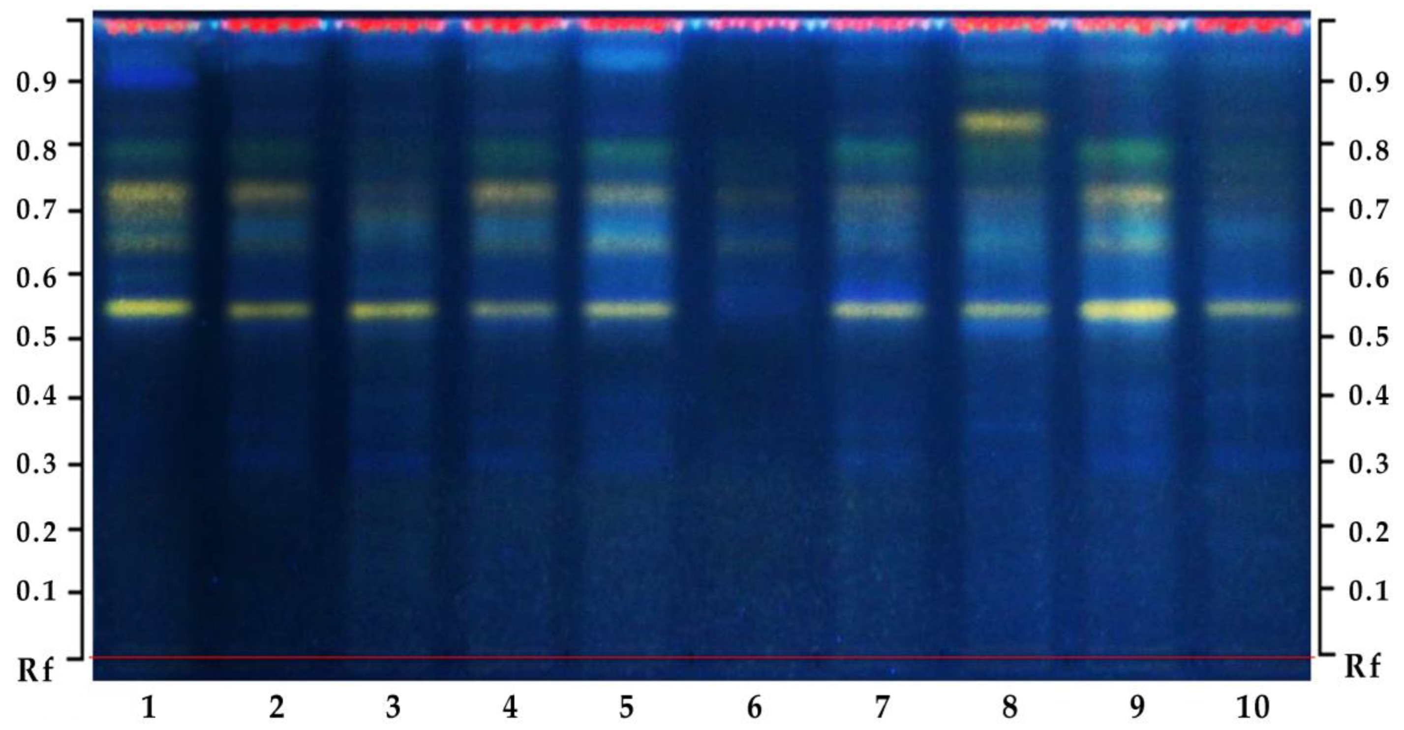
3.4.5. Biochemical Profiling of Plant Extracts with High-Performance Thin-Layer Chromatography

Biochemical profiling of oak leaf extracts was performed by the method of HPTLC on silica gel G60 plates (Merck Chemicals GmbH, Darmstadt, Germany). Separation of general phenolic compounds and flavonoids was performed in solvent systems: ethyl acetate—formic acid—acetic acid—water (v/v/v/v—100:11:11:25).
The standard (Quercetin, Rutin, Chlorogenic acid) solutions (3.0 µL of each concentration 1 mg·mL−1) were applied to the plates. The derivatization was performed with 0.5% NP reagent (1.0 g diphenylborinic acid aminoethyl ester dissolved in 200 mL ethyl acetate) and 1% PEG 400, followed by heating (5 min at 105 °C). Phenolic substances on the chromatogram were detected using UV light at 366 nm. The retention factor (Rf) of individual compounds was determined photodensitometrically using the software Sorbfil TLC ver. 2.3.0.2994 (JSC Sorbopolymer). The Rf value is equal to the distance traveled by the individual compound divided by the distance traveled by the mobile phase front (Appendix A, Figure A1 and Figure A2).

3.5. Field Testing of Bacteria on One-Year Seedlings of Q. robur

Field testing of bacteria was carried out on one-year seedlings of Q. robur. The plants were watered with the suspension of active isolates (Q2, Q7) and their mixture (Q2 + Q7) twice with a gap of three weeks. Additionally, after four weeks the leaves of annual oak plants were inoculated by endophytes and epiphytic and pathogenic bacteria in accordance with the scheme: a—control, b—Bacillus subtilis (Q2), c—Bacillus amyloliquefaciens (Q7), d—Q2/Q7 (mix 1:1), e—Pectobacterium spp., f—Pseudomonas spp., g—Lelliottia amnigena (Q5), h—Delftia acidovorans (Q6), i—epiphytic bacteria EpQ1, j—epiphytic bacteria EpQ2. Over the next months, observations of depigmentation of leaves and necrotic zones were observed.

3.6. Photo Documentation, Digital and Statistical Data Processing

Photo documentation and processing of digital images were performed with the specialized program Image-Pro Premier 9.0. The statistical significance of the differences between the values (p < 0.05) was determined by the analysis of the variance (ANOVA) method in the XLSTAT (Addinsoft Inc., New York, NY, USA, 2010). The data were compared using Tukey’s posterior test. The SigmaPlot 12.0 program was used for regression analysis.

4. Conclusions

Ten samples of endophytes were isolated from the tissues of immature acorn embryos of perennial trees Quercus robur, among which bacteria of the genus Bacillus were the dominant morphotypes. Based on the results of comparing the sequenced 16S rRNA genes (16S rDNA sequences) with the sequences deposited in GenBank, four types of endophytic bacteria were identified: Bacillus amyloliquefaciens, Bacillus subtilis, Delftia acidovorans, and Lelliottia amnigena. Endophytic dominants Bacillus amyloliquefaciens and Bacillus subtilis had different antagonistic activities in relation to phytopathogenic micromycetes that cause mycosis in oak plants. Bacillus amyloliquefaciens BSQ7-PSTQR-0920 showed high antifungal activity against phytopathogenic micromycetes Fusarium tricinctum, Botrytis cinerea, and Sclerotinia sclerotiorum using a modified method of microorganism antagonism analysis.
The isolation of endophytes from acorn tissues at the initial stages of their development and subsequent screening for pectinase activity made it possible to quickly obtain promising strains of potential PGP bacteria that are able to accelerate the regeneration of damaged tissues, have relatively high antifungal activity, and are tolerantly perceived by common oak seedlings.
Bacillus amyloliquefaciens and Bacillus subtilis bacteria inoculation was accompanied by a decrease in the total amount of polyphenols in the leaves of oak seedlings. Phytopathogenic bacteria Pectobacterium and Pseuodomonas, on the contrary, caused an increase in the concentration of polyphenols in plants by 2.0 and 2.2 times, respectively. An increase in the content of soluble phenols under the influence of biotic stress indicates the mobilization of the protective reserves of the plant organism; however, the index of the ratio of the antioxidants to phenols is an equally informative marker of the balance of the plant-microbial system. High values may indicate significant antioxidant potential of polyphenols in the leaves. The increased index of the ratio of antioxidant activity to the total content of phenols against the background of a decrease in the total pool of phenolic compounds in the leaves of oak seedlings, caused by inoculation with isolates of Bacillus amyloliquefaciens and Bacillus subtilis, may be a sign of the economic operation of the secondary synthesis system, which becomes possible under the conditions of a balanced plant-microbial system.
Therefore, the selection of natural endophytes of the PGPB class from the reproductive organs of certain plant species to create balanced plant-microbial systems within one genus or species has broad prospects, as it is based on the co-adaptation of a plant organism and a bacterial complex that has undergone long-term natural selection.

Author Contributions

Conceptualization, S.B., A.L. and V.B.; Methodology, A.L., V.B. and L.Z.; Software, A.L., L.Z. and O.S.; Analysis, S.B., A.L., V.B. and L.Z.; Investigation, S.B., A.L., V.B., L.Z. and O.S.; Resources, Y.M. and A.B.; Writing—original draft preparation, S.B., A.L., V.B. and L.Z.; Writing—review and editing, S.B., A.L., V.B., L.Z., Y.M., O.S. and A.B.; Visualization, S.B., A.L., V.B. and L.Z.; Supervision, A.L., Y.M. and A.B.; Project administration, A.L. and S.B.; Funding acquisition, A.L., S.B., Y.M. and A.B. All authors have read and agreed to the published version of the manuscript.

Funding

The research was carried out with the funds of the Ministry of Education and Culture of Ukraine: grant No. 110/540 (0117U004402) 2017–2019 “Biotechnological ways of preserving the gene pool of historically valuable centuries-old trees” (leader—S. Bilous); grant No. 37 (Basic funding) “Agrarian sciences and veterinary medicine” 2021 (leader—A. Likhanov); and grant No. 110/14-pr-2022 “Innovative solutions for measuring the quality of hardwood wood for forecasting and organizing the integrated use of forest resources” (leader—R. Vasylyshyn, co-leader—A. Likhanov, co-inventor—S. Bilous).

Data Availability Statement

Not Applicable.

Acknowledgments

The authors thank Maryna Kapustina at University of North Carolina at Chapel Hill; Alexander Goushcha, IOP United States; and Elijah Kelsey, United States for support.

Conflicts of Interest

The authors declare no conflict of interest. The funders had no role in the design of the study; in the collection, analyses, or interpretation of data; in the writing of the manuscript, or in the decision to publish the results.

Appendix A

Figure A1. Results of testing the fungistatic action of endophytic bacteria Bacillus subtilis (Q2) (i)—Bacillus amyloliquefaciens (Q7) in relation to: (ac)—Botrytis cinerea, (df)—Sclerotinia sclerotiorum, (gi)—Fusarium tricinctum.
Plants 12 01352 g0a1
Figure A2. Chromatogram of methanol extracts of common oak leaves after inoculation of endophytic, phytopathogenic and epiphytic bacteria: 1—control, 2—Bacillus subtilis (Q2), 3—Bacillus amyloliquefaciens (Q7), 4—Q2/Q7 (mix 1:1), 5—Pectobacterium spp., 6—Pseudomonas spp., 7—Lelliottia amnigena (Q5), 8—Delftia acidovorans (Q6), 9—EpQ1, 10—EpQ2.
Plants 12 01352 g0a2

References

  1. Bonan, G.B. Forests and climate change: Forcings, feedbacks, and the climate benefits of forests. Science 2008, 320, 1444–1449. [Google Scholar] [CrossRef] [PubMed] [Green Version]
  2. Shvidenko, A.; Buksha, I.; Krakovska, S.; Lakyda, P. Vulnerability of Ukrainian forests to climate change. Sustainability 2017, 9, 1152. [Google Scholar] [CrossRef] [Green Version]
  3. Seidl, R.; Thom, D.; Kautz, M.; Martin-Benito, D.; Peltoniemi, M.; Vacchiano, G.; Wild, J.; Ascoli, D.; Petr, M.; Honkaniemi, J.; et al. Forest disturbances under climate change. Nat. Clim. Chang. 2017, 7, 395–402. [Google Scholar] [CrossRef] [PubMed] [Green Version]
  4. Leach, J.E.; Triplett, L.R.; Argueso, S.T.; Trivedi, P. Communication in the phytobiome. Cell 2017, 169, 587–596. [Google Scholar] [CrossRef] [PubMed] [Green Version]
  5. Gupta, A.; Mishra, R.; Rai, S.; Bano, A.; Pathak, N.; Fujita, M.; Kumar, M.; Hasanuzzaman, M. Mechanistic Insights of Plant Growth Promoting Bacteria Mediated Drought and Salt Stress Tolerance in Plants for Sustainable Agriculture. Int. J. Mol. Sci. 2022, 23, 3741. [Google Scholar] [CrossRef]
  6. Smith, S.A.; Tank, D.C.; Boulanger, L.A.; Bascom-Slack, C.A.; Eisenman, K.; Kingery, D.; Babbs, B.; Fenn, K.; Greene, J.S.; Hann, B.D.; et al. Bioactive endophytes warrant intensified exploration and conservation. PLoS ONE 2008, 3, e3052. [Google Scholar] [CrossRef] [Green Version]
  7. Halecker, S.; Wennrich, J.-P.; Rodrigo, S.; Andrée, N.; Rabsch, L.; Baschien, C.; Steinert, M.; Stadler, M.; Surup, F.; Schulz, B. Fungal endophytes for biocontrol of ash dieback: The antagonistic potential of Hypoxylon rubiginosum. Fungal Ecol. 2020, 45, 100918. [Google Scholar] [CrossRef]
  8. Khosro, M.; Yousef, S. Bacterial Biofertilizers for Sustainable Crop Production: A Review. ARPN J. Agric. Biol. Sci. 2012, 7, 307–316. [Google Scholar]
  9. Witzell, J.; Martín, J.A.; Blumenstein, K. Ecological Aspects of Endophyte-Based Biocontrol of Forest Diseases. In Advances in Endophytic Research; Verma, V.C., Gange, A.C., Eds.; Springer: Berlin, Germany, 2014; pp. 321–333. [Google Scholar] [CrossRef]
  10. Lei, Z.; Xiao, P.S.; Jian, S.W.; Hai, Y. Endophytic bacterial effects on seeds germination and mobilization of reserves ammodendron biofolium yan. Pak. J. Bot. 2017, 49, 2029–2035. [Google Scholar]
  11. Cregger, M.A.; Carper, D.L.; Christel, S.; Doktycz, M.J.; Labbé, J.; Michener, J.K.; Dove, N.C.; Johnston, E.R.; Moore, J.A.M.; Vélez, J.M.; et al. Plant–Microbe Interactions: From Genes to Ecosystems Using Populus as a Model System. Phytobiomes 2021, 5, 29–38. [Google Scholar] [CrossRef]
  12. Suhag, M. Potential of Biofertilizers to Replace Chemical Fertilizers. Int. Adv. Res. J. Sci. Eng. Technol. 2016, 3, 163–167. [Google Scholar]
  13. Frank, A.C.; Saldierna Guzmán, J.P.; Shay, J.E. Transmission of bacterial endophytes. Microorganisms 2017, 10, 70. [Google Scholar] [CrossRef] [PubMed] [Green Version]
  14. Mohite, B. Isolation and characterization of indole acetic acid (IAA) producing bacteria from rhizospheric soil and its effect on plant growth. J. Soil Sci. Plant Nutr. 2013, 13, 638–649. [Google Scholar] [CrossRef]
  15. Karthik, M.; Pushpakanth, P.; Krishnamoorthy, R.; Senthilkumar, M. Endophytic bacteria associated with banana cultivars and their inoculation effect on plant growth. J. Hortic. Sci. Biotechnol. 2017, 92, 568–576. [Google Scholar] [CrossRef]
  16. Somal, M.K.; Karnwal, A. Effect of stress tolerance endophytic bacteria on the growth of Rheum emodi under abiotic stress. Plant Biol. 2022, 36000522. [Google Scholar] [CrossRef] [PubMed]
  17. Fort, T.; Pauvert, C.; Zanne, A.E.; Ovaskainen, O.; Caignard, T.; Barret, M.; Compant, S.; Hampe, A.; Delzon, S.; Vacher, C. Maternal effects shape the seed mycobiome in Quercus petraea. New Phytol. 2021, 230, 1594–1608. [Google Scholar] [CrossRef]
  18. Nguyen, M.H.; Yong, J.H.; Sung, H.J.; Lee, J.K. Screening of Endophytic Fungal Isolates Against Raffaelea quercus-mongolicae Causing Oak Wilt Disease in Korea. Mycobiology 2020, 48, 484–494. [Google Scholar] [CrossRef]
  19. Shestibratov, K.A.; Barano, O.Y.; Subbotina, N.M.; Lebedev, V.G.; Panteleev, S.V.; Krutovsky, K.V.; Padutov, V.E. Early Detection and Identification of the Main Fungal Pathogens for Resistance Evaluation of New Genotypes of Forest Trees. Forests 2018, 9, 732. [Google Scholar] [CrossRef] [Green Version]
  20. Yang, A.; Juzwik, J. Use of nested and real-time PCR for the detection of Ceratocystis fagacearum in the sapwood of diseased Oak species in Minnesota. Plant Dis. 2017, 101, 480–486. [Google Scholar] [CrossRef] [Green Version]
  21. Oliveira, A.L.M.; Urquiaga, S.; Döbereiner, J.; Baldani, J.I. The effect of inoculating endophytic N2-fixing bacteria on micropropagated sugarcane plants. Plant Soil 2002, 242, 205–215. [Google Scholar] [CrossRef]
  22. Iniguez, A.L.; Dong, Y.; Triplett, E.W. Nitrogen Fixation in Wheat Provided by Klebsiella pneumoniae 342. Mol. Plant-Microbe Interact. 2004, 17, 1078–1085. [Google Scholar] [CrossRef] [PubMed] [Green Version]
  23. Chalk, P.M.; Lam, S.K.; Chen, D. The significance of endophytic and phyllospheric N2 fixation in forest trees: Evi-dence from stable (15N) and radioactive (13N) tracer studies. Trees 2022, 36, 1179. [Google Scholar] [CrossRef]
  24. Sessitsch, A.N.; Hardoim, P.A.; Döring, J.; Weilharter, A.; Krause, A.N.; Woyke, T.A.; Mitter, B.; Hauberg-Lotte, L.; Friedrich, F.; Rahalkar, M.; et al. Functional characteristics of an endophyte community colonizing rice roots as revealed by metagenomic analysis. Mol. Plant-Microbe Interact. MPMI 2012, 25, 28–36. [Google Scholar] [CrossRef] [PubMed] [Green Version]
  25. Ambrose, C.; Varghese, C.; Subhash, J. Endophytic bacteria as a source of novel antibiotics: An overview. Pharmacogn. Rev. 2013, 7, 11–16. [Google Scholar]
  26. Hardoim, P.R.; van Overbeek, L.S.; Berg, G.; Pirttilä, A.M.; Compant, S.; Campisano, A.; Döring, M.; Sessitsch, A. The hidden world within plants: Ecological and evolutionary considerations for defining functioning of microbial endophytes. Microb. Mol. Biol. Rev. 2015, 79, 293–320. [Google Scholar] [CrossRef] [PubMed] [Green Version]
  27. Wani, Z.A.; Ashraf, N.; Mohiuddin, T.; Riyaz-Ul-Hassan, S. Plant-endophyte symbiosis, an ecological perspective. Appl. Microbiol. Biotechnol. 2015, 99, 2955–2965. [Google Scholar] [CrossRef] [PubMed]
  28. Pandey, S.S.; Singh, S.; Pandey, H.; Srivastava, M.; Ray, T.; Soni, S.; Pandey, A.; Shanker, K.; Babu, C.S.V.; Banerjee, S.; et al. Endophytes of Withania somnifera modulate in planta content and the site of withanolide biosynthesis. Sci. Rep. 2018, 8, 5450. [Google Scholar] [CrossRef]
  29. García-Fraile, P.; Menéndez, E.; Rivas, R. Role of bacterial biofertilizers in agriculture and forestry. AIMS Bioeng. 2015, 2, 183–205. [Google Scholar] [CrossRef]
  30. Díaz Herrera, S.; Grossi, C.; Zawoznik, M.; Groppa, M.D. Wheat seeds harbour bacterial endophytes with potential as plant growth promoters and biocontrol agents of Fusarium graminearum. Microbiol. Res. 2016, 186–187, 37–43. [Google Scholar] [CrossRef]
  31. Borodai, V.; Kolomiiets, Y.; Likhanov, A.; Zelena, L.; Butsenko, L.; Shemetun, K.; Churilov, A.; Blume, Y. The Growth-promoting and Antipathogenic Effects of Microorganisms Isolated from Solanum nigrum L. and Inoculated in Solanum lycopersicum L. Open Agric. J. 2022, 16, e2208180. [Google Scholar] [CrossRef]
  32. Shahzad, R.; Waqas, M.; Khan, A.; Al-Hosni, K.; Kang, S.; Seo, C.; Lee, I. Indoleacetic acid production and plant growth promoting potential of bacterial endophytes isolated from rice (Oryza sativa L.) seeds. Acta Biol. Hung. 2017, 68, 175–186. [Google Scholar] [CrossRef] [PubMed] [Green Version]
  33. Orozco-Mosqueda, M.D.C.; Rocha-Granados, M.D.C.; Glick, B.R.; Santoyo, G. Microbiome engineering to improve biocontrol and plant growth-promoting mechanisms. Microbiological Res. 2018, 208, 25–31. [Google Scholar] [CrossRef]
  34. Rabiey, M.; Hailey, L.E.; Roy, S.R.; Grenz, K.; Al-Zadjali, M.A.; Barrett, G.A.; Jackson, R.W. Endophytes vs tree pathogens and pests: Can they be used as biological control agents to improve tree health? Eur. J. Plant Pathol. 2019, 155, 711–729. [Google Scholar] [CrossRef] [Green Version]
  35. Christian, N.; Herre, E.A.; Mejia, L.C.; Clay, K. Exposure to the leaf litter microbiome of healthy adults protects seedlings from pathogen damage. Proc. R. Soc. B Biol. Sci. 2017, 284, 20170641. [Google Scholar] [CrossRef] [PubMed] [Green Version]
  36. Sarmiento, C.; Zalamea, P.; Dalling, J.; Davis, A.; Stump, S.; U’Ren, J.; Arnold, A. Soilborne fungi have host affinity and host-specific effects on seed germination and survival in a lowland tropical forest. Proc. Natl. Acad. Sci. USA 2017, 114, 11458–11463. [Google Scholar] [CrossRef] [Green Version]
  37. Ridout, M.E.; Schroeder, K.L.; Hunter, S.S.; Styer, J.; Newcombe, G. Priority effects of wheat seed endophytes on a rhizosphere symbiosis. Symbiosis 2019, 78, 19–31. [Google Scholar] [CrossRef]
  38. Uroz, S.; Buee, M.; Murat, C.; Frey-Klett, P.; Martin, F. Pyrosequencing reveals a contrasted bacterial diversity between oak rhizosphere and surrounding soil. Environ. Microbiol. 2010, 2, 281–288. [Google Scholar] [CrossRef] [PubMed]
  39. Kim, M.; Singh, D.; Lai-Hoe, A.; Go, R.; Abdul Rahim, R.; Ainuddin, A.N. Distinctive phyllosphere bacterial communities in tropical trees. Microb. Ecol. 2012, 63, 674–681. [Google Scholar] [CrossRef]
  40. Tanaka, A.; Tapper, B.A.; Popay, A.; Parker, E.J.; Scott, B. A symbiosis expressed non-ribosomal peptide synthetase from a mutualistic fungal endophyte of perennial ryegrass confers protection to the symbiotum from insect herbivory. Mol. Microbiol. 2005, 57, 1036–1050. [Google Scholar] [CrossRef]
  41. Vega, F.E.; Posada, F.; Aime, M.C.; Pava-Ripoll, M.; Infante, F.; Rehner, S.A. Entomopathogenic fungal endophytes. Biol. Control 2008, 46, 72–82. [Google Scholar] [CrossRef] [Green Version]
  42. Lugtenberg, B.J.J.; Caradus, J.R.; Johnson, L.J. Fungal endophytes for sustainable crop production. FEMS Microbiol. Ecol. 2016, 92, 194. [Google Scholar] [CrossRef] [PubMed]
  43. Lata, R.; Chowdhury, S.; Gond, S.K.; White, J.F., Jr. Induction of abiotic stress tolerance in plants by endophytic microbes. Lett. Appl. Microbiol. 2018, 66, 268–276. [Google Scholar] [CrossRef] [PubMed] [Green Version]
  44. Lasa, A.V.; Maslinova, T.; Baldrian, P.; Fernandez-Lopez, M. Bacteria from the endosphere and rhizosphere of Quercus spp. use mainly cell wall-associated enzymes to decompose organic matter. PLoS ONE 2019, 14, e0214422. [Google Scholar] [CrossRef] [PubMed] [Green Version]
  45. Matsala, M.; Bilous, A.; Feshchenko, R.; Matiashuk, R.; Bilous, S.; Kovbasa, Y. Spatial and compositional structure of European oak urban forests in Kyiv city, Ukraine. J. For. Sci. Link Is Disabl. 2021, 67, 143–153. [Google Scholar] [CrossRef]
  46. Borruso, L.; Wellstein, C.; Bani, A.; Casagrande Bacchiocchi, S.; Margoni, A.; Tonin, R.; Zerbe, S.; Brusetti, L. Temporal shifts in endophyte bacterial community composition of sessile oak (Quercus petraea) are linked to foliar nitrogen, stomatal length, and herbivory. PeerJ 2018, 6, e5769. [Google Scholar] [CrossRef] [PubMed] [Green Version]
  47. Meaden, S.; Metcalf, C.J.E.; Koskella, B. The effects of host age and spatial location on bacterial community composition in the English oak tree (Quercus robur). Environ. Microbiol. Rep. 2016, 8, 649–658. [Google Scholar] [CrossRef] [Green Version]
  48. Pirttilä, A.M. Endophytic Bacteria in Tree Shoot Tissues and Their Effects on Host. In Endophytes of Forest Trees; Forestry Sciences; Pirttilä, A., Frank, A., Eds.; Springer: Berlin/Heidelberg, Germany, 2018; Volume 86. [Google Scholar]
  49. Rosenblueth, M.; Martínez-Romero, E. Bacterial endophytes and their interactions with hosts. Mol. Plant-Microbe Interact. MPMI 2006, 19, 827–837. [Google Scholar] [CrossRef] [Green Version]
  50. Nguyen, M.H.; Shin, K.C.; Lee, J.K. Fungal Community Analyses of Endophytic Fungi from Two Oak Species, Quercus mongolica and Quercus serrata, in Korea. Mycobiology 2021, 9, 385–395. [Google Scholar] [CrossRef]
  51. Novotný, D. Contribution to the endophytic mycobiota of aerial parts of oaks. Czech Mycol. 2022, 74, 111–121. [Google Scholar] [CrossRef]
  52. Sapp, M.; Lewis, E.; Moss, S.; Barrett, B.; Kirk, S.; Elphinstone, J.G.; Denman, S. Metabarcoding of Bacteria Associated with the Acute Oak Decline Syndrome in England. Forests 2016, 7, 95. [Google Scholar] [CrossRef] [Green Version]
  53. Brader, G.; Compant, S.; Mitter, B.; Trognitz, F.; Sessitsch, A. Metabolic potential of endophytic bacteria. Curr. Opin. Biotechnol. 2014, 27, 30–37. [Google Scholar] [CrossRef] [PubMed] [Green Version]
  54. Griffin, J.S.; Lu, N.; Sangwan, N.; Li, A.; Dsouza, M.; Stumpf, A.J.; Sevilla, T.; Culotti, A.; Keefer, L.L.; Kelly, J.J.; et al. Microbial diversity in an intensively managed landscape is structured by landscape connectivity. FEMS Microbiol. Ecol. 2017, 93, fix120. [Google Scholar] [CrossRef] [PubMed]
  55. Tashi-Oshnoei, F.; Harighi, B.; Abdollahzadeh, J. Isolation and identification of endophytic bacteria with plant growth promoting and biocontrol potential from oak trees. For. Pathol. 2017, 47, e12360. [Google Scholar] [CrossRef]
  56. Petrini, O.; Fisher, P.J. Occurrence of fungal endophyticin twigs of Salix fragilis and Quercus robur. Mycol. Res. 1990, 94, 1077–1080. [Google Scholar] [CrossRef]
  57. Kowalski, T.; Kehr, R.D. Fungal endophytes of living branch bases in several European tree species. In Endophytic Fungi in Grasses and Woody Plants, Systematics, Ecology and Evolution; Redlin, S.C., Carris, L.M., Eds.; APS: St. Paul, MN, USA, 1996; pp. 67–86. [Google Scholar]
  58. Gennaro, M.; Gonthier, P.; Nicolotti, G. Fungal endophytic communities in healthy and declining Quercus robur L. and Q. cerris L. trees in Northern Italy. J. Phytopathol. 2003, 151, 529–534. [Google Scholar] [CrossRef]
  59. Ragazzi, A.; Moricca, S.; Capretti, P.; Dellavalle, I.; Turco, E. Differences in composition of endophytic mycobiota in twigs and leaves of healthy and declining Quercus species in Italy. For. Path. 2003, 33, 31–38. [Google Scholar] [CrossRef]
  60. Fisher, P.J.; Petrini, O.; Petrini, L.E.; Sutton, B.C. Fungal endophytes from the leaves and twigs of Quercus ilex L. from England, Majorca and Switzerland. New Phytol. 1994, 127, 133–137. [Google Scholar] [CrossRef] [PubMed]
  61. Collado, J.; Platas, G.; Gonzales, I.; Peláez, F. Geographical and seasonal influences on the distribution of fungal endophytes in Quercus ilex. New Phytol. 1999, 144, 525–532. [Google Scholar] [CrossRef]
  62. Moricca, S.; Ragazzi, A. Species- and organ-specificity in endophytes colonizing healthy and declining Mediterranean oaks. Phytopathol. Mediterr. 2012, 51, 587–598. [Google Scholar]
  63. Linaldeddu, B.T.; Sirca, C.; Spano, D.; Franceschini, A. Variation of endophytic cork oak-associated fungal communities in relation to plant health and water stress. For. Pathol. 2011, 41, 193–201. [Google Scholar] [CrossRef] [Green Version]
  64. Costa, D.; Tavares, R.; Baptista, P.; Lino-Neto, T. Diversity of fungal endophytic community in Quercus suber L. under different climate scenarios. Rev. Cienc. Agrar. 2019, 41, 22–30. [Google Scholar]
  65. Ghasemi, S.; Khodaei, S.; Karimi, K.; Tavakoli, M.; Pertot, I.; Arzanlou, M. Biodiversity study of endophytic fungi associated with two Quercus species in Iran. For. Syst. 2019, 28, 12. [Google Scholar] [CrossRef]
  66. Moricca, S.; Ragazzi, A. Fungal endophytes in Mediterranean oak forests: A lesson from Discula quercina. Phytopathology 2008, 98, 380–386. [Google Scholar] [CrossRef] [PubMed] [Green Version]
  67. Terhonen, E.; Blumenstein, K.; Kovalchuk, A.; Asiegbu, F.O. Forest Tree Microbiomes and Associated Fungal Endophytes: Functional Roles and Impact on Forest Health. Forests 2019, 10, 42. [Google Scholar] [CrossRef] [Green Version]
  68. Gupta, G.; Parihar, S.S.; Ahirwar, N.K.; Snehi, S.K.; Singh, V. Plant Growth Promoting Rhizobacteria (PGPR): Current and Future Prospects for Development of Sustainable Agriculture. Microb. Biochem. Technol. 2015, 7, 96–102. [Google Scholar]
  69. Santoyo, G.; Moreno-Hagelsieb, G.; Orozco-Mosqueda Mdel, C.; Glick, B.R. Plant growth-promoting bacterial endophytes. Microbiol. Res. 2016, 183, 92–99. [Google Scholar] [CrossRef]
  70. Fernández-González, A.J.; Martínez-Hidalgo, P.; Cobo-Díaz, J.F.; Villadas, P.J.; Martínez-Molina, E.; Toro, N.; Tringe, S.G.; Fernández-López, M. The rhizosphere microbiome of burned holm-oak: Potential role of the genus Arthrobacter in the recovery of burned soils. Sci. Rep. 2017, 7, 6008. [Google Scholar] [CrossRef]
  71. Chaudhary, P.; Agri, U.; Chaudhary, A.; Kumar, A.; Kumar, G. Endophytes and their potential in biotic stress management and crop production. Front. Microbiol. 2022, 13, 933017. [Google Scholar] [CrossRef]
  72. Gathercole, L.A.P.; Nocchi, G.; Brown, N.; Coker, T.L.R.; Plumb, W.J.; Stocks, J.J.; Nichols, R.A.; Denman, S.; Buggs, R.J.A. Evidence for the Widespread Occurrence of Bacteria Implicated in Acute Oak Decline from Incidental Genetic Sampling. Forests 2021, 12, 1683. [Google Scholar] [CrossRef]
  73. Abdelfattah, A.; Wisniewski, M.; Schena, L.; Tack, A.J.M. Experimental evidence of microbial inheritance in plants and transmission routes from seed to phyllosphere and root. Environ. Microbiol. 2021, 23, 2199–2214. [Google Scholar] [CrossRef]
  74. Martínez-Arias, C.; Sobrino-Plata, J.; Medel, D.; Gil, L.; Martín, J.A.; Rodríguez-Calcerrada, J. Stem endophytes increase root development, photosynthesis, and survival of elm plantlets (Ulmus minor Mill.). J. Plant Physiol. 2021, 261, 153420. [Google Scholar] [CrossRef] [PubMed]
  75. Vujanovic, V.; Islam, M.N.; Daida, P. Transgenerational role of seed mycobiomean endosymbiotic fungal composition as a prerequisite to stress resilience and adaptive phenotypes in Triticum. Sci. Rep. 2019, 9, 18483. [Google Scholar] [CrossRef] [PubMed] [Green Version]
  76. Ahlholm, J.U.; Helander, M.; Henriksson, J.; Metzler, M.; Saikkonen, K. Environmental conditions and host genotype direct genetic diversity of Venturia ditricha, a fungal endophyte of birch trees. Evolution 2002, 56, 1566–1573. [Google Scholar] [PubMed]
  77. Khare, E.; Mishra, J.; Kumar Arora, N. Multifaceted Interactions Between Endophytes and Plant: Developments and Prospects. J. Front. Microbiol. 2018, 9, 2732. [Google Scholar] [CrossRef]
  78. Shao, J.; Li, S.; Zhang, N.; Cui, X.; Zhou, X.; Zhang, G.; Shen, Q.; Zhang, R. Analysis and cloning of the synthetic pathway of the phytohormone indole-3-acetic acid in the plant-beneficial Bacillus amyloliquefaciens SQR9. Microb. Cell Fact. 2015, 4, 130. [Google Scholar] [CrossRef] [Green Version]
  79. Keswani, C.; Singh, S.P.; Cueto, L.; García-Estrada, C.; Mezaache-Aichour, S.; Glare, T.R.; Borriss, R.; Singh, S.P.; Blázquez, M.A.; Sansinenea, E. Auxins of microbial origin and their use in agriculture. Appl. Microbiol. Biotechnol. 2020, 104, 8549–8565. [Google Scholar] [CrossRef] [PubMed]
  80. Vaitiekunaite, D.; Kuusiene, S.; Beniušyte, E. Oak (Quercus robur) Associated Endophytic Paenibacillus sp. Promotes Poplar (Populus spp.) Root Growth In Vitro. Microorganisms 2021, 9, 1151. [Google Scholar] [CrossRef]
  81. Brooks, D.S.; Gonzales, C.F.; Appel, D.N.; Filer, T.H. Evaluation of endophytic bacteria as potential biological control agents for oak wilt. Biol. Control 1994, 4, 373–381. [Google Scholar] [CrossRef]
  82. Chandrasekaran, M.; Chun, S.C. Expression of PR-protein genes and induction of defense-related enzymes by Bacillus subtilis CBR05 in tomato (Solanum lycopersicum) plants challenged with Erwinia carotovora sub sp. carotovora. Biosci. Biotechnol. Biochem. 2016, 80, 2277–2283. [Google Scholar] [CrossRef] [Green Version]
  83. Heidari, M.; Golpayegani, A. Effects of water stress and inoculation with plant growth promoting rhizobacteria (PGPR) on antioxidant status and photosynthetic pigments in basil (Ocimum basilicum L.). J. Saudi Soc. Agric. Sci. 2012, 11, 57–61. [Google Scholar] [CrossRef] [Green Version]
  84. Gusain, Y.S.; Singh, U.S.; Sharma, A.K. Bacterial mediated amelioration of drought stress in drought tolerant and susceptible cultivars of rice (Oryza sativa L.). Afr. J. Biotechnol. 2015, 14, 764–773. [Google Scholar]
  85. Verma, S.K.; White, J.F. Seed Endophytes: Biology and Biotechnology; Springer: Berlin/Heidelberg, Germany, 2019. [Google Scholar]
  86. Vasilchenko, A.S.; Poshvina, D.V.; Sidorov, R.Y.; Iashnikov, A.V.; Rogozhin, E.A.; Vasilchenko, A.V. Oak bark (Quercus sp. cortex) protects plants through the inhibition of quorum sensing mediated virulence of Pectobacterium carotovorum. World J. Microbiol. Biotechnol. 2022, 38, 184. [Google Scholar] [CrossRef] [PubMed]
  87. Liu, L.; Wen, W.; Zhang, R.; Wei, Z.; Deng, Y.; Xiao, J.; Zhang, M. Complex enzyme hydrolysis releases antioxidative phenolics from rice bran. Food Chem. 2016, 214, 1–8. [Google Scholar] [CrossRef] [PubMed]
  88. Likhanov, A.F.; Sereda, O.V.; Gryb, V.M.; Melnyk, V.I.; Osadchuk, L.S.; Yuskevych, T. Biochemical markers of vital biodestruction in common oak (Quercus robur). Biosyst. Divers. 2019, 27, 314–321. [Google Scholar] [CrossRef]
  89. Deng, Y.; Lu, S. Biosynthesis and Regulation of Phenylpropanoids in Plants. Crit. Rev. Plant Sci. 2017, 36, 257–290. [Google Scholar] [CrossRef]
  90. Carroll, G.C. Fungal endophytes in stems and leaves: From latent pathogen to mutualistic symbiont. Ecology 1988, 969, 2. [Google Scholar] [CrossRef]
  91. Carroll, G.C. Fungal associates of woody plants as insect antagonists in leaves and stems. In Microbial Mediation of Plant-Herbivore Interactions; Barbosa, P., Krischik, V.A., Jones, C.G., Eds.; John Wiley & Sons, Inc.: Hoboken, NJ, USA, 1991; pp. 253–271. [Google Scholar]
  92. Fontana, D.C.; Paula, S.; Torres, A.G.; Souza, V.; Pascholati, S.F.; Schmidt, D.; Dourado Neto, D. Endophytic Fungi: Biological Control and Induced Resistance to Phytopathogens and Abiotic Stresses. Pathogens 2021, 10, 570. [Google Scholar] [CrossRef]
  93. Radhakrishnan, R.; Hashem, A.; Abd Allah, E.F. Bacillus: A Biological Tool for Crop Improvement through Bio-Molecular Changes in Adverse Environments. Front. Physiol. 2017, 8, 667. [Google Scholar] [CrossRef] [Green Version]
  94. Bilous, S.; Prysiazhniuk, L.; Chernii, S.; Melnyk, S.; Marchuk, Y.; Likhanov, A. Genetic characterisation of centuries-old oak and linden trees using SSR markers. Folia For. Pol. 2022, 64, 58–68. [Google Scholar] [CrossRef]
  95. Borkar, S.G. Laboratory Techniques in Plant Bacteriology, 1st ed.; CRC Press: Boca Raton, FL, USA, 2017. [Google Scholar]
  96. Patyka, V.P.; Pasichnyk, L.A.; Gvozdyak, R.I. Phytopathogenic bacteria. Res. Methods 2017, 2, 432. [Google Scholar]
  97. Don, J.; Brenner, N.R.; Krieg, J.T.; Staley, G.M.; Garrity, D.R.; Boone, P.V.; Goodfellow, M.; Rainey, F.A.; Karl-Heinz, S. Bergey’s Manual of Systematic Bacteriology, 2nd ed.; Biomedical and Life Sciences; Springer: New York, NY, USA, 2005; Volume 4, p. 976. [Google Scholar] [CrossRef]
  98. Maheshwari, R. Fungi: Experimental Methods in Biology, 2nd ed.; CRC Press: Boca Raton, FL, USA, 2011. [Google Scholar]
  99. Tamura, K.; Stecher, G.; Kumar, S. MEGA11: Molecular Evolutionary Genetics Analysis version 11. Mol. Biol. Evol. 2021, 38, 3022–3027. [Google Scholar] [CrossRef] [PubMed]
  100. Singleton, V.L.; Orthofer, R.; Lamuela-Raventos, R. Analysis of Total Phenols and Other Oxidation Substrates and Antioxidants by Means of Folin-Ciocalteu Reagent. Methods Enzymol. 1999, 299, 152–178. [Google Scholar]
  101. Cimpoiu, C.; Cristea, M.; Hosu, A.; Sandru, M.; Seserman, L. Antioxidant activity prediction and classification of some teas using neutral networks. Food Chem. 2011, 127, 1323–1328. [Google Scholar] [CrossRef] [PubMed]
  102. Brand-Williams, W.; Cuvelier, M.E.; Berset, C.L.W.T. Use of a Free Radical Method to Evaluate Antioxidant Activity. LWT-Food Sci. Technol. 1995, 28, 25–30. [Google Scholar] [CrossRef]
Figure 1. The dendrogram of the genetic similarity between strain BAQ7-PSTQR-0920 and various representatives of the Bacillus genus constructed on the basis of 16S rRNA gene sequences using the Neighbor-Joining method and Kimura’s 2-parameter model. The scale bar indicates 0.005 substitutions per nucleotide position. The analyzed strain is indicated by *.
Plants 12 01352 g001
Figure 2. The phylogenetic relationships between different bacilli based on the 16S rDNA sequences. The dendrogram was built using the Neighbor-Joining method and Kimura’s 2-parameter model. The scale bar indicates 0.005 substitutions per nucleotide position. The analyzed strain is indicated by *.
Plants 12 01352 g002
Figure 3. 16S rRNA gene sequence-based dendrograms defining the phylogenetic position of the isolated bacterial strains DQ6-BEQR-0929 (a) and LQ5-BEQR-0928 (b) and their relationship with other close relative microorganisms. The isolated strains are indicated *.
Plants 12 01352 g003
Figure 4. Inoculated leaves of annual oak plants by endophytes, epiphytic and pathogenic: (a)—control, (b)—Pectobacterium spp., (c)—Pseudomonas spp., (d)—Bacillus subtilis (Q2), (e)—Bacillus amyloliquefaciens (Q7), (f)—Q2/Q7 (mix 1:1), (g)—Lelliottia amnigena (Q5), (h)—Delftia acidovorans (Q6), (i)—EpQ1, (j)—EpQ2.
Plants 12 01352 g004
Figure 5. The degree of damage to the leaf surfaces of one-year-old oak seedlings one month after inoculation with bacterial isolates; the data were compared using Tukey’s test (HSD) by one-way ANOVA: *—significant differences to the Control at the level p < 0.05; **—at the level p < 0.01.
Plants 12 01352 g005
Figure 6. Principle component analysis by phytochemical profiles of phenolic compounds in common oak leaves after their treatment with endophytic bacteria.
Plants 12 01352 g006
Figure 7. Scheme for evaluating the antifungal activity of endophytic bacteria based on indicators of linear growth of micromycete colonies: EI—endophytic isolate; GIZ—growth inhibition zone; Mcr—micromycete; R1–R4—micromycete colony radii.
Plants 12 01352 g007
Table 2. Effect of endophytic isolate on linear growth parameters (mm) of micromycetes (x ± SE, n = 4).
PhytopathogensEIR1R2R3R4R1/RavGIZ
BotrytisQ220.3 ± 0.5817.3 ± 1.0016.3 ± 0.84 a17.7 ± 0.411.2 ± 0.081.3 ± 0.11
cinereaQ722.2 ± 0.9513.1 ± 0.19 b11.0 ± 0.25 b11.6 ± 0.32 b1.9 ± 0.108.0 ± 0.46
BCIBL-2143Contr8.8 ± 0.609.0 ± 0.658.87 ± 0.468.8 ± 0.201.0 ± 0.04-
SclerotiniaQ223.5 ± 1.1015.8 ± 0.20 b13.4 ± 0.78 b13.2 ± 1.03 b1.7 ± 0.111.5 ± 0.21
sclerotiorumQ722.7 ± 1.7916.6 ± 0.37 b14.7 ± 0.47 b13.8 ± 0.24 b1.5 ± 0.124.0 ± 0.26
SIBL-2135Contr20.5 ± 1.6321.3 ± 0.8618.4 ± 1.9318.9 ± 1.221.1 ± 0.05-
FusariumQ219.5 ± 0.4713.6 ± 0.53 a12.9 ± 0.61 a12.7 ± 0.56 b1.5 ± 0.085.1 ± 0.48
tricinctumQ718.2 ± 1.237.2 ± 0.39 b7.4 ± 0.24 b7.4 ± 0.40 b2.5 ± 0.1613.2 ± 0.36
FTIBL-2151Contr20.6 ± 0.8520.0 ± 0.4919.7 ± 0.2019.4 ± 0.711.0 ± 0.03-
Note: R1–R4—radii (mm); Rav = (R2 + R3 + R4)/3; GIZ—growth inhibition zone (mm); the data were compared using Tukey’s test (HSD) by one-way ANOVA: a—significant differences to the R1 at the level p < 0.05; b—at the level p < 0.01.
Table 3. The content of phenolic compounds in the leaves of common oak plants after inoculation with endophytic bacteria (x ± SE, n = 3).
SamplesPhenols, mg·g−1Flavonoids, mg·g−1Antioxidants, μM-eqAAi/Phi *
SamplesExperimSamplesExperimSamplesExperimExperim
Control101.3 ± 1.51125.7 ± 3.843.8 ± 0.142.0 ± 0.08 196.0 ± 7.39187.1 ± 5.721.00 ± 0.01
Q298.9 ± 3.02102.0 ± 3.402.7 ± 0.10 b1.8 ± 0.06 b210.5 ± 7.94242.1 ± 7.401.59 ± 0.01 b
Q784.3 ± 2.7877.8 ± 2.591.9 ± 0.07 b1.7 ± 0.06 b174.5 ± 6.58164.8 ± 5.491.44 ± 0.01 b
Q2/Q7 (mix 1:1)143.6 ± 4.74 b104.4 ± 3.482.3 ± 0.07 b1.9 ± 0.06 b252.2 ± 9.51 a232.6 ± 7.751.51 ± 0.01 b
Pectobacterium140.5 ± 4.64 b157.3 ± 5.24 b2.4 ± 0.09 b2.3 ± 0.08 b259.6 ± 7.94 b254.1 ± 8.47 b1.10 ± 0.01 a
Pseuodomonas150.2 ± 4.96 b175.9 ± 5.86 b2.3 ± 0.07 b1.7 ± 0.06 b257.7 ± 9.72 b256.5 ± 8.55 b0.99 ± 0.01
Q5173.0 ± 5.71 b194.3 ± 6.48 b0.4 ± 0.02 b0.3 ± 0.01257.5 ± 7.87 b246.3 ± 8.21 a0.86 ± 0.01 b
Q6118.8 ± 3.9292.7 ± 3.093.2 ± 0.12 a2.2 ± 0.07 b201.4 ± 7.60163.9 ± 5.461.20 ± 0.01 b
EpQ1143.0 ± 4.72 b158.3 ± 5.28 b3.4 ± 0.13 b3.4 ± 0.11 b242.6 ± 9.15238.6 ± 7.951.02 ± 0.01
EpQ2165.9 ± 5.47 b173.6 ± 5.79 b2.5 ± 0.08 b2.4 ± 0.08 b245.7 ± 7.51247.5 ± 9.33 a0.96 ± 0.01
*—the ratio in the experiment of the amount of antioxidants (AAi) to the amount of total phenols (Phi); the data were compared using Tukey’s test (HSD) by one-way ANOVA: a—significant differences to the Control at the level p < 0.05; b—at the level p < 0.01.
Disclaimer/Publisher’s Note: The statements, opinions and data contained in all publications are solely those of the individual author(s) and contributor(s) and not of MDPI and/or the editor(s). MDPI and/or the editor(s) disclaim responsibility for any injury to people or property resulting from any ideas, methods, instructions or products referred to in the content.

