Article

Detection Papers with Metal Complexes with Triphenylmethane Dyes for the Detection of G-Series Nerve Agents (Sarin, Soman, Cyclosarin) in the Liquid Phase

by
Martin Lobotka
1,*,
Vladimír Pitschmann
1,2 and
Lukáš Matějovský
3
1
Oritest spol. s r.o., Nábřežní 90/4, 150 00 Prague, Czech Republic
2
Faculty of Biomedical Engineering, Czech Technical University in Prague, náměstí Sítná 3105, 272 01 Kladno, Czech Republic
3
Faculty of Environmental Technology, University of Chemistry and Technology, Technická 5, 166 28 Prague, Czech Republic
*
Author to whom correspondence should be addressed.
Submission received: 11 October 2019 / Revised: 20 November 2019 / Accepted: 25 November 2019 / Published: 27 November 2019

Abstract

:
The paper presents the results of the study of the possibilities of using color metal complexes to detect the presence of chemical warfare agents (CWA) in liquid or aerosol form. Aluminon/Fe3+ and Eriochrome Cyanine R/Cu2+ coordination complexes and their ability to detect CWA in liquid phase are discussed. Detection systems have been demonstrated on instances of simple detection papers exposed to drops of real CWAs. Detection papers showed a positive response to G-series nerve agents and vesicant lewisite. Other liquid CWA do not interfere and the systems are also resistant to common organic solvents and a wide range of industrial chemicals.

Graphical Abstract

1. Introduction

The most important group of chemical warfare agents (CWA) are nerve agents whose toxic effect is based on the inhibition (blockade) of the acetylcholinesterase. This enzyme is involved in the transmission of nerve excitation using the neurotransmitter acetylcholine [1]. Chemically, there are several groups of organophosphorus compounds; the most well-known are alkyl/cycloalkyl-methylphosphonofluoridates (sarin GB, soman GD, cyclosarin GF, all with P-F bond) and ethyl-(dimethylphosphoroamido) cyanidate (tabun GA, with P-CN bond).
Although the so-called G-series nerve agents do not reach the toxicity of V-series nerve agents (VX, RVX, with P-S bond), their easier preparation and physical and chemical properties increase the risk of abuse for terrorist and criminal targets [2,3]. The basic measure of protection against them is fast, high-quality, and widely available detection. Main attention is paid to their vapor detection, because vapors pose a high risk of inhalation poisoning, especially by the volatile GB (boiling point 150 °C). Much less attention is paid to their detection in the form of droplets and aerosols, which can also cause severe percutaneous poisoning; particularly dangerous are the semi-persistent agents GD and GF (boiling point 198 °C, resp. 228 °C). The simplest yet highly effective method of detection of CWA droplets and aerosols is based on the use of detection papers by visual evaluation (the naked eye) of color changes. For example, 3-WAY detection paper is currently used worldwide to detect and differentiate vesicant mustard gas (HD) and G-series and V-series nerve agents. Color changes are due to the solubility of specific dyes in liquid CWA; in the case of G-series nerve agents it is disperse yellow (CAS: 6250-23-3) providing a yellow color. The disadvantage is its very low selectivity as many organic solvents and other industrial chemicals give the same or similar color [4,5].
It is worth mentioning that CWA detection can be also realized by advanced instrumental methods, for example, Raman spectroscopy [6,7]. However, these analytical methods do not compete with inexpensive simple detection papers, which, in addition, enables to detect primary droplet and aerosols, characteristic for chemical attack or other (emergency) leak of liquid CWAs to the environment. Raman spectroscopy and other instrumental methods are suitable for subsequent control of the presence of CWA and its identification (usually after necessary sample treatment).
The strategy for further development of detection papers enabling the detection of G-series nerve agents can be based on the application of various chemosensors that have been developed in a number of workplaces around the world in recent years [8,9,10,11]. A special group of these chemosensors are color metal complexes with some organic ligands that decompose and discolor by the action of organophosphorus CWA [9,12,13,14,15,16,17,18]. In the past, the study of these complexes was mainly focused on detection in the gas phase, but they can also be very useful for solving problems of detection of droplets and aerosols.
The aim of this paper is to report the results of research into the possibility of preparing detection papers for liquid G-series nerve agents, which are based on color complexes of metal cations with triphenylmethane dyes, in this case Aluminon and Eriochrome Cyanine R. The complexes of these dyes are well known in analytical chemistry [19,20], but their use for detecting highly toxic organophosphorus substances is new and appears to be promising.

2. Experimental Section

2.1. Chemicals and Materials

Ferric chloride hexahydrate, copper(II) chloride, Aluminon (C.I. 43810, ALU), Eriochrome Cyanine R (C.I. 43820, ECR), glycerol (all Sigma-Aldrich, St. Louis, MO, USA), and distilled water were used to prepare detection papers.
To check the functionality of the detection papers, these CWA were used: Tabun (GA), sarin (GB), soman (GD), VX agent, sulphur mustard (HD), nitrogen mustards (HN-1 and HN-2), lewisite (L), lewisite analogs (L-2, L-3), diphosgene (DP), all Military Research Institute, Brno, Czech Republic), and simulation agent (mimic) diethyl chlorophosphate (DCP, Sigma-Aldrich).
White cellulose cardboard paper with a nominal basis weight of 300 g/m2 and a thickness of 1 mm and ashless filter paper for quantitative analysis Whatman 589/3 were used as a carrier of the color complex (Whatman, Kent, UK).
An Accumet AB-150 pH meter with a combined electrode (Fischer Scientific, Pardubice, Czech Republic) were used for the pH measurement. An LMG 173 portable tristimulus colorimeter was used to assess the stability of the detection paper (Dr. Lange, Dusseldorf, Germany). Spectrophotometric measurement (absorption spectra) were performed using a spectrophotometer Aquamate (Thermo Spectronic, Cambridge, UK).

2.2. Preparation of Detection Papers

Based on the evaluation of the pilot tests, detection papers based on two complexes were prepared for further research: ALU/Fe3+ and ECR/Cu2+. The 100 × 50 mm filter paper blocks were immersed in 100 mL of the impregnation solution containing:
1
0.05% of ALU, 0.02% of ferric chloride hexahydrate, and 10% of glycerol in distilled water,
2
0.15% of ECR, 0.5% of copper(II) chloride, and 10% of glycerol in distilled water.
After impregnation the carrier with the solution of the complexes, the paper was dried at 20 °C for 24 h.

2.3. Functionality Testing

Droplets of test substances (CWA, simulants, interfering chemicals) with a volume of 10 µL were dosed on the detection paper by a micropipette and changes in its color were observed visually (by the naked eye). The evaluation was performed within 2 min by default. Each test was repeated 3 times (for CWA on 2 lots).
The detection papers were stored in the laboratory freely in the air at 21–23 °C for 6 months. A part of the sample was stored in an oven at 80 °C for 1 month to assess the thermal stability of the detection system. Changes in their appearance and functionality on DCP droplets were monitored at regular intervals. The change in appearance was followed by the tristimulus colorimeter using the ΔE parameter, which can be expressed by the equation ΔE = [(ΔL*)2 + (Δa *)2 + (Δb*)2]1/2, where ΔL*, Δa*, and Δb* are differences between individual values of L*, a*, and b* of standard and controlled color (L* represents the neutral brightness axis; a*, the chromatic green-red axis; and b*, the chromatic blue-yellow axis).

3. Results and Discussion

3.1. Pilot Tests

The research was focused on metal complexes triphenylmethane dyes that contain carboxyl groups as electron donors. Al3+, Fe3+, Zn2+ and Cu2+ were tested as metal cations, and then ALU, ECR, and xylenol orange (XO) were tested as ligands. The selection of complexes, without further modification, is shown in Table 1. It is evident that the complexes mentioned in this table reacted most frequently to GB, and on the contrary they did not respond to VX and HD at all (except for XO/Zn2+). Part of the complexes gave a positive response to L.

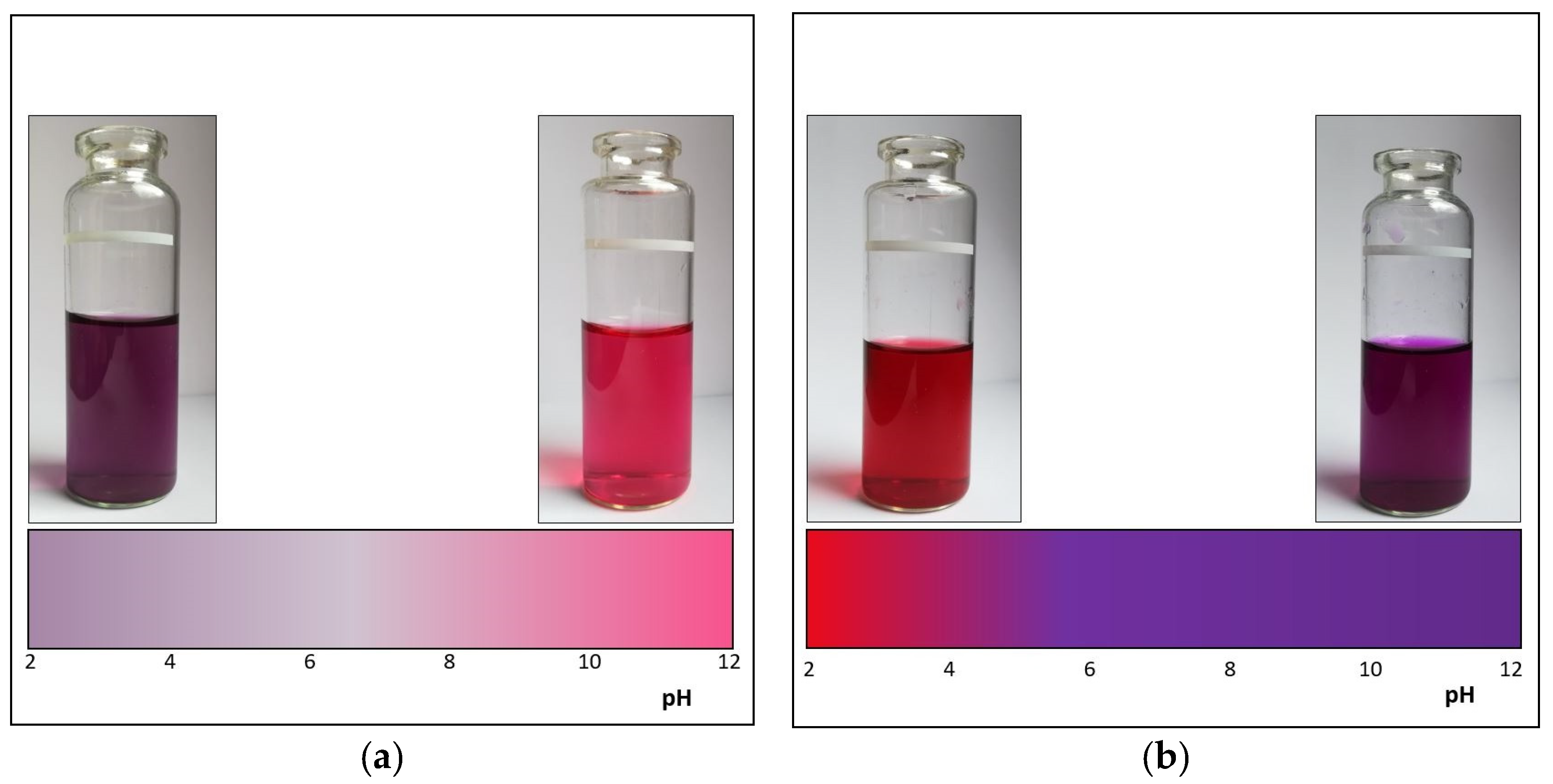
3.2. pH Study of Color Complexes

For metal complexes, color dependence on pH is typical. This has also been confirmed in the color complexes of triphenylmethane dyes, potentially suitable for CWA detection. Taking into account the results given in Table 1, two complexes covering the pH 2–12 region, namely the ALU/Fe3+ and ECR/Cu2+ complexes (chemical structures of selected dyes ALU and ECR are shown in Figure 1), were selected for further experiments on the basis of a pH study in aqueous solutions. For analytical purposes, the acid form of ALU/Fe3+ and the neutral and alkaline forms of ECR/Cu2+ are suitable. The pH-dependent color changes of both complexes are illustrated in Figure 2.
Increased attention has been paid to the possibilities of eliminating the effect of pH on the stability and functionality of the complexes. The problem is that the complexes behave differently on the carrier (cellulose paper), than in solution. However, this has no major impact on the practical application of the proposed method because the (total) discoloration of the complex is used as an analytical signal, not just a change in its color.

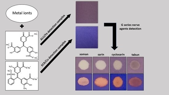
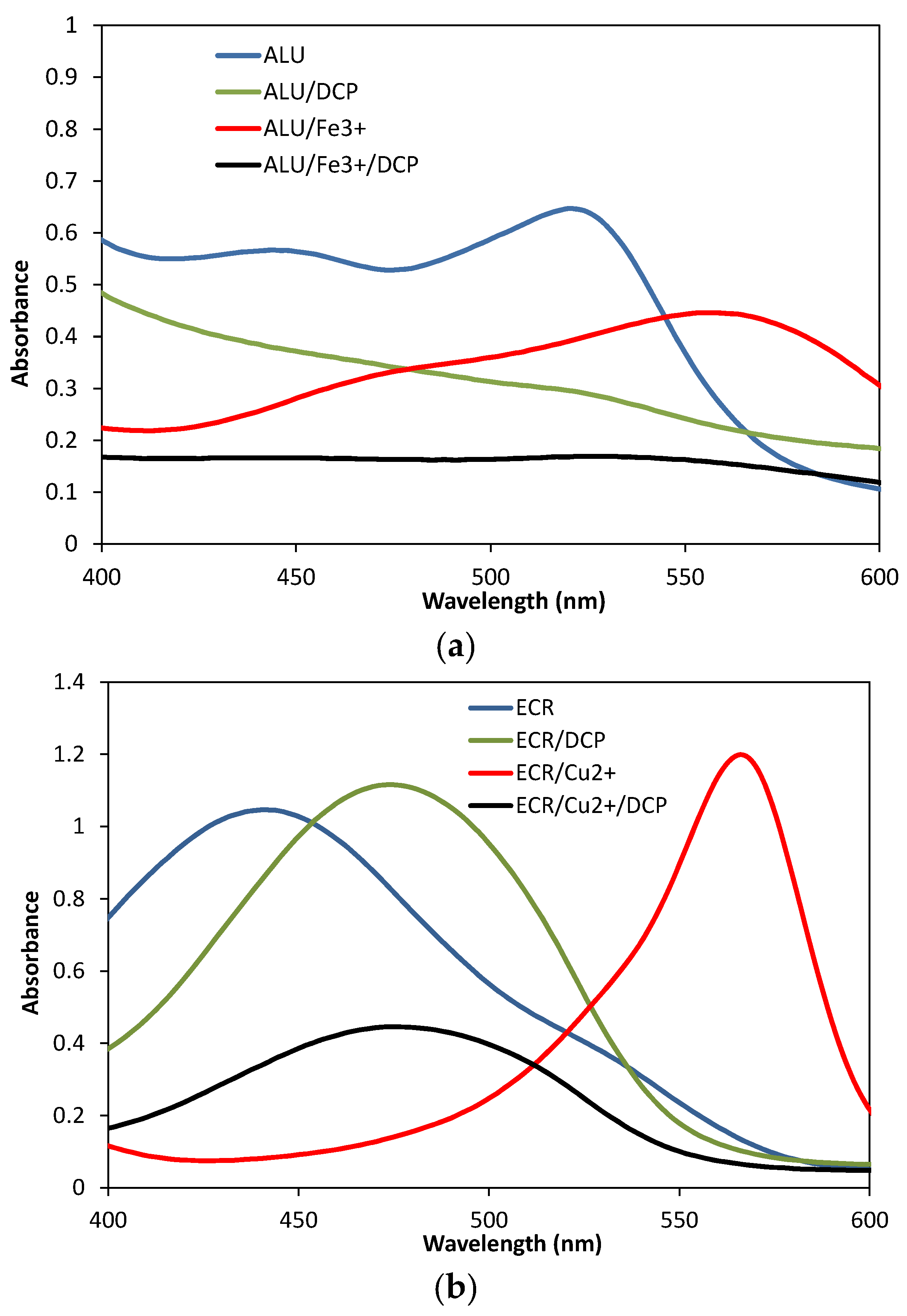
3.3. Detection/Reaction Principle

The detection system itself is likely to consist of several forms of metal complexes that cannot be described by one exact structure; various forms of aqua complexes of triphenylmethane dyes and other structures have been previously described [21,22,23,24,25,26]. The structure of the complexes will depend (among other things) on the pH of the environment. The principle of detecting G nerve agents is based on their reaction (or set of reactions, including esterification) with ALU/Fe3+ and ECR/Cu2+ complexes resulting in their decomposition associated with discoloration. This is shown in Figure 3, which contains the absorption spectra of the dyes (ALU, ECR), their metal complexes (ALU/Fe3+, ECR/Cu2+), and their reaction products with DCP. This decomplexation was experimentally confirmed in the ALU/Fe3+ system by the presence of free Fe3+ ions through an analytical reaction leading to Prussian blue (Figure 4). It was found that the rate and degree of decomplexation was significantly increased by the addition of high-boiling alcohols (glycerol, triethylene glycol, polyethylene glycol), but the mechanism of their effect has not yet been explained in detail.

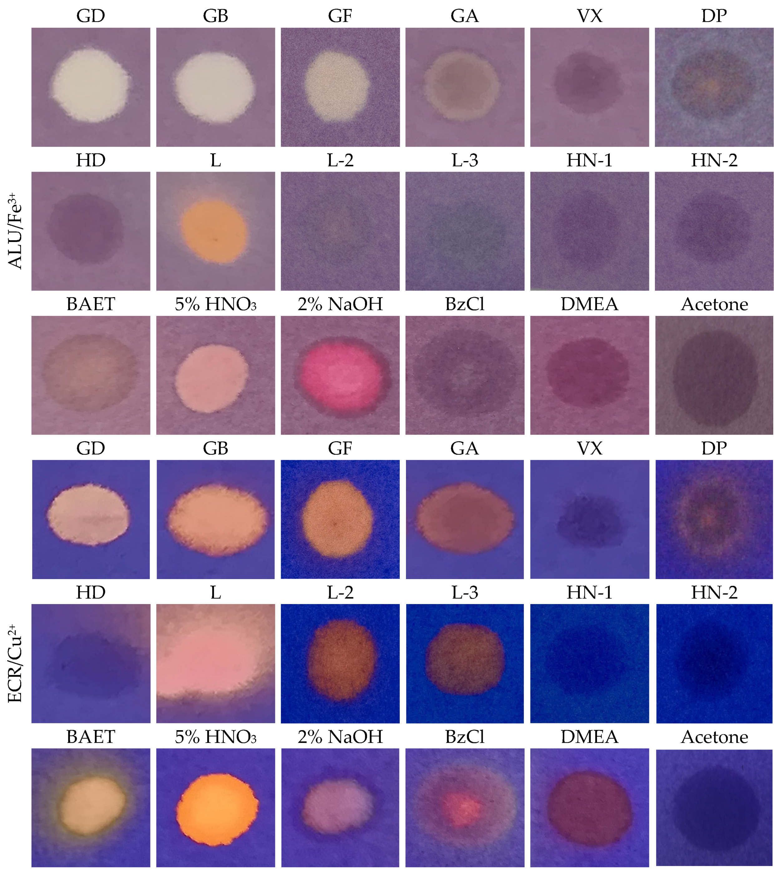
3.4. Color Reactions with CWA, and Interferences

Detection papers with ALU/Fe3+ and ECR/Cu2 complexes have been tested on virtually all major CWA in liquid form. As it can be seen in Figure 5, particularly positive reactions with both complexes were found for nerve agents GB, GD, and GF (all with P-F bond). The detection paper with the ALU/Fe3+ complex was very well distinguished from vesicant L, which gave a distinctive orange color, while GB, GD, and GF caused complete discoloration. GA (with P-CN bond) gave a positive response only to ECR/Cu2+ complex detection paper, but less clearly than other G nerve agents. HD, HN-1, HN-2, and VX did not respond to any detection paper; DP reacted in an indistinctive way.
Both systems are inert to conventional organic solvents (ethanol, methanol, acetone, acetonitrile, toluene, chloroform, liquid hydrocarbons, petrol, carbon disulphide) as well as to water; no color change was observed. In both systems there is a clear interference of acids and bases (in the case of ALU/Fe3+ the effect of bases corresponds well with the pH study (see Figure 2)). The influence of some industrial chemicals selected for their structural similarity to some CWA was different for each detection paper variant. As shown in Figure 5, the detection paper with the ALU/Fe3+ complex was clearly more resistant to these interferences.
From experimental data it can be partially concluded that the proposed method has good selectivity (the distinction of G-type nerve substances from other CWAs is significant) and relatively resistant to interferences that may occur in real conditions. In this aspect, the proposed method is much more selective and resistant than current commercial solutions. However, a detailed study of the entire spectrum of potential interferences (including various chemical mixtures) is planned as part of the industrial development of new detection paper.

3.5. Color Stability

The stability of the coloration (discoloration) at the site of exposure is very high, no reversible reaction was observed even after several hours. The discolored stains are also visible after the immersion of the detection paper in the water for 24 h.

3.6. Limit of Detection

The detection limit was expressed as the concentration of DCP, when a positive reaction was visible within 2 min after dispensing 10 µL of the solution. This condition was also realized in a 3.5% DCP solution in hexane, i.e., about 0.35 mg of DCP (Figure 6). A detection limit of less than 0.35 mg can be achieved by extending the color change evaluation time (>2 min). Just to assume: LD50 (percutaneously, liquid) of GA is 1500 mg, GB 1700 mg, GD and GF 350 mg (DCP estimated in the order of 10,000 mg). In general, the rate of discoloration depends on the analyte concentration. The undiluted DCP reacted almost immediately (within 5 s), with its dilution the rate of discoloration decreased, (the time needed to discolor the detection paper increased). In this context, it is worth noting that the decomplexation due to DCP is faster than the decomplexation due to G nerve agents with the structure of alkyl/cycloalkyl-methylphosphonofluoridates (as a result of the difference in reactivity of P-Cl and P-F bonds). ECR/Cu2+ detection paper is completely discolored (white) due to DCP, unlike G nerve agents, where pale pink to orange color appears.

3.7. Stability of Detection Papers

ALU/Fe3+ and ECR/Cu2+ detection papers stored for 6 months at laboratory temperature retained their original appearance and functionality. Interesting results were obtained when they were stored at 80 °C. Figure 7 shows considerable differences in the thermal stability (appearance) of the detection papers and a very good stability of the ALU/Fe3+ system. This finding corresponds to the observed functional stability (Figure 8). The ALU/Fe3+ detection paper provided a complete discoloration almost immediately after the application of 10 µL of DCP, regardless of the heat load time; the rate and degree of discoloration were virtually unchanged from the standard (stored at laboratory temperature). The ECR/Cu2+ detection paper showed a decrease in functionality compared to the standard; discoloration was slower and less marked (no complete discoloration).

4. Conclusions

Metal complexes represent a promising developmental direction in the field of qualitative detection of CWA in liquid or aerosol form. The proposed simple detection systems, ALU/Fe3+ and ECR/Cu2+, consisting of commonly used and cheap reagents, were able to detect G nerve agents with very good selectivity; only lewisite reacted differently from other potential CWA. Taking into account the structure of positively reacting substances, it is likely that other compounds with P-X bond (X = F, Cl) will also react similarly, i.e., for example, extremely toxic substances from the Novichok group [4]. In particular, ALU/Fe3+ detection paper is suitable for the detection of these substances in the liquid phase even in extremely difficult climatic conditions. The relatively high stability of the metal complexes allows wide modifications, where substances (such as glycerol or other hydroxy compounds) can be added to the color indicators to accelerate or enhance color change. This offers further possibilities in determining or adjusting the selectivity of the reactions. Such a solution represents a certain departure from the established detection means which use mostly chemically reactive compounds, and in the case of organophosphates also biochemical reactions.

Author Contributions

Conceptualization: V.P. and M.L.; Methodology: V.P., M.L. and L.M.; Formal analysis: M.L. and L.M.; Validation: VP, M.L. and L.M.; Writing—manuscript preparation: M.L. and V.P.; Writing—review and editing: V.P., M.L. and L.M; Resources: V.P.; project supervisor: V.P.

Funding

The work was funded in the framework of a security research project of the Ministry of Interior of the Czech Republic, No. VI20172019101.

Conflicts of Interest

The authors declare that they have no competing interest.

References

  1. Costanzi, S.; Machado, J.H.; Mitchell, M. Nerve agents: What they are, how they work, how to counter them. ACS Chem. Neurosci. 2018, 9, 873–885. [Google Scholar] [CrossRef] [PubMed]
  2. Potential Military Chemical (Biological) Agents and Compounds; Field Manual FM 3-11.9; Exidyne: Wentzeville, MO, USA, 2005.
  3. Franke, S. Lehrbuch der Militärchemie; Militärverlag der DDR: Berlin, Germany, 1977; Volume 2v. [Google Scholar]
  4. Halámek, E.; Kobliha, Z.; Pitschmann, V. Analysis of Chemical Warfare Agents; University of Defence: Brno, Czech Republic, 2009. [Google Scholar]
  5. Thoraval, D.; Bovenkamp, J.W. Paper Chemical Agent Detectors, EP 0 334 668 B1, 23.12. 1992. Available online: https://www.google.com/url?sa=t&rct=j&q=&esrc=s&source=web&cd=3&ved=2ahUKEwix0eLj6onmAhXhx6YKHdJhANcQFjACegQIBRAC&url=https%3A%2F%2Fpatentimages.storage.googleapis.com%2Fpdfs%2F57050eab456edfc3d6e9%2FEP0334668B1.pdf&usg=AOvVaw3RvGj0EAJta9K2rwuhUDtY (accessed on 28 November 2019).
  6. Galan-Freyle, N.J.; Figueroa-Navedo, A.M.; Pacheco-Londoño, Y.C.; Ortiz-Rivera, W.; Pacheco-Londoño, L.C.; Hernández-Rivera, S.P. Chemometrics-enhanced fiber optic Raman detection, discrimination and quantification of chemical agents simulants concealed in commercial bottles. Anal. Chem. Res. 2014, 2, 15–22. [Google Scholar] [CrossRef]
  7. Hu, G.; Xiong, W.; Luo, H.; Shi, H.; Li, Z.; Shen, J.; Fang, X.; Xu, B.; Zhang, J. Raman spectroscopic detection for simulants of chemical warfare agents using a spatial heterodyne spectrometer. Appl. Spectrosc. 2018, 72, 151–158. [Google Scholar] [CrossRef] [PubMed]
  8. Royo, S.; Martinez-Máñez, R.; Sancenón, F.; Costero, A.M.; Parra, M.; Gil, S. Chromogenic and fluorogenic reagens for chemical warfare nerve agents detection. Chem. Commun. 2007, 4839–4847. [Google Scholar] [CrossRef]
  9. Chen, L.; Wu, D.; Yoon, J. Recent advances in the developments of chromophore-based chemosensors for nerve agents and phosgene. ACS Sens. 2018, 3, 27–43. [Google Scholar] [CrossRef]
  10. Kangas, M.J.; Burks, R.M.; Atwater, J.; Lukowicz, R.M.; Williams, P.; Holmes, A.E. Colorimetric sensor arrays for the detection and identification of chemical weapons and explosives. Crit. Rev. Anal. Chem. 2017, 47, 138–153. [Google Scholar] [CrossRef]
  11. Pitschmann, V.; Matějovský, L.; Lunerová, K.; Dymák, M.; Urban, M.; Králík, L. Detection papers with chromogenic chemosensors for direct visual detection and distinction of liquid chemical warfare agents. Chemosensors 2019, 7, 30. [Google Scholar] [CrossRef]
  12. Knapton, D.; Burnworth, M.; Rowan, S.J.; Weder, C. Fluorescent organometallic sensor for the detection of chemical warfare-agent mimics. Angew. Chem. Int. Ed. 2006, 45, 5825–5829. [Google Scholar] [CrossRef]
  13. Sarkar, S.; Mondal, A.; Tiwari, A.K.; Shunmugam, R. Unique emission from norbornene derived terpyridine—A selective chemodosimeter for G-type nerve agent surrogates. Chem. Commun. 2012, 48, 4223–4225. [Google Scholar] [CrossRef]
  14. Ordroneau, L.; Carella, A.; Pohanka, M.; Simonato, J.P. Chromogenic detection of sarin by discolouring decomplexation of a metal coordination complex. Chem. Commun. 2013, 49, 8946–8948. [Google Scholar] [CrossRef]
  15. Maza, W.A.; Vetromile, C.M.; Kim, C.; Xu, X.; Zhang, X.P.; Larsen, R.W. Spectroscopic investigation of the noncovalent association of nerve agent simulant diisopropyl methylphosphonate (DIMP) with zinc (II) porphyrins. J. Phys. Chem. A 2013, 117, 11308–11315. [Google Scholar] [CrossRef] [PubMed]
  16. Borba-Bon, A.; Costero, A.M.; Gil, S.; Sancenón, F.; Martinez-Máñez, R. Chromo-fluorogenic BODIPY complexes for selective of V-type nerve agent surrogates. Chem. Commun. 2014, 50, 13289–13291. [Google Scholar] [CrossRef] [PubMed]
  17. Kumar, V.; Raviraju, G.; Rana, H.; Rao, V.K.; Gupta, A.K. Highly selective and sensitive chromogenic detection of nerve agents (sarin, tabun and VX): A multianalyte detection approach. Chem. Commun. 2017, 53, 12954–12957. [Google Scholar] [CrossRef] [PubMed]
  18. Sheet, K.S.; Sen, B.; Khatua, S. Organoiridium(III) complexes as luminiscence color swithing probes for selective detection of nerve agent simulant in solution and vapor phase. Inorg. Chem. 2019, 58, 3635–3645. [Google Scholar] [CrossRef] [PubMed]
  19. Ishida, R.; Narita, H.; Tonosaki, K. Copper(II)-2,2´-bipyridine-eriochrome cyanine R ternary complex and its application to the spectrophotometric determination of copper(II). Bull. Chem. Soc. Jpn. 1978, 51, 1751–1754. [Google Scholar] [CrossRef]
  20. Dacres, H.; Narayanaswamy, R. Evaluation of cooper(II) eriochrome cyanine R (ECR) complex immobilized in anion exchange membrane as a potential nitric oxide optical sensor. Aust. J. Chem. 2008, 61, 189–196. [Google Scholar] [CrossRef]
  21. Dapson, R.; Horobin, R.W.; Kierman, J. Hematoxylin shortages: Their causes and duration, and other dyes that can replace hemalum in routine hematoxylin and eosin staining. Biotech. Histochem. 2010, 85, 55–63. [Google Scholar] [CrossRef]
  22. Sarsam, L.A.; Bashir, W.A. Spectrophotometric determination of scandium(III) with eriochrome cyanine R and cetylpyridinium chloride—Application to waters and synthetic alloys. J. Raf. Sci. 2009, 20, 48–65. [Google Scholar]
  23. Langmyhr, F.J.; Stumpe, T. Complex formation of iron(III) with eriochrome cyanine R. Anal. Chim. Acta 1965, 32, 535–543. [Google Scholar] [CrossRef]
  24. Majeed, A.; Asma, D.R.N.; Sana-Ullah, M. Spectrophotometric determination of iron with xylenol orange. J. Chem. Soc. Pak. 1996, 18, 197–200. [Google Scholar]
  25. Mizuguchi, H.; Yotsuyanagi, T. Visual threshold detection of trace metal ions using a bi-functional metallochromic reagent. Anal. Sci. 2001, 17, i1687–i1689, Supplement. [Google Scholar]
  26. Barghouthi, Z.; Amereih, S. Spectrophotometric determination of fluoride in drinking water using aluminium complexes of triphenylmethane dyes. Water SA 2012, 38, 543–548. [Google Scholar] [CrossRef]
Figure 1. Structures of selected triphenylmethane dyes: ALU (a) and ECR (b).
Chemosensors 07 00059 g001
Figure 2. Change of ALU/Fe3+ (a) and ECR/Cu2+ (b) metal complexes depending on the pH of aqueous solution.
Chemosensors 07 00059 g002
Figure 3. Absorption spectra: (a) ALU, ALU/Fe3+, and their reaction products with DCP; (b) ECR, ECR/Cu2+, and their reaction products with DCP.
Chemosensors 07 00059 g003
Figure 4. Simple proof of free Fe3+ ions using the Prussian blue method: (a) Unexposed detection paper ALU/Fe3+, (b) drop of potassium ferrocyanide, (c) drop of diethyl chlorophosphate (DCP), (d) drop of DCP and potassium ferrocyanide.
Chemosensors 07 00059 g004
Figure 5. Of CWA reactions and selected chemicals with detection paper ALU/Fe3+ and ECR/Cu2+ (BAET = 2-(butylamino)ethanethiol, BzCl = benzoyl chloride, DMEA = N,N-dimethylethanolamine).
Chemosensors 07 00059 g005
Figure 6. Dependence of discoloration of detection papers (ΔE) on DCP concentrations (measured by tristimulus colorimeter after 2 min of reaction).
Chemosensors 07 00059 g006
Figure 7. Stability (ALU/Fe3+, ECR/Cu2+, detection papers appearance) at 80 °C; ΔE measured by tristimulus colorimeter.
Chemosensors 07 00059 g007
Figure 8. Tested with DCP after heat load at 80 °C within 28 days.
Chemosensors 07 00059 g008
Table 1. Pilot tests of chemical warfare agents (CWAs) with metal complexes with triphenylmethane dyes the without addition of glycerol (- no change).
ComplexOriginal ColourChange of Colour with CWA in Liquid Phase
GAGBVXHDL
ALU/Al3+Red-lightening---
ALU/Fe3+violet-lightening--orange
ALU/Zn2+Red-lightening---
ECR/Cu2+Blueorangeorange--orange
ECR/Al3+violetorangeorange--orange
XO/Fe3+Blue-pink--orange
XO/Al3+Pink-yellow---
XO/Zn2+RedyellowyellowPinkpink-

