Article

Pinus roxburghii and Nauplius graveolens Extracts Elevate Apoptotic Gene Markers in C26 Colon Carcinoma Cells Induced in a BALB/c Mouse Model

1
Nutrition and Food Science Department, Food Industries and Nutrition Research Institute, National Research Centre, Cairo 12622, Egypt
2
Pharmacognosy Department, Pharmaceutical and Drug Industries Research Institute, National Research Centre, Cairo 12622, Egypt
3
Hormones Department, Medical Research and Clinical Studies Institute, National Research Centre, Cairo 12622, Egypt
4
Biochemistry Department, Faculty of Agriculture, Cairo University, Giza 12613, Egypt
5
Department of Food Engineering, Faculty of Agriculture, Ataturk University, Erzurum 25240, Türkiye
6
Laboratory of Food Chemistry, Department of Chemistry, National and Kapodistrian University of Athens Zografou, 15771 Athens, Greece
7
Department of Food Technology, Food Industries and Nutrition Research Institute, National Research Centre, Dokki, Cairo 12622, Egypt
*
Authors to whom correspondence should be addressed.
Submission received: 11 August 2022 / Revised: 21 September 2022 / Accepted: 28 September 2022 / Published: 1 October 2022
(This article belongs to the Special Issue Novel Applications of Separation Technology)

Abstract

:
The present study aimed to evaluate the chemopreventive potential of Pinus roxburghii branch (P. roxburghii) and Nauplius graveolens (N. graveolens) extracts against human colorectal cancer (CRC) induced by C26 murine cells in a BALB/c mouse model. Real-time qRT-PCR was used to evaluate the apoptotic pathway by measuring the relative mRNA expression levels of the Bcl-2, Bax, Cas3, NF-κB, and PI3k genes. At the termination of the 30-day period, blood samples were collected to assay the biomarkers. The results showed a significant increase (p < 0.05) in the levels of TGF-β, CEA, CA19-9, malondialdehyde, ALT, AST, ALP, urea, and creatinine in the positive control compared to the negative control group. In addition, the glutathione reductase activity and total antioxidant activity were reduced in the positive control compared to the negative control. The biomarkers mentioned above were restored to almost normal levels after administering a safe dose (1/10) of a lethal dose of P. roxburghii and N. graveolens extracts. Administration of one-tenth of the LD50 of P. roxburghii and N. graveolens extracts caused a significant upregulation of the expression of Bax and Cas-3 and downregulation of the Bcl-2, NF-ĸB, and PI3k genes vs. the GAPDH gene as a housekeeping gene compared to the control group. Furthermore, the Bax/Bcl-2 ratio increased upon treatment. After administration of P. roxburghii and N. graveolens at a safe dose (1/10) of a lethal dose, the results showed improvement in both body weight gain and a significant decrease (p < 0.05) in tumor volume. Histopathological changes supported these improvements. Conclusively, the research outputs show that P. roxburghii and N. graveolens extracts can be utilized as potential chemopreventive agents for CRC treatment by stimulating cancer cell apoptosis and suppressing CRC survival and proliferation.

1. Introduction

Cancer disease is a significant public health concern, as it is considered the second leading cause of mortality annually after cardiovascular death incidences around the world [1,2]. In 2020, colorectal cancer (CRC) accounted for approximately 10% and 9.4% of cancer incidences and cancer causative deaths worldwide, respectively [3]. Based on global future perspectives, CRC incidences are predicted to exceed 3.2 million by 2040. CRC cases increase due to continuous exposure to hazardous environmental factors attributed to lifestyle shifts and diet-related habits [4]. Currently, the most applied cancer treatments include chemotherapy, radiotherapy, immunotherapy, and surgery. Almost all these treatments involve many deleterious side effects for normal cells together with cancer cells. Consequently, novel treatments with minimal side effects are necessary for cancer control and prevention [5]. More efforts need to be made to explore new alternative anticancer agents that would ultimately slow, reverse, or even prevent cancer cell progression without affecting normal cell function and integrity [6,7].
For ancient decades, herbal remedies derived from naturally grown plants were proven to be highly effective in treating many diseases [8,9,10]. Despite the continuous increase in herbal therapy popularity around the globe, very little is known about the composition of these remedies’ active compounds, and hence, minimal information is known about their mode of action either on a cellular or molecular basis [10,11]. As one of the major categories of herbal remedies, edible plants are considered a rich source of phytochemicals; however, their efficacy in cancer treatment and prevention has not yet been deciphered [12]. Inevitably, investigating plant-based therapeutic agents’ chemical and cytotoxic characteristics could shed light on applying some of these plant therapies as novel natural anticancer drugs [13]. Recently, phytochemicals were proven to directly influence cancer cell cycle arrest, proliferation enhancement, and apoptosis initiation [7]. Further studies suggested that the role of phytochemicals is primarily initiated by delaying or even halting the transformation of a healthy cell into undifferentiated, malignant cells [14,15].
Pinus roxburghii (P. roxburghii) belongs to the Pinaceae family and is famously known as chir pine and cultivated in El-Orman Botanical Garden, Egypt. P. roxburghii is an ancient medicinal plant with multi pharmaceutical and medicinal properties, including anti-inflammatory, analgesic, antidyslipidemic, antioxidant, antibacterial, antifungal, and anticancer activities, respectively [16,17,18,19]. Previous studies have demonstrated that P. roxburghii has various bioactive constituents such as phenolics, flavonoids, tannins, beta-carotene, and lycopene [20]. Moreover, different polyphenolic compounds isolated from the branch of P. roxburghii include catechin, kaempferol, protocatechuic acid, caffeic acid, and gallocatechin [21]. Nauplius graveolens (Forssk.) Wiklund (N. graveolens) (synonyms. Asteriscus graveolens, Bubonium graveolens, Odontospermum graveolens) is one of the species of the Asteraceae family. It is known as Tafss [22], and naturally grows in the inland desert of Wadi Feiran, South Sinai, Egypt. The N. graveolens plant displays medicinal and pharmacological characteristics, including antioxidant, anti-inflammatory, antimicrobial, and antitumor activities [23,24,25]. Phytochemical investigations of the N. graveolens extract revealed the diverse distribution of phytoconstituents including polyphenolics, flavonoids, tannins, coumaric, and chlorogenic acid as the major phenolic compounds [23]. Moreover, N. graveolens contained a high content of flavonoids and sesquiterpene lactone asteriscunolide isomers; naturally occurring Asteriscunolide A enhances apoptosis in a tumor cell line [26].
Targeting apoptosis could be the main approach in cancer treatment and a strong, active area of research. Apoptosis, one of the main mechanisms controlling cancer cell progression, is mainly triggered by the activation, or silencing of caspases, Bcl2, Bax, NF-κB, and PI3k genes in response to cell injury, which in turn are responsible for the activation or inactivation of certain cellular substrates, eventually leading to either cell death or survival according to the degree of cell injury [27,28,29]. Several murine models have been developed to study CRC pathogenesis, preferably reflecting CRC chemoprevention or the most critical chemo-treatment aspects. Selecting the most appropriate murine models will maximize the upscaling of therapies from in vitro laboratory trials to in vivo clinical practice. Over the last three decades, colon-26 and adenocarcinoma mouse models (C26 model) have been used for research on the natural history of carcinomas and antitumor therapy to study the effects of several antitumor agents [30,31,32]. Although previous studies have classified the major phytoconstituents which may responsible for the anticancer activity of the two plant extracts, the actual assessment of those compounds was not evaluated in vivo in any of these studies. Therefore, the aim of the present study was to assess the in vivo anticancer activities of P. roxburghii and N. graveolens extracts in a BALB/c mouse model.

2. Material and Methods

2.1. Chemicals

Methanol, ethanol, dimethyl sulfoxide (DMSO), and paraformaldehyde (PFA) were obtained from Sigma Chemical Company (St. Louis, MO, USA). RPMI 1640 medium, fetal bovine serum, and trypsin-EDTA were procured from Gibco, Life Technologies Limited (Paisley, UK).

2.2. Extract Preparation

The extraction of P. roxburghii and N. graveolens plants was carried out by maceration method with 90% aqueous methanol. P. roxburghii was grown and obtained from the Ministry of Agriculture, Orman botanical garden, Giza (Authentication number: 2151). After removing the foliar leaves, the branch was cut with scissors into small pieces, which were allowed to dry at 40 °C. Aliquots of 75 g of finely ground flour were extracted with 450 mL of absolute methanol. The methanolic extract was filtered, and the filtrate was concentrated on a Büchi rotary evaporator R-114 (Büchi Labortechnik AG, Flawil, Switzerland). The residue was freeze-dried using a Snijders Freeze Dryer (Tilburg, Holland) and saved in sterile vials at −20 °C. N. graveolens was collected from the inland desert of Wadi Feiran, South Sinai, Egypt (Authentication number: 3187). Both plants were identified by the pharmacognosy department, National Research Centre, Giza, Egypt according to taxonomic classification by Boulos (2002) [33]. The whole N. graveolens plant was treated similarly to P. roxburghii [34]. The extraction yields of P. roxburghii and N. graveolens plants were 5.2 and 5.5 g/75 g of dry material, respectively.

2.3. Composition of Basal Diet

The basal diet consists of corn starch (46.5%), casein (14%), soybean oil (4%), fiber (5%), mineral mixture (3.5%), and vitamin mixture (1%) as described in AIN-93 M [35].

2.4. Determination of the Median Lethal Dose (LD50)

The median lethal dose (LD50) of P. roxburghii and N. graveolens extracts was evaluated according to Wilbrandt [36]. Briefly, different P. roxburghii and N. graveolens extract doses of up to 2000 mg/kg body weight in DMSO were orally administered to male albino mice (six mice per group), and a group of six mice was given the respective amount of DMSO orally and left as a control. The median lethal dose (LD50) of the methanolic extracts of P. roxburghii accounted for 1708.3 mg/kg body weight, while that of N. graveolens was 1562.5 mg/kg body weight. The 1/10 LD50 of P. roxburghii and N. graveolens extracts accounted for 170.83 and 156.25 mg/kg body weight, respectively [34].

2.5. Animals

Twenty-four male BALB/c mice weighing 20–25 g were purchased from Schistosome Biological Supply Centre (SBSC) at Theodore Bilharz Research Institute (TBRI), Imbaba, Giza, Egypt. All the animals were acclimatized for a week under standard husbandry conditions. The animals had been fed a standard pellet diet and water ad libitum. Animal handling and experimental procedures were conducted by the Guidelines of Care Laboratory Animals, National Research Centre, and approved by the Research Ethical Committee and the national legislation on lab Animal Rescue and Usage (Ethical Code: 15/097; 4 February 2021).

2.6. Induction of C26 Murine Cells in BALB/c Mice

Murine colon-26 cells were cultured in RPMI 1640 medium supplemented with fetal bovine serum (10%) (Gibco, Life Technologies Limited, Paisley, UK) in a humidified atmosphere (5% CO2) at 37 °C. Cells were collected using trypsin-EDTA (0.25%) to obtain a single-cell suspension. Cells were counted using a hemocytometer and pelleted via centrifugation at 25 °C (500× g for 5 min). Mice were injected subcutaneously on the dorsal side with 1 × 10⁶ cells (Figure 1), as described by [37,38].

2.7. Measurement of Body Wasting

Body mass was monitored weekly after inoculation. Tumor size was measured using a Verniercaliper, and tumor volume was measured using the formula V = (length × width2)/2 in mm3 [39,40].

2.8. Experimental Design

Twenty-four male BALB/c mice were divided into four groups as follows:
Group (1): Negative control received a basal diet.
Group (2): Positive control received a basal diet and was injected intraperitoneally with a respective amount of DMSO.
Group (3): Tumor mice received a basal diet and were injected intraperitoneally with a dose equivalent to 1/10 LD50 of extract of P. roxburghii branch day after day.
Group (4): Tumor mice received a basal diet and were injected intraperitoneally with a dose equivalent to 1/10 LD50 of N. graveolens extract day after day.
Tumor volumes were measured weekly for four consecutive weeks (one month).
The diet was withdrawn at the end of the one-month feeding trial, and all animals were fasted overnight (12 h). The mice were anesthetized by intramuscular injection with ketamine chloride (24 mg/kg body weight). Blood was sampled from the orbital sinus of the eye and collected in clean, dry test tubes. The serum was separated by centrifugation (1500× g, 10 min, 4 °C). The serum obtained was used for various biochemical estimations.

2.9. Changes in Body Weight (BW) and Feed Efficiency Ratio (FER)

Body weight and food consumption were recorded by Hsu et al. [41] by the following formula:
Changes in body weight = final body weight − initial body weight
Feed efficiency ratio (FER) = Body weight gain, g/Food intake, g

2.10. Biochemical Analyses

Liver function was assessed by measuring liver enzyme activities, such as alanine aminotransferase (ALT), aspartate aminotransferase (AST), and alkaline phosphatase (ALP), using a kit (Biodiagnostic, Egypt). Serum concentrations of urea and creatinine, the two indicators of kidney function, were determined using kits (Biodiagnostic, Egypt). The assays followed the manufacturer’s protocol based on published techniques [42,43,44,45]. Glutathione reductase, total antioxidant capacity, and malondialdehyde (MDA) were determined colorimetrically using a kit (Biodiagnostics, Egypt) described by [46,47,48]. The TGF-β level was assayed using an ELISA kit purchased from Sunlong Biotech Co., Ltd., China. Serum CA 19-9 was assessed using the Cancer Antigen CA 19-9 ELISA Kit (Abcam, Cambridge, UK). Serum carcinoembryonic antigen (CEA) was determined using an ELISA kit (enzyme-linked immunosorbent assay) (Abcam, Cambridge, UK). These assays were carried out based on the manufacturer’s instructions.

2.11. RNA Extraction and cDNA Synthesis

RNA was extracted from tissues using the RNeasy mini kit (Qiagen, Hilden, Germany) as designed by the manufacturer. Total RNA purity and concentration were detected by a Nanodrop UV spectrophotometer. cDNA was synthesized using a cDNA synthesis kit (Intron Biotechnology, Seongnam-si, Korea) as stated in the manufacturer’s protocol.

2.12. Quantitative Real-Time PCR

Gene expression was tested on the genes Bax, Bcl-2, Cas3, PI3K, and NF-κB, and GAPDH was used as a housekeeping gene. The specific primers used for quantitative PCR were designed through NCBI BLAST before purchasing from Thermo Fisher Scientific (Waltham, MA, USA) (https://blast.ncbi.nlm.nih.gov/Blast.cgi, accessed on 1 May 2021), and their sequences are presented in Table 1. A real-time PCR cycler (QIAGEN, Rotor-Gene Q, Valencia, CA) was used to detect the synthesized cDNA copy number. PCR mixtures were set up in a 25 µL final volume, containing 12.5 µL SYBR Premix Ex Taq TM (Thermo Fisher Scientific, Waltham, Massachusetts, USA), 6.5 µL distilled water, 0.5 µL sense primer (0.2 mM), 0.5 µL antisense primer (0.2 mM), and 5 µL of cDNA template. The reaction program was designed with three steps. The first denaturation step was performed at 95.0 °C for 3 min. The second step consisted of 40 cycles, each cycle divided into (a) 95.0 °C for 15 s; (b) 55.0–60.0 °C for 30 s; and (c) 72.0 °C for 30 s. The third step consisted of several cycles, which started at 60.0 °C and increased approximately 0.5 °C every 10 s up to 95.0 °C. At the end of each qRT-PCR cycle, a melting curve analysis was designed at 95.0 °C, then 55.0–60.0 °C, followed by 95.0 °C to check the primer quality. Each sample was prepared in triplicate, and the variability of the samples was evaluated using geometric standard deviations. The geometric mean of the triplicate run for each gene was normalized to the GAPDH geometric mean.

2.13. Histopathological Examination

Tumors were separated and fixed in 4% paraformaldehyde (PFA) for 24 h. The tissues were washed with tap water, followed by sequential soaking in diluted methanol, ethanol, and absolute ethyl alcohol for dehydration. The specimens were cleared in xylene, embedded in paraffin, and left in a hot air oven at 56 °C for 24 h. Paraffin bee wax tissue blocks were prepared for sectioning at 4 microns at room temperature by a sledge microtome (SLEE medical, Nieder-Olm, Germany). The final tissue sections were embedded on glass slides, deparaffinized, and stained with hematoxylin and eosin for examination by a light microscope [49].

2.14. Statistical Analyses

Statistical analysis was implemented using GraphPad Prism software 8.0 (San Diego, CA, USA). The results are expressed as the mean ± standard error of the mean (SEM) of three independent experiments. Data are presented as the means ± SDs and estimated by one-way analysis of variance (ANOVA) performed using SPSS 19.0 (SPSS Ltd., Surrey, UK). Duncan’s multiple range test (DMRT) was used to detect individual comparisons. Differences were considered significant at p < 0.05.

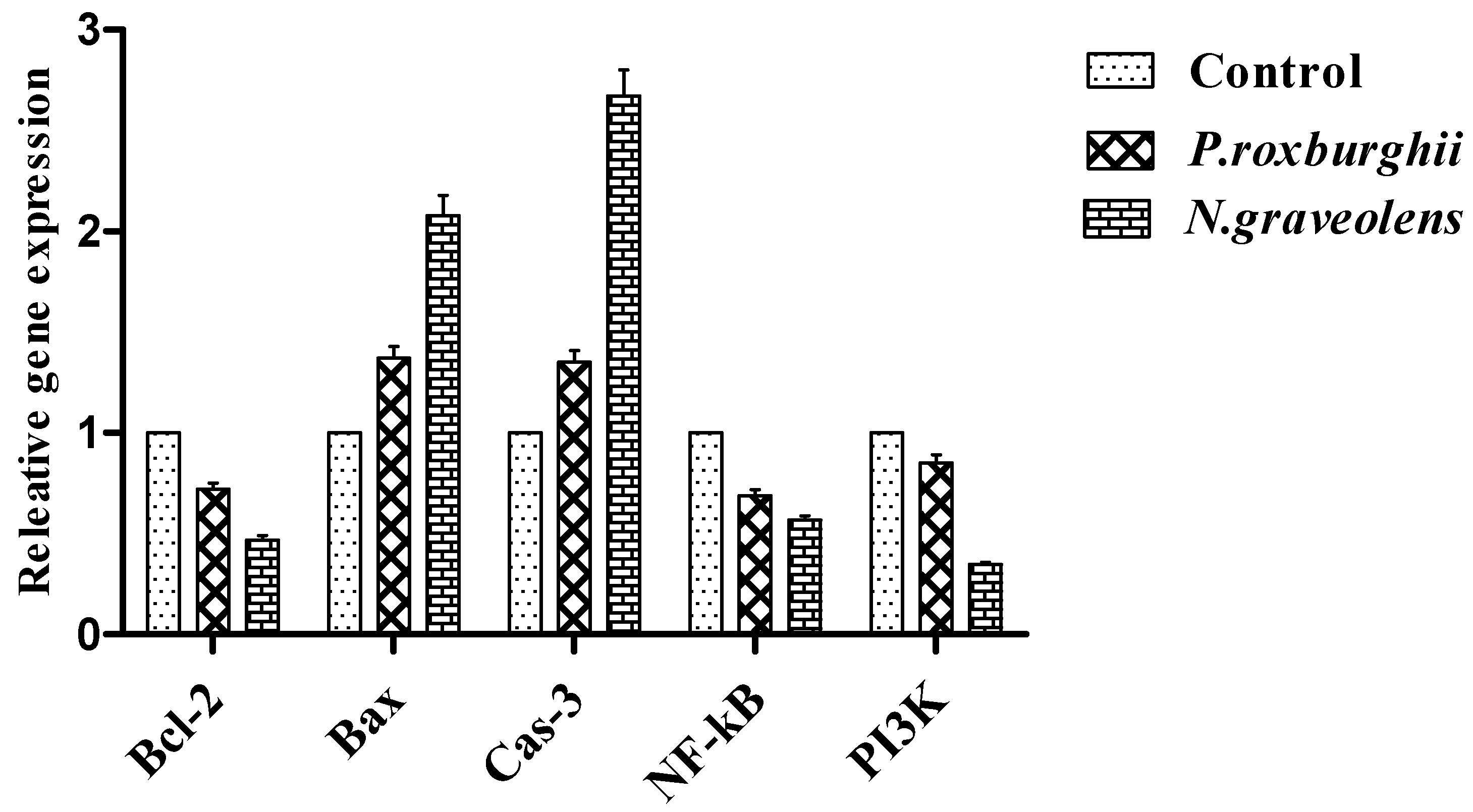
3. Results and Discussion

The data in Figure 2 illustrate the effect of P. roxburghii and N. graveolens extracts on the tumor growth of mice. The colon tumor volume increased in the positive control during the baseline scan and the first week. In the positive control, tumor volume increased significantly in the second, third, and fourth weeks. Tumor volume can be considered a critical prognostic factor for assessing several types of cancers [50]. Tullie et al. [51] reported a significant correlation between tumor volume and survival rate. Huang et al. [52] explained that large-volume tumors were proportionally correlated with increased cancer risk at both the cellular and molecular levels. Furthermore, data reporting that tumor volume was a potent independent factor revealed the prognosis of several types of cancers, such as lung, nasopharyngeal, neck, head, and other malignant tumors [53,54]. Administration of one-tenth of the LD50 of P. roxburghii and N. graveolens extracts showed a significant decrease in tumor volume every subsequent week.
The data in Table 2 represent the effect of P. roxburghii and N. graveolens extracts on the nutritional status of mice. During the development of cancer in the BALB/C mouse model, there was a significant decrease in body weight gain, food intake, and the food efficiency ratio in the positive control (19.36, 177.54, and 0.10, respectively) compared to the negative control (27.11, 189.21, and 0.14, respectively). Generally, body weight decrease is a common side effect in CRC patients. Consequently, research studies have reported that weight control is an essential criterion in CRC survival [55]. Weight loss was significantly linked to increasing colorectal cancer and mortality [56]. However, Diculescu et al. [57], stated that the weight loss index is an independent prediagnostic factor for colorectal cancer progression. Barber et al. [58] also found that body weight loss caused by cancer may be due to anorexia, substrate metabolism deficiencies, and hypermetabolism. Administration of 1/10 LD50 of P. roxburghii and N. graveolens extracts showed improvement in body weight gain, food intake, and feed efficiency ratio by 24.52, 25.23, 184.37, 186.29, 0.13, and 0.13, respectively.
The data presented in Figure 3 show the results of serum alanine aminotransferase (ALT), aspartate aminotransferase (AST), and alkaline phosphatase (ALP) activities in the studied groups. The data exhibited a significant increase in serum ALT, AST, and ALP activities in the positive control compared to the negative control. The activities of ALT, AST, and ALP were significantly decreased after administration of 1/10 LD50 of P. roxburghii and N. graveolens extracts. The conversion of normal cells to cancer cells and cancer cell proliferation lead to abnormal serum enzyme synthesis [59]. ALT, AST, and ALP are major indicator enzymes for liver diagnosis and management [60]. ALT and AST are found inside hepatocytes and are released when the liver is damaged. Liver infection is usually associated with mitochondrial damage, which leads to the immediate release of ALT and AST into the blood circulation and eventually a dramatic increase in these enzyme levels [61]. ALT and AST have a vital role in the prognosis of some cancer types [62,63].
Serum ALT, AST, and ALP activities were significantly increased in the positive control, representing the degree of severity of hepatic tissue injury reported by Ahn et al. [64]. The alteration of hepatocyte plasma membrane permeability in hepatocyte cancer cells leads to leakage of cellular enzymes. This ultimately increases the concentrations of these markers in the serum and decreases them in hepatocytes. The activities of ALT and AST were significantly lowered after administration of 1/10 LD50 of P. roxburghii and N. graveolens extracts. Alkaline phosphatase (ALP) is an enzyme that has been proven to be involved in the transport of metabolites across cellular membranes, proteins and certain enzyme synthesis, glycogen metabolism, and other secretary activities [65]. ALP activity measurement is used as a tumor marker in the early detection and diagnosis of cancer [66]. Lakshmi and Subramanian [67] observed that an elevated level of ALP activity was detected in cancer-bearing animals because of a disturbance in the secretory activity or transport of metabolites of cancer cells. The activity of ALP was significantly decreased after administration of 1/10 LD50 of P. roxburghii and N. graveolens extracts.
The data in Figure 4 show the serum urea and creatinine levels in the different groups. Serum urea and creatinine levels increased significantly in the positive control group compared to the negative control group. Meanwhile, the administration of 1/10 LD50 of P. roxburghii and N. graveolens extracts significantly decreased serum urea and creatinine levels. Impaired kidney function is one of the most detectable characteristics in cancer patients. Elevated urea and creatinine levels in the bloodstream of cancer patients, which eventually occur due to renal cell degeneration, are vital in cancer disease diagnosis and management [68,69,70]. The results showed that blood serum creatinine and urea levels were significantly increased in colon cancer-induced mouse models. The results corresponded with those of Raj et al. [71], who reported a detectable increase in serum creatinine levels in breast cancer-induced rats. Increased creatinine levels were also detected in the plasma of cervical cancer patients, as reported by Koji et al. [72], and were considered the major risk factor for cervical cancer. In contrast, a significant reduction in serum creatinine and urea levels was detected after administration of 1/10 LD50 of P. roxburghii and N. graveolens extracts. These findings corresponded with the results of Falconer et al. [73]. They observed a significant increase in the creatinine level in all cancer types compared to the normal subjects.
The data presented in Figure 5, Figure 6 and Figure 7 illustrate the activity of glutathione reductase, total antioxidant capacity, and malondialdehyde levels. The data showed a significant decrease in glutathione reductase activity in the positive control compared to the negative control. Administration of 1/10 LD50 of P. roxburghii and N. graveolens extracts significantly increases serum GR activity. Moreover, the total antioxidant level decreased significantly in the positive control compared to the negative control. Administration of 1/10LD50 of P. roxburghii and N. graveolens extracts showed a significant increase in serum TAC levels. In contrast, a significant increase was observed in serum malondialdehyde levels in the positive control compared to the negative control. Administration of 1/10 LD50 of P. roxburghii and N. graveolens extracts significantly decreased serum MDA levels. Antioxidant activity can be estimated in humans, which reflects the disturbance in redox equilibrium in body fluids, tissues, or organs under different pathological conditions, including tumor development [74]. Generally, oxidative stress and inflammation are associated with substrate metabolic disturbances because of an environmental effect that eventually leads to the development of CRC [75]. The results showed lower TAC levels in the positive control group than in the negative control group, indicating a weakened antioxidant barrier resulting from overproduction of free radicals. Administration of 1/10 LD50 of P. roxburghii and N. graveolens extracts showed a significant increase in the TAC level.
The process of colorectal cancer initiation and progression involves the interaction of various physiological factors, including the overproduction of reactive oxygen species (ROS) and malondialdehyde (MDA) formation. ROS are excessively produced in gastrointestinal tract chronic infection because of lipid peroxidation that eventually leads to cellular protein damage and carcinogenesis induction [76,77]. Malondialdehyde (MDA) (final product of the lipoperoxidation process) is a strong electrophilic compound that interacts with cell nucleophiles to form MDA oligomers that can be used for cancer detection and as a prediagnostic index for CRC patients [78]. MDA is a major molecule responsible for the highest mutagenicity and carcinogenicity [79]. In the present study, the MDA level was significantly increased in the positive control compared to the negative control group. Administration of 1/10 LD50 of P. roxburghii and N. graveolens extracts showed a significant decrease in the MDA level. Glutathione reductase (GR) is the most important cellular antioxidant enzyme. In its reduced form, GR can capture reactive oxygen and nitrogen species and then participate in the control of redox homeostasis. GR usually accumulates in cellular regions of high electron flux, where reactive species are generated. Mayo et al. [80] reported that ordinary free radicals, such as hydroxyl, alkoxyl, superoxide anion, and peroxyl radicals, may alter the GR structures, resulting in the inactivation of the enzyme and eventually lowering its activity. In CRC cases, GR content might be exhausted, which subjects the patients to redox imbalance and oxidative damage [81]. The level of GR activity was significantly decreased in the positive control compared to the control group. Administration of 1/10 LD50 of P. roxburghii and N. graveolens extracts significantly increased GR activity.
The data presented in Figure 8, Figure 9 and Figure 10 illustrate the effect of P. roxburghii and N. graveolens extracts on transforming growth factor-beta (TGF-β) and serum carcinoembryonic antigen (CEA) and cancer antigen (CA19-9) levels in different groups. The results revealed a significant elevation in serum TGF-β levels in the positive control compared to the negative control. Administration of 1/10 LD50 of P. roxburghii and N. graveolens extracts showed a significant decrease in serum TGF-ß levels. Furthermore, a significant increase was observed in serum CEA levels in the positive control compared to the negative control. Administration of one-tenth of the LD50 of P. roxburghii and N. graveolens extracts showed a significant decrease in serum CEA levels. The data showed a significant increase in the level of CA19-9 in the positive control compared to the negative control. Administration of one-tenth of the LD50 of P. roxburghii and N. graveolens extracts significantly lowered serum CA19-9 levels.
According to Jung et al. [82], transforming growth factor-beta (TGF-β) is a signaling pathway for controlling cell growth, differentiation, multiplication, homeostasis, and apoptosis. Although TGF-β is responsible for signaling the arrest of epithelial cell growth in normal tissues, it enhances tumor cell development in tissues with cancer progression [83]. Carcinoembryonic antigen (CEA) is a membrane-bound glycoprotein expressed by cancer and, to some level, by normal epithelial cells of the gastrointestinal tract. CEA is currently recognized as a tumor biomarker for the analytical detection and diagnosis of colon cancer (CC) [84]. An increased blood level of CEA indicates metastasis and cancer prognosis [85]. CEA is implicated in various biological processes of neoplasia, such as cell adhesion, metastasis, suppression of cellular immune mechanisms, and inhibition of apoptosis [86]. Cancer antigen 19-9 (CA19-9) is used as a tumor marker and plays a vital role in the process of tumor progression [87]. It has also been detected as a prognostic biomarker for colorectal cancer [88,89]. Combining three tumor markers, TGF-β, CEA, and CA19-9, provides a more sensitive method for colorectal cancer detection and prognosis. The results of the current study revealed that there was a significant increase in serum TGF-β, CEA, and CA19.9 levels in the positive control compared to the negative control group. Administration of 1/10 LD50 of P. roxburghii and N. graveolens extracts decreased serum TGF-β, CEA, and CA19-9 levels.
The results of the expression of the Bcl-2, Bax, Cas3, NF-kB, and PI3k genes in the different groups are shown in Figure 11. Real-time qRT–PCR was used to evaluate the expression of several apoptotic genes, including Bcl-2, Bax, cleaved Cas3, NF-κB, and PI3k. Administration of one-tenth of the LD50 of P. roxburghii and N. graveolens extracts caused a significant upregulation of the expression of the Bax gene and downregulation of the Bcl-2 gene. The Bax/Bcl-2 ratio was increased upon treatment after administration of one-tenth of the LD50 of P. roxburghii and N. graveolens, with ratios of 2.01 and 1.91, respectively. In addition, the Cas-3 gene was upregulated after administration of one-tenth of the LD50 of P. roxburghii and N. graveolens extracts. Meanwhile, administration of one-tenth of the LD50 of P. roxburghii and N. graveolens extracts activated apoptosis by downregulating the expression of the NF-kB and PI3K genes.
Previous studies demonstrated that these apoptotic proteins are crucial in tumor suppression, arrest, and finally, execution of cancer cells [90,91]. Phenolic compounds may play a vital role in the down- and upregulation of the selected genes under study.
Phytochemical analysis of both P. roxburghii and N. graveolens extracts showed that they are highly rich in catechin, chlorogenic acid, and gallic acid. Other polyphenol compounds, such as p-coumaric, syringic, caffeic, and p-hydroxybenzoic acids, were detected in both extracts. Cinnamic acid, rosmarinic acid, and apigenin-7-glucoside were undetected in the P. roxburghii extract, which was rich in other compounds, such as ferulic and protocatechuic acids [20,21,23]. Similarly, Granado-Serrano et al. [92], found that epicatechin induced NF-κB, PI3K/AKT, and ERK signaling in HepG2 cells. Moradzadeh et al. [93] reported that epicatechin increased the Bax/Bcl-2 ratio, downregulated PI3K, Akt, and Bcl-2, and upregulated p53, p21, caspase-3, and caspase-9 in a human breast cancer cell line (T47D). Gallic acid suppresses cancer cell proliferation and triggers apoptosis in a dual process through the downregulation of Bcl-2 and upregulation of Bax [94]. Sajid et al. [95], observed that the essential oil of P. roxburghii inhibited the expression of NF-κB-regulated gene products implicated in cell survival (Bcl-2, Bcl-xL, c-Myc) and MMP-9. Intriguingly, in the current study, the chlorogenic acid and rutin found in the N. graveolens extract may exhibit antitumor properties similar to the results obtained by [96,97]. Moreover, N. graveolens has a good source of apigenin-7-glucoside; apigenin intake blocks the phosphorylation and further degradation of nuclear factor of kappa light polypeptide gene stimulator in the B-cell inhibitor alpha (IκBα) by the inhibitor of NF-κB kinase (IKK) activation, which in turn leads to the inhibition of NF-κB activation. Apigenin intake downregulated Bcl-2 and Bcl-xL and suppressed NF-κB activation [98]. Rosmarinic acid intake downregulated the expression of Bcl-2 and elevated Bax in human colon adenocarcinoma (HT-29) [99]. Li et al. [100] found that rosmarinic acid downregulated the expression of Bcl-2 and upregulated the expression of Bax in breast cancer stem-like cancer (BCSCs). The other chemical compounds were detected in P. roxburghii and N. graveolens extracts, including p-hydroxybenzoic acid, caffeic, syringic, vanillic, p-coumaric, and kaempferol. Caffeic acid supplementation led to cell cycle modulation, suppression of colony formation, alterations in the expression of caspases, and induction of apoptosis [101]. Abaza et al. [102] found that syringic acid has antimitogenic and chemosensitizing activities against human colorectal cancer, thus halting the cell cycle and inducing apoptosis. Vanillic acid suppressed angiogenesis and proliferation which led to cell cycle arrest in HCT-116 cells [103]. Moreover, supplementation with p-coumaric acid induced apoptosis through modulation of the Bax/Bcl-2 ratio and improvement in detoxification [104].

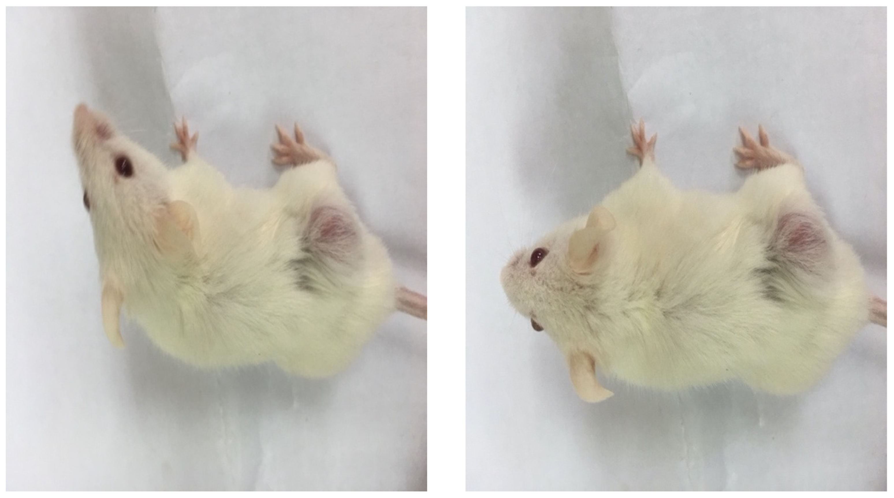
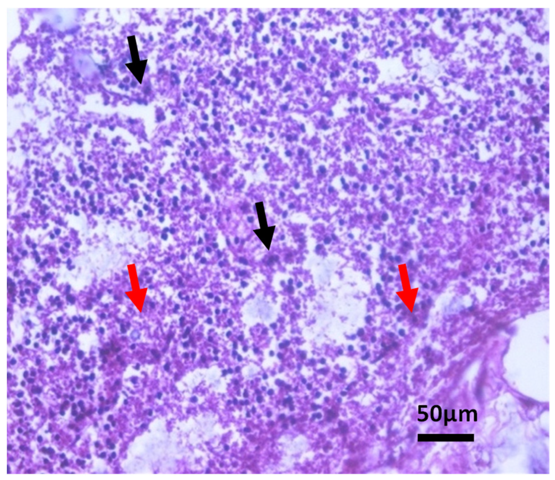
Histopathological Observations of Tissues

The histopathological examination of muscle fiber sections from the control group revealed normal muscle fiber (black arrow) and entangling fibrous tissue (red arrow) (HE, ×400) (Figure 12). Positive control sections showed malignant growth formed of sheets of malignant epithelial cells with large nuclei and an increased the nucleocytoplasmic ratio (black arrow), showing moderate anaplasia (Figure 13). Sections of mice administered one-tenth the LD50 of P. roxburghii extract are illustrated in Figure 14 and showed a scattered sheet of dysplastic epithelial cells with rounded nuclei with an increased nucleocytoplasmic ratio (black arrow), with many apoptotic cells and necrosis (red arrows).
Sections of mice administered one-tenth the LD50 of N. graveolens extract are illustrated in Figure 15 and showed a scattered sheet of epithelial cells with rounded nuclei with an increased nucleocytoplasmic ratio (black arrow), with many degenerated/necrotic and/or apoptotic cells (red arrows).

4. Conclusions

The research outputs of the current study concluded that administration of one-tenth of the LD50 of P. roxburghii and N. graveolens extracts possess anti-proliferative activity by suppressing the tumor growth rate in the BALB/c mouse model. General enhancement in liver and kidney functions, antioxidant activity, and reduction in tumor markers blood levels were detected in the BALB/c mouse model after the administration of the selected dose. Furthermore, the apoptotic cell cycle was enhanced by optimizing the regulation of various apoptotic gene marker expression levels. These improvements were supported by histopathological changes. However, further clinical trials are needed to be performed to confirm these extracts as a potential anticancer drug.

Author Contributions

Methodology, M.G., K.M., A.A.A.-R., A.A.Z., A.M.H., A.S.A.-A. and H.Z.H.; software and validation, M.G., M.S.A.-L. and A.A.Z.; formal analysis, M.G., F.A.A., A.A.A.-R. and A.S.A.-A., investigation, M.G. and A.A.Z.; resources, M.G., H.Z.H., A.A.A.-R., A.M.H. and A.A.Z.; data curation, M.G. and A.A.Z.; writing—original draft preparation, M.G., F.A.A., A.A.A.-R. and A.A.Z.; writing—review and editing, C.P., M.G., H.Z.H., K.M., A.A.Z. and F.O.; visualization, A.A.Z. and M.G.; project administration, H.Z.H., K.M., M.G., F.A.A., A.A.Z., M.G. and C.P.; funding acquisition, A.A.Z. and M.G. All authors have read and agreed to the published version of the manuscript.

Funding

This research received no external funding.

Institutional Review Board Statement

The animal study was reviewed and approved by the Ethical Committee of Medical Research at National Research Centre, Cairo, Egypt.

Data Availability Statement

Not applicable.

Acknowledgments

We thank the National Research Centre, Cairo, Egypt and the National and Kapodistrian University of Athens.

Conflicts of Interest

The authors declare no conflict of interest.

References

  1. Mathers, C.D.; Loncar, D. Projections of Global Mortality and Burden of Disease from 2002 to 2030. PLoS Med. 2006, 3, 442. [Google Scholar] [CrossRef] [PubMed] [Green Version]
  2. Ferlay, J.; Ervik, M.; Lam, F.; Colombet, M.; Mery, L.; Pineros, M.; Znaor, A.; Soerjomataram, I.; Bray, F. Global Cancer Observatory: Cancer Today. Lyon, France: International Agency for Research on Cancer. Available online: https://gco.iarc.fr/today (accessed on 1 December 2020).
  3. World Health Organization (WHO). Global Health Estimates 2020: Deaths by Cause, Age, Sex, by Country and by Region, 2000–2019; WHO: Geneva, Switzerland, 2020; Available online: https://www.who.int/data/gho/data/themes/mortality-and-global-health-estimates/ghe-leading-causes-of-death (accessed on 11 December 2020).
  4. Wiseman, M. The second World Cancer Research Fund/American Institute for Cancer Research expert report. Food, nutrition, physical activity, and the prevention of cancer: A global perspective. Proc. Nutr. Soc. 2008, 67, 253–256. [Google Scholar] [CrossRef] [PubMed] [Green Version]
  5. Tsao, A.S.; Edward, S.K.; Hong, W.K. Chemoprevention of cancer. CA Cancer J. Clin. 2004, 54, 150–180. [Google Scholar] [CrossRef] [PubMed] [Green Version]
  6. Surh, Y.J.; Na, H.K.; Lee, S.S. Transcription factors and mitogen-activated protein kinases as molecular targets for chemoprevention with anti-inflammatory phytochemicals. Biofactors 2004, 21, 103–108. [Google Scholar] [CrossRef]
  7. Gonzalez de Mejia, E.; Song, Y.S.; Ramirez-Mares, M.V.; Kobayashi, H. Effect of yerba mate (Ilex paraguariensis) tea on topoisomerase inhibition and oral carcinoma cell proliferation. J. Agric. Food. Chem. 2005, 53, 1966–1973. [Google Scholar] [CrossRef]
  8. Yano, H.; Mizoguchi, A.; Fukuda, K. The herbal medicine sho-saiko-to inhibits proliferation of cancer cell lines by inducing apoptosis and arrest at the G0/G1 phase. Cancer Res. 1994, 54, 448–454. [Google Scholar]
  9. Oubre, A.Y.; Carlson, T.J.; King, S.R.; Reaven, G.M. From plant to patient: An ethnomedical approach to the identification of new drugs for the treatment of NIDDM. Diabetologia 1997, 40, 614–617. [Google Scholar] [CrossRef]
  10. Gao, H.; Lamusta, J.; Zhang, W. F Tumor cell selective cytotoxicity and apoptosis induction by an herbal preparation from brucea javanica. N. Am. J. Med. Sci. 2011, 4, 62–66. [Google Scholar] [CrossRef]
  11. Zahri, S.; Razavi, S.M.; Niri, F.H.; Mohammadi, S. Induction of programmed cell death by Prangos uloptera, a medicinal plant. Biol. Res. 2009, 42, 517–522. [Google Scholar] [CrossRef] [Green Version]
  12. Russo, M.; Spagnuolo, C.; Tedesco, I.; Bilotto, S.; Russo, G.L. The flavonoid quercetin in disease prevention and therapy: Facts and fancies. Biochem. Pharmacol. 2011, 83, 6–15. [Google Scholar] [CrossRef]
  13. Zadi, M.Y.; Heidari, M.; Akbarpour, M.; Mirjalili, M.H.; Zeinali, A.; Parsa, M. In vitro cytotoxic activity of the essential oil of Dorema ammoniacum D. Don. Middle East J. Sci. Res. 2011, 7, 511–514. [Google Scholar]
  14. Tamimi, R.M.; Hankinson, S.E.; Campos, H.; Spiegelman, D.; Zhang, S.; Colditz, G.A. Plasma carotenoids, retinol, and tocopherols and risk of breast cancer. Am. J. Epidemiol. 2005, 161, 153–160. [Google Scholar] [CrossRef]
  15. Shen, T.; Khor, S.C.; Zhou, F.; Duan, T.; Xu, Y.Y.; Zheng, Y.F. Chemoprevention by lipid-soluble tea polyphenols in diethylnitrosamine/phenobarbital-induced hepatic pre- cancerous lesions. Anticancer. Res. 2014, 34, 683–693. [Google Scholar]
  16. Kaushik, D.; Kumar, A.; Kaushik, P.; Rana, A.C. Analgesic and anti-inflammatory activity of P. roxburghii Sarg. Advanc. Pharmacol. Sci. 2012, 2012, 245431. [Google Scholar]
  17. Anju, P.; Srivastava, A.K.; Singhal, B.; Mishra, S.K.; Srivastava, S.; Lakshmi, V. Antidyslipidemic and antioxidant activity of Pinus roxburghii needles. Med. Chem. Res. 2011, 20, 1589–1593. [Google Scholar]
  18. Qadir, M.; Shah, W.A. GC-MS analysis, antibacterial, antioxidant and anticancer activity of essential oil of P. roxburghii from Kashmir, India. Int. J. Res. Pharm. Chem. 2014, 4, 228–232. [Google Scholar]
  19. Satyal, P.; Paudel, J.; Raut, A.; Noura, S.; Setzer, N. Volatile constituents of P. roxburghii from Nepal. Phcog. Res. 2013, 5, 43–48. [Google Scholar]
  20. Sharma, A.; Goyal, R.; Sharma, L. Potential biological efficacy of Pinus plant species against oxidative, inflammatory and microbial disorders. BMC Altern. Complement. Med. 2016, 6, 35. [Google Scholar] [CrossRef] [Green Version]
  21. Rawat, U.; Srivastava, B.; Semwal, S.; Sati, O.P. Xanthones from Pinus roxburghii. J. Indian. Chem. Soc. 2009, 83, 391–392. [Google Scholar] [CrossRef]
  22. Cheriti, A.; Saad, A.; Belboukhari, N.; Ghezali, S. The essential oil composition of Bubonium graveolens (Forssk.) Maire from the Algerian Sahara. Flavour. Fragr. J. 2007, 22, 286–288. [Google Scholar] [CrossRef]
  23. Ramdane, F.; Essid, R.; Mkadmini, K.; Hammami, M.; Fares, N.; Mahammed, M.H.; El Ouassis, D.; Tabbene, O.; Limam, F.; Hadj, M.D. Phytochemical composition and biological activities of Asteriscus graveolens (Forssk) extracts. Process. Biochem. 2017, 56, 186–192. [Google Scholar] [CrossRef]
  24. Chaib, F.; Allali, H.; Bennaceur, M.; Flamini, G. Chemical composition and antimicrobial activity of essential oils from the aerial parts of Asteriscus graveolens (forssk.) less. and Pulicaria incisa (lam.) dc: Two asteraceae herbs growing wild in the hoggar. Chem. Biodiv. 2017, 14, 1700092. [Google Scholar] [CrossRef] [PubMed]
  25. Aouissi, H.; Gourine, N.; Wang, H.; Chen, X.I.; Bombarda, I.; Boudjeniba, M.; Yousfi, M. Chemical composition, antioxidative, antimicrobial and anti-cancer activities of Asteriscus graveolens (Forssk) essential oil. Oriental. Pharm. Experim. Med. 2018, 18, 217–223. [Google Scholar] [CrossRef] [Green Version]
  26. Negrín, G.; Eiroa, J.L.; Morales, M.; Triana, J.; Quintana, J.; Estévez, F. Naturally occurring asteriscunolide A induces apoptosis and activation of mitogen-activated protein kinase pathway in human tumor cell lines. Mol. Carcinog. 2010, 49, 488–499. [Google Scholar] [CrossRef]
  27. Tsujimoto, Y.; Finger, L.R.; Yunis, J.; Nowell, P.C.; Croce, C.M. Cloning of the chromosome breakpoint of neoplastic B cells with the t(14; 18) chromosome translocation. Science 1984, 226, 1097–1099. [Google Scholar] [CrossRef]
  28. Reed, J.C. Bcl-2 family proteins: Regulators of apoptosis and chemoresistance in haematologic malignancies. Semin. Haematol. 1997, 34, 9–19. [Google Scholar]
  29. Ghobrial, I.M.; Witzig, T.E.; Adjei, A.A. Targeting apoptosis pathways in cancer therapy. CA Cancer J. Clin. 2005, 55, 178–194. [Google Scholar] [CrossRef] [Green Version]
  30. Kamm, Y.J.; Peters, G.J.; Hull, W.E.; Punt, C.J.; Heerschap, A. Correlation between 5-fluorouracil metabolism and treatment response in two variants of C26 murine colon carcinoma. Br. J. Cancer 2003, 89, 754–762. [Google Scholar] [CrossRef] [Green Version]
  31. Schastak, S.; Yafai, Y.; Geyer, W.; Kostenich, G.; Orenstein, A.; Wiedemann, P. Initiation of apoptosis by photodynamic therapy using a novel positively charged and water-soluble near infra-red photosensitizer and white light irradiation. Methods Find Exp. Clin. Pharmacol. 2008, 30, 17–23. [Google Scholar] [CrossRef]
  32. Aulino, P.; Berardi, E.; Cardillo, V.M.; Rizzuto, E.; Perniconi, B.; Ramina, C. Molecular, cellular and physiological characterization of the cancer cachexia-inducing C26 colon carcinoma in mouse. BMC Cancer 2010, 10, 363. [Google Scholar] [CrossRef]
  33. Boulos, L. Flora of Egypt 3: Compositae-Verbenaceae; Al Hadara Publishing: Cairo, Egypt, 2002. [Google Scholar]
  34. Gad, M.; Hassouna, H.Z.; Mahmoud, K.; Abd-Rabou, A.A.; Abdel-Azeem, A.S.; Hegazy, A.H.; Adel-Lattife, M.S.; Ahmed, F.A. Preliminary study on the toxicological impacts of P. roxburghii and N. graveolens extracts on albino mice. Egypt. J. Chem. 2021, 64, 3489–3498. [Google Scholar]
  35. Reeves, P.G.; Nielsen, F.H.; Fahey, G.C. AIN-93 purified diet for laboratory rodents: Final report of american institute of nutrition ad hoc writing committee on the reformulation of the AIN-76A rodent diet. J. Nutr. 1993, 123, 1939–1951. [Google Scholar] [CrossRef] [PubMed] [Green Version]
  36. Wilbrandt, W. Behrens methods for calculation of LD50. Arzneimittel-Forschung 1952, 2, 501–503. [Google Scholar] [PubMed]
  37. Tanaka, Y.; Eda, H.; Fujimoto, k.; Tanaka, T.; Ishikawa, T.; Ishitsuka, H. Anticachectic activity of 5′-Deoxy-5- fluorouridine in a murine tumor cachexia model, colon 26 adenocarcinoma. Cancer Res. 1990, 50, 4528–4532. [Google Scholar] [PubMed]
  38. Fujimoto-Ouchi, K.; Tamura, S.; Mori, K.; Tanaka, Y.; Ishitsuka, H. Establishment and characterization of cachexia- inducing and -non-inducing clones of murine colon 26 carcinoma. Exper. Cancer 1995, 61, 522–528. [Google Scholar] [CrossRef]
  39. Euhus, D.M.; Hudd, C.; LaRegina, M.C.; Johnson, F.E. Tumor measurement in the nude mouse. J. Surg. Oncol. 1986, 31, 229–234. [Google Scholar] [CrossRef]
  40. Tomayko, M.M.; Reynolds, C.P. Determination of subcutaneous tumor size in athymic (nude) mice. Cancer Chemother. Pharmacol. 1989, 24, 148–154. [Google Scholar] [CrossRef]
  41. Hsu, H.W.; Sutton, N.E.; Banjo, M.O.; Satterlee, L.D.; Kendrick, J.G. The C-PER and assays for protein quality. J. Food Technol. 1978, 12, 69–74. [Google Scholar]
  42. Reitman, S.; Frankel, S.A. Colorimetric method for the determination of serum glutamic oxaloacetic and glutamic pyruvic transaminases. Am. J. Clin. Path. 1957, 28, 56–62. [Google Scholar] [CrossRef]
  43. Rec, G.S. Optimized standard colorimetric methods. Serum Alkaline phosphatase (DGKC). J. Clin. Chem. Clin. Biochem. 1972, 10, 182. [Google Scholar]
  44. Patton, C.J.; Crouch, S.R. Spectrophotometric and kinetics investigations of the Berthelot reaction for the determination of ammonia. Analyti. Chem. 1977, 49, 464–469. [Google Scholar] [CrossRef]
  45. Faulkner, N.R.; King, J.W. Fundamental of Clinical Chemistry, 2nd ed.; Tietz, N.W., Ed.; Sannders: Philadelphia, PA, USA, 1976; pp. 994–998. [Google Scholar]
  46. Goldberg, D.M.; Spooner, R.J. Methods of Enzymatic Analysis, 3rd ed.; Bergmeyer, H.-U., Ed.; Verlog. Chem. Weinheim: Deerfield Beach, FL, USA, 1983; Volume 3, pp. 258–265. [Google Scholar]
  47. Koracevic, D.; Koracevic, G.; Djordjevic, V.; Andrejevic, S.; Cosic, V. Method for the measurement of antioxidant activity in human fluids. J. Clin. Pathol. 2001, 54, 356–361. [Google Scholar] [CrossRef] [PubMed] [Green Version]
  48. Satoh, K. Serum lipid peroxide in cerebrovascular disorders determined by a new colorimetric method. Clin. Chem. Acta 1978, 90, 37–43. [Google Scholar]
  49. Bancroft, D.; Stevens, A.; Turner, R. Theory and Practice of Histological Techniques, 4th ed.; Churchill Livingstone: Edinburgh, UK; London, UK; Melbourne, Australia, 1996. [Google Scholar]
  50. International Union against Cancer. TNM Classification of Malignant Tumours, 5th ed.; Sobin, L.H., Wittekind, C., Eds.; Wiley-Liss: New York, NY, USA, 1997. [Google Scholar]
  51. Tullie, L.G.; Sohn, H.M.; Zylstra, J. A role for tumor volume assessment in resectable esophageal cancer. Ann. Surg. Oncol. 2016, 23, 3063–3070. [Google Scholar] [CrossRef] [PubMed]
  52. Huang, A.C.; Postow, M.A.; Orlowski, R.J. T-cell invigoration to tumour burden ratio associated with anti-PD-1 response. Nature 2017, 545, 60–65. [Google Scholar] [CrossRef] [Green Version]
  53. Alexander, B.M.; Othus, M.; Caglar, H.B.; Allen, A.M. Tumor volume is a prognostic factor in non-small-cell lung cancer treated with chemoradiotherapy. Int. J. Radiat. Oncol. Biol. Phys. 2011, 79, 1381–1387. [Google Scholar] [CrossRef]
  54. Rutkowski, T. The role of tumor volume in radiotherapy of patients with head and neck cancer. Radiat. Oncol. 2014, 9, 23. [Google Scholar] [CrossRef] [Green Version]
  55. Campbell, P.T.; Newton, C.C.; Newcomb, P.A. Association between body mass index and mortality for colorectal cancer survivors: Overall and by tumor molecular phenotype. Cancer Epidemiol. Biomark. Prev. 2015, 24, 1229–1238. [Google Scholar] [CrossRef] [Green Version]
  56. Baade, P.D.; Meng, X.; Youl, P.H. The impact of body mass index and physical activity on mortality among patients with colorectal cancer in Queensland, Australia. Cancer Epidemiol. Biomark. Prev. 2011, 20, 1410–1420. [Google Scholar] [CrossRef] [Green Version]
  57. Diculescu, M.; Iacob, R.; Iacob, S.; Croitoru, A.; Becheanu, G.; Popeneciu, V. The importance of histopathological and clinical variables in predicting the evolution of colon cancer. Rom. J. Gastroenterol. 2002, 11, 183–189. [Google Scholar]
  58. Barber, M.D.; Ross, J.A.; Fearon, K.C. Disordered metabolic response with cancer and its management. World J. Surg. 2000, 24, 681–689. [Google Scholar] [CrossRef] [PubMed]
  59. Ferreira, L.M.; Hebrant, A.; Dumont, J.E. Metabolic reprogramming of the tumor. Oncogene 2012, 31, 3999–4011. [Google Scholar] [CrossRef] [PubMed] [Green Version]
  60. Giannini, E.G.; Testa, R.; Savarino, V. Liver enzyme alteration: A guide for clinicians. CMAJ 2005, 172, 367–379. [Google Scholar] [CrossRef] [PubMed] [Green Version]
  61. Okuda, M.; Li, K.; Beard, M.R. Mitochondrial injury, oxidative stress, and antioxidant gene expression are induced by hepatitis C virus core protein. Gastroenterology 2002, 122, 366–375. [Google Scholar] [CrossRef] [PubMed] [Green Version]
  62. Liu, X.; Meng, Q.H.; Ye, Y. Prognostic significance of pretreatment serum levels of albumin, LDH and total bilirubin in patients with non-metastatic breast cancer. J. Carcinog. 2015, 36, 243–248. [Google Scholar] [CrossRef] [Green Version]
  63. Chen, S.L.; Li, J.P.; Li, L.F. Elevated preoperative serum alanine aminotransferase/aspartate aminotransferase (alt/ast) ratio is associated with better prognosis in patients undergoing curative treatment for gastric adenocarcinoma. Int. J. Mol. Sci. 2016, 17, 911. [Google Scholar] [CrossRef] [PubMed]
  64. Ahn, J.H.; Kim, S.B.; Yun, M.R.; Lee, J.S.; Kang, Y.K.; Kim, W.K. Alternative therapy and abnormal liver function during adjuvant chemotherapy in breast cancer patients. J. Korean Med. Sci. 2004, 19, 397–400. [Google Scholar] [CrossRef]
  65. Pfeiffer-Guglielmi, B.B.; Dombert, S.; Jablonka, V.; Hausherr, V.; Thriel, C.; Schobel, N.; Jansen, R.P. Axonal and dendritic localization of mRNAs for glycogen-metabolizing enzymes in cultured rodent neurons. BMC. Neurosci. 2014, 15, 15–70. [Google Scholar] [CrossRef] [Green Version]
  66. Yasuda, Y.; Kawamura, K.; Ichikado, K.; Yoshioka, M. Alkaline phosphatase flare phenomenon following epidermal growth factor receptor-tyrosine kinase inhibitor treatment of non-small cell lung cancer: Report of a case and case review. Respir. Med. Case Rep. 2014, 13, 51–53. [Google Scholar] [CrossRef] [Green Version]
  67. Lakshmi, A.; Subramanian, S. Chemotherapeutic effect of tangeretin, a poly methoxylated flavone studied in 7, 12-dimethylbenz (a) anthracene-induced mammary carcinoma in experimental rats. Biochimie 2014, 99, 96–109. [Google Scholar] [CrossRef]
  68. Gross, J.L.; De Azevedo, M.J.; Silveiro, S.P.; Canani, L.H.; Caramori, M.L.; Zelmanovitz, T. Diabetic nephropathy: Diagnosis, prevention, and treatment. Diabetes Care 2005, 28, 164–176. [Google Scholar] [CrossRef] [PubMed] [Green Version]
  69. Faull, R. Prescribing in renal disease. Aust. Prescr. 2007, 30, 17–20. [Google Scholar] [CrossRef]
  70. Yener, Y.; Yerlikaya, F.H.; Toker, A. Evaluation of some renal function parameters in rats treated with acrylamide. AJAVS 2016, 2, 1–8. [Google Scholar]
  71. Raj, G.; Chidambaram, R.; Varunkumar, K.; Ravikumar, V.; Pandi, M. Chemopreventive potential of fungal taxol against 7,12-dimethylbenz [a] anthracene induced mammary gland carcinogenesis in Sprague Dawley rats. Eur. J. Pharmacol. 2015, 767, 108–118. [Google Scholar]
  72. Koji, K.; Shiro, H.; Mitsuo, T.; Hideyuki, A. Serum creatinine level for cervical cancer. J. Oncol. 1998, 28, 546–550. [Google Scholar]
  73. Falconer, I.S.; Slater, C.; Ross, J.A.; Preston, T.; Fearon, K. Albumin synthetic rates, the acute phase response and cyrolanes in pancreatic cancer. J. Surg. 1995, 82, 682. [Google Scholar]
  74. Erel, O.A. A novel automated method to measure total antioxidant response against potent free radical reactions. Clin. Biochem. 2004, 37, 112–119. [Google Scholar] [CrossRef]
  75. Perse, M.; Cerar, A. Morphological and molecular alterations in 1,2 dimethylhydrazine and azoxymethane induced colon carcinogenesis in rats. J. Biomed. Biotechnol. 2011, 2011, 473964. [Google Scholar] [CrossRef]
  76. Mena, S.; Ortega, A.; Estrela, J.M. Oxidative stress in environmental-induced carcinogenesis. Mutat. Res. Toxicol. Environ. Mutagen. 2009, 674, 36–44. [Google Scholar] [CrossRef]
  77. Reuter, S.; Gupta, S.C.; Chaturvedi, M.M.; Aggarwal, B.B. Oxidative stress, inflammation, and cancer: How are they linked? Free Radic. Biol. Med. 2010, 49, 1603–1616. [Google Scholar] [CrossRef] [Green Version]
  78. Fang, C.; Dupertuis, Y.M.; Pichard, C. Role of polyunsaturated fatty acids and lipid peroxidation on colorectal cancer risk and treatments. Curr. Opin. Clin. Nutr. Metab Care 2012, 15, 99–106. [Google Scholar]
  79. Ayala, A.; Munoz, M.F.; Arguelles, S. Lipid peroxidation: Production, metabolism, and signaling mechanisms of malondialdehyde and 4-Hydroxy-2-Nonenal. Oxid. Med. Cell. Longev. 2014, 2014, 360438. [Google Scholar] [CrossRef] [PubMed] [Green Version]
  80. Mayo, J.C.; Tan, D.X.; Sainz, R.M.; Lopez-Burillo, S.; Reiter, R.J. Oxidative damage to catalase induced by peroxyl radicals: Functional protection by melatonin and other antioxidants. Free Radic. Res. 2003, 37, 543–553. [Google Scholar] [CrossRef] [PubMed]
  81. Cvorovic, J.; Tramer, F.; Granzotto, M.; Candussio, L.; Decorti, G.; Passamonti, S. Oxidative stress-based cytotoxicity of delphinidin and cyanidin in colon cancer cells. Arch. Biochem. Biophys. 2010, 501, 151–157. [Google Scholar] [CrossRef]
  82. Jung, B.; Staudacher, J.J.; Beauchamp, D. Transforming growth factor beta superfamily signaling in development of colorectal cancer. Gastroenterology 2017, 152, 36–52. [Google Scholar] [CrossRef] [Green Version]
  83. Principe, D.R.; Doll, J.A.; Bauer, J.; Jung, B.; Munshi, H.G.; Bartholin, L.; Pasche, B.; Lee, C.; Grippo, P.J. TGF-β: Duality of Function Between Tumor Prevention and Carcinogenesis. J. Natl. Cancer Inst. 2014, 106, djt369. [Google Scholar] [CrossRef]
  84. Li, Y.H.; Cao, Z.; Jiao, S.B.; Pakala, D.N.; Sirigiri, R.; Li, W.; Kumar, R.; Mishra, L. Carcinoembryonic antigen interacts with TGF- receptor and inhibits TGF-ß signaling in colorectal cancers. Cancer Res. 2010, 70, 8159–8168. [Google Scholar] [CrossRef] [Green Version]
  85. Bast, R.C.P.; Ravdin, D.F.; Hayes, S.; Bates, H.J.R.; Fritsche, J.M.; Jessup, N.; Kemeny, G.Y.; Locker, R.G.; Mennel and Somerfield, M.R. Update of recommendations for the use of tumor markers in breast and colorectal cancer: Clinical practice guidelines of the american society of clinical oncology. J. Clin. Oncol. 2000, 19, 1865–1878. [Google Scholar] [CrossRef]
  86. Taheri, M.; Saragovi, M.U.; Stanners, C.P. The adhesion and differentiation- inhibitory activities of the immunoglobulin superfamily member, carcinoembryonic antigen, can be independently blocked. J. Biol. Chem. 2003, 278, 14632–14639. [Google Scholar] [CrossRef] [Green Version]
  87. Del Villano, B.C.; Brennan, S.; Brock, P.; Bucher, C.; Liu, V.; McClure, M.; Rake, B.; Space, S.; Westrick, B.; Schoemaker, H., Jr.; et al. Radio immunometric assay for a monoclonal antibody- defined tumor marker, CA 19-9. Clin. Chem. 1983, 29, 549–552. [Google Scholar] [CrossRef]
  88. Filella, X.; Molina, R.; Grau, J.J.; Piqué, J.M.; Garcia-Valdecasas, J.C.; Astudillo, E.; Biete, A.; Bordas, J.M.; Novell, A.; Campo, E.; et al. Prognostic value of CA19-9 levels in colorectal cancer. Ann. Surg. 1992, 216, 55–59. [Google Scholar] [CrossRef] [PubMed]
  89. Nakayama, T.; Watanabe, M.; Teramoto, T.; Kitajima, M. CA19-9 as a predictor of recurrence in patients with colorectal cancer. J. Surg. Oncol. 1997, 66, 238–243. [Google Scholar] [CrossRef]
  90. Delbridge, A.R.D.; Valente, L.J.; Strasser, A. The role of the apoptotic machinery in tumor suppression. Cold Spring Harb. Perspect. Biol. 2012, 4, a008789. [Google Scholar] [CrossRef] [PubMed]
  91. Guo, X.X.; Li, Y.; Sun, C.; Jiang, D.; Lin, Y.J.; Jin, F.X.; Lee, S.K.; Jin, Y.H. P53-dependent Fas expression is critical for Gin- senoside Rh2 triggered caspase-8 activation in HeLa cells. Protein Cell 2014, 5, 224–234. [Google Scholar] [CrossRef] [PubMed] [Green Version]
  92. Granado-Serrano, A.B.; Martin, M.A.; Goya, L. Time- course regulation of survival pathways by epicatechin on HepG2 cells. J. Nutr. Biochem. 2009, 20, 115–124. [Google Scholar] [CrossRef] [Green Version]
  93. Moradzadeh, M.; Hosseini, A.; Erfanian, S.; Rezaei, H. Epigallocatechin-3-gallatepromotesapoptosisin human breast cancer T47D cells through down-regulation of PI3K/AKT and Telomerase. Pharmacol. Rep. 2017, 69, 924–928. [Google Scholar] [CrossRef]
  94. Zhang, T.; Ma, L.; Wu, P.; Li, W.; Li, T.; Gu, R.; Dan, X.; Li, Z.; Fan, X.; Xiao, Z. Gallic acid has anticancer activity and enhances the anticancer effects of cisplatin in non small cell lung cancer A549 cells via the JAK/STAT3 signaling pathway. Oncol. Rep. 2019, 41, 1779–1788. [Google Scholar] [CrossRef] [Green Version]
  95. Sajid, A.; Manzoor, Q.; Iqbal, M.; Tyagi, A.K.; Adil Sarfraz, R.; Sajid, A. Proxburghii essential oil anticancer activity and chemical composition evaluation. EXCLI J. 2018, 17, 233–245. [Google Scholar]
  96. Ben Sghaier, M.; Pagano, A.; Mousslim, M.; Ammari, Y.; Kovacic, H.; Luis, J. Rutin inhibits proliferation, attenuates superoxide production and decreases adhesion and migration of human cancerous cells. Biomed. Pharmacother. 2016, 84, 1972–1978. [Google Scholar] [CrossRef]
  97. Gouthamchandra, K.; Sudeep, H.V.; Venkatesh, B.J.; Shyam Prasad, K. Chlorogenic acid complex (CGA7), standardized extract from green coffee beans exerts anticancer effects against cultured human colon cancer HCT-116 cells. Food. Sci. Hum. Wellness 2017, 6, 147–153. [Google Scholar] [CrossRef]
  98. Shukla, S.; Shankar, E.; Fu, P.; MacLennan, G.T.; Gupta, S. Suppression of NF-κB and NF-κB-Regulated gene expression by apigenin through IκBα and IKK pathway in TRAMP mice. PLoS ONE 2015, 10, e0138710. [Google Scholar] [CrossRef] [PubMed] [Green Version]
  99. Sharmila, R.; Manoharan, S. Anti-tumor activity of rosmarinic acid in 7,12-dimethylbenz(a)anthracene (DMBA) induced skin carcinogenesis in Swiss albino mice. Indian J. Exp. Biol. 2012, 50, 187–194. [Google Scholar] [PubMed]
  100. Li, H.; Zhang, Y.; Chen, H.H.; Huang, E.; Zhuang, H.; Li, D.; Ni, F. Rosmarinic acid inhibits stem-like breast cancer through hedgehog and Bcl-2/Bax signaling pathways. Phcog Mag. 2019, 15, 600–606. [Google Scholar] [CrossRef]
  101. Pelinson, L.P.; Assmann, C.E.; Palma, T.V.; da Cruz, I.B.M.; Pillat, M.M.; Manica, A.; Stefanello, N.; Weis, G.C.C.; de Oliveira Alves, A.; de Andrade, C.M. Antiproliferative and apoptotic effects of caffeic acid on SK-Mel-28 human melanoma cancer cells. Mol. Biol. Rep. 2019, 46, 2085–2092. [Google Scholar] [CrossRef] [PubMed]
  102. Abaza, M.S.; Al-Attiyah, R.; Bhardwaj, R.; Abbadi, G.; Koyippally, M.; Afzal, M. Syringic acid from Tamarix aucheriana possesses antimitogenic and chemo-sensitizing activities in human colorectal cancer cells. Pharm. Biol. 2013, 51, 1110–1124. [Google Scholar] [CrossRef] [PubMed] [Green Version]
  103. Gong, J.; Zhou, S.; Yang, S. Vanillic Acid Suppresses HIF-1alpha Expression via Inhibition of mTOR/p70S6K/4E- BP1 and Raf/MEK/ERK Pathways in Human Colon Cancer HCT-116 Cells. Int. J. Mol. Sci. 2019, 20, 465. [Google Scholar] [CrossRef] [Green Version]
  104. Sharma, S.H.; Rajamanickam, V.; Nagarajan, S. Supplementation of p-coumaric acid exhibits chemopreventive effect via induction of Nrf2 in a short-term preclinical model of colon cancer. Eur. J. Cancer Prev. 2019, 28, 472–482. [Google Scholar] [CrossRef]
Figure 1. Tumor growth in BALB/c mice.
Separations 09 00277 g001
Figure 2. Average tumor volume per week in different groups.
Separations 09 00277 g002
Figure 3. Serum ALT, AST, and ALP activities in different groups.
Separations 09 00277 g003
Figure 4. Levels of serum urea and creatinine in different groups.
Separations 09 00277 g004
Figure 5. Serum glutathione reductase activity in different groups.
Separations 09 00277 g005
Figure 6. Level of serum total antioxidant capacity in different groups.
Separations 09 00277 g006
Figure 7. Level of serum malondialdehyde in different groups.
Separations 09 00277 g007
Figure 8. Level of serum TGF-β in different groups.
Separations 09 00277 g008
Figure 9. Level of serum CEA in different groups.
Separations 09 00277 g009
Figure 10. Level of serum CA19-9 in different groups.
Separations 09 00277 g010
Figure 11. Effect of P. roxburghii and N. graveolens extracts on Bcl-2, Bax, Cas3, NF-kB, and PI3k genes relative to the GAPDH gene in BALB/c mice.
Separations 09 00277 g011
Figure 12. Sections of muscle fibers from the control group showed normal muscle fibers (black arrow) and entangling fibrous tissue (red arrow) (HE, ×400).
Separations 09 00277 g012
Figure 13. Sections of the positive control group showed malignant growth formed of sheets of malignant epithelial cells with large nuclei with an increased nucleocytoplasmic ratio (black arrow), showing a moderate degree of anaplasia (HE, ×400).
Separations 09 00277 g013
Figure 14. Sections treated with P. roxburghii extract showed a scattered sheet of dysplastic epithelial cells with rounded nuclei with an increased nucleocytoplasmic ratio (black arrow), with many apoptotic cells and necrosis (red arrows) (HE, ×400).
Separations 09 00277 g014
Figure 15. Sections treated with N. graveolens extract showed a scattered sheet of epithelial cells with rounded nuclei with an increased nucleocytoplasmic ratio (black arrow), with many degenerated/necrotic and/or apoptotic cells (red arrows) (HE, ×400).
Separations 09 00277 g015
Table 1. Primer sequences used for qRT-PCR.
PrimersSequences
Bcl-2F: 5′TGGGATGCCTTTGTGGAAC 3′
R: 5′CATATTTGTTTGGGGCAGGTC3′
BaxF: 5′TGCTACAGGGTTTCATCCAG3′
R: 5′ATCCACATCAGCAATCATCC3′
Cas-3F: 5′GCTGGACTGCGGTATTGAGA3′
R: 5′CCATGACCCGTCCCTTGA3′
NF-kBF: 5′GCAAAGGGAACATTCCGATAT3′
R: 5′GCGACATCACATGGAAATCTA3′
PI3kF: 5′GTGTCAGCGCTCTCCGCC3′
R: 5′CTGATAATTGATGTAGG3′
GAPDHF: 5′CATTCAAGACCGGACAGAGG3′
R: 5′ACATACTCAGCACCAGCATCACC3′
Table 2. Nutritional parameters of different groups.
Parameters
Groups
Body Weight Gain (BWG) (g)Food Intake (FI) (g)Feed Efficiency Ratio (FER)
Negative control27.11 ± 0.37 a189.21 ± 0.53 a0.14
Positive control19.36 ± 0.64 c177.54 ± 0.66 c0.10
P. roxburghii24.52 ± 0.58 b184.37 ± 0.71 b0.13
N. graveolens25.23 ± 0.45 b186.29 ± 0.49 b0.13
All values are represented as the mean ± S.E. Means with different letters are significantly different (p < 0.05).
Publisher’s Note: MDPI stays neutral with regard to jurisdictional claims in published maps and institutional affiliations.

