Article

Synthesis, Structures and Chemical Reactivity of Dithiolato-Bridged Ni-Fe Complexes as Biomimetics for the Active Site of [NiFe]-Hydrogenases

Department of Chemistry, State Key Laboratory of Elemento-Organic Chemistry, College of Chemistry, Nankai University, Tianjin 300071, China
*
Author to whom correspondence should be addressed.
Submission received: 22 May 2022 / Revised: 20 June 2022 / Accepted: 21 June 2022 / Published: 24 June 2022
(This article belongs to the Special Issue Inorganics: 10th Anniversary)

Abstract

:
To develop the structural and functional modeling chemistry of [NiFe]-H2ases, we have carried out a study regarding the synthesis, structural characterization and reactivity of a new series of [NiFe]-H2ase model complexes. Thus, treatment of diphosphine dppb-chelated Ni complex (dppb)NiCl2 (dppb = 1,2-(Ph2P)2C6H4) with (dppv)Fe(CO)2(pdt) (dppv = 1,2-(Ph2P)2C2H2, pdt = 1,3-propanedithiolate) and NaBF4 gave dicarbonyl complex [(dppb)Ni(pdt)Fe(CO)2(dppv)](BF4)2 ([A](BF4)2). Further treatment of [A](BF4)2 with Me3NO and Bu4NCN or KSCN afforded t-cyanido and t-isothiocyanato complexes [(dppb)Ni(pdt)Fe(CO)(t-R)(dppv)]BF4 ([1]BF4, R = CN; [2]BF4, R = NCS), respectively. While azadiphosphine MeN(CH2PPh2)2-chelated t-hydride complex [MeN(CH2PPh2)2Ni(pdt)Fe(CO)(t-H)(dppv)]BF4 ([3]BF4) was prepared by treatment of dicarbonyl complex [MeN(CH2PPh2)2Ni(pdt)Fe(CO)2(dppv)](BF4)2 ([B](BF4)2) with Me3NO and 1.5 MPa of H2, treatment of dicarbonyl complex [B](BF4)2 with Me3NO (without H2) in pyridine resulted in formation of a novel monocarbonyl complex [MeN(CH2PPh2)2Ni(SCHCH2CH2S)Fe(CO)(dppv)]BF4 ([4]BF4) via the unexpected sp3 C-H bond activation reaction. Furthermore, azadiphosphine PhN(CH2PPh2)2-chelated µ-mercapto complex [PhN(CH2PPh2)2Ni(pdt)Fe(CO)(µ-SH)(dppv)]BF4 ([5]BF4) was prepared by treatment of dicarbonyl complex [PhN(CH2PPh2)2Ni(pdt)Fe(CO)2(dppv)](BF4)2 ([C](BF4)2) with Me3NO and H2S gas, whereas treatment of azadiphosphine Ph2CHN(CH2PPh2)2-chelated dicarbonyl complex [Ph2CHN(CH2PPh2)2Ni(pdt)Fe(CO)2(dppe)](BF4)2 ([D](BF4)2, dppe = 1,2-(Ph2P)2C2H4) with Me3NO⋅2H2O gave rise to µ-hydroxo complex [Ph2CHN(CH2PPh2)2Ni(pdt)Fe(CO)(µ-OH)(dppe)]BF4 ([6]BF4). All the possible pathways for formation of the new model complexes are briefly discussed, and their structures were fully characterized by various spectroscopic techniques and for six of them by X-ray crystallography.

1. Introduction

Hydrogenases (H2ases) are a class of biological enzymes that catalyze the reversible interconversion of H2 ⇌ 2H+ + 2e in various microorganisms such as bacteria, archaea, and some eukaryotes [1,2,3]. According to the metal composition in their active site, H2ases are generally divided into three main groups [NiFe]-H2ases [4,5,6], [FeFe]-H2ases [7,8,9] and [Fe]-H2ase [10,11,12]. Among the three enzymes, [NiFe]-H2ases are the oldest and most widely distributed in nature [13,14].
X-ray crystallographic study revealed that the active site of [NiFe]-H2ases consists of two metal centers in which the Ni center is coordinated by two terminal cysteinate ligands, the Fe center is coordinated by one terminal CO/two terminal CN ligands, and the two metal centers are combined together by two bridging cysteinate ligands [15,16,17,18]. To date, the H2 activation function catalyzed by the [NiFe]-H2ase active site is known to involve several states, such as the Ni-SIa, Ni-R and Ni-C states [19,20,21]. While Ni-SIa, and Ni-R states all contain a diamagnetic butterfly NiIIFeII(µ-S)2 core, the Ni-C state contains a paramagnetic NiIIIFeII(µ-S)2 core. In addition, the Ni-R, and Ni-C states involve an additional µ-H ligand, while the Ni-SIa state does not have any other bridging ligand except the two µ-S ligands in its NiIIFeII(µ-S)2 core (Figure 1).
The well-elucidated active site structure and the identified states of the active site during H2 activation catalyzed by [NiFe]-H2ases have promoted researchers to synthesize a wide variety of [NiFe]-H2ase models [22,23,24,25,26,27,28,29,30,31,32,33,34,35,36,37,38,39,40,41,42,43,44,45]. In recent years, we have prepared some [NiFe]-H2ase model complexes and some of them were proved to be functional models of [NiFe]-H2ases [35,37,38,42,43,44,45]. To develop the biomimetic chemistry of [NiFe]-H2ases and to understand the reversible H2 activation catalyzed by [NiFe]-H2ases, we further designed and synthesized a series of new dithiolato-bridged [NiFe]-H2ase biomimetics. In this article, we report their synthetic procedures, structural characterization and chemical reactivity.

2. Results and Discussion

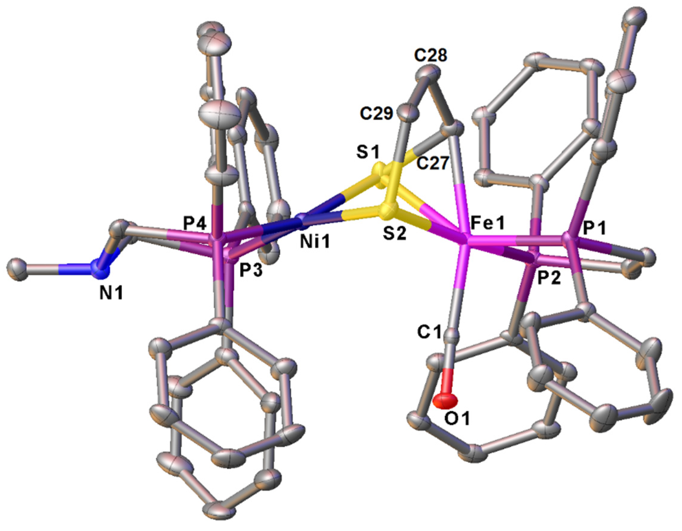
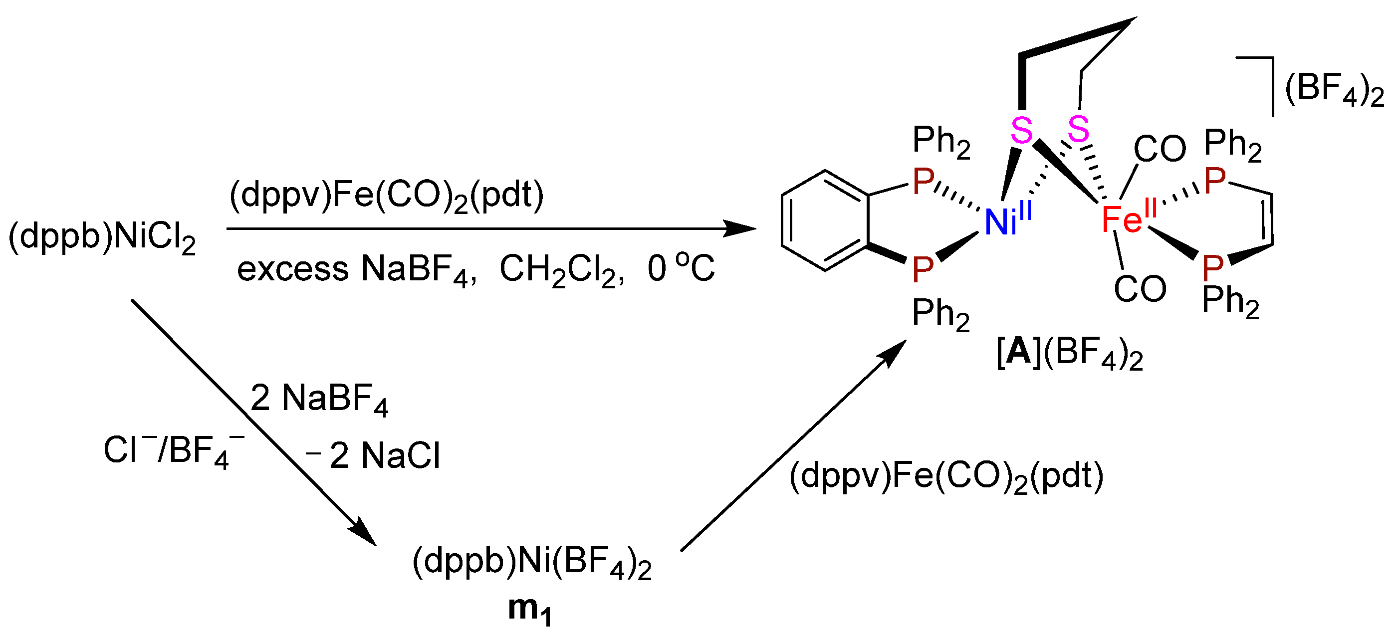
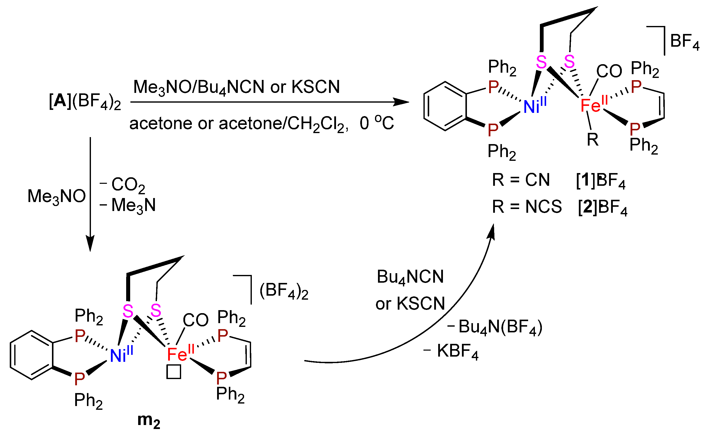
2.1. Synthesis and Characterization of Diphosphine dppb-Chelated Dicarbonyl Complex[(dppb)Ni(pdt)Fe(CO)2(dppv)](BF4)2 ([A](BF4)2) and Monocarbonyl Complexes [(dppb)Ni(pdt)Fe(CO)(t-R)(dppv)]BF4 ([1]BF4, R = CN; [2]BF4, R = NCS)

We found that diphosphine dppb (dppb = 1,2-(Ph2P)2C6H4)-chelated dicarbonyl complex [A](BF4)2 could be prepared by treatment of a CH2Cl2 solution of mononuclear Ni complex (dppb)NiCl2 with one equimolar Fe complex (dppv)Fe(CO)2(pdt) (dppv = 1,2-(PPh2)2C2H2, pdt = 1,3-propanedithiolate) and a large excess of NaBF4 in 80% yield (Scheme 1). The formation of dicarbonyl complex [A](BF4)2 might be suggested to involve two elementary reaction steps: (i) the doubly anionic Cl/BF4 exchange between (dppb)NiCl2 and NaBF4 to give intermediate m1; and (ii) coordination of the two S atoms in pdt ligand of (dppv)Fe(CO)2(pdt) to Ni atom of the resulting intermediate m1 to afford the final product [45] (Scheme 1).
Dicarbonyl complex [A](BF4)2 is an air-stable orange-red solid. The IR spectrum of [A](BF4)2 shows one very strong absorption band at 1997 cm−1 for its terminal carbonyls. The 31P{1H} NMR spectrum displays one singlet at 56.9 ppm for its two P atoms in the Ni-bound dppb ligand and one singlet at 65.1 ppm for its two P atoms in the Fe-bound dppv ligand, respectively. In addition, the 1H NMR and 13C{1H} NMR spectra are in good agreement with its structure shown in Scheme 1.
The molecular structure of [A](BF4)2 was determined by X-ray crystallography. As shown in Figure 2, this complex comprises one dication [(dppb)Ni(pdt)Fe(CO)2(dppv)]2+ and two BF4 monoanions. In its dication, a dithiolato pdt ligand is bridged between the NiFe centers to form a butterfly NiIIFeII(µ-S)2 core with a “hinge” angle 45.88° between the two Ni1S1Fe1 and Ni1S2Fe1 planes. While the Ni center adopts a distorted square-planar geometry, the Fe center adopts a pseudo-octahedral geometry. The Ni⋅⋅⋅⋅Fe distance (3.1875 Å) in [A](BF4)2 is much longer than that (2.57 Å) in D. Volgaris Miyazaki F. [NiFe]-H2ase [19] and much longer than the sum (2.56 Å) of Ni and Fe atom covalent radii [46]. This implies that there isn′t any metal-metal bonding interaction between its NiFe centers.
After dicarbonyl complex [A](BF4)2 was prepared, we further prepared the diphosphine dppb-chelated monocarbonyl complexes [1]BF4 and [2]BF4. Thus, when complex [A](BF4)2 was treated with one equiv of decarbonylation agent Me3NO in acetone followed by treatment of the resulting mixture with one equiv of Bu4NCN or KSCN, the corresponding t-cyanido and t-isothiocyanato complexes [1]BF4 and [2]BF4 were produced in 68% and 63% yields, respectively (Scheme 2).
A possible pathway suggested for the formation of complexes [1]BF4 and [2]BF4 is shown in Scheme 2, which involves the following two reaction steps. The first step involves decarbonylation of dicarbonyl complex [A](BF4)2 under the action of decarbonylating agent Me3NO to give 5-coordinate intemediate m2 with a vacant site trans to its pdt ligand [47]. The second step involves the nucleophilic attack of the negatively-charged C atom in CN group of Bu4NCN or the neucleophic attack of the paired electrons on N atom in NCS group of KSCN (note that the SCN group in KSCN is a well-known ambidentate ligand) [48] at the positively-charged Fe atom followed by loss of one molecule of Bu4N(BF4) or KBF4 to produce the final t-cyanido and t-isothiocyanato complexes.
Both [1]BF4 and [2]BF4 are air-stable orange-red solids. The IR spectra of the two complexes show one very strong absorption band at 1956 and 1950 cm−1 for their terminal carbonyls and one additional absorption band at 2067 cm−1 for the N=C=S ligand in [2]BF4. In addition, the 13C{1H} NMR spectra of [1]BF4 and [2]BF4 exhibit one singlet at 214.4 and 215.5 ppm for their terminal carbonyl C atoms, respectively. The 31P{1H} NMR spectra of the two complexes, similar to dicarbonyl complex [A](BF4)2, display one singlet at 51.9/55.3 ppm for their two P atoms in the Ni-bound dppb ligands and one singlet at 76.6/73.6 ppm for their two P atoms in the Fe-bound dppv ligands, respectively. The assignment of the lower field singlets to the two P atoms in the dppv ligands is based on the fact that the 31P{1H} NMR signal of (dppv)(CO)2Fe(pdt) determined under the same conditions was found in the lower field at 79.3 ppm.
The molecular structures of [1]BF4 and [2]BF4 were confirmed by X-ray crystal diffraction analysis. As shown in Figure 3 and Figure 4, the two complexes are isostructural. Both of them consist of one monocation [(dppb)Ni(pdt)Fe(CO)(CN)(dppv)]+ or [(dppb)Ni(pdt)Fe(CO)(NCS)(dppv)]+ and one BF4 monoanion. The NiFe centers of [1]BF4 and [2]BF4 are bridged by a dithiolato pdt ligand to form a butterfly NiIIFeII(µ-S)2 core and the “hinge” angles between their Ni1S1Fe1 and Ni1S2Fe1 planes are 45.97 and 47.16°, respectively. The C2≡N1 bond length in [1]BF4 is 1.144 Å, whereas the N1=C2 and C2=S3 bond lengths in [2]BF4 are 1.145 Å and 1.626 Å, respectively. The Ni⋅⋅⋅⋅Fe distances of [1]BF4 (3.2106 Å) and [2]BF4 (3.1570 Å) are very close to that of [A](BF4)2. Therefore, like [A](BF4)2, they do not have any Ni⋅⋅⋅⋅Fe metal-metal bonding interaction. Notably, complex [2]BF4 is the first prepared and crystallographically characterized N=C=S ligand-containing [NiFe]-H2ase model, although some CN ligand-containing [NiFe]-H2ase models were previously reported by other groups [24,49].

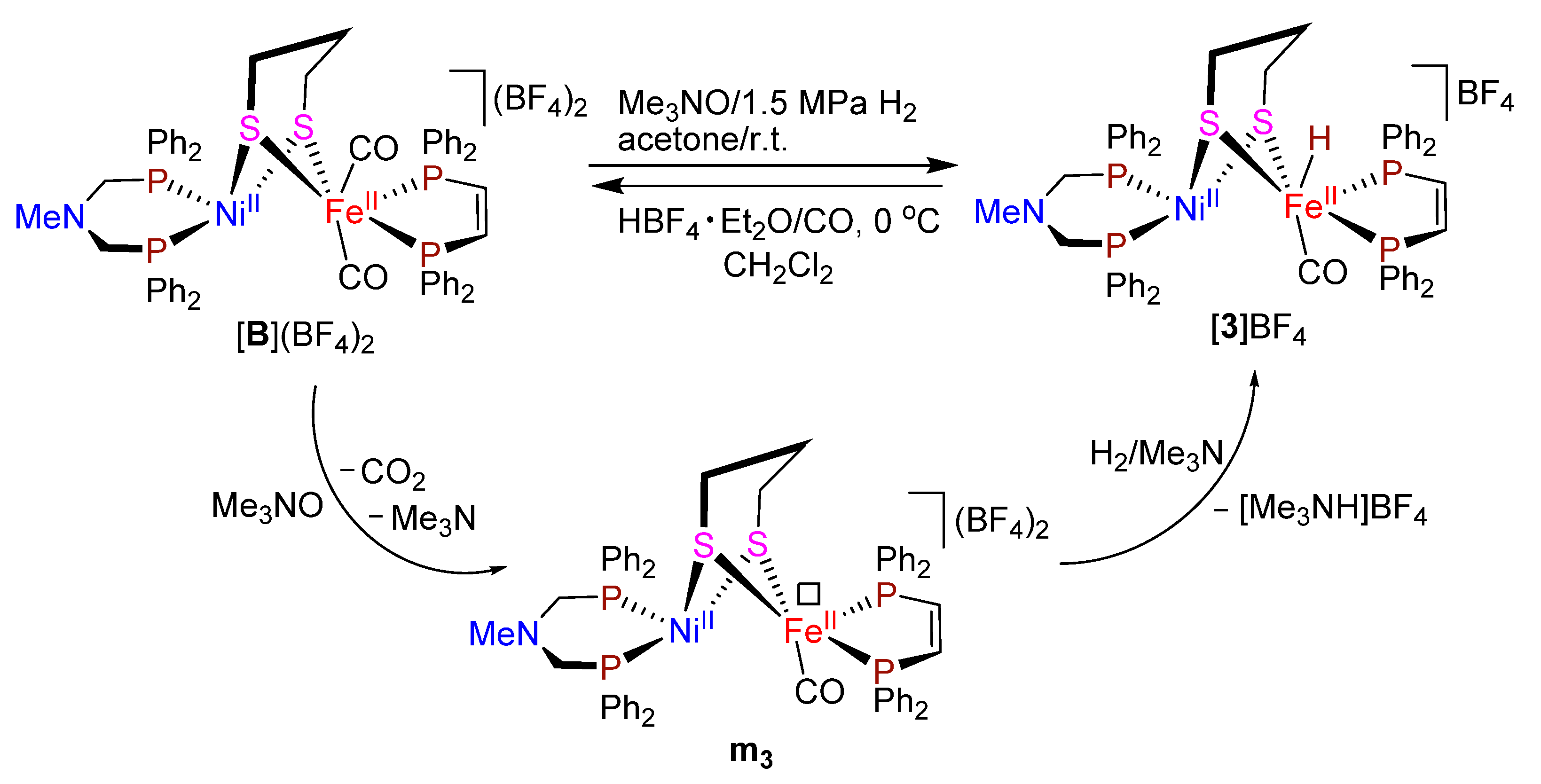
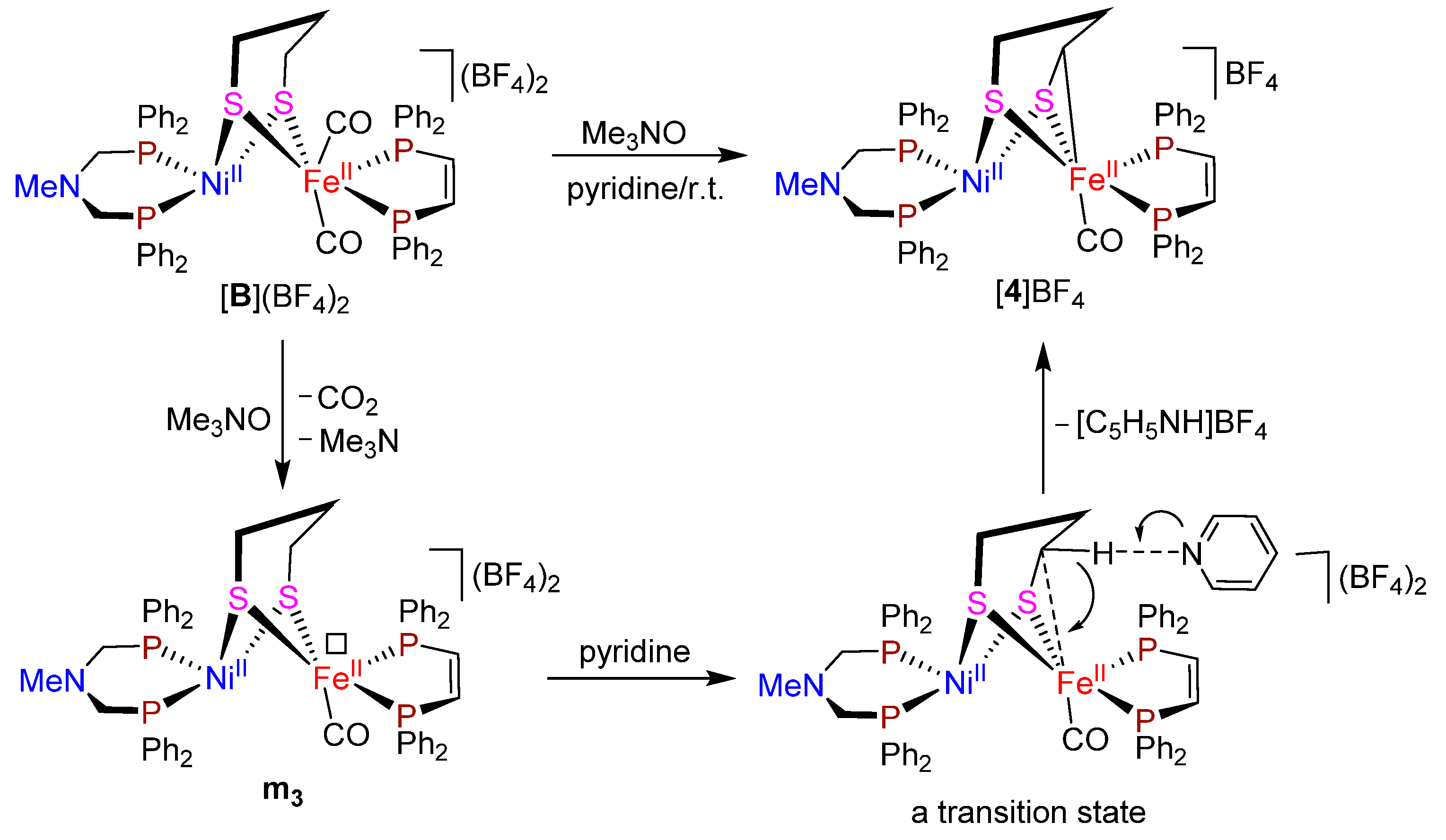
2.2. Reactions of MeN(CH2PPh2)2-Chelated Dicarbonyl Complex [MeN(CH2PPh2)2Ni(pdt)Fe(CO)2(dppv)](BF4)2 ([B](BF4)2) Leading to Monocarbonyl Complexes [MeN(CH2PPh2)2Ni(pdt)Fe(CO)(t-H)(dppv)]BF4 ([3]BF4) and [MeN(CH2PPh2)2Ni(SCHCH2CH2S)Fe(CO)(dppv)]BF4 ([4]BF4)

In 2017, we reported the preparation of an azadiphosphine PhN(CH2PPh2)2-chelated t-hydride [NiFe]-H2ase model [42]. In order to see if the azadiphosphine MeN(CH2PPh2)2-chelated dicarbonyl complex [B](BF4)2 [45] (an analogue of the diphosphine dppb-chelated dicarbonyl complex [A](BF4)2) could also activate H2 to give the corresponding MeN(CH2PPh2)2-chelated t-hydride complex [3]BF4, we carried out the reaction of [B](BF4)2 with one equiv of decarbonylating agent Me3NO and 1.5 MPa of H2; as a result, the expected t-hydride complex [3]BF4 was produced in 78% yield, indicating that complex [B](BF4)2 possesses the H2 activation function (Scheme 3). Similar to the previously reported PhN(CH2PPh2)2-chelated t-hydride complex [42], the formation of the MeN(CH2PPh2)2-chelated t-hydride complex [3]BF4 might be suggested to involve two reaction steps. The first step involves the formation of decarbonylating intermediate m3 with a vacant site cis to its pdt ligand for H2 coordination. The second step involves heterolytic cleavage of the coordinated H2 under the assistance of in situ generated proton acceptor Me3N to give the final t-hydride product (Scheme 3). In addition, it should be noted that when t-hydride complex [3]BF4 was treated with excess HBF4⋅Et2O and with bubbling CO gas, it could be converted to dicarbonyl complex [B](BF4)2 in 81% yield (Scheme 3).
More interestingly, except H2 activation, dicarbonyl complex [B](BF4)2 was further found to have the sp3 C-H bond activation function [50]. Thus, when dicarbonyl complex [B](BF4)2 was treated in pyridine with an equimolar Me3NO (without H2), a novel sp3 C-Fe bond-containing monocarbonyl complex [4]BF4 was produced via the intramolecular sp3 C-H bond activation of [B](BF4)2 in 73% yield (Scheme 4). It is interesting to note that complex [B](BF4)2 is the first [NiFe]-H2ase model to have the C-H bond activation function.
As shown in Scheme 4, the formation of complex [4]BF4 might be suggested to include three reaction steps. The first step involves the formation of 5-coordinate intermediate m3 as indicated in Scheme 3. The second step includes a transition state formed by interaction of one sp3 C-H bond directly attached to S atom of the pdt ligand in intermediate m3 with pyridine N atom and the positively-charged Fe atom. The final step affords product [4]BF4 accompanied by formation of the pyridinium salt [C5H5NH]BF4.
While t-hydride complex [3]BF4 is an air-stable deep-green solid, the sp3 C-Fe bond-containing complex [4]BF4 is an air-stable grey-black solid. The IR spectra of the two complexes show one very strong absorption band at 1912 and 1911 cm−1 for their terminal carbonyls, respectively. The 31P{1H} NMR spectrum of [3]BF4 exhibits one singlet at 7.2 ppm for its two P atoms in the Ni-bound azadiphosphine ligand and another singlet at 91.4 ppm for its two P atoms in the Fe-bound dppv ligand. In addition, [4]BF4 displays two doublets at 3.9/11.4 ppm for its two P atoms in the Ni-bound azadiphosphine ligand and two doublets at 93.2/94.7 ppm for its two P atoms in the Fe-bound dppv ligands since it is an asymmetric molecule. The 1H NMR spectrum of [3]BF4 displays one singlet at 2.54 ppm for its CH3N group and one triplet at −4.19 ppm with J = 74 Hz for its terminal hydride, which is very close to that (−4.27 ppm, J = 74 Hz) displayed by its azadiphosphine PhN(CH2PPh2)2-chelated analogue [42].
Fortunately, the molecular structure of [4]BF4 was successfully determined by X-ray crystallography (Figure 5). This molecule is composed of one monocation [MeN(CH2PPh2)2Ni(SCHCH2CH2S)Fe(CO)(dppv)]+ and one BF4 monoanion. In its monocation there is one sp3 C-Fe bond, namely the C27-Fe1 bond formed by interaction of one sp3 C-H bond attached directly to S atom of the bridging pdt ligand with pyridine N atom and the positively-charged Fe atom. The C27-Fe1 bond length is 2.067 Å, whereas the Ni⋅⋅⋅⋅Fe distance is 2.9751 Å. While the Ni atom adopts a distorted square-planar geometry, the Fe atom has a pseudo-octahedral geometry. The “hinge” angle between its two Ni1S1Fe1 and NiS2Fe1 planes is 49.14°, which is obviously larger than that (45.97°) in monocarbonyl complex [1]BF4 or that (47.16°) in complex [2]BF4, presumably owing to formation of the intramolecular C27-Fe1 bond in monocarbonyl complex [4]BF4.

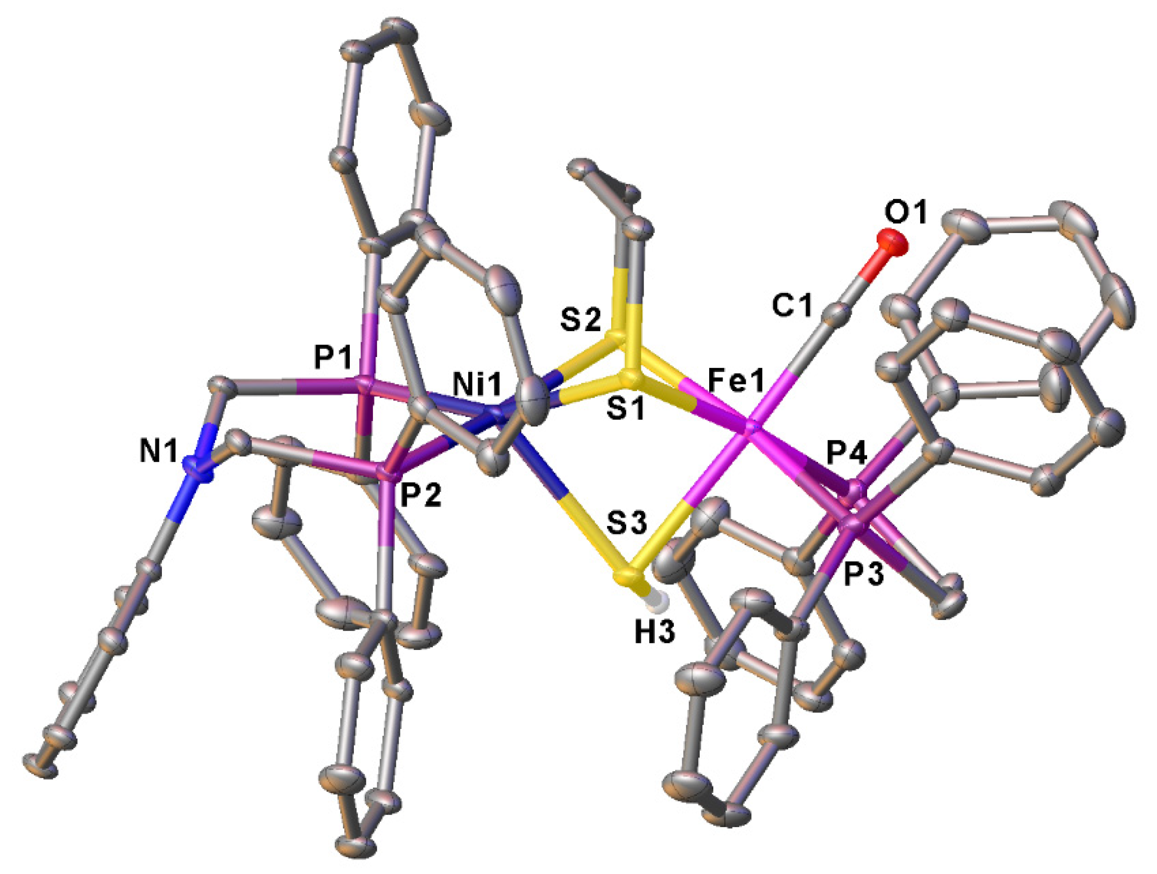
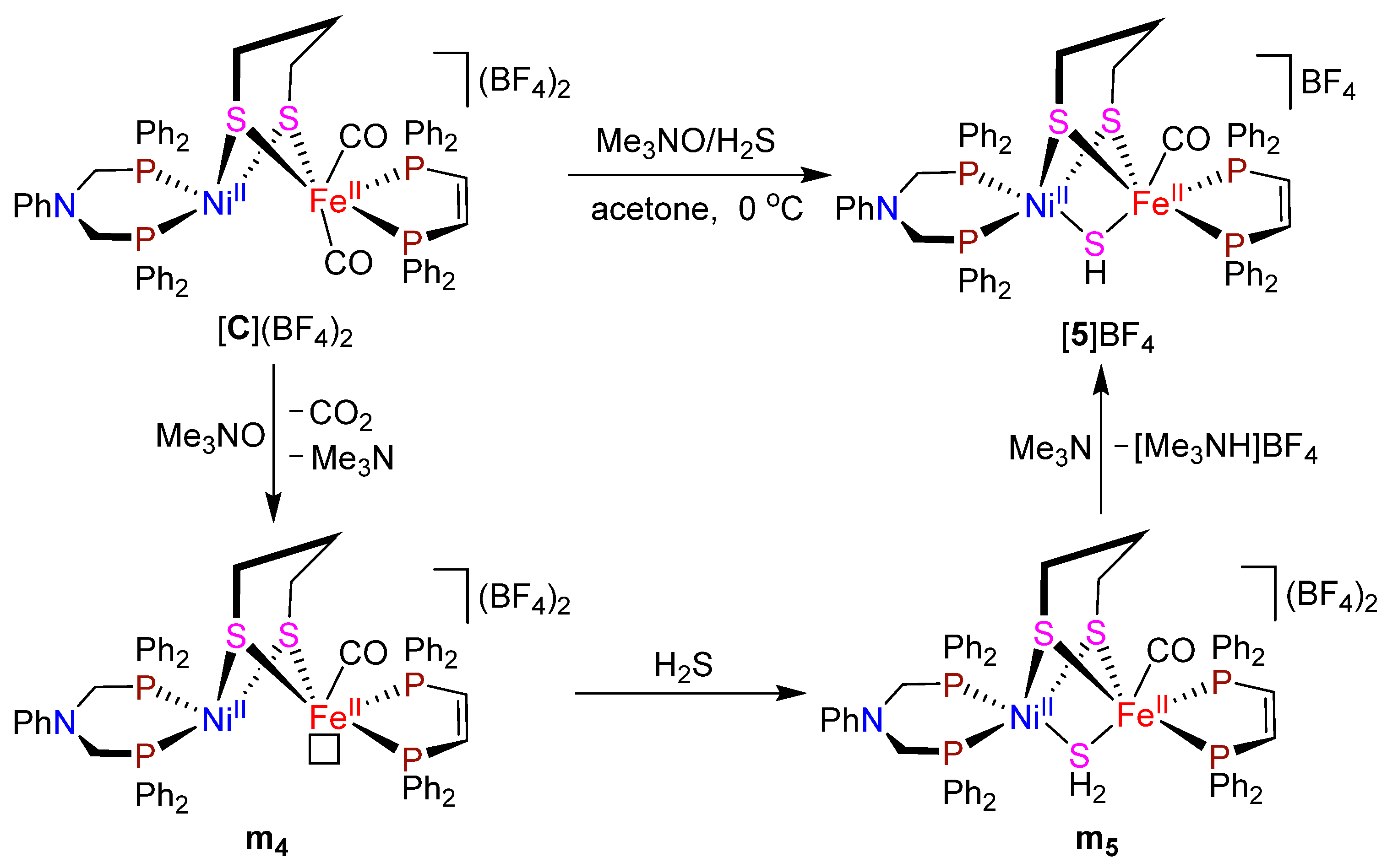
2.3. Reaction of Azadiphosphine PhN(CH2PPh2)2-Chelated Dicarbonyl Complex [PhN(CH2PPh2)2Ni(pdt)Fe(CO)2)(dppv)](BF4)2 ([C](BF4)2) Leading to Monocarbonyl Complex [PhN(CH2PPh2)2Ni(pdt)Fe(CO)(µ-SH)(dppv)]BF4 ([5]BF4)

The azadiphosphine PhN(CH2PPh2)2-chelated µ-mercapto complex [5]BF4 was found to be prepared by reaction of the corresponding dicarbonyl complex [PhN(CH2PPh2)2Ni(pdt)Fe(CO)2(dppv)](BF4)2 ([C](BF4)2) [42], an analogue of the diphosphine dppb-chelated dicarbonyl complex [A](BF4)2, with one equiv of decarbonylating agent Me3NO in acetone followed by treatment of the resulting mixture with bubbling H2S gas in nearly quantitative yield (Scheme 5).
A possible pathway for the formation of µ-mercapto complex [5]BF4 is shown in Scheme 5. The first step in Scheme 5 is similar to that shown in Scheme 2 to involve the formation of 5-coordinate intermediate m4. The second step involves coordination of one molecule of H2S to the positively-charged Fe center of m4 to afford intermediate m5. The final step involves deprotonation from µ-SH2 ligand with the aid of in situ generated proton acceptor Me3N to produce [5]BF4.
µ-Mercapto complex [5]BF4 is an air-stable brown solid. The IR spectrum of [5]BF4 displays one very strong absorption band at 1938 cm−1 for its terminal carbonyl. The 1H NMR spectrum exhibits one triplet at −2.89 ppm for its µ-SH ligand. The 31P{1H} NMR spectrum exhibits two singlets at −1.3 and 73.6 ppm for two P atoms in its azadiphosphine and dppv ligands, respectively.
The X-ray crystallographic study indicated that complex [5]BF4 contains one monocation [PhN(CH2PPh2)2Ni(pdt)Fe(CO)(µ-SH)(dppv)]+ and one monoanion BF4 (Figure 6). While the Ni center adopts a distorted square-pyramidal geometry, the Fe center adopts a pseudo-octahedral geometry. The most striking feature of [5]BF4 is to contain a bridging µ-SH ligand, which is unsymmetrically bridged between its NiFe centers with the Ni1−S3 and Fe1−S3 distances being 2.5968 and 2.3444 Å, respectively. It should be noted that complex [5]BF4 is the first prepared and crystallographically characterized µ-SH ligand-containing [NiFe]-H2ase model.

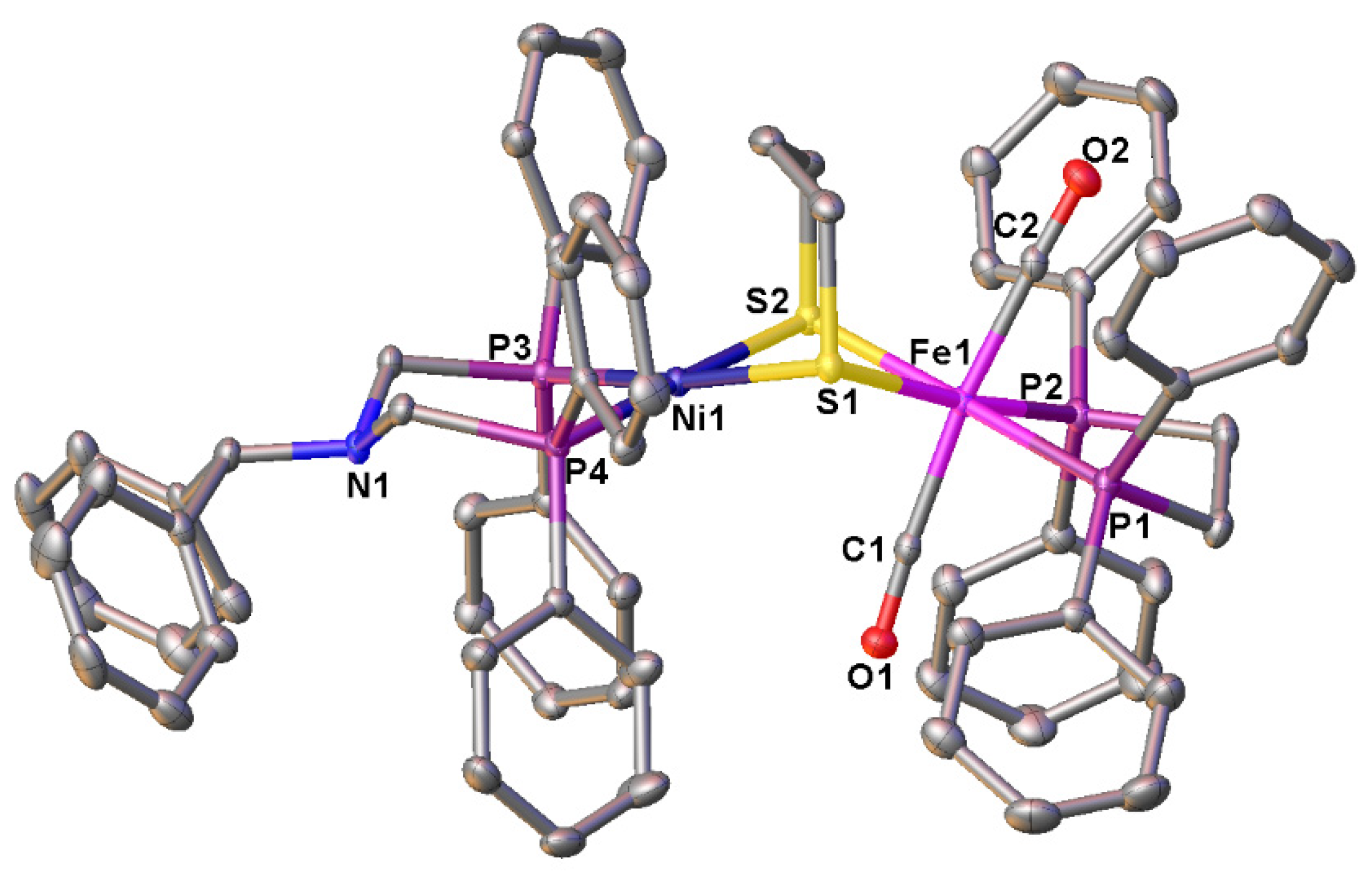
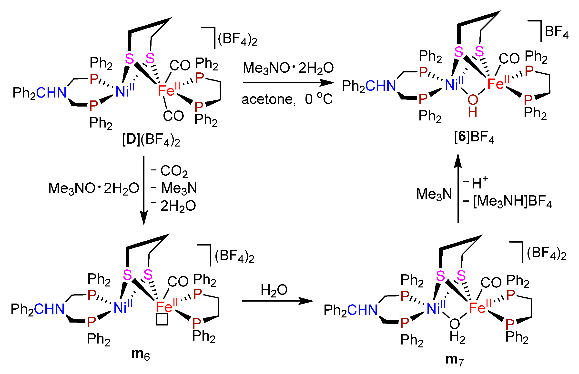
2.4. Synthesis and Characterization of Azadiphosphine Ph2CHN(CH2PPh2)2-Chelated Dicarbonyl Complex [Ph2CHN(CH2PPh2)2Ni(pdt)Fe(CO)2(dppe)](BF4)2 ([D](BF4)2) and Monocarbonyl Complex [Ph2CHN(CH2PPh2)2Ni(pdt)Fe(CO)(µ-OH)(dppe)]BF4 ([6]BF4)

Similar to the preparation of diphosphine dppb-chelated complex [A](BF4)2, the azadiphosphine Ph2CHN(CH2PPh2)2-chelated complex [D](BF4)2 was found to be prepared by treatment of a CH2Cl2 solution of mononuclear Ni complex [Ph2CHN(CH2PPh2)2]NiCl2 with 1 equiv of mononuclear Fe complex (dppe)Fe(CO)2(pdt) (dppe = 1,2-(Ph2P)2C2H4) and a large excess of NaBF4 in 80% yield. Furthermore, we found that when complex [D](BF4)2 was treated with 1 equiv of the hydrated trimethylamine oxide, an azadiphosphine Ph2CHN(CH2PPh2)2-chelated µ-hydroxo complex [6]BF4 was isolated in 65% yield (Scheme 6). Similar to the previously reported azadiphosphine PhN(CH2PPh2)2-chelated µ-hydroxo analogue [42], the formation of µ-hydroxo complex [6]BF4 could be suggested to include the following three reaction steps. The first step involves decarbonylation of dicarbonyl complex [D](BF4)2 to give intermediate m6 with a vacant coordination site trans to its pdt ligand [47]. The second step involves coordination of m6 with one molecule of in situ liberated H2O from Me3NO⋅2H2O to afford µ-aqua intermediate m7. The third step involves deprotonation of the µ-aqua intermediate m7 with the aid of Me3N to afford µ-hydroxo complex [6]BF4 (Scheme 6).
While dicarbonyl complex [D](BF4)2 is an air-stable orange-red solid, µ-hydroxo complex [6]BF4 is an air-stable orange-red solid. The IR spectra of [D](BF4)2 and [6]BF4 display one very strong absorption band at 1983 and 1917 cm−1 for their terminal carbonyls, respectively. The 31P{1H} NMR spectra of [D](BF4)2 and [6]BF4 exhibit one singlet at 4.9 and 2.2 ppm for their two P atoms attached to the Ni-bound azadiphosphine ligand and one singlet at 56.8 and 65.8 ppm for their two P atoms attached to the Fe-bound dppe ligand, respectively. The 1H NMR spectra of [D](BF4)2 and [6]BF4 display one singlet at 4.93 and 4.51 ppm for the methyne H atom in their CHPh2 groups, respectively. In addition, µ-hydroxo complex [6]BF4 exhibits another singlet at −3.39 ppm for its bridging µ-hydroxo ligand, which is slightly downfield shifted relative to the −3.66 ppm displayed by the previously reported azadiphosphine PhN(CH2PPh2)2-chelated µ-hydroxo analogue [42].
The molecular structure of [D](BF4)2 was unequivocally confirmed by X-ray crystal diffraction analysis (Figure 7). The structure of this molecule is very similar to that of dicarbonyl complex [A](BF4)2. For example, it contains one dication Ph2CHN(CH2PPh2)2Ni(pdt)Fe(CO)2(dppe)]2+ and two BF4 monoanion. The dithiolato pdt ligand is bridged between its NiFe centers to construct a butterfly NiIIFeII(µ-S)2 core. While the Ni center adopts a distorted square-planar geometry, the Fe center takes a pseudo-octahedral geometry. The Ni⋅⋅⋅⋅Fe distance (3.2957 Å) in this molecule is very close to that (3.1875 Å) of [A](BF4)2.

3. Experimental

3.1. General Comments

All reactions were performed using standard Schlenk and vacuum-line techniques under an atmosphere of highly purified N2 or argon. While CH2Cl2 was distilled under argon from CaH2, acetone was distilled from anhydrous K2CO3. Pyridine, NaBF4, Me3NO, Me3NO⋅2H2O, Bu4NCN, KSCN, HBF4⋅Et2O (50−55% in Et2O) and other reagents were available commercially and used as received. H2S gas was produced from reaction of NaHS and H3PO4. (dppb)NiCl2 (dppb = 1,2-(Ph2P)2C6H4) [51], (dppv)Fe(CO)2(pdt) (dppv = 1,2-(Ph2P)2C2H2, pdt = 1,3-propanedithiolate) [52], (dppe)Fe(CO)2(pdt) (dppe = 1,2-(Ph2P)2C2H4) [52], [MeN(CH2PPh2)2Ni(pdt)Fe(CO)2(dppv)](BF4)2 ([B](BF4)2) [45] and [PhN(CH2PPh2)2Ni(pdt)Fe(CO)2(dppv)](BF4)2 ([C](BF4)2) [42] were prepared according to the published procedures. 1H, 13C{1H} and 31P{1H} NMR spectra were obtained on a Bruker Avance 400 NMR spectrometer. IR spectra were recorded on a Bruker tensor 27 infrared spectrophotometer. Elemental analyses were performed on an Elementar Vario EL analyzer. Melting points were determined on a SGW X-4 melting point apparatus with a microscope and were uncorrected.

3.2. Synthesis of Model Complexes

[(dppb)Ni(pdt)Fe(CO)2(dppv)](BF4)2 ([A](BF4)2): A 100 mL three-necked flask fitted with a magnetic stir-bar, two serum caps, and a nitrogen inlet tube was charged with (dppb)NiCl2 (0.288 g, 0.50 mmol), (dppv)Fe(CO)2(pdt) (0.307, 0.50 mmol), NaBF4 (0.550 g, 5.0 mmol) and CH2Cl2 (30 mL). The mixture was stirred at 0 °C for 5 h. Solvent was removed at reduced pressure and the residue was subjected to column chromatography (silica gel). Elution with CH2Cl2/acetone (v/v = 12:1) developed a major orange-red band, from which [A](BF4)2 (0.517 g, 80%) was obtained as an orange-red solid, mp 177 °C (dec). Anal. calcd for C61H52B2F8FeNiO2P4S2: C, 56.65; H, 4.05. Found: C, 56.39; H, 4.54. IR (KBr disk): νCO 1997 (vs) cm1. 1H NMR (400 MHz, acetone-d6): δ 2.16–2.20, 2.60–2.69 (2m, 6H, CH2CH2CH2), 7.28–7.85 (m, 44H, C6H4, 8C6H5), 8.68–8.84 (m, 2H, CH=CH) ppm. 13C{1H} NMR (100 MHz, acetone-d6): δ 33.3, 34.8, 37.8 (3s, CH2CH2CH2), 130.7–135.8 (m, C6H4, C6H5), 139.7–151.2 (m, CH=CH), 207.9, 208.3 (2s, C≡O) ppm. 31P{1H} NMR (162 MHz, acetone-d6): δ 56.9 (s, NiP2), 65.1 (s, FeP2) ppm.
[(dppb)Ni(pdt)Fe(CO)(t-CN)(dppv)]BF4 ([1]BF4): A 50 mL three-necked flask fitted with a magnetic stir-bar, two serum caps, and a nitrogen inlet tube was charged with [A](BF4)2 (0.129 g, 0.10 mmol), Me3NO (7.5 mg, 0.10 mmol) and acetone (10 mL). The mixture was stirred at 0 °C for 15 min and then a CH2Cl2 (5 mL) solution of Bu4NCN (0.027 mg, 0.10 mmol) was added. After the new mixture was stirred at 0 °C for 1 h. solvent was removed at reduced pressure to leave a residue, which was subjected to silica gel column chromatography. Elution with CH2Cl2/acetone (v/v = 15:1) developed a major orange-red band, from which [1]BF4 (0.082 g, 68%) was obtained as an orange-red solid, mp 180–182 °C. Anal. calcd for C61H52BF4FeNNiOP4S2: C, 60.83; H, 4.35; N, 1.16. Found: C, 60.59; H, 4.54; N, 1.07. IR (KBr disk): νCO 1956 (vs) cm1. 1H NMR (400 MHz, acetone-d6): δ 2.21, 2.82 (2s, 6H, CH2CH2CH2), 7.16–8.46 (m, 46H, C6H4, 8C6H5, CH=CH) ppm. 13C{1H} NMR (100 MHz, acetone-d6): δ 24.4, 32.3, 37.8 (3s, CH2CH2CH2), 128.5–136.8 (m, C6H4, C6H5), 141.7–150.7 (m, CH=CH), 214.4 (s, C≡O) ppm. 31P{1H} NMR (162 MHz, acetone-d6): δ 51.9 (s, NiP2), 76.6 (s, FeP2) ppm.
[(dppb)Ni(pdt)Fe(CO)(t-SCN)(dppv)]BF4 ([2]BF4): The same procedure as that for preparation of [1]BF4 was followed, except that Bu4NCN was replaced by KSCN (9.7 mg, 0.10 mmol). [2]BF4 (0.078 g, 63%) was obtained as an orange-red solid, mp 155 °C (dec). Anal. calcd for C61H52BF4FeNNiOP4S3: C, 59.25; H, 4.24; N, 1.13. Found: C, 59.45; H, 4.17; N, 1.07. IR (KBr disk): νN=C=S 2067 (s); νC≡O 1950 (vs) cm1. 1H NMR (400 MHz, CDCl3): δ 2.06–2.65 (m, 6H, CH2CH2CH2), 7.14–8.03 (m, 46H, C6H4, 8C6H5, CH=CH) ppm. 13C{1H} NMR (100 MHz, CDCl3): δ 30.2, 37.1 (2s, CH2CH2CH2), 127.0–134.4 (m, C6H4, C6H5), 140.3–149.0 (m, CH=CH), 215.5 (s, C≡O) ppm. 31P{1H} NMR (162 MHz, acetone-d6): δ 55.3 (s, NiP2), 73.6 (s, FeP2) ppm.
[MeN(CH2PPh2)2Ni(pdt)Fe(CO)(t-H)(dppv)]BF4 ([3]BF4): In an argon-filled glove box, a mixture of [MeN(CH2PPh2)2Ni(pdt)Fe(CO)2(dppv)](BF4)2 ([B](BF4)2) (0.128 g, 0.10 mmol), Me3NO (7.5 mg, 0.10 mmol), and acetone (3 mL) was added to a 30 mL autoclave’s inner sleeve (made of PTFE) containing a magnetic stir-bar. The reaction mixture was stirred at 0 °C for 10 min and then it was frozen by inserting the sleeve into liquid nitrogen. After the autoclave was sealed, the head space of the sleeve was evacuated and was refilled with 1.5 MPa of H2. The frozen reaction mixture was thawed and then the reaction mixture was stirred at room temperature for 4 h. Solvent was removed at reduced pressure to give a residue, which was subjected to column chromatography (silica gel G). Elution with CH2Cl2/acetone (v/v = 8:1) developed a deep-green band, from which [3]BF4 (0.090 g, 78%) was obtained as a deep-green solid, mp 119 °C (dec). Anal. calcd for C57H56BF4FeNNiOP4S2: C, 59.00; H, 4.86; N, 1.21. Found: C, 58.84; H, 5.15; N, 1.34. IR (KBr disk): νC≡O 1912 (vs) cm1. 1H NMR (400 MHz, acetone-d6): δ −4.19 (t, J = 74 Hz, 1H, Fe-H), 1.31–1.95 (m, 6H, CH2CH2CH2), 2.54 (s, 3H, CH3N), 3.43–3.79 (m, 4H, CH2NCH2), 7.24–7.95 (m, 42H, 8C6H5, CH=CH) ppm. 13C{1H} NMR (100 MHz, CDCl3): δ 28.7, 31.9, 34.8 (3s, CH2CH2CH2), 49.8 (s, CH3N), 55.5–55.9 (m, CH2NCH2), 127.0–134.4 (m, C6H5), 148.4–149.1 (m, CH=CH), 215.4 (s, C≡O) ppm. 31P{1H} NMR (162 MHz, CDCl3): δ 7.2 (s, NiP2), 91.4 (s, FeP2) ppm.
[MeN(CH2PPh2)2Ni(SCHCH2CH2S)Fe(CO)(dppv)]BF4 ([4]BF4): A 100 mL three-necked flask fitted with a magnetic stir-bar, two serum caps, and a nitrogen inlet tube was charged with [B](BF4)2 (0.127 g, 0.10 mmol), Me3NO (7.5 mg, 0.10 mmol) and pyridine (50 mL). The mixture was stirred at room temperature for 3 h and then hexane (250 mL) was added to give a precipitate, which was subjected to column chromatography (silica gel). Elution with CH2Cl2/acetone (v/v = 20:1) developed one black band, from which [4]BF4 (0.085 g, 73%) was obtained as a grey-black solid, mp 158–159°. Anal. calcd for C57H54BF4FeNNiOP4S2: C, 59.10; H, 4.70; N, 1.21. Found: C, 58.83; H, 4.71; N, 1.13. IR (KBr disk): νCO 1911(vs) cm1. 1H NMR (400 MHz, CDCl3): δ 2.31–2.77, 3.34–3.74 (2m, 12H, CHCH2CH2, CH3N, CH2NCH2), 6.60–7.89 (m, 42H, 8C6H5, CH=CH) ppm. 13C{1H} NMR (100 MHz, CD2Cl2): δ 28.9–31.1 (m, SCH2CH2, CH3N), 49.6, 50.5 (2s, CH2NCH2), 60.7 (s, SCHFe), 122.4–138.8 (m, C6H5), 146.9–153.1 (m, CH=CH), 214.6 (s, C≡O) ppm. 31P{1H} NMR (162 MHz, acetone-d6): δ 3.9, 11.4 (2d, NiP2), 93.2, 94.7 (2d, FeP2) ppm.
[PhN(CH2PPh2)2Ni(pdt)Fe(CO)(µ-SH)(dppv)]BF4 ([5]BF4): A 50 mL three-necked flask fitted with a magnetic stir-bar, two serum caps, and a nitrogen inlet tube was charged with [C](BF4)2 (0.134 g, 0.10 mmol), Me3NO (7.5 mg, 0.10 mmol) and acetone (10 mL). The mixture was stirred for 15 min at 0 °C and then dry H2S gas was bubbled into the mixture for 1 h. Solvent was removed at reduced pressure and the residue was subjected to column chromatography (silica gel). Elution with CH2Cl2/acetone (v/v = 12:1) developed a major brown band, from which [5]BF4 (0.123 g, 98%) was obtained as a brown solid, mp 180 °C (dec). Anal. calcd for C62H58BF4FeNNiOP4S3: C, 59.36; H, 4.66; N, 1.12. Found: C, 59.20; H, 4.90; N, 1.21. IR (KBr disk): νC≡O 1938 (vs) cm1. 1H NMR (400 MHz, acetone-d6): δ −2.89 (t, J = 8.0 Hz, 1H, SH), 1.77–2.83 (m, 6H, CH2CH2CH2), 3.97–4.30 (m, 4H, CH2NCH2), 6.52–7.82 (m, 45H, 9C6H5), 8.05–8.19 (m, 2H, CH=CH) ppm. 13C{1H} NMR (100 MHz, acetone-d6): δ 26.2, 29.2, 33.8 (3s, CH2CH2CH2), 51.9, 52.1 (2s, CH2NCH2), 116.9–134.2 (m, C6H5), 148.3–150.8 (m, CH=CH), 214.2 (s, C≡O) ppm. 31P{1H} NMR (162 MHz, acetone-d6): δ −1.3 (s, NiP2), 73.6 (FeP2) ppm.
[Ph2CHN(CH2PPh2)2Ni(pdt)Fe(CO)2(dppe)](BF4)2 ([D](BF4)2):
(i) Ph2CHN(CH2PPh2)2: A 100 mL three-necked flask fitted with a magnetic stir-bar, two serum caps, and a reflux condenser topped with a nitrogen inlet tube was charged with polyformaldehyde (0.213 g, 7.10 mmol) and toluene (30 mL). After the mixture was stirred and slowly warmed to 70 °C, Ph2CHNH2 (0.646 g, 3.53 mmol) and Ph2PH (1.35 mL, 7.75 mmol) were added. The new mixture was stirred at 70 °C for 7 h and then solvent was removed at reduced pressure to leave a residue. The residue was dissolved in EtOH (50 mL). After the solution was cooled in a refrigerator for over 1 h, the white precipitate was filtered out and washed sequentially with EtOH and Et2O to give Ph2CHN(CH2PPh2)2 (1.636 g, 80%) as a white solid. Anal. calcd for C39H35NP2: C, 80.81; H, 6.09; N, 2.42. Found: C, 80.95; H, 5.95; N, 2.19. IR (KBr disk): νP-N-P 871 (m) cm1. 1H NMR (400 MHz, CDCl3): δ 3.84 (s, 4H, CH2NCH2), 5.48 (s, CHPh2), 7.18–7.46 (m, 30H, 6C6H5) ppm. 13C{1H} NMR (100 MHz, CDCl3): δ 55.2 (s, CH2NCH2), 72.5 (s, CHPh2), 126.9–141.5 (m, C6H5) ppm. 31P{1H} NMR (162 MHz, CDCl3): δ −29.3 (s, PPh) ppm.
(ii) [Ph2CHN(CH2PPh2)2]NiCl2: A 100 mL three-necked flask fitted with a magnetic stir-bar, two serum caps, and a nitrogen inlet tube was charged with NiCl2⋅6H2O (1.188 g, 5.00 mmol) and EtOH (20 mL). To this stirred solution was slowly added a CH2Cl2 (10 mL) solution of Ph2CHN(CH2PPh2)2 (2.898 g, 5.00 mmol). After the new mixture was stirred at room temperature for 6 h, solvent was removed at reduced pressure to leave a residue, which was washed sequentially with EtOH and Et2O to give [Ph2CHN(CH2PPh2)2]NiCl2 (2.802 g, 79%) as an orange-red solid, mp 207 °C (dec). Anal. calcd for C39H35Cl2NNiP2: C, 66.05; H, 4.97; N, 1.97. Found: C, 66.10; H, 4.75; N, 1.71. IR (KBr disk): νP-N-P 871 (m) cm1. 1H NMR (400 MHz, CD2Cl2): δ 3.26 (s, 4H, CH2NCH2), 4.32 (s, CHPh2), 6.84–7.94 (m, 30H, 6C6H5) ppm. 13C{1H} NMR (100 MHz, acetone-d6): δ 53.1–54.8 (m, CH2NCH2), 78.8 (s, CHPh2), 127.7–139.4 (m, C6H5) ppm. 31P{1H} NMR (162 MHz, acetone-d6): δ 5.9 (s, PPh2) ppm.
(iii) [Ph2CHN(CH2PPh2)2Ni(pdt)Fe(CO)2(dppe)](BF4)2 ([D](BF4)2): A 100 mL three-necked flask fitted with a magnetic stir-bar, two serum caps, and a nitrogen inlet tube was charged with [Ph2CHN(CH2PPh2)2]NiCl2 (0.355 g, 0.50 mmol), (dppe)Fe(CO)2(pdt) (0.308, 0.50 mmol), NaBF4 (0.550 g, 5.0 mmol) and CH2Cl2 (30 mL). After the mixture was stirred at 0 °C for 3 h, solvent was removed at reduced pressure and then the residue was subjected to column chromatography (silica gel). Elution with CH2Cl2/acetone (v/v = 2:1) developed an orange-red band, from which [D](BF4)2 (0.571 g, 80%) was obtained as an orange-red solid, mp 170 °C (dec). Anal. calcd for C70H65B2F8FeNNiO2P4S2: C, 58.86; H, 4.59; N, 0.98. Found: C, 58.83; H, 4.71; N, 1.13. IR (KBr disk): νCO 1983 (vs) cm1. 1H NMR (400 MHz, acetone-d6): δ 2.38–3.99 (m, 14H, CH2CH2CH2, PCH2CH2P, CH2NCH2), 4.93 (s, 1H, CHPh2), 6.88–8.00 (m, 50H, 10C6H5) ppm. 13C{1H} NMR (100 MHz, acetone-d6): δ 23.3–37.7 (m, CH2CH2CH2, PCH2CH2P), 53.0 (s, CH2NCH2), 78.1 (CHPh2), 126.8–139.8 (m, C6H5), 209.4, 210.3 (2s, C≡O) ppm. 31P{1H} NMR (162 MHz, acetone-d6): δ 4.9 (s, NiP2), 56.8 (s, FeP2) ppm.
[Ph2CHN(CH2PPh2)2Ni(pdt)Fe(CO)(µ-OH)(dppe)]BF4 ([6]BF4): A 50 mL three-necked flask fitted with a magnetic stir-bar, two serum caps, and a nitrogen inlet tube was charged with [D](BF4)2 (0.143 g, 0.10 mmol), Me3NO⋅2H2O (0.011 g, 0.10 mmol) and acetone (20 mL) under nidrogen. The mixture was stirred at 0 °C for 4 h. Solvent was removed at reduced pressure and the residue was subjected to column chromatography (silica gel). Elution with CH2Cl2/acetone (v/v = 10:1) developed an orange-red band, from which [6]BF4 (0.087 g, 65%) was obtained as an orange-red solid, mp 183–185 °C. Anal. calcd for C69H66BF4FeNNiO2P4S2: C, 62.28; H, 5.00; N, 1.05. Found: C, 62.11; H, 5.03; N, 1.06. IR (KBr disk): νCO 1917 (vs) cm1. 1H NMR (400 MHz, acetone-d6): δ −3.39 (s 1H, OH), 2.20–3.99 (m, 14H, CH2CH2CH2, PCH2CH2P, CH2NCH2), 4.51 (s, 1H, CHPh2), 6.66–8.33 (m, 50H, 10C6H5) ppm. 13C{1H} NMR (100 MHz, acetone-d6): δ 27.8, 32.3, 37.9 (3s, CH2CH2CH2, PCH2CH2P), 55.2 (s, CH2NCH2), 78.9 (CHPh2), 127.9–140.9 (m, C6H5), 217.3 (s, C≡O) ppm. 31P{1H} NMR (162 MHz, acetone-d6): δ 2.2 (s, NiP2), 65.8 (s, FeP2) ppm.

3.3. Crystal Structure Determinations of Models [A](BF4)2, [1]BF4, [2]BF4, [4]BF4, [5]BF4 and [D](BF4)2

While single crystals of [A](BF4)2, [1]BF4, [2]BF4 and [D](BF4)2 for X-ray diffraction analysis were grown by slow diffusion of n-hexane into their CH2Cl2 solutions at room temperature, those of [4]BF4 and [5]BF4 were grown by slow diffusion of n-hexane into their acetone solutions at room temperature. A single crystal of [A](BF4)2, [1]BF4, [2]BF4, [4]BF4 or [5]BF4 was mounted on a SuperNova, Dual, Cu at zero, AtlasS2 diffractometer, and data were collected using a confocal monochromator with Cu Kα radiation (λ = 1.54184 Å) in the ω scanning mode at the temperature of 137 K, 151.6 K, 152 K, 100 K and 293 K, respectively. A single crystal of [D](BF4)2 was mounted on a Rigaku Pilatus 200K diffractometer, and data were collected using a confocal monochromator with Mo Kα radiation (λ = 0.71073 Å) in the ω scanning mode at the temperature of 113 K. Data collection, reduction, and absorption correction were performed by the CRYSTALCLEAR program [53]. the structures were solved by direct methods using the SHELXT program [54,55,56] and refined by full-matrix least-squares techniques (SHELXL) [56] on F2. Hydrogen atoms were located by using the geometric method. Details of the crystal data, data collections, and structure refinements are summarized in Tables S1–S3 of the Supporting Information.

4. Conclusions

Based on the preparation of diphosphine or azadiphosphine-chelated dicarbonyl [NiFe]-H2ase models [A](BF4)2−[D](BF4)2, we have further synthesized the corresponding monocarbonyl [NiFe]-H2ase models [1]BF4−[6]BF4 via CO transformation reactions of dicarbonyl complexes [A](BF4)2−[D](BF4)2. All the new models [A](BF4)2, [D](BF4)2 and [1]BF4−[6]BF4 have been fully characterized by various spectroscopic methods, and particularly for some of them by X-ray crystallography. Of particular interest are (i) dicarbonyl complex [B](BF4)2 not only possesses the H2 activation function to give t-hydride complex [3]BF4, but also possesses the sp3 C-H bond activation function to afford the novel sp3 C-Fe bond-containing complex [4]BF4; and (ii) monocarbonyl complexes [2]BF4 and [5]BF4 are the first prepared and crystallographically characterized t-isothiocyanato and µ-mercapto ligand-containing [NiFe]-H2ase models. We believe that the studied results reported here will promote further development of the structural and functional modeling chemistry of [NiFe]-H2ases.

Supplementary Materials

The following supporting information can be downloaded at: https://0-www-mdpi-com.brum.beds.ac.uk/article/10.3390/inorganics10070090/s1, IR, 1H NMR, 13C{1H} NMR and 31P{1H} NMR spectra of all the model complexes (Figures S1–S32); crystal data and structure refinement for [A](BF4)2, [1]BF4, [2]BF4, [4]BF4, [5]BF4 and [D](BF4)2) (Tables S1–S3).

Author Contributions

L.-C.S. designed and directed the research. S.C., X.-F.H., Z.-Q.Z., Y.-P.W. and Y.-X.D. synthesized and characterized all the new compounds. L.-C.S. acquired funding and wrote the manuscript. All authors have read and agreed to the published version of the manuscript.

Funding

This work was financially supported by the National Natural Science Foundation of China (21772106) and the Ministry of Science and Technology of China (973 program 2014CB845604).

Institutional Review Board Statement

Not applicable.

Informed Consent Statement

Not applicable.

Data Availability Statement

Accession codes CCDC 2173844 for [A](BF4)2, CCDC 2173845 for [1]BF4)2, CCDC 2173846 for [2]BF4, CCDC 2173847 for [4]BF4, CCDC 2173848 for [5]BF4 and CCDC 2173849 for [D](BF4)2 contain the supplementary crystallographic data for this paper. These data can be obtained free of charge from The Cambridge Crystallographic Data Centre via www.ccdc.cam.ac.uk/data_request/cif (accessed on 20 May 2022).

Conflicts of Interest

The authors declare no conflict of interest.

References

  1. Adams, M.W.W.; Stiefel, E.I. Biological Hydrogen Production: Not So Elementary. Science 1998, 282, 1842–1843. [Google Scholar] [CrossRef] [PubMed]
  2. Cammack, R. Hydrogenase sophistication. Nature 1999, 397, 214–215. [Google Scholar] [CrossRef] [PubMed]
  3. Schilter, D.; Camara, J.M.; Huynh, M.T.; Hammes-Schiffer, S.; Rauchfuss, T.B. Hydrogenase Enzymes and Their Synthetic Models: The Role of Metal Hydrides. Chem. Rev. 2016, 116, 8693–8749. [Google Scholar] [CrossRef] [Green Version]
  4. Albracht, S.P.J. Nickel hydrogenases: In search of the active site. Biochim. Biophys. Acta 1994, 1188, 167–204. [Google Scholar] [CrossRef] [Green Version]
  5. Bouwman, E.; Reedijk, J. Structural and functional models related to the nickel hydrogenases. Coord. Chem. Rev. 2005, 249, 1555–1581. [Google Scholar] [CrossRef]
  6. Fontecilla-Camps, J.C.; Volbeda, A.; Cavazza, C.; Nicolet, Y. Structure/Function Relationships of [NiFe]- and [FeFe]-Hydrogenases. Chem. Rev. 2007, 107, 4273–4303. [Google Scholar] [CrossRef]
  7. Nicolet, Y.; Lemon, B.J.; Fontecilla-Camps, J.C.; Peters, J.W. A novel FeS cluster in Fe-only hydrogenases. Trends Biochem. Sci. 2000, 25, 138–143. [Google Scholar] [CrossRef]
  8. Frey, M. Hydrogenases: Hydrogen-Activating Enzymes. ChemBioChem 2002, 3, 153–160. [Google Scholar] [CrossRef]
  9. Evans, D.J.; Pickett, C.J. Chemistry and the hydrogenases. Chem. Soc. Rev. 2003, 32, 268–275. [Google Scholar] [CrossRef]
  10. Shima, S.; Thauer, R.K. A Third Type of Hydrogenase Catalyzing H2 Activation. Chem. Rec. 2007, 7, 37–46. [Google Scholar] [CrossRef]
  11. Shima, S.; Pilak, O.; Vogt, S.; Schick, M.; Stagni, M.S.; Meyer-Klaucke, W.; Warkentin, E.; Thauer, R.K.; Ermler, U. The Crystal Structure of [Fe]-Hydrogenase Reveals the Geometry of the Active Site. Science 2008, 321, 572–575. [Google Scholar] [CrossRef] [PubMed] [Green Version]
  12. Wright, J.A.; Turrell, P.J.; Pickett, C.J. The Third Hydrogenase: More Natural Organometallics. Organometallics 2010, 29, 6146–6156. [Google Scholar] [CrossRef]
  13. Volbeda, A.; Charon, M.-H.; Piras, C.; Hatchikian, E.C.; Frey, M.; Fontecilla-Camps, J.C. Crystal structure of the nickel-iron hydrogenase from Desulfovibrio gigas. Nature 1995, 373, 580–587. [Google Scholar] [CrossRef] [PubMed]
  14. Vignais, P.M.; Billond, B.; Meyer, J. Classification and phylogeny of hydrogenases. FEMS Microbiol. Rev. 2001, 25, 455–501. [Google Scholar] [CrossRef]
  15. Volbeda, A.; Garcin, E.; Piras, C.; De Lacey, A.L.; Fernandez, V.M.; Hatchikian, E.C.; Frey, M.; Fontecilla-Camps, J.C. Structure of the [NiFe] Hydrogenase Active Site: Evidence for Biologically Uncommon Fe Ligands. J. Am. Chem. Soc. 1996, 118, 12989–12996. [Google Scholar] [CrossRef]
  16. Higuchi, Y.; Ogata, H.; Miki, K.; Yasuoka, N.; Yagi, T. Removal of the bridging ligand atom at the Ni–Fe active site of [NiFe] hydrogenase upon reduction with H2, as revealed by X-ray structure analysis at 1.4 Å resolution. Structure 1999, 7, 549–556. [Google Scholar] [CrossRef]
  17. Matias, P.M.; Soares, C.M.; Saraiva, L.M.; Coelho, R.; Morais, J.; Le Gall, J.; Carrondo, M.A. [NiFe] hydrogenase form Desulfovibrio desulfuricans ATCC 27774: Gene sequencing, three-dimensional structure determination and refinement at 1.8 Å and modelling studies of its interaction with the tetrahaem cytochrome c3. J. Biol. Inorg. Chem. 2001, 6, 63–81. [Google Scholar] [CrossRef]
  18. Volbeda, A.; Martin, L.; Cavazza, C.; Matho, M.; Faber, B.W.; Roseboom, W.; Albracht, S.P.J.; Garcin, E.; Rousset, M.; Fontecilla-Camps, J.C. Structural differences between the ready and unready oxidized states of [NiFe] hydrogenases. J. Biol. Inorg. Chem. 2005, 10, 239–249. [Google Scholar] [CrossRef]
  19. Ogata, H.; Nishikawa, K.; Lubitz, W. Hydrogens detected by subatomic resolution protein crystallography in a [NiFe] hydrogenase. Nature 2015, 520, 571–574. [Google Scholar] [CrossRef]
  20. Wang, H.; Yoda, Y.; Ogata, H.; Tanaka, Y.; Lubitz, W. A strenuous experimental journey searching for spectroscopic evidence of a bridging nickel–iron–hydride in [NiFe] hydrogenase. J. Synchrotron Rad. 2015, 22, 1334–1344. [Google Scholar] [CrossRef] [Green Version]
  21. Ogo, S. H2 and O2 activation by [NiFe]hydrogenases—Insights from model complexes. Coord. Chem. Rev. 2017, 334, 43–53. [Google Scholar] [CrossRef]
  22. Bose, M.; Li, Z.; Matsumoto, T.; Tatsumi, K. A Dithiolato and Hydrido Bridged (CO/CN)Fe−Ni Complex with Unprotected CN: A Model for the [Ni−R] State of the [Ni−Fe] Hydrogenase Active Site. Inorg. Chem. 2020, 59, 968–971. [Google Scholar] [CrossRef] [PubMed]
  23. Lindenmaier, N.J.; Wahlefeld, S.; Bill, E.; Szilvási, T.; Eberle, C.; Yao, S.; Hildebrandt, P.; Horch, M.; Zebger, I.; Driess, M. An S-Oxygenated [NiFe] Complex Modelling Sulfenate Intermediates of an O2-Tolerant Hydrogenase. Angew. Chem. Int. Ed. 2017, 56, 2208–2211. [Google Scholar] [CrossRef] [PubMed]
  24. Perotto, C.U.; Sodipo, C.L.; Jones, G.J.; Tidey, J.P.; Blake, A.J.; Lewis, W.; Davies, E.S.; McMaster, J.; Schröder, M. Heterobimetallic [NiFe] Complexes Containing Mixed CO/CN Ligands: Analogs of the Active Site of the [NiFe] Hydrogenases. Inorg. Chem. 2018, 57, 2558–2569. [Google Scholar] [CrossRef]
  25. Osterloh, F.; Saak, W.; Haase, D.; Pohl, S. Synthesis, X-ray structure and electrochemical characterisation of a binuclear thiolate bridged Ni-Fe-nitrosyl complex, related to the active site of NiFe hydrogenase. Chem. Commun. 1997, 979–980. [Google Scholar] [CrossRef]
  26. Song, L.-C.; Lu, Y.; Zhu, L.; Li, Q.-L. Dithiolato- and Diselenolato-Bridged Nickel-Iron Biomimetics for the Active Site of [NiFe]Hydrogenases. Organometallics 2017, 36, 750–760. [Google Scholar] [CrossRef]
  27. Jiang, J.; Maruani, M.; Solaimanzadeh, J.; Lo, W.; Koch, S.A.; Millar, M. Synthesis and Structure of Analogues for the Ni-Fe Site in Hydrogenase Enzymes. Inorg. Chem. 2009, 48, 6359–6361. [Google Scholar] [CrossRef] [PubMed]
  28. Sellmann, D.; Geipel, F.; Lauderbach, F.; Heinemann, F.W. [(C6H4S2)Ni(µ−‘S3’)Fe(CO)(PMe3)2]: A Dinuclear [NiFe] Complex Modeling the [(RS)2Ni(µ-SR)2Fe(CO)(L)2] Core of [NiFe] Hydrogenase Centers. Angew. Chem. Int. Ed. 2002, 41, 632–634. [Google Scholar] [CrossRef]
  29. Smith, M.C.; Barclay, J.E.; Cramer, S.P.; Davies, S.C.; Gu, W.-W.; Hughes, D.L.; Longhurst, S.; Evans, D.J. Nickel-iron-sulfur complexes: Approaching structural analogues of the active sites of NiFe-hydrogenase and carbon monoxide dehydrogenase/acetyl-CoA synthase. J. Chem. Soc., Dalton Trans. 2002, 13, 2641–2647. [Google Scholar] [CrossRef]
  30. Verhagen, J.A.W.; Lutz, M.; Spek, A.L.; Bouwman, E. Synthesis and Characterisation of New Nickel-Iron Complexes with an S4 Coordination Environment around the Nickel Centre. Eur. J. Inorg. Chem. 2003, 2003, 3968–3974. [Google Scholar] [CrossRef] [Green Version]
  31. Li, Z.; Ohki, Y.; Tatsumi, K. Dithiolato-Bridged Dinuclear Iron-Nickel Complexes [Fe(CO)2(CN)2(µ-SCH2CH2CH2S)Ni(S2CNR2)]- Modeling the Active Site of [NiFe] Hydrogenase. J. Am. Chem. Soc. 2005, 127, 8950–8951. [Google Scholar] [CrossRef] [PubMed]
  32. Sellmann, D.; Lauderbach, F.; Heinemann, F.W. Trinuclear [NiFe] Clusters as Structural Models for [NiFe] Hydrogenase Active Sites. Eur. J. Inorg. Chem. 2005, 2005, 371–377. [Google Scholar] [CrossRef]
  33. Ogo, S.; Ichikawa, K.; Kishima, T.; Matsumoto, T.; Nakai, H.; Kusaka, K.; Ohhara, T. A Functional [NiFe]Hydrogenase Mimic That Catalyzes Electron and Hydride Transfer from H2. Science 2013, 339, 682–684. [Google Scholar] [CrossRef] [PubMed]
  34. Ohki, Y.; Yasumura, K.; Ando, M.; Shimokata, S.; Tatsumi, K. A model for the CO-inhibited form of [NiFe] hydrogenase: Synthesis of (CO)3Fe(μ-StBu)3Ni{SC6H3-2,6-(mesityl)2} and reversible CO addition at the Ni site. Proc. Natl. Acad. Sci. USA 2010, 107, 3994–3997. [Google Scholar] [CrossRef] [Green Version]
  35. Song, L.-C.; Li, J.-P.; Xie, Z.-J.; Song, H.-B. Synthesis, Structural Characterization, and Electrochemical Properties of Dinuclear Ni/Mn Model Complexes for the Active Site of [NiFe]-Hydrogenases. Inorg. Chem. 2013, 52, 11618–11626. [Google Scholar] [CrossRef]
  36. Brazzolotto, D.; Wang, L.; Tang, H.; Gennari, M.; Queyriaux, N.; Philouze, C.; Demeshko, S.; Meyer, F.; Orio, M.; Artero, V.; et al. Tuning Reactivity of Bio-inspired [NiFe]-Hydrogenase Models by Ligand Design and Modeling the CO Inhibition Process. ACS Catal. 2018, 8, 10658–10667. [Google Scholar] [CrossRef]
  37. Song, L.-C.; Chen, W.; Feng, L. Two heterodinuclear NiFe-based sulfenate complexes mimicking an S-oxygenated intermediate of an O2-tolerant [NiFe]-H2ase: Synthesis, structures, and reactivity. New J. Chem. 2020, 44, 14015–14023. [Google Scholar] [CrossRef]
  38. Song, L.-C.; Feng, L.; Lu, Y.; Yang, X.-Y. Synthesis, Structures and Reactivity of [NiFe]-H2ase Mimics Containing One Square-Planar N2S2 Ligand Bridged between Their Ni/Fe Centers through One or Two S Atoms. Organometallics 2021, 40, 508–519. [Google Scholar] [CrossRef]
  39. Lubitz, W.; Tumas, W. Hydrogen: An Overview. Chem. Rev. 2007, 107, 3900–3903. [Google Scholar] [CrossRef]
  40. Simmons, T.R.; Berggren, G.; Bacchi, M.; Fontecave, M.; Artero, V. Mimicking hydrogenases: From biomimetics to artificial enzymes. Coord. Chem. Rev. 2014, 270–271, 127–150. [Google Scholar] [CrossRef]
  41. Ogo, S.; Kishima, T.; Yatabe, T.; Miyazawa, K.; Yamasaki, R.; Matsumoto, T.; Ando, T.; Kikkawa, M.; Isegawa, M.; Yoon, K.-S.; et al. [NiFe], [FeFe], and [Fe]hydrogenase models from isomers. Sci. Adv. 2020, 6, eaaz8181. [Google Scholar] [CrossRef] [PubMed]
  42. Song, L.-C.; Yang, X.-Y.; Cao, M.; Gao, X.-Y.; Liu, B.-B.; Zhu, L.; Jiang, F. Dithiolato-Bridged Nickel-Iron Complexes as Models for the Active Site of [NiFe]-Hydrogenases. Chem. Commun. 2017, 53, 3818–3821. [Google Scholar] [CrossRef] [PubMed]
  43. Song, L.-C.; Yang, X.-Y.; Gao, X.-Y.; Cao, M. Nickel-Iron Dithiolato Hydrides Derived from H2 Activation by Their µ-Hydroxo Ligand-Containing Analogues. Inorg. Chem. 2019, 58, 39–42. [Google Scholar] [CrossRef] [PubMed]
  44. Song, L.-C.; Liu, B.-B.; Liu, W.-B.; Tan, Z.-L. Heterodinuclear nickel(II)–iron(II) azadithiolates as structural and functional models for the active site of [NiFe]-hydrogenases. RSC Adv. 2020, 10, 32069–32077. [Google Scholar] [CrossRef] [PubMed]
  45. Song, L.-C.; Gao, X.-Y.; Liu, W.-B.; Zhang, H.-T.; Cao, M. Synthesis, Characterization, and Reactions of Functionalized Nickel-Iron Dithiolates Related to the Active Site of [NiFe]-Hydrogenases. Organometallics 2018, 37, 1050–1061. [Google Scholar] [CrossRef]
  46. Cordero, B.; Gómez, V.; Platero-Prats, A.E.; Revés, M.; Echeverría, J.; Cremades, E. Barragán, F.; Alvarez, S. Covalent radii revisited. Dalton Trans. 2008, 21, 2832–2838. [Google Scholar] [CrossRef]
  47. Manor, B.C.; Rauchfuss, T.B. Hydrogen Activation by Biomimetic [NiFe]-Hydrogenase Model Containing Protected Cyanide Cofactors. J. Am. Chem. Soc. 2013, 135, 11895–11900. [Google Scholar] [CrossRef] [Green Version]
  48. Collman, J.P.; Hegedus, L.S. Principles and Applications of Organotransition Metal Chemistry; University Science Books: Mill Valley, CA, USA, 1980. [Google Scholar]
  49. Tanino, S.; Li, Z.; Ohki, Y.; Tatsumi, K. A dithiolate-bridged (CN)2(CO)Fe-Ni complex reproducing the IR bands of [NiFe] hydrogenase. Inorg. Chem. 2009, 48, 2358–2360. [Google Scholar] [CrossRef]
  50. Shang, R.; Ilies, L.; Nakamura, E. Iron-Catalyzed C−H Bond Activation. Chem. Rev. 2017, 117, 9086–9139. [Google Scholar] [CrossRef]
  51. Gray, L.R.; Higgins, S.J.; Levason, W.; Webster, M. Co-ordination chemistry of higher oxidation states. Part 8. Nickel(III) complexes of bi- and multi-dentate phosphorus and arsenic ligands; crystal and molecular structure of [Ni(Ph2PCH2CH2PPh2)Br3]·C6H5Me. J. Chem. Soc. Dalton Trans. 1984, 459–467. [Google Scholar] [CrossRef]
  52. Carroll, M.E.; Chen, J.; Gray, D.E.; Lansing, J.C.; Rauchfuss, T.B.; Schilter, D.; Volkers, P.I.; Wilson, S.R. Ferrous Carbonyl Dithiolates as Precursors to FeFe, FeCo, and FeMn Carbonyl Dithiolates. Organometallics 2014, 33, 858–867. [Google Scholar] [CrossRef] [PubMed]
  53. CrystalClear and CrystalStructure; Rigaku and Rigaku Americas: The Woodlands, TX, USA, 2007.
  54. Sheldrick, G.M. SHELXS-97 and SHELXL-97, Program for Crystal Structure Solution and Refinement; University of Göttingen: Göttingen, Germany, 1997. [Google Scholar]
  55. Sheldrick, G.M. A short history of SHELX. Acta Crystallogr. Sect. A Found. Crystallogr. 2008, 64, 112–122. [Google Scholar] [CrossRef] [PubMed] [Green Version]
  56. Sheldrick, G.M. SHELXT-Integrated space-group and crystal-structure determination. Acta Crystallogr. Sect. A Found. Adv. 2015, 71, 3–8. [Google Scholar] [CrossRef] [PubMed] [Green Version]
Figure 1. Some states of [NiFe]-H2ases associated with a suggested catalytic cycle for H2 activation.
Inorganics 10 00090 g001
Scheme 1. Synthesis of dicarbonyl complex [A](BF4)2.
Inorganics 10 00090 sch001
Figure 2. Molecular structure of [A](BF4)2 with thermal ellipsoids drawn at a 30% probability level. All H atoms and two BF4 anions are omitted for the sake of clarity. Selected bond lengths (Å) and angles (°): Ni⋅⋅⋅⋅Fe 3.1875, Ni1-S1 2.2301(11), Ni1-S2 2.2314(12), Fe1-S1 2.3106(11), Fe1-S2 2.3235(11), Fe1-P3 2.2330(12), Fe1-P4 2.2426(12), Ni1-P1 2.1585(12), Ni1-P2 2.1647(12); S1-Ni1-S2 84.03(4), P1-Ni1-S1 175.93(5), P1-Ni1-P2 87.54(4), S1-Fe1-S2 80.24(4), P3-Fe1-S1 92.74(4), P3-Fe1-P4 87.34(4).
Inorganics 10 00090 g002
Scheme 2. Synthesis of t-cyanido complex [1]BF4 and t-isothiocyanato complex [2]BF4.
Inorganics 10 00090 sch002
Figure 3. Molecular structure of [1]BF4 with thermal ellipsoids drawn at a 30% probability level. All H atoms and one BF4 anion are omitted for the sake of clarity. Selected bond lengths (Å) and angles (°): Ni⋅⋅⋅⋅Fe 3.2106, Ni1-S1 2.2500(8), Ni1-S2 2.2258(8), Fe1-S1 2.3360(8), Fe1-S2 2.3186(8), Fe1-P3 2.2226(8), Fe1-P4 2.2203(8), Ni1-P1 2.1667(8), C2-N1 1.144(4); S1-Ni1-S2 83.72(3), P1-Ni1-S1 173.76(3), P1-Ni1-P2 88.50(3), S1-Fe1-S2 79.84(3), P3-Fe1-S1 99.10(3), P3-Fe1-P4 86.13(3).
Inorganics 10 00090 g003
Figure 4. Molecular structure of [2]BF4 with thermal ellipsoids drawn at a 30% probability level. All H atoms and one BF4 anion are omitted for the sake of clarity. Selected bond lengths (Å) and angles (°): Ni⋅⋅⋅⋅Fe 3.1570, Ni1-S1 2.2343(19), Ni1-S2 2.235(2), Fe1-S1 2.3390(19), Fe1-S2 2.3199(18), Fe1-P3 2.232(2), Ni1-P1 2.169(2), C2-N1 1.145(9), C2-S3 1.626(7); S1-Ni1-S2 85.00(7), P1-Ni1-S1 93.09(8), P1-Ni1-P2 89.30(8), S1-Fe1-S2 80.79(6), P3-Fe1-S1 174.89(8), P3-Fe1-P4 86.85(7).
Inorganics 10 00090 g004
Scheme 3. Synthesis of t-hydride complex [3]BF4.
Inorganics 10 00090 sch003
Scheme 4. Synthesis of the sp3 C-Fe bond-containing monocarbonyl complex [4]BF4.
Inorganics 10 00090 sch004
Figure 5. Molecular structure of [4]BF4 with thermal ellipsoids drawn at a 30% probability level. All H atoms and one BF4anion are omitted for the sake of clarity. Selected bond lengths (Å) and angles (°): Ni⋅⋅⋅⋅Fe 2.9751, Ni1-S1 2.1805(14), Ni1-S2 2.2103(14), Fe1-S1 2.3330(14), Fe1-S2 2.2755(14), Fe1-P1 2.1735(14), Fe1-P2 2.1969(13), Ni1-P3 2.2005(14), Ni1-P4 2.1718(14); S1-Ni1-S2 88.67(5), P3-Ni1-S1 89.95(5), P3-Ni1-P4 93.19(5), S1-Fe1-S2 83.48(5), P1-Fe1-S1 148.21(6), P1-Fe1-P2 86.71(5).
Inorganics 10 00090 g005
Scheme 5. Synthesis of µ-mercapto complex [5]BF4.
Inorganics 10 00090 sch005
Figure 6. Molecular structure of [5](BF4) with thermal ellipsoids drawn at a 30% probability level. All H atoms except that on the µ-SH ligand and one BF4 anion are omitted for the sake of clarity. Selected bond lengths (Å) and angles (°): Ni⋅⋅⋅⋅Fe 3.1002, Ni1-S1 2.2668(10), Ni1-S3 2.5968(9), Fe1-S1 2.2798(9), Fe1-S3 2.3444(9), Fe1-P3 2.2168(10), Fe1-P4 2.2201(10), Ni1-P1 2.1919(10), Ni1-P2 2.2093(9); S1-Ni1-S2 81.12(3), P1-Ni1-S1 159.20(4), P1-Ni1-P2 95.80(4), S1-Fe1-S2 79.76(3), P3-Fe1-S1 93.34(4), P3-Fe1-P4 86.95(4).
Inorganics 10 00090 g006
Scheme 6. Synthesis of dicarbonyl complex [D](BF4)2 and µ-hydroxo complex [6]BF4.
Inorganics 10 00090 sch006
Figure 7. Molecular structure of [D](BF4)2 with thermal ellipsoids drawn at a 30% probability level. All H atoms and two BF4 anions are omitted for the sake of clarity. Selected bond lengths (Å) and angles (°): Ni⋅⋅⋅⋅Fe 3.2957, Ni1-S1 2.2409(9), Ni1-S2 2.2329(9), Fe1-S1 2.3456(9), Fe1-S2 2.3013(9), Fe1-P1 2.2498(10), Fe1-P2 2.2667(10), Ni1-P3 2.1921(10), Ni1-P4 2.2063(9); S1-Ni1-S2 81.86(3), P3-Ni1-S1 170.33(4), P3-Ni1-P4 93.97(3), S1-Fe1-S2 78.21(3), P1-Fe1-S1 99.26(4), P1-Fe1-P2 87.87(4).
Inorganics 10 00090 g007
Publisher’s Note: MDPI stays neutral with regard to jurisdictional claims in published maps and institutional affiliations.

