Article

Comparison between Some Phenotypic and Genotypic Methods for Assessment of Antimicrobial Resistance Trend of Bovine Mastitis Staphylococcus aureus Isolates from Bulgaria

1
Department of Veterinary Microbiology, Infectious and Parasitic Diseases, Faculty of Veterinary Medicine, Trakia University, 6000 Stara Zagora, Bulgaria
2
Department of Obstetrics, Reproduction and Reproductive Disorders, Faculty of Veterinary Medicine, Trakia University, 6000 Stara Zagora, Bulgaria
3
Department of Internal Diseases, Faculty of Veterinary Medicine, Trakia University, 6000 Stara Zagora, Bulgaria
4
Department of Pharmacology, Animal Physiology and Physiological Chemistry, Faculty of Veterinary Medicine, Trakia University, 6000 Stara Zagora, Bulgaria
5
Department of Medical Microbiology, Medical University of Sofia, Faculty of Medicine, 1431 Sofia, Bulgaria
*
Author to whom correspondence should be addressed.
Submission received: 27 June 2022 / Revised: 26 July 2022 / Accepted: 29 July 2022 / Published: 31 July 2022
(This article belongs to the Special Issue Resistant Staphylococci in Animals)

Abstract

:

Simple Summary

The aim of the study was to assess the resistance of bovine mastitis Staphylococcus aureus isolates to antimicrobials by comparison of laboratory methods and statistical analysis. For this, 546 mastitis milk samples at 14 farms from 9 districts in Bulgaria were examined. A total of 92 Staphylococcus aureus strains were isolated and identified. The results showed high resistance to sulfadimethoxine (87%), followed by resistance to penicillin (33.7%), erythromycin (13%), streptomycin (8.7%), tetracycline (6.5%) and gentamicin (1.1%). The comparison of the methods demonstrated more than 90% agreement for 9 tested antimicrobial drugs, hence proving reliability of results from monitoring of resistance. Considering the detected discrepancies for some of isolates, an integral evaluation through standard and modern molecular methods for Staphylococcus aureus is recommended. The results from this study add to the microbiology laboratory experience and strategies for mastitis prevention and control programmes.

Abstract

The aim of this study was to assess the resistance of bovine mastitis S. aureus isolates from farms in Bulgaria to different classes of chemotherapeutic drugs by comparison of some phenotypic and genotypic methods by means of Cohen’s kappa statistics. The study comprised 546 milk samples from subclinical and clinical mastitis at 14 farms from 9 districts in the country. A total of 92 Staphylococcus aureus strains were isolated from tested samples and identified by nuc PCR. The results demonstrated high levels of resistance to sulfadimethoxine (87%), followed by resistance to penicillin (33.7%), erythromycin (13%), streptomycin (8.7%), tetracycline (6.5%) and gentamicin (1.1%). The comparison of both phenotypic tests with respect to 9 antimicrobials revealed strong agreement with kappa coefficient 0.836. An almost complete agreement was evidenced between phenotypic resistance to penicillin and blaZ gene presence, to methicillin with mecA gene, to tetracycline with tet genes, but the agreement between erythromycin resistance and erm genes presence was moderate. This study was the first to demonstrate discrepancy between the behaviour to cefoxitin in the disk diffusion test and oxacillin in the MIC test for an isolate shown to carry the mecA gene in the subsequent genetic analysis. Considering the detected discrepancies for some of isolates, an integral evaluation through phenotypic and molecular methods for monitoring of antimicrobial resistance of Staphylococcus aureus is recommended.

1. Introduction

Staphylococcus aureus (S. aureus) is among the most commonly isolated bacterial pathogens causing mild to severe and life-threatening infections in men [1,2]. S. aureus is described as agent of various infections in many domestic and wild animal species that may be transmitted to humans [3]. In cows, it is determined as causative agent of about one-third of udder inflammations–the main global health concern in dairy cattle farming [4]. Regardless of mastitis type (subclinical, clinical or chronic), farmers incur serious economic losses. The costs differ according to mastitis agent and lactation period [5]. For example, coagulase-negative staphylococci considered as minor pathogens cause no or low losses from milk yield compared to mastitis caused by S. aureus or Escherichia coli [5,6]. The progress in modern veterinary medicine and the new generation of diagnostic techniques assign great importance to intestinal microbiota and its protective role against development of S. aureus mastitis [7]. The milk microbiota, its interaction with microbial pathogens and possible effects on mastitis course are also investigated [8,9].
Apart the financial aspect of bovine udder inflammations, their public health impact is not less important. Non-prudent use of antibiotics for treatment and control of mastitis results in higher risk from selection of resistant S. aureus, including multiresistant ones, that may spread along the food chain [10,11,12]. Emergence of allergies and dysbacteriosis are other possible negative effects [13]. The detection of methicillin-resistant S. aureus (MRSA) isolates from cows with mastitis is especially alarming, as they often carry genetic determinants of resistance to antimicrobial drug classes other than beta-lactams [12,14,15,16,17]. Furthermore, the infections caused by these strains are refractory [18]. Several studies reported also the presence of methicillin-sensitive S. aureus (MSSA) resistant to erythromycin, tetracyclines, aminoglycosides, used in cattle farming for treatment of various infections, including intramammary ones [12,19,20]. The studies investigating the sensitivity of bovine mastitis S. aureus in Bulgaria at the phenotypic level are few, moreover data on the prevalence of genetic determinants of antibiotic resistance are not reported, which was the main incentive to undertake the present study.
S. aureus mechanisms of resistance to above mentioned antibiotics are well acknowledged. The blaZ gene, responsible for production of β-lactamase, is closely associated to emergence of resistance to penicillin [21]. Methicillin resistance is determined by the mecA gene, encoding a homologue of penicillin-binding protein PBP2 (PBP2a or PBP2’) whose affinity to β-lactams is low [22]. The resistance of erythromycin is due mainly to erm genes, and resistance to tetracyclines is encoded by tet genes [23,24].
The aim of this study was to assess the resistance of bovine mastitis S. aureus isolates from farms in Bulgaria to different classes of chemotherapeutic drugs by comparison of some phenotypic and genotypic methods by means of Cohen’s kappa statistics.

2. Materials and Methods

2.1. Sampling and Primary Identification of Staphylococcus spp. Isolates

A total of 546 milk samples from cows with subclinical and clinical mastitis were selected in 2019–2021 after screening for increased cell elements content with Mastitis test NK (Biovet, Ivanovice na Nané, Czech Republic) following manufacturer’s instructions. In addition to the increased somatic cell content in the tested quarters, inclusion criteria for sampling subclinical mastitis milk were both the absence of concurrent disease and dry period in the animal. Exclusion criterion for clinical mastitis sampling was the pre-treatment of the animal with either systemic or intramammary chemotherapeutic drug. From positive milk quarters, individual samples were collected for bacteriological examination in line with approved practices [25]. In the lab, 10 μL from each milk sample were routinely inoculated on tryptic soy agar (TSA, Sigma-Aldrich, Darmstadt, Germany) supplemented with 5% defibrinated sheep blood (TSBA), MacConkey agar (Himedia, Mumbai, India) for detection of Gram-negative bacterial microflora and mannitol salt phenol-red agar (Sigma-Aldrich) as a selective medium for presumptive identification of S. aureus. Plates were incubated aerobically for 24–48 h at 35 °C. Identification of Staphylococcus spp. was based on the absence of growth on McConkey agar, growth on blood agar, mannitol salt phenol-red agar and colonial characteristics of the isolates on the blood agar–size, morphology, presence of haemolysis. Fermentation of mannitol on the selective medium was also considered. After obtaining pure culture, Gram staining, catalase/oxidase tests and coagulase production in tube coagulase test with rabbit plasma (Hi Media, Mumbai, India) were carried out.

2.2. PCR Identification of S. aureus Isolates

DNA from presumptive S. aureus isolates was isolated with QIAmp DNA mini kit (Quiagen, Hilden, Germany) according to the protocol for Gram-positive bacteria. For cell wall lysis, the bacterial depot was treated with buffer containing 20 mg/mL lysozyme, 20 mM Tris HCl with pH 8.0, 2 mM EDTA and 1.2% Triton. DNA concentration was determined by UV-Vis spectrophotometry (Cary 60, Agilent Technologies, Santa Clara, CA, USA). Extracted DNA was stored at −20 °C until analysis. Isolates were identified through PCR amplification of species-specific sequences from the nuc gene of S. aureus with primers generating 359 bp amplicons [26]. The sequence of primers was as follows: au-F3 TCGCTTGCTATGATTGTGG, au-nucR GCCAATGTTCTACCATAGC. The reaction volume of 20 μL contained 0.25 μM of each primer, 1x PCR buffer, 1.5 mM MgCl2, 0.2 mM dNTPs, 1 U/rxn Taq polymerase (Canvax, Córdoba, Spain), 1 μL DNA. PCR temperature details are presented in Table S1. PCR products were run in 2% agarose gel (Sigma-Aldrich, Burlington, MA, USA) stained with 10 ng/mL ethidium bromide (Sigma-Aldrich, Merck KGaA, Saint Louis, MO, USA).

2.3. Antimicrobial Susceptibility Testing

The sensitivity of S. aureus isolates to chemotherapeutic drugs at the phenotypic level was determined by the disk diffusion test as per CLSI [27] and by minimum inhibitory concentrations (MIC) using the serial dilutions micromethod with commercial plates (Trek Diagnostic Systems Ltd., East Grinstead, UK). The following antimicrobial drugs and concentrations were used in the former test: penicillin 10E, ampicillin 10 μg, amoxicillin/clavulanic acid 20/10, cefoxitin 30 μg, cephalothin 30 μg, ceftiofur 30 μg, streptomycin 10 μg, gentamicin 10 μg, tetracycline 30 μg, erythromycin 15 μg, enrofloxacin 5 μg, co-trimoxazole (sulfa/trimethoprim 23.75/1.25), penicillin/novobiocin 10/30 μg and pirlimycin 2 μg. The disks were provided by HiMedia (Mumbai, India), except for those containing penicillin, penicillin/novobiocin, ceftiofur and pirlimycin, purchased, respectively, from BB-NCIPD (Sofia, Bulgaria), BioLab (Budapest, Hungary) and Oxoid (Basingstoke, UK). Bacterial inocula from 18-20-h cultures of isolates on TSBA were prepared by the direct colony suspension method in physiological saline and brought to 0.5 McFarland standard (Densilameter II, Erba Lachema, Brno, CZ). Inoculated plates were incubated aerobically for 24 h at 35 °C. After measurement of growth inhibition zones, isolates were determined as sensitive, resistant and intermediate; the latter were considered as resistant from clinical point of view. A reference S. aureus ATCC 25923 strain was used as control.
Minimum inhibitory concentrations (MIC) were determined on Sensititre mastitis plate format CMV1AMAF with two-fold dilutions of selected antimicrobial drugs presented in Table S2. Plates were inoculated with 50 μL standardised inoculum prepared according to the former method and diluted in cation adjusted Mueller-Hinton broth to achieve a final concentration of approximately 5 × 10⁵ cfu/mL. After sealing with adhesive foil provided by the manufacturer, plates were aerobically incubated at 35°C for 24 h. After the incubation, results were read manually, considering first positive growth controls (3 wells) for each of isolates. MICs were interpreted as the lowest antimicrobial drug concentration inhibiting visible growth. The reference strains S. aureus ATCC 29213 and Escherichia coli ATCC 25922 were used as controls for each batch of plates. The interpretation of results from both phenotypic methods was carried out according to CLSI criteria [27]

2.4. Detection of Genes Encoding Resistance to Antibiotics

S. aureus isolates resistant and sensitive to beta-lactams, tetracyclines and erythromycin were analysed for presence of mecA, blaZ, tetM, tetK, ermB and ermC genes by means of conventional PCR. The sequences of primers expected PCR product size and their origin are listed in [19,28] Both singleplex and multiplex PCR reactions were performed. In the first variant, the PCR protocol described for nuc gene detection was followed. A multiresistant methicillin-resistant S. aureus DSM 29134 provided by the German Collection of Microorganisms and Cell Cultures GmbH served as positive control.
The multiplex reaction was performed with two primer pairs–blaZ/ermC and blaZ/tetK. The optimum hybridisation temperature of primers was selected through a gradient PCR within the range 49.9–56.1 °C. The reaction included 35 cycles with the following concentrations of reagents for the first primer pair: 0.5 μM of each primer, 1x PCR σyϕep, 2 mM MgCl2, 0.3 mM dNTPs, 1.5 U/rxn Taq polymerase (Canvax, Spain), 4 μL DNA and molecular biology grade water (Sigma-Aldrich, USA) up to 20 μL. For the second primer pair, reagent concentrations were: 0.5 μM of each primer, 1x PCR σyϕep, 1.5 mM MgCl2, 0.2 mM dNTPs, 1 U/rxn Taq polymerase, 4 μL DNA and water up to 20 μL Temperature conditions and exposures comprised single denaturation at 95 °C for 5 min, denaturation at 95 °C for 30 cek, annealing at 55 °C for the first reaction mix and 52 °C for the second reaction mix for 35 s, elongation at 72 °C for 60 s and final amplification at 72 °C for 7 min.
Gel electrophoresis was performed with 2% agarose (Sigma-Aldrich, Burlington, MA, USA) stained with 10 ng/mL ethidium bromide (Sigma-Aldrich, USA), 1 × TBE buffer, DNA ladder (100 bp, BrightMax, Córdoba, Spain) at 120 V, 400 mA for 30 min. The gel was visualised using UV transilluminator (ImageQuant 150, GE Healthcare, Taipei, Taiwan) at 240/260 nm.

2.5. Statistical Analysis

The agreement of results from the behaviour of isolates against chemotherapeutic drugs by the disk diffusion method and MIC method were compared by Cohen’s kappa statistic and interpreted according to McHugh [29]. The Mueller-Hinton broth dilution micromethod was used as gold standard for comparisons [30]. The sensitivity and specificity of PCR tests for detection of genes encoding resistance to antibiotics and the corresponding behaviour at the phenotypic level were determined by formulas described by Martin [31]. Cohen’s kappa statistic was used to determine the agreement between phenotypic resistance and presence of genes of resistance to beta-lactams, erythromycin and tetracyclines. Statistically significant differences in the resistance against the tested antibacterial drugs in the investigated farms were assessed by ANOVA test, followed by Bonferoni post-hoc test. Statistycal evaluation of the results was performed with a specialized software (STATISTICA for Windows 10.0, StatSoft, Inc., Tulsa, Oklahoma, USA).

3. Results

3.1. Primary Identification of S. aureus Isolates

A total of 92 presumptive S. aureus strains were isolated from tested 546 milk samples from cows with subclinical and clinical mastitis. The results from primary identification tests of the staphylococcal isolates are presented in Table S4. With the exception of 3 isolates, all demonstrated haemolytic activity on blood agar with sheep red blood cells: 63/68.5%-double haemolysis and 26/28.3%-beta haemolysis. Gram-stained microscopic preparations demonstrated Gram-positive cocci arranged in grape-like clusters. Tests for production of catalase and oxidase were positive and negative, respectively. The pigmentation of colonies, mannitol fermentation on mannitol-salt agar and coagulase production of isolates were variable. A major part of isolates produced colonies with yellowish tint –69/75%, the rest were not pigmented. Seven isolates gave a mild positive mannitol fermentation reaction (7.6%), and another 8 were negative in the free coagulase tube test (8.7%). The strains originated from 14 farms located in 9 administrative districts and 4 regions in Bulgaria (Table S5).

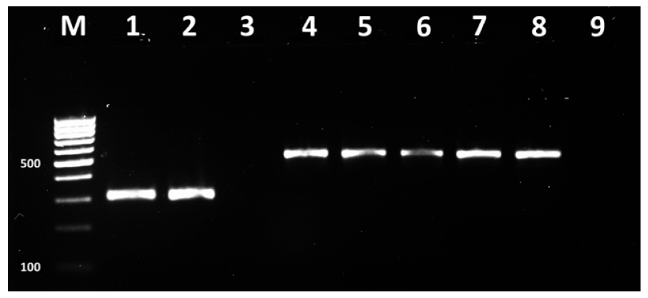
3.2. PCR Identification of S. aureus Isolates

Presumptive S. aureus isolates were precisely identified at the species level by conventional PCR. The results demonstrated 359 bp amplification products, specific for the nuc gene of S. aureus in all tested isolates (Figure 1).

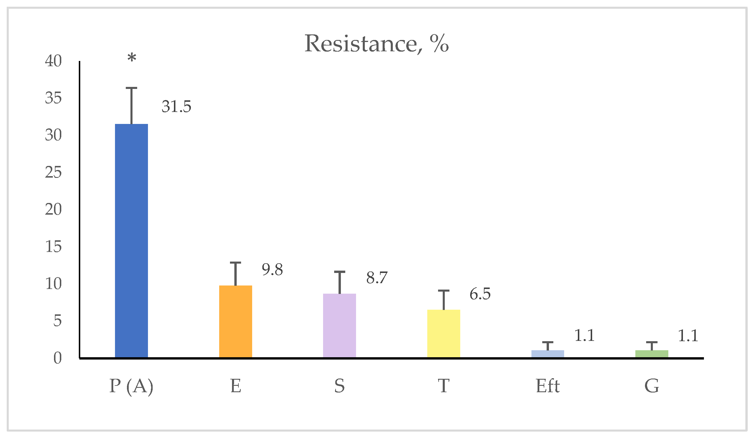
3.3. Antimicrobial Susceptibility Testing

In this case, 32 (34.8%) of the 92 S. aureus isolates tested by the disk diffusion method demonstrated resistance against selected antimicrobial drugs. Resistant isolates originated from 9 farms in 4 regions of the country. At the farm level, the resistance of S. aureus to tested antimicrobials varied from 0% to 75%. In 5/14 (35.7 %) of farms, resistance to none of used chemotherapeutics was detected. These farms were located in Pazardzhik. Stara Zagora, Sliven, Burgas and Kardzhali districts, situated in the South central and Southeastern regions of Bulgaria (Table S5).
The highest, statistically significant, resistance rate was found out against penicillins-29/92 (31.5% of all isolates, p < 0.0001). There were not statistically significant differences in the resistance against the other tested antimicrobials between the farms. The resistance rate to erythromycin was 9/92 (9.8%, p > 0.05), to streptomycin–8/92 (8.7%, p > 0.05), to tetracyclines–6/92 (6.5%, p > 0.05), to gentamicin and ceftiofur–1/92 (1.1%, p > 0.05), respectively. S. aureus isolates resistant to the other tested antimicrobials drugs were not found out (Figure 2).
The resistance patterns of S. aureus isolates are presented in Table S6 and depicted in Figure S1. Most of isolates were resistant to a single chemotherapeutic drug class-3 isolates to streptomycin, 18 to penicillin (ampicillin) or to two antibiotic classes-3 isolates resistant to penicillin (ampicillin) and erythromycin and one: to penicillin (ampicillin) and tetracycline. Multiresistance to three and more groups of antimicrobial drugs was exhibited by 7 isolates (7.6%) and the following patterns were observed: penicillin (ampicillin), tetracycline and erythromycin (n = 2), (P (A)-T-E); penicillin (ampicillin), streptomycin and erythromycin (n = 2), (P (A)-S-E); penicillin (ampicillin), streptomycin, tetracycline and erythromycin (n = 2), (P (A)-S-T-E); 1 isolate was resistant to penicillin (ampicillin), ceftiofur, streptomycin, gentamicin and tetracycline (P (A)-EFT-S-G-T). Multiresistant isolates were isolated only from cows with clinical mastitis.
Data about minimum inhibitory concentrations of the 10 chemotherapeutic drugs in the mastitis pathogens plate for S. aureus are presented in Table 1.
In most isolates determined as sensitive in the disk diffusion test, MIC values of chemotherapeutics in the plate were within the reference ranges. There were isolates, whose growth was not inhibited by the highest concentration of an antimicrobial in the plate. For example, no inhibition was detected from penicillin, ampicillin, erythromycin, tetracycline and sulfadimethoxine in 6.5%, 5.4%, 8.7%, 6.5% and 87% of tested isolates. For them, respective MIC90 values were 2 µg/mL, 4 µg/mL, 1 µg/mL, 1 µg/mL and >256 µg/mL.
Table 2 presents the data from the general comparison between the results from the disk diffusion and MIC tests for 9 chemotherapeutic drugs against bovine mastitis S. aureus isolates. The agreement between both tests was 92.39% with kappa coefficient 0.836 [95% confidence interval (CI) 0.719–0.952; standard error (SE) of kappa–0.059].
With respect to penicillin (Table S7) and ampicillin (Table S8), data were similar: agreement 98.91%, kappa coefficient 0.975, 95% CI 0.925(6)–1.000, SE 0.025 (0.000).
The results for cefoxitin/oxacillin were the same as for ampicillin, and those for ceftiofur–the same as for penicillin. The comparison of tests with respect to erythromycin is shown in Table S9. The results demonstrated 96.74% agreement with kappa coefficient 0.83(95% CI 0.662–1.000; SE of kappa 0.090).
For tetracycline (Table S10), the tests agreed completely: 100%; kappa coefficient 1.000 (95% CI 1.000–1.000; SE 0.000). The results of cephalotin, penicillin/novobiocin and pirlimycin corresponded to those for tetracycline.

3.4. Detection of Resistance Genes

The singleplex PCR assays demonstrated amplification products with size 310 bp, 517 bp (Figure 3), 190 bp and 169 bp (Figure 4), corresponding to, mecA, blaZ genes and ermC and tetK genes, respectively, in 2% agarose gel ((Sigma-Aldrich, USA) stained with 10 ng/mL ethidium bromide (Sigma-Aldrich, USA).
Out of the 29 isolates resistant to penicillin, all possessed the blaZ gene. BlaZ gene was not detected in 61 out of all 63 penicillin-sensitive isolates. The sensitivity and specificity of PCR assay for detection of resistance to penicillin were 100% and 96.8%, respectively. Among the 12 isolates resistant to erythromycin, the erm gene was found out in 7, furthermore, it was detected in 2 of susceptible isolates; therefore, the sensitivity of PCR for detected of resistance to erythromycin was 58.3%, and specificity–97.5%. With regard to resistance to methicillin and tetracycline, both the sensitivity and specificity of the reaction were 100%.
The results from the subsequent multiplex PCR analysis corresponded entirely to those of reactions with single primer pairs (Figure 5).
The agreement between phenotypic resistance to penicillin and the presence of the blaZ gene was 97.83% with kappa coefficient 0.951 (95% CI 0.883–1.000; SE of kappa 0.035). The agreement between phenotypic and genotypic tests for erythromycin was 92.39% with kappa 0.625 (95% CI 0.371–0.878; SE of kappa 0.129); for methicillin and tetracycline–kappa 1.000; 100% agreement.

4. Discussion

The first step for characterisation of bacterial strains is isolation of pure culture and precise identification by means of contemporary methods with high sensitivity and specificity. The performed primary identification of bovine mastitis S. aureus isolates in this study showed variable results from key tests, e.g., presence of haemolysins, pigment production, mannitol fermentation, rabbit plasma coagulation, which was in line with previous observations of ours [32,33]. In a number of small-scale microbiological labs, the identification of S. aureus at the species level is based on the free coagulase tube test. It is not considered a definitive test, as coagulase-negative S. aureus strains are also isolated, probably due to mutations in the coa gene–an important epidemiological marker for detection of different S. aureus variants [34]. Thus, such S. aureus isolates may be erroneously classified as coagulase-negative staphylococci (CNS), which usually cause self-limiting udder infections [35]. The wrong diagnosis can influence the sufficiency of measures for mastitis control. On the other hand, coagulase-positive staphylococci other than S. aureus (S. intermedius, S. pseudintermedius, S. shleiferi subsp. coagulans) may be also involved in the etiology of mastitis [32,36,37]. In this study, all presumptive S. aureus isolates after basic biochemical tests were identified with conventional PCR based on the nuc gene, encoding thermonuclease, including isolates with atypical behaviour. The selected gene is strongly conservative and may be successfully used for precise identification of S. aureus, as confirmed by other research reports [19,26,38,39].
The testing of antimicrobial susceptibility of S. aureus isolates as well as of other causative agents is essential element of mastitis control strategies [40]. Selected antimicrobials comprised drugs used for treatment of mastitis in our country as well as preparations non-registered for use: pirlimycin or ceftiofur, erythromycin under the form of intramammary infusion, as well as penicillin/novobiocin to investigate the trends in antimicrobial resistance of this bacterial species, isolated from mastitic milk in Bulgaria. In this case, 14 substances were tested in the first method, MICs were determined for 10 antimicrobials, and comparisons of S. aureus behaviour in both phenotypic tests were made for 9 antimicrobial drugs included in the Sensititre mastitis plate format. The results determined 32 isolates (34.8%) as resistant to at least one of antimicrobial drugs by the disk diffusion test, whereas the MIC test identified 35 isolates (38%); therefore, a strong agreement between the two tests was found out with kappa value of 0.836. The resistance rates of isolates varied among the farms, districts and regions from 0% to 75%, which required a differentiated approach in programmes for mastitis prevention and control. For penicillin and ampicillin, both tests showed an almost perfect agreement with kappa coefficient 0.975. Disagreements referred to two different isolates. One isolate was interpreted as resistant by the disk diffusion test: 20 mm inhibition zone (IZ), but sensitive in the MIC test (0.12 µg/mL), and another isolate–sensitive to ampicillin by the disk diffusion test (39 mm IZ), but resistant by MIC (8 µg/mL). It should be noted that tests were performed under strictly controlled conditions to eliminate factors that would lead to imprecision in detection of results [27]; also, tests were repeated in independent experiments when inconsistent results for respective isolates were obtained. The paradoxical findings can be most probably due to the Eagle phenomenon–the ability of microorganisms to survive and replicate at concentrations higher than their optimum minimum bactericidal concentration–effect described for S. aureus against penicillin and other beta-lactams [41]. Discrepancies between the disk diffusion test and MIC determined by the E-test were reported by another study [42] having tested the sensitivity of S. aureus isolates from mammary glands of cows and sheep, the authors detected error rates of 3.3% for penicillin and 3.8% for ampicillin. Disagreement was demonstrated by Khalili et al. [43] in testing S. aureus and Enterococcus spp. susceptibility to beta-lactams, glycopeptide antibiotics and clindamycin. Before being reported as sensitive to penicillin, S. aureus strains should be tested for production of beta-lactamase [27]. That is why this study tested isolates with phenotypic resistance and those sensitive to penicillin for the presence of the blaZ gene. The results showed a high sensitivity and specificity of the PCR method for blaZ gene detection compared to phenotypic tests with almost perfect agreement (kappa coefficient 0.951), in line with data from other studies [19,44]. The established 33.7% resistance to penicillin after the genetic analysis (n = 31/92 blaZ positive isolates) was relatively low compared to reported rates from Estonia (61.4%), England (36 to 46%), Korea (38.6 to 78.8%), some farms in China (>60%); northern Ethiopia (>90%), southeastern Brazil (60.7%), Comarca Lagunera region of Mexico (97%) etc. [45,46,47,48,49,50,51]. Lower rates of resistance to penicillin were reported from Chinese dairy farms in Shandong, Jiangsu and Guangdong provinces–24.2% [12], farms in East Poland–23.6% [52], Switzerland–14% [53] and others. In our country, Nikolova et al. [54] have tested the susceptibility of Staphylococcus spp. isolates from two dairy cattle farms in northeastern Bulgaria against different antimicrobials. The authors identified 8 coagulase-positive staphylococci, which were 100% sensitive to tested antimicrobial drugs at farm 1 whereas at farm 2–18.2% were resistant to amoxicillin: a rate comparable to our results.
A surprising finding of this study was the detection of a methicillin-resistant S. aureus isolate from clinical bovine mastitis, which was initially clearly defined as sensitive to cefoxitin by the disk diffusion test on the basis of the 22 mm IZ. A zone of the same size was observed also with the MRSA strain DSM 29134, used as reference strain in the PCR assay, which was also atypical. Subsequent analyses demonstrated resistance to oxacillin with MIC > 4 µg/mL and presence of the mecA gene in both strains. To our best knowledge, this is the first report for isolation of MRSA from a cow with mastitis in Bulgaria, confirmed to carry the mecA gene. Additionally, this clinical isolate showed an unique resistance profile: to penicillin (ampicillin), ceftiofur, streptomycin, gentamicin and tetracycline by the disk diffusion test and susceptibility to amoxillin/clavulanic acid. It was determined as sensitive to erythromycin by both phenotypic tests, but according to the PCR assay, carried the ermC gene. Further detailed studies of this isolate at the molecular level are necessary to establish its SCCmec type as well as full genome sequencing for better understanding of antimicrobial resistance mechanisms. Oxacillin-sensitive mecA positive S. aureus are reported in the literature [55,56], yet the present study is the first to report disagreement between the behaviour to cefoxitin in the disk diffusion test (used as surrogate in oxacillin resistance testing) and that to oxacillin in the MIC test.
With respect to erythromycin, the agreement of results of both phenotypic tests was strong (kappa coefficient 0.839), yet the agreement of phenotypic test and the presence of erm genes was only moderate (kappa coefficient 0.625). From all 12 detected erythromycin-resistant isolates (9 in the disk diffusion and MIC tests; another 3 only in the MIC test), seven strains carried the ermC gene whereas the ermB gene was absent in either resistant or sensitive isolates. The observed sensitivity of the PCR method for detection of genes encoding resistance to erythromycin–58.3% was much lower than the 100% sensitivity reported by Gao et al. [19] for detection of ermC/ermB genes. This was probably due to the presence of ermA gene that was not tested in our isolates and is a limitation of this study. Nevertheless, the prevalence of erm genes in different research reports is highly variable. For example, Ghanbari et al. [23] observed the highest prevalence of ermC followed by ermB and ermA genes among tested human S. aureus isolates; in addition, they did not detect any erm gene in two strains with inducible MLSB phenotype. Another study [57] reported 60% prevalence of the ermA gene among erythromycin-resistant S. aureus isolates. The interpretation of resistance to macrolides should be careful and integral as the different resistance mechanisms produce a different phenotypic profile [58]. At the farm level, the resistance to erythromycin varied from 0 to 46.2%, taking into account that in Bulgaria, this antibiotic is not applied as intramammary infusion. The common use of tylosin in veterinary medical practice may have resulted in horizontal transfer of genetic elements conferring cross resistance in S. aureus to this antimicrobial drug class [59]. In addition, resistance was confirmed in both subclinical and clinical mastitis isolates.
The results for the behaviour of isolates to tetracycline agreed almost perfectly between the two phenotypic tests, as well as with the presence of tet genes, among which only the tetK gene was found out. The resistance rate to tetracycline in all tested isolates was low (6.5%) compared to other reports [49,50,60]. In the different farms however, resistance rates varied from 0 to 50% which was not surprising at the background of the broad use of this antibiotic in cattle farming, including for treatment of mastitis. Rates from 0 to 30.8% in farms were obtained also with respect to streptomycin (in the disk diffusion test), used in combination with penicillin as intramammary infusion–an inevitable effect of the selective pressure of antimicrobial drugs on bacteria [18].
A perfect agreement on susceptibility of S. aureus with kappa value 1.000 was obtained from the two phenotypic tests for cephalothin, penicillin/novobiocin and pirlimycin. With regard to ceftiofur, included in Sensititre mastitis plate format, disagreement referred only to one isolate exhibiting intermediate sensitivity in the disk diffusion test and therefore classified as resistant, but then showing susceptibility with MIC = 2 µg/mL. Against sulfadimethoxine, the isolates from the present study showed high resistance rates: 87% were not inhibited at concentrations of 256 µg/mL, conversely to the behaviour of S. aureus strains in the study of Freu et al. [50] reporting 100% susceptibility. In this study, the sensitivity to amoxicillin/clavulanic acid, fluoroquinolones (enrofloxacin) and co-trimoxazole, tested by the disk diffusion method was 100%, contrary to other research data [39,49,61].

5. Conclusions

Bovine mastitis S. aureus isolates in the present study demonstrated preserved sensitivity to most of tested antimicrobial drug classes, high resistance to sulfadimethoxine and moderately high resistance to penicillinase-sensitive penicillin (33.7%). The comparative analysis between the disk diffusion test used in smaller veterinary diagnostic laboratories and MIC determination by the serial dilution micromethod applied in labs handling a greater number of samples showed more than 90% agreement for 9 tested antimicrobial drugs, hence proving reliability of results from phenotypic monitoring of resistance. By reason of observed discrepancies between the behaviour of some isolates in the phenotypic tests, e.g., against penicillin, ampicillin, cefoxitin/oxacillin and erythromycin, integral evaluation of resistance patterns with inclusion of molecular biological methods is advised. This approach resulted in detection of a cefoxitin-sensitive but oxacillin-resistant mecA-positive S. aureus isolate, as well as of few isolates sensitive in the disk diffusion test but resistant in the MIC test and vice versa. We believe that the results from this study will contribute to the experience of microbiological diagnostic labs and strategies implemented in mastitis prevention and control programmes.

Supplementary Materials

The following supporting information can be downloaded at: https://0-www-mdpi-com.brum.beds.ac.uk/article/10.3390/vetsci9080401/s1, Table S1: Nuc PCR temperature conditions; Table S2: Classes of antimicrobial drugs included in the mastitis pathogens testing plate (Trek Diagnostic Systems Ltd., UK), their concentrations and susceptible MIC breakpoints according to CLSI [27]; Table S3: Sequence of primers, targeted genes, expected size of PCR products and their source; Table S4: Primary identification of bovine mastitis Staphylococcus spp. Isolates; Table S5: Distribution of isolates resistant in the Bauer-Kirby disk diffusion test by regions, districts and farms; Table S6: Resistance patterns of bovine mastitis S. aureus isolates resistant in the Bauer-Kirby disk diffusion test by regions, districts and farms; Figure S1: Some resistance patterns depicted by the disk diffusion test; Table S7: Comparison between disk diffusion and MIC methods for testing resistance to penicillin; Table S8: Comparison between disk diffusion and MIC methods for testing resistance to ampicillin; Table S9: Comparison between disk diffusion and MIC methods for testing resistance to erythromycin; Table S10. Comparison between disk diffusion and MIC methods for testing resistance to tetracycline.

Author Contributions

Conceptualization, N.R.; methodology, N.R., A.M. and I.S.; software, N.R.; validation, N.R., A.M. and I.S.; formal analysis, N.R.; investigation, N.R., N.V., A.R., A.M. and I.S.; resources, N.R.; data curation, N.R. and I.S.; writing—original draft preparation, N.R.; writing—review and editing, N.V., A.R., A.M. and I.S.; visualization, N.R.; supervision, A.M.; project administration, N.R.; funding acquisition, N.R. All authors have read and agreed to the published version of the manuscript.

Funding

This research was funded by Trakia University, Faculty of Veterinary Medicine, Stara Zagora, Bulgaria, grant number 11 FVM/02.06.2020.

Institutional Review Board Statement

Not applicable.

Informed Consent Statement

Not applicable.

Data Availability Statement

The data presented in this study can be found in the manuscript.

Acknowledgments

We thank veterinarians for the sample collection.

Conflicts of Interest

The authors declare no conflict of interest.

References

  1. Tong, S.Y.; Davis, J.S.; Eichenberger, E.; Holland, T.L.; Fowler, V.G., Jr. Staphylococcus aureus infections: Epidemiology, pathophysiology, clinical manifestations, and management. Clin. Microbiol. Rev. 2015, 28, 603–661. [Google Scholar] [CrossRef] [PubMed] [Green Version]
  2. Taylor, T.A.; Unakal, C.G. Staphylococcus Aureus. In StatPearls. Treasure Island (FL): StatPearls. Available online: https://0-www-ncbi-nlm-nih-gov.brum.beds.ac.uk/books/NBK441868/ (accessed on 14 February 2022).
  3. Haag, A.F.; Fitzgerald, J.R.; Penadés, J.R. Staphylococcus aureus in Animals. Microbiol. Spectr. 2019, 7, 3. [Google Scholar] [CrossRef] [PubMed]
  4. Botrel, M.A.; Haenni, M.; Morignat, E.; Sulpice, P.; Madec, J.Y.; Calavas, D. Distribution and antimicrobial resistance of clinical and subclinical mastitis pathogens in dairy cows in Rhone- Alpes, France. Foodborne Pathog. Dis. 2010, 7, 479–487. [Google Scholar] [CrossRef] [PubMed]
  5. Heikkilä, A.M.; Liski, E.; Pyörälä, S.; Taponen, S. Pathogen-specific production losses in bovine mastitis. J. Dairy Sci. 2018, 101, 9493–9504. [Google Scholar] [CrossRef] [PubMed] [Green Version]
  6. Hertl, J.A.; Schukken, Y.H.; Welcome, F.L.; Tauer, L.W.; Gröhn, Y.T. Pathogen-specific effects on milk yield in repeated clinical mastitis episodes in Holstein dairy cows. J. Dairy Sci. 2014, 97, 1465–1480. [Google Scholar] [CrossRef] [Green Version]
  7. Hu, X.; Guo, J.; Zhao, C.; Jiang, P.; Maimai, T.; Yanyi, L.; Cao, Y.; Fu, Y.; Zhang, N. The gut microbiota contributes to the development of Staphylococcus aureus-induced mastitis in mice. ISME J. 2020, 14, 1897–1910. [Google Scholar] [CrossRef]
  8. Taponen, S.; McGuinness, D.; Hiitiö, H.; Simojoki, H.; Zadoks, R.; Pyörälä, S. Bovine milk microbiome: A more complex issue than expected. Vet. Res. 2019, 50, 44. [Google Scholar] [CrossRef] [Green Version]
  9. Kaczorowski, Ł.; Powierska-Czarny, J.; Wolko, Ł.; Piotrowska-Cyplik, A.; Cyplik, P.; Czarny, J. The Influence of Bacteria Causing Subclinical Mastitis on the Structure of the Cow’s Milk Microbiome. Molecules 2022, 27, 1829. [Google Scholar] [CrossRef]
  10. Ruegg, P.; Oliveira, L.; Jin, W.; Okwumabua, O. Phenotypic antimicrobial susceptibility and occurrence of selected resistance genes in gram-positive mastitis pathogens isolated from Wisconsin dairy cows. J. Dairy Sci. 2015, 98, 4521–4534. [Google Scholar] [CrossRef] [PubMed]
  11. Santos, R.I.; Zuninob, P.M.; Gila, A.D.; Laportc, A.; Hirigoyenc, D.J. Antibiotic resistance of Staphylococcus aureus associated with subclinical and clinical mastitis in Uruguay during an eight-year period. Austral. J. Vet. Sci. 2017, 49, 191–194. [Google Scholar] [CrossRef] [Green Version]
  12. Liu, K.; Tao, L.; Li, J.; Fang, L.; Cui, L.; Li, J.; Meng, X.; Zhu, G.; Bi, C.; Wang, H. Characterization of Staphylococcus aureus Isolates From Cases of Clinical Bovine Mastitis on Large-Scale Chinese Dairy Farms. Front. Vet. Sci. 2020, 7, 580129. [Google Scholar] [CrossRef] [PubMed]
  13. Soares, B.S.; Melo, D.A.; Motta, C.C.; Marques, V.F.; Barreto, N.B.; Coelho, S.M.O.; Coelho, I.S.; Souza, M.M.S. Characterization of virulence and antibiotic profile and agr typing of Staphylococcus aureus from milk of subclinical mastitis bovine in State of Rio de Janeiro. Arq. Bras. Med. Vet. Zootec. 2017, 69, 843–850. [Google Scholar] [CrossRef] [Green Version]
  14. Srednik, M.E.; Crespi, E.; Testorelli, M.F.; Puigdevall, T.; Pereyra, A.M.D.; Rumi, M.V.; Caggiano, N.; Gulone, L.; Mollerach, M.; Gentilini, E.R. First isolation of a methicillin-resistant Staphylococcus aureus from bovine mastitis in Argentina. Vet. Anim. Sci. 2019, 7, 100043. [Google Scholar] [CrossRef] [PubMed]
  15. Shrestha, A.; Bhattarai, R.K.; Luitel, H.; Karki, S.; Basnet, H.B. Prevalence of methicillin-resistant Staphylococcus aureus and pattern of antimicrobial resistance in mastitis milk of cattle in Chitwan, Nepal. BMC Vet. Res. 2021, 17, 239. [Google Scholar] [CrossRef] [PubMed]
  16. Kadlec, K.; Entorf, M.; Peters, T. Occurrence and Characteristics of Livestock-Associated Methicillin-Resistant Staphylococcus aureus in Quarter Milk Samples From Dairy Cows in Germany. Front. Microbiol. 2019, 10, 1295. [Google Scholar] [CrossRef] [PubMed] [Green Version]
  17. Qolbaini, E.N.; Khoeri, M.M.; Salsabila, K.; Paramaiswari, W.T.; Tafroji, W.; Artika, I.M.; Safari, D. Identification and antimicrobial susceptibility of methicillin-resistant Staphylococcus aureus-associated subclinical mastitis isolated from dairy cows in Bogor, Indonesia. Vet. World 2021, 14, 1180–1184. [Google Scholar] [CrossRef] [PubMed]
  18. Schnitt, A.; Tenhagen, B.A. Risk Factors for the Occurrence of Methicillin-Resistant Staphylococcus aureus in Dairy Herds: An Update. Foodborne Pathog. Dis. 2020, 17, 585–596. [Google Scholar] [CrossRef] [Green Version]
  19. Gao, J.; Ferreri, M.; Liu, X.Q.; Chen, L.B.; Su, J.L.; Han, B. Development of multiplex polymerase chain reaction assay for rapid detection of Staphylococcus aureus and selected antibiotic resistance genes in bovine mastitic milk samples. J. Vet. Diagn. Investig. 2011, 23, 894–901. [Google Scholar] [CrossRef] [Green Version]
  20. Klibi, A.; Jouini, A.; Gómez, P.; Slimene, K.; Ceballos, S.; Torres, C.; Maaroufi, A. Molecular Characterization and Clonal Diversity of Methicillin-Resistant and -Susceptible Staphylococcus aureus Isolates of Milk of Cows with Clinical Mastitis in Tunisia. Microb. Drug Resist. 2018, 24, 8. [Google Scholar] [CrossRef]
  21. Llarrull, L.; Mobashery, S. Dissection of events in the resistance to β-lactam antibiotics mediated by the protein BlaR1 from Staphylococcus aureus. Biochemistry 2012, 51, 4642–4649. [Google Scholar] [CrossRef] [Green Version]
  22. Williams, M.C.; Dominguez, S.R.; Prinzi, A.; Lee, K.; Parker, S.K. Reliability of mecA n Predicting Phenotypic Susceptibilities of Coagulase-Negative Staphylococci and Staphylococcus aureus. Open Forum Infect. Dis. 2020, 12, ofaa553. [Google Scholar] [CrossRef] [PubMed]
  23. Ghanbari, F.; Ghajavand, H.; Havaei, R.; Jami, M.S.; Khademi, F.; Heydari, L.; Shahin, M.; Havaei, S.A. Distribution of erm genes among Staphylococcus aureus isolates with inducible resistance to clindamycin in Isfahan, Iran. Adv. Biomed. Res. 2016, 5, 62. [Google Scholar] [CrossRef] [PubMed]
  24. Bruce, S.A.; Smith, J.T.; Mydosh, J.L.; Ball, J.; Needle, D.B.; Gibson, R.; Andam, C.P. Shared antibiotic resistance and virulence genes in Staphylococcus aureus from diverse animal hosts. Sci. Rep. 2022, 12, 4413. [Google Scholar] [CrossRef] [PubMed]
  25. Markey, B.; Leonar, F.; Archambault, M.; Cullinane, A.; Maguire, D. Clinical Veterinary Microbiology, 2nd ed.; Mosby, Elsevier Ltd.: Edinburgh, UK, 2013; pp. 105–118. [Google Scholar]
  26. Sasaki, T.; Tsubakishita, S.; Tanaka, Y.; Sakusabe, A.; Ohtsuka, M.; Hirotaki, S.; Kawakami, T.; Fukata, T.; Hiramatsu, K. Multiplex-PCR method for species identification of coagulase-positive staphylococci. J. Clin. Microbiol. 2010, 48, 765–769. [Google Scholar] [CrossRef] [PubMed] [Green Version]
  27. CLSI. Performance Standards for Antimicrobial Disk and Dilution Susceptibility Tests for Bacteria Isolated from Animals, 4th ed.; Approved Guideline: Document M100-S25; Clinical and Laboratory Standards Institute: Wayne, PA, USA, 2018. [Google Scholar]
  28. Zhang, K.; Sparling, J.; Chow, B.L.; Elsayed, S.; Hussain, Z.; Church, D.L.; Gregson, D.B.; Louie, T.; Conly, J.M. New quadriplex PCR assay for detection of methicillin and mupirocin resistance and simultaneous discrimination of Staphylococcus aureus from coagulase-negative staphylococci. J. Clin. Microbiol. 2004, 42, 4947–4955. [Google Scholar] [CrossRef] [PubMed] [Green Version]
  29. McHugh, M.L. Interrater reliability: The kappa statistic. Biochem. Med. 2012, 22, 276–282. [Google Scholar] [CrossRef]
  30. Camargo, C.H.; Mondelli, A.L.; Bôas, P.J. Comparison of teicoplanin disk diffusion and broth microdilution methods against clinical isolates of Staphylococcus aureus and S. epidermidis. Braz. J. Microbiol. 2011, 42, 1265–1268. [Google Scholar] [CrossRef] [Green Version]
  31. Martin, S.W. Estimating disease prevalence and the interpretation of screening test results. Prev. Vet. Med. 1984, 2, 463–472. [Google Scholar] [CrossRef]
  32. Rusenova, N.; Gebreyes, W.; Koleva, M.; Mitev, J.; Penev, T.; Vasilev, N.; Miteva, T. Comparison of three methods for routine detection of Staphylococcus aureus isolated from bovine mastitis. Kafkas Univ. Vet. Fak. Derg. 2013, 19, 709–712. [Google Scholar]
  33. Rusenova, N.V.; Rusenov, A.G. Detection of Staphylococcus aureus among coagulase positive staphylococci from animal origin based on conventional and molecular methods. Mac. Vet. Rev. 2017, 40, 29–36. [Google Scholar] [CrossRef] [Green Version]
  34. Effendi, M.H.; Hisyam, M.; Hastutiek, P.; Tyasningsih, W. Detection of coagulase gene in Staphylococcus aureus from several dairy farms in East Java, Indonesia, by polymerase chain reaction. Vet. World 2019, 12, 68–71. [Google Scholar] [CrossRef] [PubMed] [Green Version]
  35. Phophi, L.; Petzer, I.M.; Qekwana, D.N. Antimicrobial resistance patterns and biofilm formation of coagulase-negative Staphylococcus species isolated from subclinical mastitis cow milk samples submitted to the Onderstepoort Milk Laboratory. BMC Vet. Res. 2019, 15, 420. [Google Scholar] [CrossRef] [PubMed]
  36. Freitas Guimarães, F.F.; Nóbrega, D.B.; Richini-Pereira, V.B.; Marson, P.M.; Figueiredo Pantoja, J.C.; Langoni, H. Enterotoxin genes in coagulasenegative and coagulase-positive staphylococci isolated from bovine milk. J. Dairy Sci. 2013, 96, 2866–2872. [Google Scholar] [CrossRef] [Green Version]
  37. Karzis, J.; Petzer, I.M.; Naidoo, V.; Donkin, E.F. The spread and antimicrobial resistance of Staphylococcus aureus in South African dairy herds—A review. Onderstepoort J. Vet. Res. 2021, 88, e1–e10. [Google Scholar] [CrossRef] [PubMed]
  38. Monistero, V.; Graber, H.U.; Pollera, C.; Cremonesi, P.; Castiglioni, B.; Bottini, E.; Ceballos-Marquez, A.; Lasso-Rojas, L.; Kroemker, V.; Wente, N.; et al. Staphylococcus aureus Isolates from Bovine Mastitis in Eight Countries: Genotypes, Detection of Genes Encoding Different Toxins and Other Virulence Genes. Toxins 2018, 10, 247. [Google Scholar] [CrossRef] [Green Version]
  39. Mbindyo, C.M.; Gitao, G.C.; Plummer, P.J.; Kulohoma, B.W.; Mulei, C.M.; Bett, R. Antimicrobial Resistance Profiles and Genes of Staphylococci Isolated from Mastitic Cow’s Milk in Kenya. Antibiotics 2021, 10, 772. [Google Scholar] [CrossRef]
  40. McDougall, S.; Hussein, H.; Petrovski, K. Antimicrobial resistance in Staphylococcus aureus, Streptococcus uberis and Streptococcus dysgalactiae from dairy cows with mastitis. N. Z. Vet. J. 2014, 62, 68–76. [Google Scholar] [CrossRef] [PubMed]
  41. Prasetyoputri, A.; Jarrad, A.M.; Cooper, M.A.; Blaskovich, M.A.T. The Eagle Effect and Antibiotic-Induced Persistence: Two Sides of the Same Coin? Trends Microbiol. 2019, 27, 339–354. [Google Scholar] [CrossRef]
  42. Pengov, A.; Ceru, S. Antimicrobial drug susceptibility of Staphylococcus aureus strains isolated from bovine and ovine mammary glands. J. Dairy Sci. 2003, 86, 3157–3163. [Google Scholar] [CrossRef]
  43. Khalili, H.; Soltani, R.; Negahban, S.; Abdollahi, A.; Gholami, K. Reliability of Disk Diffusion Test Results for the Antimicrobial Susceptibility Testing of Nosocomial Gram-positive Microorganisms: Is E-test Method Better? Iran J. Pharm. Res. 2012, 11, 559–563. [Google Scholar] [PubMed]
  44. Lúcio, É.C.; Gouveia, G.V.; Costa, M.M.; Oliveira, M.B.; Mota, R.A.; Pinheiro Junior, J.W. Phenotypic and genotypic characteristics and resistance profile of Staphylococcus spp. from bovine mastitis. Acta Sci. Vet. 2020, 48, 1759. [Google Scholar] [CrossRef]
  45. Kalmus, P.; Aasmäe, B.; Kärssin, A.; Orro, T.; Kask, K. Udder pathogens and their resistance to antimicrobial agents in dairy cows in Estonia. Acta Vet. Scand. 2011, 53, 4. [Google Scholar] [CrossRef] [PubMed] [Green Version]
  46. Hendriksen, R.S.; Mevius, D.J.; Schroeter, A.; Teale, C.; Meunier, D.; Butaye, P.; Franco, A.; Utinane, A.; Amado, A.; Moreno, M.; et al. Prevalence of antimicrobial resistance among bacterial pathogens isolated from cattle in different European countries: 2002-2004. Acta Vet. Scand. 2008, 50, 28. [Google Scholar] [CrossRef] [Green Version]
  47. Nam, H.-M.; Lee, A.-L.; Jung, M.-N.K.; Jang, G.-C.; Wee, S.-H.; Lim, S.-K. Antimicrobial susceptibility of staphylococus aureus and characterization of methicillin-resistant staphylococcus aureus isolates from bovine mastitis in Korea. Foodborne Pathog. Dis. 2011, 8, 231–238. [Google Scholar] [CrossRef] [PubMed]
  48. Cheng, J.; Qu, W.; Barkema, H.W.; Nobrega, D.B.; Gao, J.; Liu, G.; Buck, J.; Kastelic, J.P.; Sun, H.; Han, B. Antimicrobial resistance profiles of 5 common bovine mastitis pathogens in large Chinese dairy herds. J. Dairy Sci. 2019, 102, 2416–2426. [Google Scholar] [CrossRef] [Green Version]
  49. Kalayu, A.A.; Woldetsadik, D.A.; Woldeamanuel, Y.; Wang, S.H.; Gebreyes, W.A.; Teferi, T. Burden and antimicrobial resistance of S. Aureus in dairy farms in Mekelle, Northern Ethiopia. BMC Vet. Res. 2020, 16, 20. [Google Scholar] [CrossRef]
  50. Freu, G.; Tomazi, T.; Filho, A.F.S.; Heinemann, M.B.; Santos, M.V. Antimicrobial Resistance and Molecular Characterization of Staphylococcus aureus Recovered from Cows with Clinical Mastitis in Dairy Herds from Southeastern Brazil. Antibiotics 2022, 11, 424. [Google Scholar] [CrossRef] [PubMed]
  51. Mora-Hernández, Y.; Vera Murguía, E.; Stinenbosch, J.; Hernández Jauregui, P.; van Dijl, J.M.; Buist, G. Molecular typing and antimicrobial resistance profiling of 33 mastitis-related Staphylococcus aureus isolates from cows in the Comarca Lagunera region of Mexico. Sci. Rep. 2021, 11, 6912. [Google Scholar] [CrossRef] [PubMed]
  52. Szweda, P.; Schielmann, M.; Frankowska, A.; Kot, B.; Zalewska, M. Antibiotic resistance in Staphylococcus aureus strains isolated from cows with mastitis in eastern Poland and analysis of susceptibility of resistant strains to alternative nonantibiotic agents: Lysostaphin, nisin and polymyxin B. J. Vet. Med. Sci. 2014, 76, 355–362. [Google Scholar] [CrossRef] [PubMed] [Green Version]
  53. Käppeli, N.; Morach, M.; Corti, S.; Eicher, C.; Stephan, R.; Johler, S. Staphylococcus aureus related to bovine mastitis in Switzerland: Clonal diversity, virulence gene profiles, and antimicrobial resistance of isolates collected throughout 2017. J. Dairy Sci. 2019, 102, 3274–3281. [Google Scholar] [CrossRef] [PubMed] [Green Version]
  54. Nikolova, M.; Urumova, V.; Liuzkanov, M.; Sabev, S. Studies on the causes of bacterial mastities in two dairy bovine farms in northeastern Бulgaria and their sensitivity to antimicrobial products. Tradit. Mod. Vet. Med. 2021, 6, 21–27. [Google Scholar]
  55. Kumar, V.A.; Steffy, K.; Chatterjee, M.; Sugumar, M.; Dinesh, K.R.; Manoharan, A.; Karim, S.; Biswas, R. Detection of oxacillin-susceptible mecA-positive Staphylococcus aureus isolates by use of chromogenic medium MRSA ID. J. Clin. Microbiol. 2013, 51, 318–319. [Google Scholar] [CrossRef] [Green Version]
  56. Liu, J.L.; Li, T.M.; Zhong, N.; Wang, X.; Jiang, J.; Zhang, W.X.; Tang, R.; Guo, Y.J.; Liu, Y.; Hu, J.; et al. Current status of oxacillin-susceptible mecA-positive Staphylococcus aureus infection in Shanghai, China: A multicenter study. J. Microbiol. Immunol. Infect. 2021, 54, 1070–1077. [Google Scholar] [CrossRef] [PubMed]
  57. Saderi, H.; Emadi, B.; Owlia, P. Phenotypic and genotypic study of macrolide, lincosamide and streptogramin B (MLSB) resistance in clinical isolates of Staphylococcus aureus in Tehran, Iran. Med. Sci. Monit. 2011, 17, BR48–BR53. [Google Scholar] [CrossRef] [Green Version]
  58. Miklasińska-Majdanik, M. Mechanisms of Resistance to Macrolide Antibiotics among Staphylococcus aureus. Antibiotics 2021, 10, 1406. [Google Scholar] [CrossRef] [PubMed]
  59. Feßler, A.T.; Wang, Y.; Wu, C.; Schwarz, S. Mobile macrolide resistance genes in staphylococci. Plasmid 2018, 99, 2–10. [Google Scholar] [CrossRef]
  60. Martini, C.; Lange, C.; Brito, M.; Ribeiro, J.; Mendonça, L.; Vaz, E. Characterisation of penicillin and tetracycline resistance in Staphylococcus aureus isolated from bovine milk samples in Minas Gerais, Brazil. J. Dairy Res. 2017, 84, 202–205. [Google Scholar] [CrossRef] [PubMed]
  61. Bissong, M.E.A.; Ateba, C.N. Genotypic and Phenotypic Evaluation of Biofilm Production and Antimicrobial Resistance in Staphylococcus aureus Isolated from Milk, North West Province, South Africa. Antibiotics 2020, 9, 156. [Google Scholar] [CrossRef] [PubMed] [Green Version]
Figure 1. PCR identification of S. aureus isolates on the basis of nuc gene. M–100 bp molecular marker (BrightMax 100–1000 bp, Canvax, Spain); 1–positive control S. aureus ATCC 25923; 2-8–tested isolates; 9-negative control (water).
Vetsci 09 00401 g001
Figure 2. Percentage distribution of S. aureus isolates from bovine mastitis, resistant to tested chemotherapeutic drugs in the Bauer-Kirby disk diffusion test. Legend: P-penicillin, A-ampicillin, E-erythromycin, S-streptomycin, T-tetracycline, Eft-ceftiofur, G–gentamicin. * Statistically significantly higher resistance against penicillin in comparison to the other antibiotics in the investigated farms.
Vetsci 09 00401 g002
Figure 3. PCR demonstrating mecA and blaZ genes in bovine mastitis S. aureus isolates from Bulgaria. M–DNA ladder 100 bp (BrightMax 100–1000 bp, Canvax, Spain), 1–MRSA DSM 29134 mecA positive control, 2–mecA positive isolate, 3–negative control of first reaction mix; 4–MRSA DSM 29134 blaZ positive control, 5-8–blaZ positive isolates, 9–negative control of second reaction mix.
Vetsci 09 00401 g003
Figure 4. PCR demonstrating ermC and tetK genes in bovine mastitis S. aureus isolates from Bulgaria. M–DNA ladder 100 bp, 1–4–ermC positive isolates, 5-negative control of first reaction mix; 6–8–tetK positive isolates, 9-negative control of second reaction mix.
Vetsci 09 00401 g004
Figure 5. Multiplex PCR demonstrating the presence of blaZ/tetK and blaZ/ermC genes in some S. aureus isolates. 4, 9–negative controls of first and second reaction mix.
Vetsci 09 00401 g005
Table 1. Distribution of minimum inhibitory concentrations of 10 antimicrobials included in Sensititre Mastitis plate format to S. aureus isolates from bovine mastitis in Bulgaria.
AntimicrobialsDistribution (%) of MIC (µg/mL)NIMIC50MIC90
0.120.250.51248163264128256
PEN68.54.38.75.44.30.02.2-----6.50.122
AMP64.13.312.07.62.23.32.2-----5.40.124
ERY-18.568.53.31.10.0------8.70.51
OXA+----98.91.1-------22
PIRL--54.345.70.00.0-------0.51
TET---90.23.30.00.0-----6.511
CEP----98.91.10.00.0-----22
XNL--7523.91.10.0-------0.51
SDM--------2.20.01.19.887>256>256
P/N---1000.00.00.0------11
Legend: PEN-penicillin, AMP-ampicillin, ERY-erythromycin, Oxa+-oxacillin + 2%NaCl, RIRL–pirlimycin, TET-tetracycline, CEP–cephalothin, XNL-ceftiofur, SDM–sulphadimethoxine, P/N–penicillin/novobiocin. Susceptibility breakpoints are highlighted in light blue whereas resistance breakpoints: in dark blue (CLSI, 2018). Intermediate isolates were interpreted as resistant. NI = the growth of the isolate is not inhibited by the highest concentration of the antimicrobial agent in the plate. MIC50 (μg/mL) = concentration inhibiting 50% of isolates. MIC90 (μg/mL) = concentration inhibiting 90% of isolates.
Table 2. Comparison between disk diffusion and minimum inhibitory concentration methods for testing resistance of bovine mastitis S. aureus isolates to 9 chemotherapeutic drugs (penicillin, ampicillin, cefoxitin/oxacillin, cephalotin, ceftiofur, erythromycin, tetracycline, penicillin/novobiocin, pirlimycin).
Minimum Inhibitory ConcentrationTotal
N Sensitive IsolatesN Resistant Isolates
Disk diffusion test
N sensitive isolates
55560
Disk diffusion test
N resistant isolates
23032
Total573592
Publisher’s Note: MDPI stays neutral with regard to jurisdictional claims in published maps and institutional affiliations.

