Article

Pilot Study: Assessing the Expression of Serum Lactate Dehydrogenase and Peripheral Leukocyte Ratios in Canine Oral Malignant Melanoma

by
Ben Murray
and
Kelly L. Bowlt Blacklock
*
Hospital for Small Animals, Royal (Dick) School of Veterinary Studies, University of Edinburgh, Easter Bush Campus, Midlothian EH25 9RG, UK
*
Author to whom correspondence should be addressed.
Submission received: 17 June 2022 / Revised: 27 July 2022 / Accepted: 28 July 2022 / Published: 9 August 2022
(This article belongs to the Special Issue Advances in Understanding Spontaneously Occurring Melanoma in Animals)

Abstract

:

Simple Summary

This study aimed to assess the expression of novel blood biomarkers in dogs with oral malignant melanoma, an aggressive and common oral cancer in dogs. The results of this pilot study suggest that the concentrations of lactate dehydrogenase, which can be easily measured with blood samples, are significantly higher in dogs with oral malignant melanoma. These blood biomarkers have been shown to be of prognostic value in human melanoma patients, meaning that they can act to predict the potential behaviour of the tumour. Therefore, research into our canine patients should be viewed as being potentially very valuable, as the discovery of easily measurable prognostic biomarkers could significantly further not only our understanding of the underlying physiology of melanoma itself, but also change the way veterinary surgeons investigate and treat the disease in the future. This study lays the foundations for further, more extensive investigation, into the topic.

Abstract

Measurement of blood biomarkers such as lactate dehydrogenase (LDH) and peripheral leukocyte ratios have been shown to be of prognostic value in human melanoma patients. Previous veterinary studies have demonstrated that changes in these values are detectable in multiple canine cancer patients. However, to the authors’ knowledge, no studies have yet demonstrated an increase in LDH in canine oral malignant melanoma patients, nor has the effect of metastasis on LDH levels been explored. This retrospective pilot study included 18 dogs, of which 10 were healthy controls, 5 OMM patients with metastasis and 3 without metastasis. Serum LDH was measured and pre-treatment peripheral leucocyte ratios were calculated. LDH was measurable within all patient groups and a statistically significant difference in LDH levels was detected between patients with OMM and healthy controls (p < 0.05); however, no significant difference was detected between patients with or without metastatic disease. This study suggests that serum LDH levels are significantly increased in dogs with OMM compared to healthy controls, paving the way for further research to investigate the prognostic value of this biomarker.

1. Introduction

Oral malignant melanoma (OMM) is the most common oral neoplasm in the dog, accounting for 30–40% of all canine oral malignancies [1,2,3]. There is no known sex predilection and it is typically a disease of older dogs, most often diagnosed in Scottish terriers, golden retrievers, chow chows, poodles and dachshunds [4]. The site of tumour growth is most commonly located in the gingiva but can be found anywhere within the oral cavity including the internal lip, tongue and hard palate [1]. OMM is aggressive and locally invasive with variable rates of metastasis reported, ranging from 30.3% to 74% to the local lymph nodes and 14.0% to 92% to the lungs and other organs [3]. Melanomas have an extremely diverse and unreliable spectrum of biological behaviour. The biological behaviour of OMM can be predicted using several clinical, biochemical and histopathological factors including anatomic site, size, clinical stage, mitotic index, Ki67 expression, degree of pigmentation, nuclear atypia and immunohistochemistry [3,4,5,6,7].
The treatment for patients with OMM begins with local disease control, of which en bloc surgical excision alongside mandibular and medial retropharyngeal lymphadenectomy is prioritised in most cases [1,8]. Adjunctive therapies may include radiotherapy, electrochemotherapy, immunotherapy or chemotherapy where appropriate [3]. Many recent veterinary studies have focussed on the immunogenicity of melanoma, with novel adjuvant immunotherapies showing initially encouraging results [9,10]. The median survival time for dogs with OMM treated using surgery alone are 511 to 874 days, 160 to 818 days and 168 to 207 days for stages I–III, respectively, with metastatic disease being the most common cause of death [3].
Recently within human medical oncology, there has been an increased interest in the use of circulating blood biomarkers, such as lactate dehydrogenase (LDH), as prognostic indicators of disease progression and treatment effectiveness [11,12]. LDH catalyses the conversion of pyruvate to lactate during anaerobic respiration in normal and neoplastic cells, with increased serum LDH levels believed to reflect high metabolic activity within the hypoxic and highly active cellular environment of malignant tumours [13,14]. To this end, assessment of LDH has been included in the updated American Joint Committee on Cancer melanoma staging system, with multiple studies having confirmed a correlation between serum LDH levels and decreased survival times in human patients with advanced stage melanoma [15,16,17]. Within Veterinary Medicine, studies have shown significant elevations in serum LDH levels in dogs with lymphoma, mammary gland and oral tumours compared to healthy controls [13,14,18,19].
As well as LDH levels, recent human studies have shown blood neutrophil to lymphocyte ratios (NLR) and lymphocyte to monocyte ratios (LMR) to be of prognostic value in various cancers including melanoma, with an increased NLR and decreased LMR representing negative prognostic indicators [20,21,22]. These haematological changes are theorised to reflect the effect of central inflammation on the development and progression of cancer, with lymphocytes aiding in the production of an immune response against the tumour and monocytes conversely being recruited to produce growth factors and cytokines within the tumour leading to immunosuppression and angiogenesis, creating optimal conditions for growth within the tumour [6]. The prognostic impact of these ratios has also been assessed in several canine malignancies including lymphoma, soft tissue sarcoma, osteosarcoma, mast cell tumours, and OMM. Although results vary between studies, multiple papers have demonstrated these ratios to be of prognostic value in veterinary patients [23,24,25,26].
To this author’s knowledge, no previous studies have assessed serum levels of LDH specifically in patients with OMM, nor have they assessed the effect of metastasis on serum LDH levels. The aim of this pilot study was to compare LDH levels from stored frozen serum in OMM patients against those of healthy controls, as well as to assess whether there was a statistically significant difference in LDH, NLR and LMR values between patients with or without evidence of metastasis.

2. Materials and Methods

2.1. Patient Selection Criteria

Records from the Hospital for Small Animals, University of Edinburgh were searched for client-owned dogs with a confirmed histological diagnosis of OMM between June 2012 and December 2021. Dogs with confirmed OMM through histopathology and that had undergone full clinical staging to determine the presence of metastasis to the local lymph nodes and lungs were considered eligible for entry into this study. Clinical staging included lymph node assessment through either fine needle aspiration (FNA) or histopathology when available as well as head and thorax CT scan, or alternatively 3-view thoracic radiography. Inclusion criteria included availability of pre-treatment haematology and leukocyte differential counts taken at patient presentation as well as evidence of full clinical staging to assess for the presence of metastasis. Patients were only included in the non-metastatic group if full lymph node histopathology was available to rule out metastasis. Not all patients included in this study had frozen serum samples for assessment of LDH but were included purely for assessment of leukocyte ratios. Exclusion criteria included those patients with any evidence of hepatic or cardiac disease either at presentation or in the previous month that may interfere with LDH values, as well as patients that had received corticosteroids within the past month that may interfere with leukocyte differential counts.
Data retrieved from patient records for each dog included signalment (age, breed and sex), tumour location, evidence of lymph node metastasis at initial staging, evidence of lung metastasis at initial staging, method of staging (FNA vs. histopathology/CT vs. X-ray), date of sample collection and pre-treatment haematology results including leukocyte counts.
Ethical approval for this study was granted through the Veterinary Ethical Review Committee, University of Edinburgh (126.21). Written forms signed by owners prior to recruitment for this study confirmed owners’ consent to data collection as well as storage and use of spare blood from clinical sampling for research purposes.
Control values for LDH were measured from the frozen serum of clinically healthy patients with no evidence of recent or concurrent disease on history or clinical examination as a part of the University of Edinburgh biobank programme.

2.2. Evaluation of Serum LDH

After collection in serum tubes, samples were centrifuged at 2500 rpm for 10 min at room temperature. Serum samples were then stored at −80 °C in a clinical biobank within 24 h of collection. For the purpose of this study, frozen samples were defrosted at room temperature on a lab roller mixer and immediately run through Beckman Coulter AU480 analyser (Beckman Coulter Ltd, London, UK) using their reagent, category number OSR6126. Results were displayed in U/L. The normal range for LDH at our laboratory was 21–212 U/L. Given that localised or metastatic disease could be present, total serum LDH was measured, rather than individual isoenzyme values which have been shown to vary based on the affected organ [27].

2.3. Evaluation of NLR and LMR

Haematological analysis including leukocyte differentials and complete blood count, was performed on whole blood in ethylenediaminetetraacetic (EDTA) anti-coagulant at the time of patient presentation prior to any treatment. All samples were run either at the Easter Bush Veterinary Pathology Unit using a Siemens Advia 2120 (Siemens Healthcare GmbH, Erlangen, Germany), or in-house using a Procyte Dx machine (IDEXX Laboratories, Inc., Westbrook, ME, USA). As measurements were taken using multiple analysers, standard reference intervals were used when considering leukocyte counts, shown in Table 1. NLR and LMR were calculated using ratios of the absolute count of neutrophil to lymphocytes and the absolute count of lymphocytes to monocytes.

2.4. Statistical Analysis

Statistical analysis was performed using Minitab® (version 20.3) with statistical significance set to p < 0.05. Analysis was applied to explore the relationship between serum LDH values of control, non-metastatic and metastatic groups, as well as assess for difference in NLR and LMR in non-metastatic and metastatic groups. One way ANOVA testing was used to compare values between groups.

3. Results

3.1. Patient Population

A total of 15 patients with OMM were considered eligible for this study. Of the 15 patients, 8 had stored frozen serum available for LDH measurement and 14 had pre-treatment haematological data available. One patient did not have pre-treatment haematological data but was included in this study for use in LDH groups only. In addition to this, a total of 10 healthy canine patients were included as a control group for LDH measurement. Patient details are included in Table 2.
The median age of all dogs in the sample population was 10 years (range 5–13 years). 9/15 patients had evidence of metastasis to the local lymph nodes (9/9) or lungs (1/9), whereas 6/15 had no evidence of metastasis. Lymph node metastasis was detected by FNA in 5/9 cases and histopathology in 4/9 cases. All non-metastatic cases had local metastasis ruled out through lymph node histopathology. One dog displayed a mild neutrophilia (13.22 × 10⁹) and two patients displayed a mild lymphopenia (0.52 × 10⁹ and 0.65 × 10⁹).

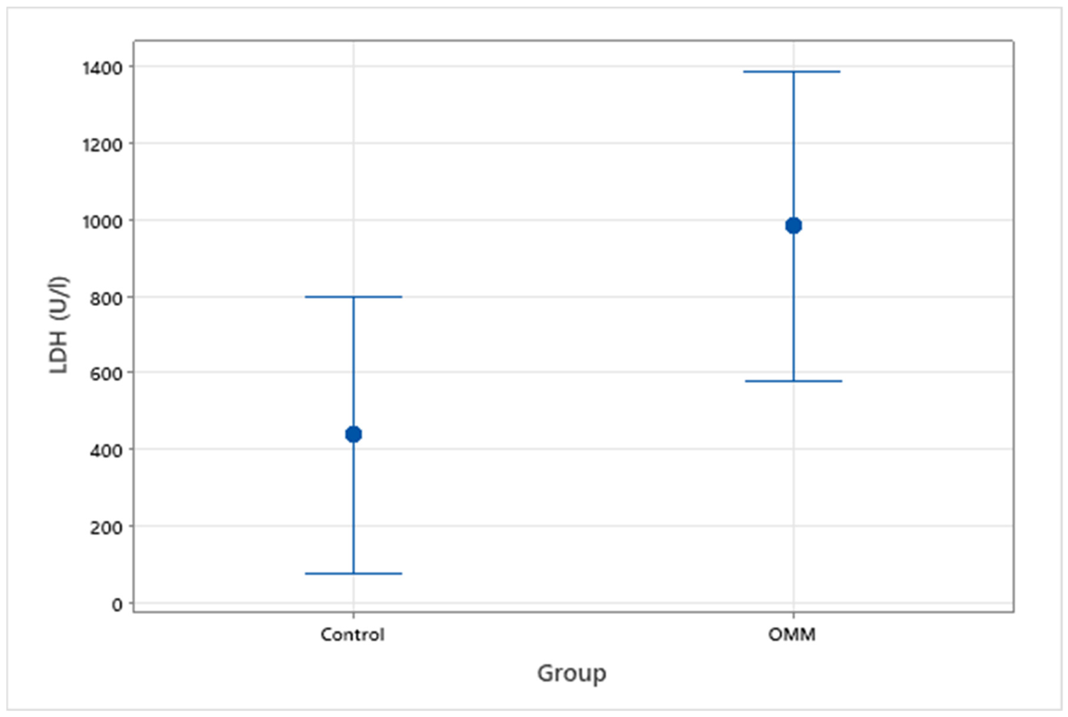
3.2. Serum LDH Values between Groups

Serum LDH was measured in a total of 18 patients, 5 with known metastasis to the local lymph nodes or lungs, 3 without evidence of metastasis and 10 control patients. The median average serum LDH value for the healthy control group was 299 U/L (range 228–1368 U/L) compared to 748 U/L (range 198–2032 U/L) for the OMM group (Figure 1). Within the OMM group, average LDH values were 1612 U/L (range 327–1762 U/L) and 611 U/L (range 198–2032 U/L) for non-metastatic and metastatic groups, respectively. There was a significant difference between all OMM patients and the control group (p = 0.049), however there was not a statistically significant difference between OMM patients with or without metastasis (p = 0.40) (Figure 2).

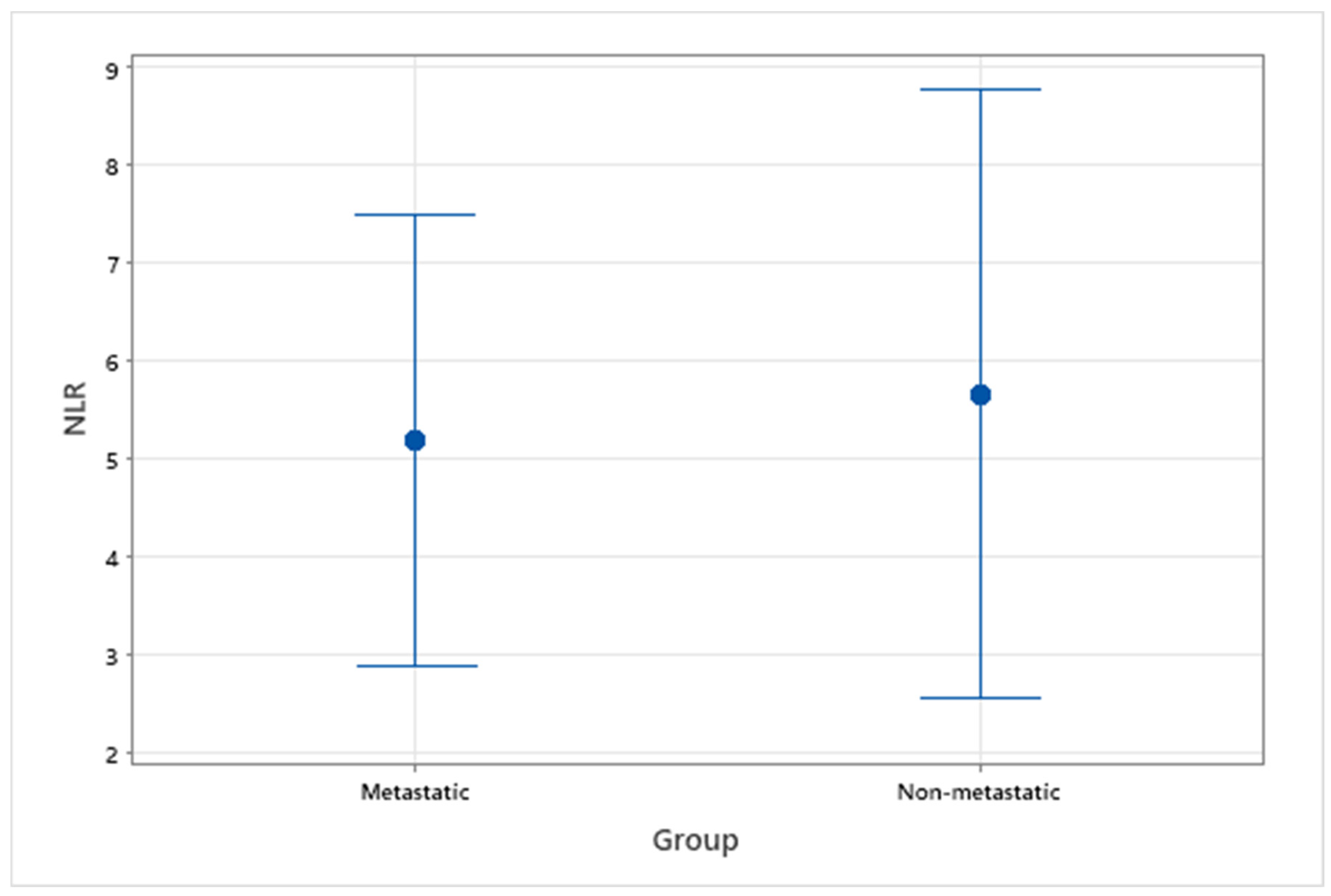
3.3. NLR and LMR between Groups

NLR and LMR from pre-treatment haematological data were assessed in 14 patients with OMM, 9 with evidence of metastasis and 5 without evidence of metastasis. A healthy control group was not available when assessing this variable, so analysis was focussed on comparison between metastatic and non-metastatic groups. The median NLR and LMR for all OMM patients with or without metastasis were 4.83 (range 1.14–11.57) and 3.83 (range 1.11–7.67), respectively. For patients without metastasis, median NLR and LMR were 4.29 (range 1.14–11.57) and 2.43 (range 1.11–5.25), respectively. For patients with metastasis, median NLR and LMR were 5.37 (range 1.94–9.11) and 4.00 (range 1.5–7.67), respectively. There was not a statistically significant difference between metastatic and non-metastatic groups for NLR (p = 0.80) or LMR (p = 0.46) (Figure 3 and Figure 4).

4. Discussion

The purpose of this retrospective study was to assess whether statistically significant differences in serum LDH could be detected between canine patients with OMM and healthy controls, as well as to assess whether significant differences existed between those patients with and without evidence of metastasis. In addition to this, we aimed to assess the effect of metastasis on NLR and LMR values in canine OMM patients. Previous studies have investigated serum LDH values in dogs with any oral tumour however to this author’s knowledge, no study had focussed on serum LDH values in OMM, nor had they assessed the effect of metastasis on LDH levels [19]. One recent study had assessed NLR and LMR in canine OMM patients and found no statistically significant correlation with any current accepted prognostic indicators including clinical stage [6]. Given the known prognostic value of LDH, NLR and LMR in human malignant melanoma patients, further investigation into their prognostic impact is warranted in our canine patients. The use of these values in veterinary medicine is an appealing idea, as all can be easily and affordably measured from routine blood samples, often taken during clinical staging [11,12,15].
It has been shown that even in well perfused, normoxaemic masses, energy is often obtained through anaerobic rather than aerobic respiration through the so called ‘Warburg effect’. It has therefore been proposed that increased serum LDH in cancer patients acts as a reflection of increased glycolytic activity in neoplastic cells, where LDH acts to drive anaerobic respiration through catalysing the conversion of pyruvate to lactate whilst regenerating NADH to NAD+ [28].
Similarly to LDH, the use of peripheral leukocyte ratios such as NLR and LMR has been investigated widely in human medicine with recent studies demonstrating their role as prognostic indicators in a number of human malignancies including malignant melanoma. Studies have shown an increased NLR is often seen in patients with a greater clinical stage and a decrease in LMR associated with shorter survival times [21,22]. The association between systemic inflammation and cancer is well recognised. Although the anti-tumour effects of neutrophils has been clearly demonstrated, relative neutrophilia has been shown to increase the release of a number of inflammatory markers including growth factors, anti-apoptotic markers and pro-angiogenic markers, all aiding tumour growth and progression [20,29]. Lymphocytes also play a key role against cancer in the body, acting through both humoral and cellular anti-tumour immune responses [30]. Low lymphocyte counts are frequently seen in advanced human cancer patients and have been correlated with poorer overall survival [22]. Conversely, monocytes recruited by tumours can act to release growth factors, promote angiogenesis and release immunosuppressive cytokines, all acting to produce a favourable tumour microenvironment [22]. Previous veterinary studies have largely demonstrated promising results when assessing leukocyte ratios as prognostic indicators in various malignancies including lymphoma, mast cell tumours, feline injection site sarcomas and osteosarcomas [23,24,25,26]. One recent study evaluated pre-treatment NLR and LMR in canine OMM patients but found no correlation with any known prognostic factors or indeed survival time, suggesting these ratios may be of little use as prognostic tools in canine OMM [6].
In this current study, there was a statistically significant difference in serum LDH levels between OMM patients and healthy controls. However, no significant difference in serum LDH was detected in OMM patients with or without metastasis. These data suggest that serum LDH could potentially act as a biomarker in canine OMM patients, in agreement with previous studies assessing its use in other canine malignancies. Similarly to LDH, no statistically significant differences were detected in NLR and LMR levels between canine OMM patients with or without evidence of metastasis. This supports the previous study by Camerino et al., which demonstrated no difference in NLR or LMR with increasing clinical stage in OMM patients [6].
This study had several limitations which may have contributed to a type 2 error. Firstly, our sample size was limited, especially with regard to LDH samples, where the availability of stored serum presented a major challenge. Additionally, the use of frozen LDH samples presents the potential for enzyme degradation over time, leading to potentially inaccurate readings. The retrospective nature of this study also presents a major limitation. Therefore, results obtained in this study especially with regard to serum LDH should be approach cautiously.
Given the results in this study, further investigation into the use of prognostic and diagnostic biomarkers such as LDH in canine OMM is worthwhile, with the effect of metastasis and tumour progression being of particular interest going forward. This current study, although limited in scope, acts as a pilot study, clearly demonstrating the relationship between OMM and serum LDH levels and laying the foundations for further research. In the future, prospective studies utilising a larger patient population, full histopathological assessment of lymph nodes and immediate measurement of serum LDH would be needed to assess these biomarkers further.

Author Contributions

Conceptualization, K.L.B.B.; methodology, K.L.B.B. and B.M.; software, Minitab version 20.3; statistical analysis, B.M.; writing—original draft, B.M.; writing—review and editing, K.L.B.B. and B.M.; supervision, K.L.B.B.; acquisition of funding, K.L.B.B. All authors have read and agreed to the published version of the manuscript.

Funding

This research received no external funding.

Institutional Review Board Statement

The study was conducted in accordance with the Declaration of Helsinki and approved by the ethics committee of the University of Edinburgh (code 126.21, approved 19 November 2021).

Informed Consent Statement

Written consent was obtained from the owners of patients for the collection of blood to be stored in a clinical biobank and used for research purposes, including the publication of these data in relevant scientific journals.

Data Availability Statement

The data presented in this study are available on request from the corresponding author.

Acknowledgments

The authors would like to acknowledge Darren Shaw for his input on statistical analysis in this article.

Conflicts of Interest

The authors declare no conflict of interest.

References

  1. Bergman, P.J. Canine Oral Melanoma. Clin. Tech. Small Anim. Pract. 2007, 22, 55–60. [Google Scholar] [CrossRef] [PubMed]
  2. Wingo, K. Histopathologic Diagnoses From Biopsies of the Oral Cavity in 403 Dogs and 73 Cats. J. Veter Dent. 2018, 35, 7–17. [Google Scholar] [CrossRef] [PubMed]
  3. Liptak, J.M. Oral tumors In Withrow and MacEwen’s Small Animal Clinical Oncology, 6th ed.; Vail, D.M., Thamm, D.H., Liptak, J.M., Eds.; Elsevier: St. Louis, MO, USA, 2020; pp. 432–448. [Google Scholar]
  4. Hahn, K.A.; DeNicola, D.B.; Richardson, R.C.; Hahn, E.A. Canine oral malignant melanoma: Prognostic utility of an alternative staging system. J. Small Anim. Pract. 1994, 35, 251–256. [Google Scholar] [CrossRef]
  5. Smedley, R.C.; Spangler, W.L.; Esplin, D.G.; Kitchell, B.E.; Bergman, P.J.; Ho, H.-Y.; Bergin, I.L.; Kiupel, M. Prognostic Markers for Canine Melanocytic Neoplasms. Veter Pathol. 2011, 48, 54–72. [Google Scholar] [CrossRef] [PubMed]
  6. Camerino, M.; Giacobino, D.; Iussich, S.; Ala, U.; Riccardo, F.; Cavallo, F.; Martano, M.; Morello, E.; Buracco, P. Evaluation of prognostic impact of pre-treatment neutrophil to lymphocyte and lymphocyte to monocyte ratios in dogs with oral malignant melanoma treated with surgery and adjuvant CSPG4 -antigen electrovaccination: An explorative study. Veter Comp. Oncol. 2021, 19, 353–361. [Google Scholar] [CrossRef]
  7. Bergin, I.L.; Smedley, R.C.; Esplin, D.G.; Spangler, W.L.; Kiupel, M. Prognostic Evaluation of Ki67 Threshold Value in Canine Oral Melanoma. Veter Pathol. 2011, 48, 41–53. [Google Scholar] [CrossRef] [PubMed] [Green Version]
  8. Giacobino, D.; Camerino, M.; Riccardo, F.; Cavallo, F.; Tarone, L.; Martano, M.; Dentini, A.; Iussich, S.; Lardone, E.; Franci, P.; et al. Difference in outcome between curative intent vs marginal excision as a first treatment in dogs with oral malignant melanoma and the impact of adjuvant CSPG4-DNA electrovaccination: A retrospective study on 155 cases. Veter Comp. Oncol. 2021, 19, 651–660. [Google Scholar] [CrossRef] [PubMed]
  9. Grosenbaugh, D.A.; Leard, A.T.; Bergman, P.J.; Klein, M.K.; Meleo, K.; Susaneck, S.; Hess, P.R.; Jankowski, M.K.; Jones, P.D.; Leibman, N.F.; et al. Safety and efficacy of a xenogeneic DNA vaccine encoding for human tyrosinase as adjunctive treatment for oral malignant melanoma in dogs following surgical excision of the primary tumor. Am. J. Veter Res. 2011, 72, 1631–1638. [Google Scholar] [CrossRef]
  10. Treggiari, E.; Grant, J.P.; North, S.M. A retrospective review of outcome and survival following surgery and adjuvant xenogeneic DNA vaccination in 32 dogs with oral malignant melanoma. J. Veter Med Sci. 2016, 78, 845–850. [Google Scholar] [CrossRef] [Green Version]
  11. Lim, S.Y.; Lee, J.; Diefenbach, R.; Kefford, R.; Rizos, H. Liquid biomarkers in melanoma: Detection and discovery. Mol. Cancer 2018, 17, 1–14. [Google Scholar] [CrossRef]
  12. Weinstein, D.; Leininger, J.; Hamby, C.; Safai, B. Diagnostic and prognostic biomarkers in melanoma. J. Clin. Aesthetic Dermatol. 2014, 7, 13–24. [Google Scholar]
  13. Campos, L.; Lavalle, G.; Estrela-Lima, A.; de Faria, J.M.; Guimarães, J.; Dutra; Ferreira, E.; de Sousa, L.; Rabelo; da Costa, A.V.; et al. CA15.3, CEA and LDH in Dogs with Malignant Mammary Tumors. J. Veter Intern. Med. 2012, 26, 1383–1388. [Google Scholar] [CrossRef]
  14. Marconato, L.; Crispino, G.; Finotello, R.; Mazzotti, S.; Salerni, F.; Zini, E. Serum lactate dehydrogenase activity in canine malignancies. Veter Comp. Oncol. 2009, 7, 236–243. [Google Scholar] [CrossRef] [PubMed] [Green Version]
  15. Keung, E.Z.; Gershenwald, J.E. The eighth edition American Joint Committee on Cancer (AJCC) melanoma staging system: Implications for melanoma treatment and care. Expert Rev. Anticancer Ther. 2018, 18, 775–784. [Google Scholar] [CrossRef] [PubMed]
  16. Blank, C.U.; Kelderman, S.; Van Tinteren, H.; Heemskerk, B.; Brom, R.V.D.; Hospers, G.A.; Eertwegh, A.J.V.D.; Kapiteijn, E.; De Groot, J.W.; Jansen, R.L.; et al. Serum lactate dehydrogenase (LDH) as a prognostic selection criterion for ipilimumab treatment in metastatic melanoma. J. Clin. Oncol. 2013, 31, 3036. [Google Scholar] [CrossRef]
  17. Agarwala, S.S.; Keilholz, U.; Gilles, E.; Bedikian, A.Y.; Wu, J.; Kay, R.; Stein, C.A.; Itri, L.M.; Suciu, S.; Eggermont, A.M. LDH correlation with survival in advanced melanoma from two large, randomised trials (Oblimersen GM301 and EORTC 18951). Eur. J. Cancer 2009, 45, 1807–1814. [Google Scholar] [CrossRef] [PubMed]
  18. Zanatta, R.; Abate, O.; D’Angelo, A.; Miniscalco, B.; Mannelli, A. Diagnostic and prognostic value of serum lactate dehydrogenase (LDH) and LDH isoenzymes in canine lymphoma. Veter Res. Commun. 2003, 27 (Suppl. 1), 449–452. [Google Scholar] [CrossRef] [PubMed]
  19. Choisunirachon, N.; Klansnoh, U.; Phoomvuthisarn, P.; Pisamai, S.; Thanaboonnipat, C.; Rungsi, A. The expression of serum lactate dehydrogenase in canine oral tumors. Thai J. Vet. Med. 2019, 49, 283–288. [Google Scholar]
  20. Paramanathan, A.; Saxena, A.; Morris, D.L. A systematic review and meta-analysis on the impact of pre-operative neutrophil lymphocyte ratio on long term outcomes after curative intent resection of solid tumours. Surg. Oncol. 2014, 23, 31–39. [Google Scholar] [CrossRef] [PubMed]
  21. Zaragoza, J.; Caille, A.; Beneton, N.; Bens, G.; Christiann, F.; Maillard, H.; Machet, L. High neutrophil to lymphocyte ratio measured before starting ipilimumab treatment is associated with reduced overall survival in patients with melanoma. Br. J. Dermatol. 2016, 174, 146–151. [Google Scholar] [CrossRef]
  22. Nishijima, T.F.; Muss, H.B.; Shachar, S.S.; Tamura, K.; Takamatsu, Y. Prognostic value of lymphocyte-to-monocyte ratio in patients with solid tumors: A systematic review and meta-analysis. Cancer Treat. Rev. 2015, 41, 971–978. [Google Scholar] [CrossRef] [PubMed]
  23. Chiti, L.E.; Martano, M.; Ferrari, R.; Boracchi, P.; Giordano, A.; Grieco, V.; Buracco, P.; Iussich, S.; Giudice, C.; Miniscalco, B.; et al. Evaluation of leukocyte counts and neutrophil-to-lymphocyte ratio as predictors of local recurrence of feline injection site sarcoma after curative intent surgery. Veter Comp. Oncol. 2020, 18, 105–116. [Google Scholar] [CrossRef]
  24. Sottnik, J.; Rao, S.; Lafferty, M.; Thamm, D.; Morley, P.; Withrow, S.; Dow, S. Association of Blood Monocyte and Lymphocyte Count and Disease-Free Interval in Dogs with Osteosarcoma. J. Veter Intern. Med. 2010, 24, 1439–1444. [Google Scholar] [CrossRef] [PubMed]
  25. Marconato, L.; Martini, V.; Stefanello, D.; Moretti, P.; Ferrari, R.; Comazzi, S.; Laganga, P.; Riondato, F.; Aresu, L. Peripheral blood lymphocyte/monocyte ratio as a useful prognostic factor in dogs with diffuse large B-cell lymphoma receiving chemoimmunotherapy. Veter J. 2015, 206, 226–230. [Google Scholar] [CrossRef] [PubMed]
  26. Macfarlane, M.J.; Macfarlane, L.L.; Scase, T.; Parkin, T.; Morris, J.S. Use of neutrophil to lymphocyte ratio for predicting histopathological grade of canine mast cell tumours. Veter Rec. 2016, 179, 491. [Google Scholar] [CrossRef]
  27. Kopanke, J.H.; Chen, A.V.; Brune, J.E.; Brenna, A.C.; Thomovsky, S.A. Reference intervals for the activity of lactate dehydrogenase and its isoenzymes in the serum and cerebrospinal fluid of healthy canines. Veter Clin. Pathol. 2018, 47, 267–274. [Google Scholar] [CrossRef] [PubMed] [Green Version]
  28. Forkasiewicz, A.; Dorociak, M.; Stach, K.; Szelachowski, P.; Tabola, R.; Augoff, K. The usefulness of lactate dehydrogenase measurements in current oncological practice. Cell. Mol. Biol. Lett. 2020, 25, 1–14. [Google Scholar] [CrossRef] [PubMed]
  29. Grecian, R.; Whyte, M.K.B.; Walmsley, S. The role of neutrophils in cancer. Br. Med Bull. 2018, 128, 5–14. [Google Scholar] [CrossRef] [PubMed] [Green Version]
  30. Faria, S.S.; Fernandes, P.C., Jr.; Silva, M.J.B.; Lima, V.C.; Fontes, W.; Freitas-Junior, R.; Eterovic, A.K.; Forget, P. The neutrophil-to-lymphocyte ratio: A narrative review. Ecancermedicalscience 2016, 10, 702. [Google Scholar] [CrossRef] [PubMed] [Green Version]
Figure 1. Interval plot to show serum LDH levels (U/L) in clinically healthy dogs vs. OMM patients.
Vetsci 09 00421 g001
Figure 2. Interval plot of serum LDH levels (U/L) in clinically healthy dogs, patients with OMM with metastasis, and patients with OMM without metastasis.
Vetsci 09 00421 g002
Figure 3. Interval plot of pre-treatment NLR in patients with OMM, with and without evidence of metastasis.
Vetsci 09 00421 g003
Figure 4. Interval plot of pre-treatment LMR in patients with OMM, with and without evidence of metastasis.
Vetsci 09 00421 g004
Table 1. Standard canine reference values for haematological parameters.
Haematological ParameterReference Values
White blood cell count (×109/L)6.0–15.0
Neutrophil count (×109/L)3.6–12.0
Lymphocyte count (×109/L)0.7–4.8
Monocyte count (×109/L)0–1.5
Table 2. Patient population characteristics.
PatientAge (Years)BreedSexGroupTumour LocationLN
Metastasis
Lung
Metastasis
LN
Assessment
Chest
Assessment
LDH (U/L)NLRLMR
111Cross breedFEMetastaticGingivalYesNoFNA3-view chest X-ray4422.464.00
212LabradorMEMetastaticGingivalYesNoFNACT6117.733.67
39LabradorFNMetastaticLingualYesYesFNACT1989.111.50
410Cocker
spaniel
FNMetastaticGingivalYesNoFNACT20325.374.67
510Cocker
spaniel
FNMetastaticGingivalYesNoHistopathologyCT8857.501.46
613Tibetan
terrier
MEMetastaticGingivalYesNoFNACT 1.945.33
78Golden
retriever
MEMetastaticGingivalYesNoHistopathologyCT 3.434.20
810Golden
retriever
FNMetastaticGingivalYesNoHistopathologyCT 3.047.67
99Red setterMEMetastaticGingivalYesNoHistopathology3-view chest X-ray 6.152.17
105LabradorMNNon-metastaticGingivalNoNoHistopathology3-view chest X-ray16123.405.00
118LabradorMENon-metastaticGingivalNoNoHistopathology3-view chest X-ray1762
1211LabradorFNNon-metastaticGingivalNoNoHistopathologyCT3274.292.43
1313Border
terrier
FNNon-metastaticGingivalNoNoHistopathologyCT 7.901.11
1412Bearded
collie
MNNon-metastaticGingivalNoNoHistopathology3-view chest X-ray 11.571.17
1512Cocker
spaniel
FNNon-metastaticGingivalNoNoHistopathology3-view chest X-ray 1.145.25
161Hungarian vizslaMNControl 505
1710Cross breedFEControl 294
1811LabradorMNControl 488
198Spaniel
(unspecified)
FEControl 228
202Golden
retriever
FNControl 278
214GoldenretrieverMEControl 405
223Cocker
spaniel
FNControl 303
2311LabradorFNControl 1368
241Cocker
spaniel
FNControl 235
252Golden
retriever
MNControl 290
Publisher’s Note: MDPI stays neutral with regard to jurisdictional claims in published maps and institutional affiliations.

