Introduction to Special Issue “Leaders in Cardiovascular Research, Dedicated to the Memory of Professor Adriana Gittenberger-de Groot”


Abstract
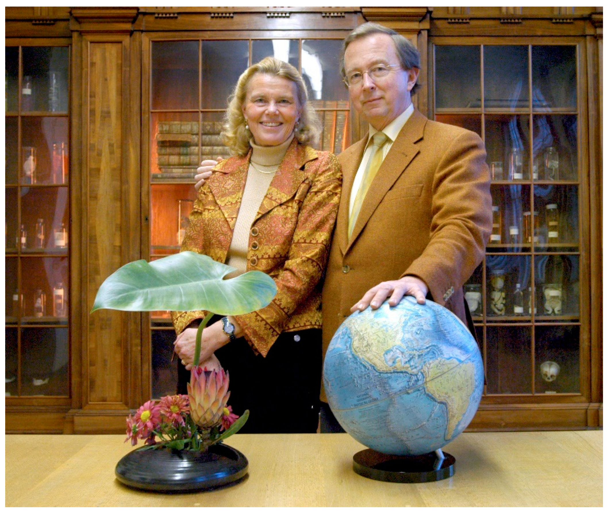
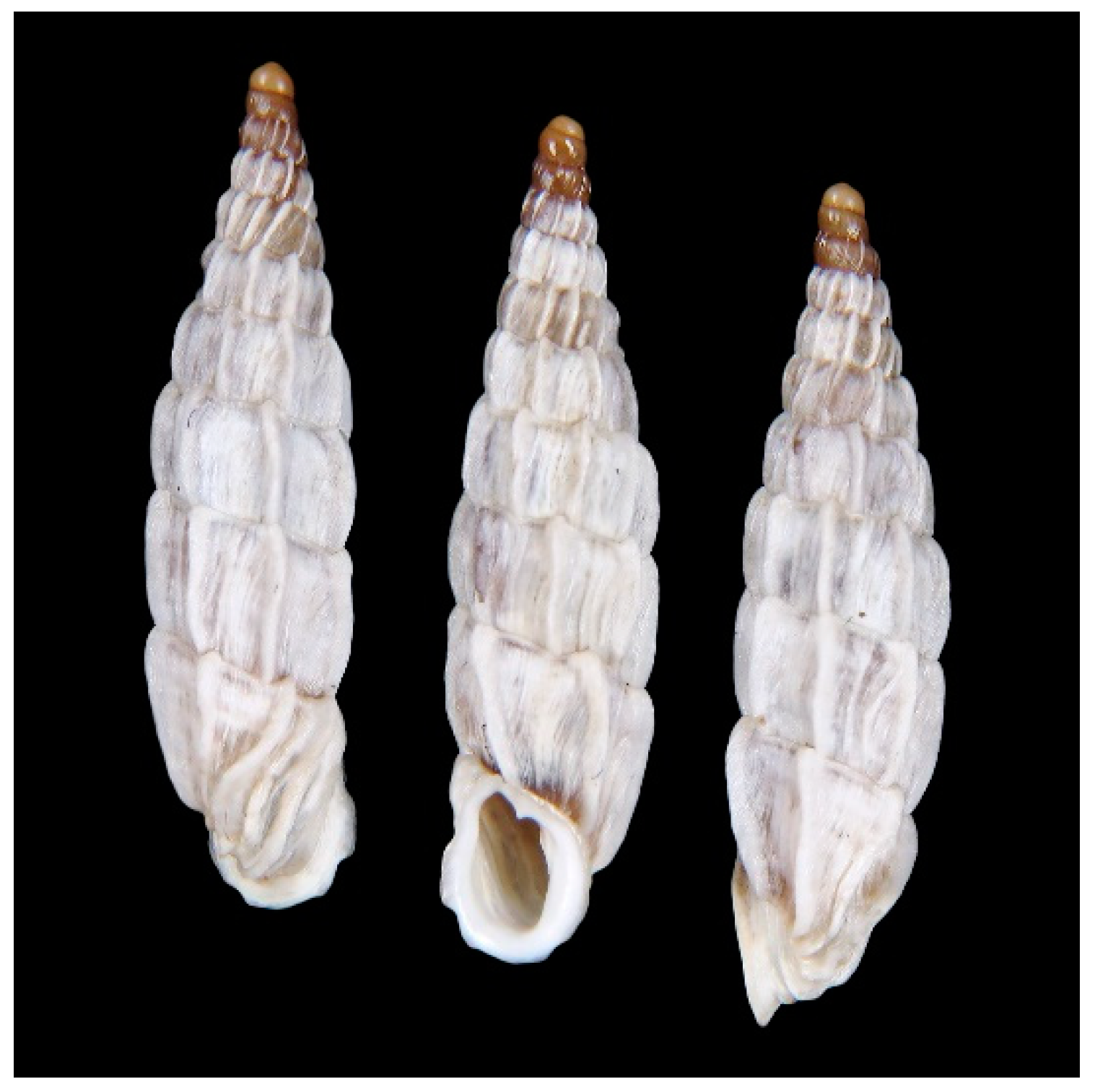
:1. Introduction: Marriage, Medicine and Malacology, Three in One [E.G.]
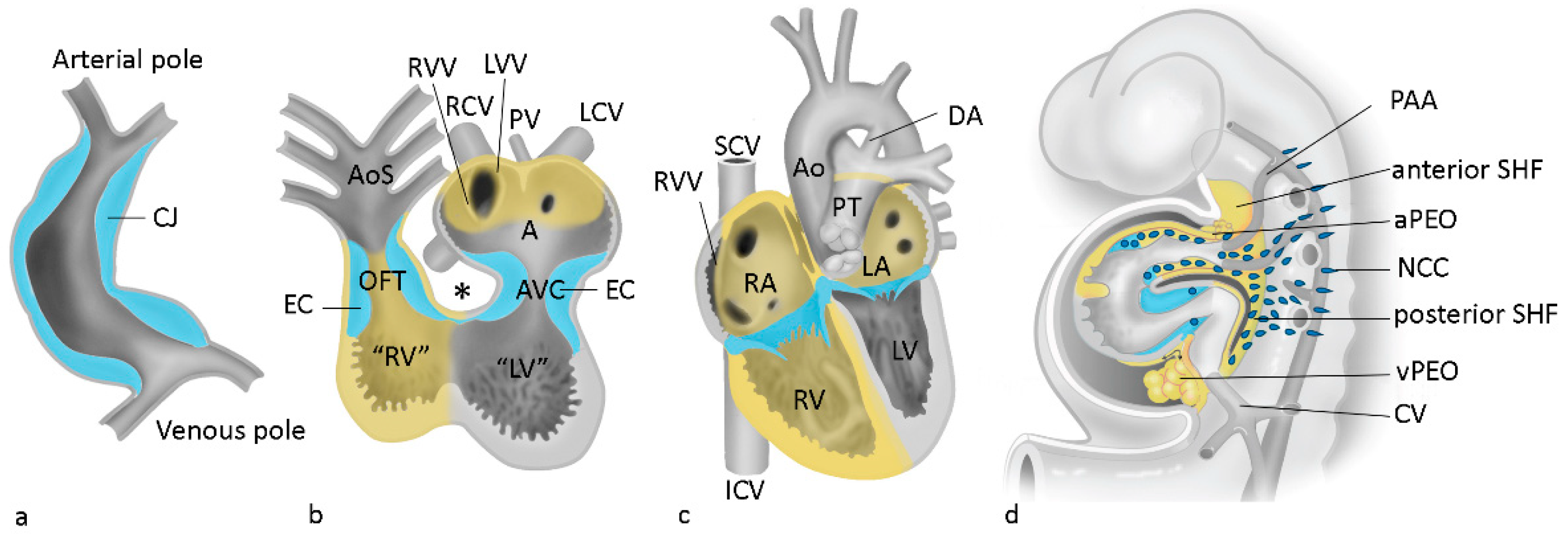
2. The Special Issue: The (Bio)medical Legacy of Adriana Gittenberger-de Groot
Author Contributions
Funding
Conflicts of Interest
References
- Hierck, B.P.; Witte, B.; Poelmann, R.E.; Gittenberger-de Groot, A.C.; Gittenberger, E. Chirality in snails is determined by highly conserved asymmetry genes. J. Molluscan. Stud. 2005, 71, 192–195. [Google Scholar] [CrossRef]
- Gittenberger-de Groot, A.C.; Bartelings, M.M.; Poelmann, R.E.; Haak, M.C.; Jongbloed, M.R.M. Embryology of the heart and its impact on understanding fetal and neonatal heart disease. Semin. Fetal. Neonatal. Med. 2013, 18, 237–244. [Google Scholar] [CrossRef] [PubMed]
- Grewal, N.; Gittenberger-de Groot, A.C.; Poelmann, R.E.; Klautz, R.J.M.; Lindeman, J.H.N.; Goumans, M.J.; Palmen, M.; Mohamed, S.A.; Sievers, H.H.; Bogers, A.J.; et al. Ascending aorta dilation in association with bicuspid aortic valve: A maturation defect of the aortic wall. J. Thorac. Cardiovasc. Surg. 2014, 148, 1583–1590. [Google Scholar] [CrossRef] [PubMed] [Green Version]
- Gittenberger-de Groot, A.C.; Calkoen, E.E.; Poelmann, R.E.; Bartelings, M.M.; Jongbloed, M.R.M. Morphogenesis and molecular considerations on congenital cardiac septal defects. Ann. Med. 2014, 46, 640–652. [Google Scholar] [CrossRef]
- Sendra, M.; Domínguez, J.N.; Torres, M. Dissecting the Complexity of Early Heart Progenitor Cells. J. Cardiovasc. Dev. Dis. 2022, 9, 5. [Google Scholar] [CrossRef]
- Peterson, J.C.; Kelder, T.P.; Goumans, M.J.T.H.; Jongbloed, M.R.M.; DeRuiter, M.C. The Role of Cell Tracing and Fate Mapping Experiments in Cardiac Outflow Tract Development, New Opportunities through Emerging Technologies. J. Cardiovasc. Dev. Dis. 2021, 8, 47. [Google Scholar] [CrossRef]
- Stefanovic, S.; Etchevers, H.C.; Zaffran, S. Outflow Tract Formation-Embryonic Origins of Conotruncal Congenital Heart Disease. J. Cardiovasc. Dev. Dis. 2021, 8, 42. [Google Scholar] [CrossRef]
- Johnson, A.-L.; Schneider, J.E.; Mohun, T.J.; Williams, T.; Bhattacharya, S.; Henderson, D.J.; Phillips, H.M.; Bamforth, S.D. Early Embryonic Expression of AP-2α Is Critical for Cardiovascular Development. J. Cardiovasc. Dev. Dis. 2020, 7, 27. [Google Scholar] [CrossRef]
- Grunert, M.; Appelt, S.; Grossfeld, P.; Sperling, S.R. The needle in the haystack—Searching for genetic and epigenetic differences in monozygotic twins discordant for tetralogy of fallot. J. Cardiovasc. Dev. Dis. 2020, 7, 55. [Google Scholar] [CrossRef]
- Kirby, M.L.; Gale, T.F.; Stewart, D.E. Neural crest cells contribute to normal aorticopulmonary septation. Science 1983, 220, 1059–1061. [Google Scholar] [CrossRef]
- Poelmann, R.E.; Gittenberger-de Groot, A.C. A subpopulation of apoptosis-prone cardiac neural crest cells targets to the venous pole: Multiple functions in heart development? Dev. Biol. 1999, 207, 271–286. [Google Scholar] [CrossRef]
- Chakrabarti, M.; Al-Sammarraie, N.; Gebere, M.G.; Bhattacharya, A.; Chopra, S.; Johnson, J.; Peña, E.A.; Eberth, J.F.; Poelmann, R.E.; Gittenberger-de Groot, A.C.; et al. Transforming Growth Factor Beta3 is Required for Cardiovascular Development. J. Cardiovasc. Dev. Dis. 2020, 7, 19. [Google Scholar] [CrossRef] [PubMed]
- Leung, C.; Engineer, A.; Kim, M.Y.; Lu, X.; Feng, Q. Myocardium-Specific Deletion of Rac1 Causes Ventricular Noncompaction and Outflow Tract Defects. J. Cardiovasc. Dev. Dis. 2021, 8, 29. [Google Scholar] [CrossRef]
- Wolters, R.; Deepe, R.; Drummond, J.; Harvey, A.B.; Hiriart, E.; Lockhart, M.M.; van den Hoff, M.J.; Norris, R.A.; Wessels, A. Role of the Epicardium in the Development of the Atrioventricular Valves and Its Relevance to the Pathogenesis of Myxomatous Valve Disease. J. Cardiovasc. Dev. Dis. 2021, 8, 54. [Google Scholar] [CrossRef] [PubMed]
- Gittenberger-de Groot, A.C.; Vrancken Peeters, M.P.; Mentink, M.M.; Gourdie, R.G.; Poelmann, R.E. Epicardium-derived cells contribute a novel population to the myocardial wall and the atrioventricular cushions. Circ. Res. 1998, 82, 1043–1052. [Google Scholar] [CrossRef] [PubMed] [Green Version]
- Poelmann, R.E.; Gittenberger-de Groot, A.C.; Goerdajal, C.; Grewal, N.; De Bakker, M.A.G.; Richardson, M.K. Ventricular septation and outflow tract development in crocodilians result in two aortas with bicuspid semilunar valves. J. Cardiovasc. Dev. Dis. 2021, 8, 132. [Google Scholar] [CrossRef]
- Zwanenburg, F.; Deruiter, M.C.; Wisse, L.J.; van Munsteren, C.J.; Bartelings, M.M.; Goumans, M.J.; Ten Harkel, A.D.; Jongbloed, M.R.; Haak, M.C. Deficient myocardial organization and pathological fibrosis in fetal aortic stenosis—Association of prenatal ultrasound with postmortem histology. J. Cardiovasc. Dev. Dis. 2021, 8, 121. [Google Scholar] [CrossRef]
- Kruithof, B.P.T.; van Wijngaarden, A.L.; Mousavi Gourabi, B.; Hjortnaes, J.; Palmen, M.; Ajmone Marsan, N. Superimposed Tissue Formation in Human Aortic Valve Disease: Differences between Regurgitant and Stenotic Valves. J. Cardiovasc. Dev. Dis. 2021, 8, 79. [Google Scholar] [CrossRef]
- Çelik, M.; Mahtab, E.A.F.; Bogers, A.J.J.C. Surgical Aortic Valve Replacement with Concomitant Aortic Surgery in Patients with Purely Bicuspid Aortic Valve and Associated Aortopathy. J. Cardiovasc. Dev. Dis. 2021, 8, 16. [Google Scholar] [CrossRef]
- Grewal, N.; Gittenberger-de Groot, A.C.; von der Thusen, L.J.; Bartelings, M.M.; DeRuiter, M.C.; Klautz, R.J.; Poelmann, R.E. The Development of the Ascending Aortic Wall in Tricuspid and Bicuspid Aortic Valve: A Process from Maturation to Degeneration. J. Clin. Med. 2020, 9, 908. [Google Scholar] [CrossRef] [Green Version]
- Çelik, M.; Milojevic, M.; Durko, A.P.; Oei, F.B.S.; Mahtab, E.A.F.; Bogers, A.J.J.C. Asymptomatic Patients with Severe Aortic Stenosis and the Impact of Intervention. J. Cardiovasc. Dev. Dis. 2021, 8, 35. [Google Scholar] [CrossRef] [PubMed]
- Grewal, N.; Velders, B.J.J.; Gittenberger-de Groot, A.C.; Poelmann, R.; Klautz, R.J.M.; Van Brakel, T.J.; Lindeman, J.H. A Systematic Histopathologic Evaluation of Type-A Aortic Dissections Implies a Uniform Multiple-Hit Causation. J. Cardiovasc. Dev. Dis. 2021, 8, 12. [Google Scholar] [CrossRef] [PubMed]
- Thiene, G.; Basso, C.; Della Barbera, M. Pathology of the Aorta and Aorta as Homograft. J. Cardiovasc. Dev. Dis. 2021, 8, 76. [Google Scholar] [CrossRef] [PubMed]
- Molin, D.G.M.; DeRuiter, M.C.; Wisse, L.J.; Azhar, M.; Doetschman, T.; Poelmann, R.E.; Gittenberger-de Groot, A.C. Altered apoptosis pattern during pharyngeal arch artery remodelling is associated with aortic arch malformations in Tgf-beta2 knock-out mice. Cardiovasc. Res. 2002, 56, 312–322. [Google Scholar] [CrossRef] [Green Version]
- van Rosendael, P.J.; Stöger, J.L.; Kiès, P.; Vliegen, H.W.; Hazekamp, M.G.; Koolbergen, D.R.; Lamb, H.J.; Jongbloed, M.R.; Egorova, A.D. The clinical spectrum of kommerell’s diverticulum in adults with a right-sided aortic arch: A case series and literature overview. J. Cardiovasc. Dev. Dis. 2021, 8, 25. [Google Scholar] [CrossRef]
- Bogers, A.J.J.C.; Gittenberger-de Groot, A.C.; Poelmann, R.E.; Peault, B.M.; Huysmans, H.A. Development of the origin of the coronary arteries, a matter of ingrowth or outgrowth? Anat. Embryol. 1989, 180, 437–441. [Google Scholar] [CrossRef]
- Gittenberger-de Groot, A.C.; Koenraadt, W.M.C.; Bartelings, M.M.; Bökenkamp, R.; DeRuiter, M.C.; Hazekamp, M.G.; Bogers, A.J.; Quaegebeur, J.M.; Schalij, M.J.; Vliegen, H.W.; et al. Coding of coronary arterial origin and branching in congenital heart disease: The modified Leiden Convention. J. Thorac. Cardiovasc. Surg. 2018, 156, 2260–2269. [Google Scholar] [CrossRef] [Green Version]
- Hazekamp, M. Coronary Anatomy in Congenital Heart Disease: The Important Contributions of Professor Dr. Adriana Gittenberger-de Groot. J. Cardiovasc. Dev. Dis. 2021, 8, 27. [Google Scholar] [CrossRef]
- Koppel, C.J.; Vliegen, H.W.; Bökenkamp, R.; Ten Harkel, A.D.J.; Kiès, P.; Egorova, A.D.; Jukema, J.W.; Hazekamp, M.G.; Schalij, M.J.; Gittenberger-de Groot, A.C.; et al. The Leiden Convention coronary coding system: Translation from the surgical to the universal view. Eur. Hear. J. Cardiovasc. Imaging 2022, 23, 412–422. [Google Scholar] [CrossRef]
- Katekaru-Tokeshi, D.I.; Jiménez-Santos, M.; Koppel, C.J.; Vliegen, H.W.; Díaz-Zamudio, M.; Castillo-Castellón, F.; Jongbloed, M.R.; Kimura-Hayama, E. Applicability of the Leiden Convention and the Lipton Classification in Patients with a Single Coronary Artery in the Setting of Congenital Heart Disease. J. Cardiovasc. Dev. Dis. 2021, 8, 93. [Google Scholar] [CrossRef]
- Engele, L.J.; Mulder, B.J.M.; Schoones, J.W.; Kiès, P.; Egorova, A.D.; Vliegen, H.W.; Hazekamp, M.G.; Bouma, B.J.; Jongbloed, M.R. The coronary arteries in adults after the arterial switch operation: A systematic review. J. Cardiovasc. Dev. Dis. 2021, 8, 102. [Google Scholar] [CrossRef] [PubMed]
- Gittenberger-de Groot, A.C.; Strengers, J.L. Histopathology of the arterial duct (ductus arteriosus) with and without treatment with prostaglandin E1. Int. J. Cardiol. 1988, 19, 153–166. [Google Scholar] [CrossRef]
- Bökenkamp, R.; DeRuiter, M.C.; van Munsteren, C.; Gittenberger-de Groot, A.C. Insights into the pathogenesis and genetic background of patency of the ductus arteriosus. Neonatology 2010, 98, 6–17. [Google Scholar] [CrossRef] [PubMed]
- Gittenberger-de Groot, A.C.; Bokenkamp, R.; Raz, V.; van Munsteren, C.; Poelmann, R.E.; Grewal, N.; DeRuiter, M.C. Progerin Expression during Normal Closure of the Human Ductus Arteriosus: A Case of Premature Ageing? Nakanishi, T., Markwald, R.R., Baldwin, H.S., Keller, B.B., Srivastava, D., Yamagishi, H., Eds.; Springer: Tokyo, Japan, 2016; pp. 245–251. [Google Scholar]
- Saito, J.; Kojima, T.; Tanifuji, S.; Kato, Y.; Oka, S.; Ichikawa, Y.; Miyagi, E.; Tachibana, T.; Asou, T.; Yokoyama, U. Transcriptome Analysis Reveals Differential Gene Expression between the Closing Ductus Arteriosus and the Patent Ductus Arteriosus in Humans. J. Cardiovasc. Dev. Dis. 2021, 8, 45. [Google Scholar] [CrossRef]
- Gittenberger-de Groot, A.C.; Moulaert, A.J.; Harinck, E.; Becker, A.E. Histopathology of the ductus arteriosus after prostaglandin E1 administration in ductus dependent cardiac anomalies. Br. Heart J. 1978, 40, 215–320. [Google Scholar] [CrossRef] [Green Version]
- Fedrigo, M.; Visentin, S.; Veronese, P.; Barison, I.; Giarraputo, A.; Cosmi, E.; Thiene, G.; Gervasi, M.T.; Basso, C.; Angelini, A. Isolated Dissection of the Ductus Arteriosus Associated with Sudden Unexpected Intrauterine Death. J. Cardiovasc. Dev. Dis. 2021, 8, 91. [Google Scholar] [CrossRef]


Publisher’s Note: MDPI stays neutral with regard to jurisdictional claims in published maps and institutional affiliations. |
© 2022 by the authors. Licensee MDPI, Basel, Switzerland. This article is an open access article distributed under the terms and conditions of the Creative Commons Attribution (CC BY) license (https://creativecommons.org/licenses/by/4.0/).
Share and Cite
Gittenberger, E.; Poelmann, R.E.; Jongbloed, M.R.M. Introduction to Special Issue “Leaders in Cardiovascular Research, Dedicated to the Memory of Professor Adriana Gittenberger-de Groot”. J. Cardiovasc. Dev. Dis. 2022, 9, 92. https://0-doi-org.brum.beds.ac.uk/10.3390/jcdd9040092
Gittenberger E, Poelmann RE, Jongbloed MRM. Introduction to Special Issue “Leaders in Cardiovascular Research, Dedicated to the Memory of Professor Adriana Gittenberger-de Groot”. Journal of Cardiovascular Development and Disease. 2022; 9(4):92. https://0-doi-org.brum.beds.ac.uk/10.3390/jcdd9040092
Chicago/Turabian StyleGittenberger, Edi, Robert E. Poelmann, and Monique R. M. Jongbloed. 2022. "Introduction to Special Issue “Leaders in Cardiovascular Research, Dedicated to the Memory of Professor Adriana Gittenberger-de Groot”" Journal of Cardiovascular Development and Disease 9, no. 4: 92. https://0-doi-org.brum.beds.ac.uk/10.3390/jcdd9040092