Case Report

Two Candida auris Cases in Germany with No Recent Contact to Foreign Healthcare—Epidemiological and Microbiological Investigations

1
Institute of Clinical Hygiene, Medical Microbiology and Infectiology, Klinikum Nürnberg, Paracelsus Medical University, 90419 Nuremberg, Germany
2
Institute of Medical Microbiology, University Hospital Essen, University of Duisburg-Essen, 45147 Essen, Germany
3
Department of Medical Microbiology and Infectious Diseases, Canisius-Wilhelmina Hospital, 6532 SZ Nijmegen, The Netherlands
4
Centre of Expertise in Mycology Radboudumc, Canisius-Wilhelmina Hospital, 6532 SZ Nijmegen, The Netherlands
*
Author to whom correspondence should be addressed.
Submission received: 18 April 2021 / Revised: 4 May 2021 / Accepted: 7 May 2021 / Published: 12 May 2021
(This article belongs to the Special Issue Candida auris 2.0)

Abstract

:
Candida auris has become a global fungal public health threat. This multidrug-resistant yeast is associated with nosocomial intra- and interhospital transmissions causing healthcare-associated infections. Here, we report on two C. auris cases from Germany. The two patients stayed in Germany for a long time before C. auris was detected during their hospitalization. The patients were isolated in single rooms with contact precautions. No nosocomial transmissions were detected within the hospital. Both C. auris isolates exhibited high minimum inhibitory concentrations (MICs) of fluconazole and one isolate additionally high MICs against the echinocandins. Microsatellite genotyping showed that both strains belong to the South Asian clade. These two cases are examples for appropriate in-hospital care and infection control without further nosocomial spread. Awareness for this emerging, multidrug-resistant pathogen is justified and systematic surveillance in European health care facilities should be performed.

1. Introduction

Candida auris has become a worldwide public health threat [1]. This fungus is associated with multidrug resistance and a high mortality. Within a hospital C. auris can survive on inanimate surfaces for long periods and nosocomial transmissions were repeatedly reported from all over the world [2]. Compared to Asia, USA and South Africa the C. auris incidence in Europe is, despite outbreaks reported in Spain and UK [3,4], low. Between January 2018 and May 2019, 349 cases were reported in the European Union/European Economic Area [5]. In Germany only a few C. auris cases have been reported so far. In a series of seven German patients, six of these cases were previously hospitalized abroad or had contact to foreign healthcare [6]. Colonization with C. auris has been detected at various body sites, including nares, groin, axilla, ears and rectum [7]. Risk factors for colonization are among others elderly age, diabetes, indwelling medical devices, immunosuppression, hemodialysis/chronic renal disease and use of broad-spectrum antibiotics and/or antifungals [8]. Receiving carbapenems and systemic fluconazole in the prior 90 days, being on a ventilator and having ≥ 1 acute care hospital visit in the prior six months were shown to be associated with C. auris colonization in New York nursing facilities with ventilator units [9]. Furthermore, contact with C. auris positive patients or their environment is an additional risk factor in healthcare institutions. Recently, C. auris emerged in an Italian hospital in context with the COVID-19 pandemic [10]. In most of the European hospitals no clear screening strategies for C. auris are established [11]. However, UK guidelines recommend that all hospitals should establish a C. auris screening policy after local risk assessment of those patients most likely to be colonized [12]. In Germany, all admitted patients with a history of hospitalization in a country with a high prevalence of multidrug-resistant (MDR) gram-negative bacteria should be screened for MDR bacteria and placed in contact precautions in a private room [11]. In contrast, similar recommendations to screen for C. auris in Germany do not exist so far.
Here we present the epidemiological, clinical, and mycological details of two C. auris cases in Germany highlighting that the time point of fungal pathogen acquisition remains open and infection control standards may prevent further transmission.

2. Methods and Results

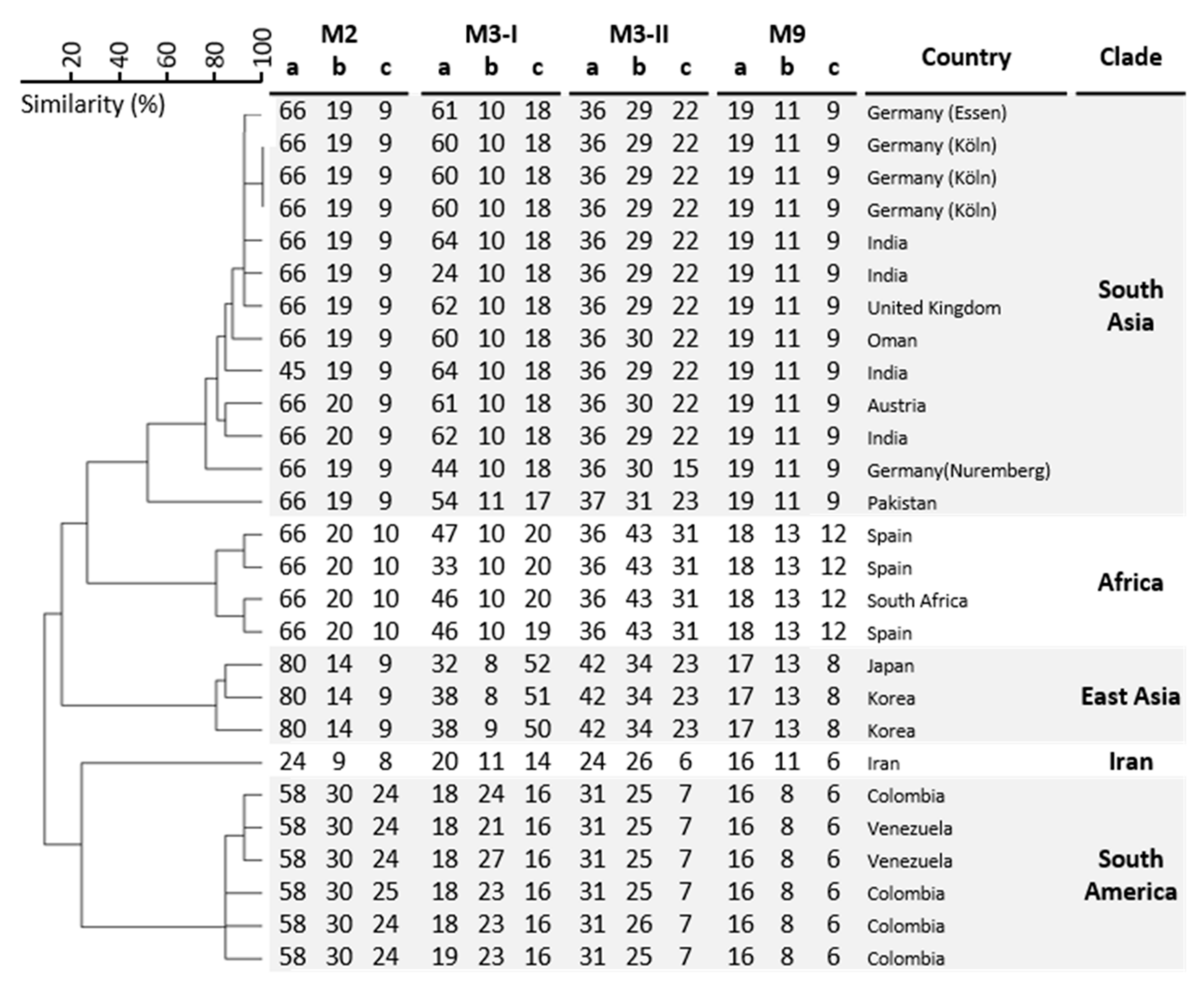
2.1. Laboratory Investigations

C. auris screening samples from patients and environmental swabs were cultured on chromogenic yeast medium (chromID, bioMèrieux, Nürtingen, Germany or Candida Brilliance, Thermo Fisher Scientific, Wesel, Germany) for 48 h at 37 °C. Identification of C. auris was done with MALDI TOF MS (Bruker, Bremen, Germany or VITEK MS, bioMèrieux, Marcy-l’Étoile, France) and confirmed by ITS sequencing as described previously [13]. Susceptibility testing was performed by broth microdilution according to CLSI, following M27-S4 [14] and one isolate was tested at the National Reference Centre for Invasive Fungal Infections, Leibniz Institute for Natural Product Research and Infection Biology—Hans Knöll Institute, Jena, Germany according to EUCAST. One C. auris isolate was selected for ERG11 and FKS1 gene amplification and sequencing [15]. Genotyping was performed by a recently developed short tandem repeat typing method [16].

2.2. Cases

Case 1: A middle-aged male patient with tetraparesis after fracture of the cervical vertebrae 4/5 in Iraq in 2015 came for further treatment to South Germany in 2018 and lived in a long-term care facility since 2019. In 2019 and 2020 the patient had several hospitalizations in Nuremberg, Germany because of pneumonia and recurrent urinary tract infections. He had an indwelling urinary catheter, decubitus ulcer and received several courses of broad-spectrum antibiotics. During his first hospital stay in February 2019, no C. auris was detected in screening swabs of rectum, groin and nose. In August 2019, C. auris was found for the first time in wound and urine screening samples at admission. During hospital stays in December 2019, April 2020 and September 2020, C. auris was found at several body sites including the ears. After C. auris was detected in August 2019, the patient was cared for in a single patient room throughout his hospitalizations. Based on results of clinical specimens there was no evidence of transmission of C. auris to other patients within the hospital. After patient discharge in April 2020 and subsequent cleaning of the patient room, inanimate surfaces close to the patient environment, e.g., bedrails, were examined for C. auris by taking a total of 20 swabs. Examining these swabs revealed no presence of yeasts. The patient never received antifungal treatment during hospitalizations. Susceptibility testing and microsatellite typing results are shown in Table 1 and Figure 1. The strain represents a new (sub)genotype within the South Asian clade.
Case 2: A middle-aged male patient underwent lung transplantation in a tertiary hospital in Essen, Germany. Bronchial cultures of donor and recipient showed no fungal growth. After transplantation, antifungal prophylaxis with voriconazole was started. Regularly performed therapeutic drug monitoring showed trough levels of voriconazole within the therapeutic range (1–4 mg/L). The patient was nursed in a single patient room during the whole stay in the hospital due to colonization with MDR pathogens (Klebsiella pneumoniae with extended spectrum beta-lactamase phenotype and resistance to quinolones, vancomycin-resistant Enterococcus faecium, carbapenem-resistant Pseudomonas aeruginosa). Post-transplant the patient suffered from CMV pneumonia and recurrent septic episodes with Pseudomonas and Klebsiella as well as episodes of multi-organ failure. While routine microbiology tracheal aspirate samples showed colonization of bronchi with Candida albicans, urine samples showed no growth of pathogens. The patient died of cardiogenic-septic shock after a six week stay on an intensive care unit on day 290 after transplantation. He was not discharged home since transplantation.
Five days before his death, yeasts were found in a urine culture without leucocyturia. At this time, only C. albicans and C. glabrata were cultured from screening samples from the respiratory tract and stool. Blood cultures always remained negative for fungi. The urine culture grew C. auris. Broth microdilution susceptibility testing showed that the C. auris isolate was resistant to fluconazole (>64 mg/L) and voriconazole (16 mg/L) (Table 1) [17]. The echinocandins micafungin and anidulafungin had an MIC of 2 mg/L. By sequencing the ERG11, an amino acid substitution was found at position 132 (Y132F). Further, the isolate was wild type at position 639 in the FKS1 gene. Immediately after identification of C. auris, all patients of the intensive care ward (n = 15) were assessed for C. auris (respiratory samples, stool, urine), but no further isolates were detected. Furthermore, C. auris was not found in routine screening or diagnostic samples of other patients on the wards where the patient stayed before. Genetically, the isolate belongs to the South Asian clade (Figure 1). The patient did not travel abroad for the last years and no other patients with C. auris could be identified, therefore the origin of this isolate remains elusive.

3. Discussion

This report shows epidemiological and microbiological details of two C. auris cases from two distinct German tertiary hospitals without a recent exposure to foreign healthcare. C. auris is rarely detected in Germany and other European countries and was so far mainly introduced from patients with contact to healthcare systems from countries with higher prevalence [11,18,19,20]. Both patients had several risk factors for C. auris colonization. Epidemiological, phenotypical, and genotyping data showed that there was no link between the patients and/or isolates, indicating two different sources. Because there were no additional infected or colonized cases with C. auris identified and environmental sampling was negative, contact isolation precautions and appropriate infection control seemed to prevent further transmissions.
Even though no C. auris case from Iraq was reported, it is possible that the first patient was already colonized with C. auris in Iraq years ago, but the fungus was never detected during previous hospital stays in Germany. C. auris may not have been detected on screening because the fungal load may have been below the limit of detection. A patient-to-patient transmission as the potential source of C. auris colonization in the long-term care facility in Germany cannot be excluded as the residents were not screened for C. auris. It has been shown earlier that C. auris can be highly prevalent in residents and in the environment of long-term care facilities in the USA [9].
Both patients were each nursed in single patient rooms during their hospital stay. Beyond basic hygiene, isolating colonized or infected patients with contact precautions, and regularly cleaning/decontaminating the environment is helpful to prevent further transmission [21,22]. The first patient was colonized with C. auris at several body sites, but no room contamination at high touch, inanimate surfaces could be found after terminal room cleaning. In addition, patient contact screening of direct contacts including those being discharged from the health care facility should be performed [22].
An open question remains whether both patients were already colonized with C. auris at hospital admission and detection was only possible due to selection factors (e.g., anti-infective therapy) during stay. There is only limited data from European hospitals on the prevalence of C. auris colonization at admission. In a study from London the prevalence of C. auris in admitted patients to the hospital was 0.04% (N = 1/2246) [3]. In a recent study from England, 921 patients from 998 admissions to eight ICUs in three major cities in 2017 to 2018 were screened for C. auris at admission [12]. No C. auris isolate was detected. It was concluded that widespread screening for C. auris in ICUs in England is not recommended and unlikely to be cost-effective. An admission screening strategy with focus on high-risk individuals based on local risk assessment was suggested [12]. Based on the current literature and our experience, a screening strategy for C. auris in patients with contact to foreign healthcare and with multiple risk factors including regional factors (e.g., outbreak in the area) can be considered in low prevalence countries. It is currently unclear, whether only an admission screening or additional repeated screening for C. auris should be conducted. A case-by-case decision may also be appropriate.
One isolate from Essen had elevated anidulafungin and micafungin MICs, but the patient never received antifungal therapy with echinocandins within the hospital. He received long treatment with voriconazole and the isolate exhibited high MICs against this tri-azole and also to fluconazole. In the USA, about 90% of C. auris isolates have been found to be resistant to fluconazole, about 30% resistant to amphotericin B, and less than 5% resistant to echinocandins [23]. We found an amino acid substitution in ERG11 at position 132 (Y132F). This substitution has been found previously in azole-resistant C. auris isolates. [15,24]. Additionally, the isolate was wildtype at position S639 in the FKS1 gene. A large study from Austria failed to find FKS1 mutations in C. albicans isolates with borderline echinocandin MICs [25]. The fact that our C. auris isolate had no mutation in the FKS1 region is congruent to observations that a mutation is mainly linked to high MICs above 4 mg/L [15]. EUCAST did not provide clinical breakpoints so far. The CDC reported tentative breakpoints for caspofungin with MICs of ≥2 mg/L as well as anidulafungin and micafungin of ≥4 mg/L [26]. A comparison of EUCAST and CLSI reference microdilution showed that six C. auris isolates, with elevated echinocandin MICs (4 mg/L), had the same results as measured by both methods for micafungin and anidulafungin (4.9%) [27].
Four major clades of C. auris (I, II, III, and IV initially originating from South Asia, East Asia, South America, and South Africa, respectively) [26] and a fifth clade reported from Iran have been described to date [28]. Our isolates were from clade I as shown by short tandem repeat typing, a method which is comparable with whole-genome sequencing [7,16]. The two described isolates differ in three markers indicating that they are not related.
This case report has some limitations. The two reported cases were retrospectively investigated and not all screening samples were performed according to the recommendations. Furthermore, for both patients the time point of acquisition of C. auris cannot be definitely determined and it cannot be excluded that an undetected hospital transmission may have occurred.
These two potentially “endemic” German C. auris cases show the importance of awareness of C. auris in hospitals in Europe not only for patients with contact to foreign healthcare environments. European health care facilities should establish local guidelines for C. auris screening for patients with risk factors and implement strict infection control measures for positive cases.

Author Contributions

Conceptualisation, J.S., J.F.M. and P.-M.R.; laboratory investigations, J.F.M. and L.K., investigations, J.S., T.S. and P.-M.R.; writing—review and editing, J.S., T.S., J.F.M., L.K. and P.-M.R.; supervision, J.S. and P.-M.R. All authors have read and agreed to the published version of the manuscript.

Funding

This research received no external funding.

Institutional Review Board Statement

Not applicable.

Informed Consent Statement

Not applicable.

Data Availability Statement

Data are available within the article.

Acknowledgments

We thank Theun de Groot, Manja Schmidt and Dirk Schmidt for excellent technical assistance. We thank the National Reference Centre for Invasive Fungal Infections, Leibniz Institute for Natural Product Research and Infection Biology—Hans Knöll Institute, Jena, Germany for providing MIC results of isolate 1.

Conflicts of Interest

The authors declare no conflict of interest.

References

  1. Meis, J.F.; Chowdhary, A. Candida auris: A global fungal public health threat. Lancet Infect. Dis. 2018, 18, 1298–1299. [Google Scholar] [CrossRef]
  2. Chowdhary, A.; Sharma, C.; Meis, J.F. Candida auris: A rapidly emerging cause of hospital-acquired multidrug-resistant fungal infections globally. PLoS Pathog. 2017, 13, e1006290. [Google Scholar] [CrossRef]
  3. Schelenz, S.; Hagen, F.; Rhodes, J.L.; Abdolrasouli, A.; Chowdhary, A.; Hall, A.; Ryan, L.; Shackleton, J.; Trimlett, R.; Meis, J.F.; et al. First hospital outbreak of the globally emerging Candida auris in a European hospital. Antimicrob. Resist. Infect. Control. 2016, 5, 35. [Google Scholar] [CrossRef] [PubMed] [Green Version]
  4. Ruiz-Gaitán, A.; Moret, A.M.; Tasias-Pitarch, M.; Aleixandre-López, A.I.; Martínez-Morel, H.; Calabuig, E.; Salavert-Lletí, M.; Ramírez, P.; López-Hontangas, J.L.; Hagen, F.; et al. An outbreak due to Candida auris with prolonged colonisation and candidaemia in a tertiary care European hospital. Mycoses 2018, 61, 498–505. [Google Scholar] [CrossRef] [PubMed] [Green Version]
  5. Plachouras, D.; Lötsch, F.; Kohlenberg, A.; Monnet, D.L. Candida auris Survey Collaborative Group. Candida auris: Epidemiological situation, laboratory capacity and preparedness in the European Union and European Economic Area*, January 2018 to May 2019. Eurosurveillance 2020, 25. [Google Scholar] [CrossRef] [Green Version]
  6. Hamprecht, A.; Barber, A.E.; Mellinghoff, S.C.; Thelen, P.; Walther, G.; Yu, Y.; Neurgaonkar, P.; Dandekar, T.; Cornely, O.A.; Martin, R.; et al. Candida auris in Germany and Previous Exposure to Foreign Healthcare. Emerg. Infect. Dis. 2019, 25, 1763–1765. [Google Scholar] [CrossRef] [PubMed] [Green Version]
  7. Yadav, A.; Singh, A.; Wang, Y.; van Haren, M.H.; Singh, A.; de Groot, T.; Meis, J.F.; Xu, J.; Chowdhary, A. Colonisation and Transmission Dynamics of Candida auris among Chronic Respiratory Diseases Patients Hospitalised in a Chest Hospital, Delhi, India: A Comparative Analysis of Whole Genome Sequencing and Microsatellite Typing. J. Fungi 2021, 7, 81. [Google Scholar] [CrossRef] [PubMed]
  8. Du, H.; Bing, J.; Hu, T.; Ennis, C.L.; Nobile, C.J.; Huang, G. Candida auris: Epidemiology, biology, antifungal resistance, and virulence. PLoS Pathog. 2020, 16, e1008921. [Google Scholar] [CrossRef]
  9. Pacilli, M.; Kerins, J.L.; Clegg, W.J.; Walblay, K.A.; Adil, H.; Kemble, S.K.; Xydis, S.; McPherson, T.D.; Lin, M.Y.; Hayden, M.K.; et al. Regional Emergence of Candida auris in Chicago and Lessons Learned from Intensive Follow-up at 1 Ventilator-Capable Skilled Nursing Facility. Clin. Infect. Dis. 2020, 71, e718–e725. [Google Scholar] [CrossRef]
  10. Di Pilato, V.; Codda, G.; Ball, L.; Giacobbe, D.R.; Willison, E.; Mikulska, M.; Magnasco, L.; Crea, F.; Vena, A.; Pelosi, P.; et al. Molecular Epidemiological Investigation of a Nosocomial Cluster of C. auris: Evidence of Recent Emergence in Italy and Ease of Transmission during the COVID-19 Pandemic. J. Fungi 2021, 7, 140. [Google Scholar] [CrossRef]
  11. Vogelzang, E.H.; Weersink, A.J.L.; van Mansfeld, R.; Chow, N.A.; Meis, J.F.; van Dijk, K. The First Two Cases of Candida auris in The Netherlands. J. Fungi 2019, 5, 91. [Google Scholar] [CrossRef] [Green Version]
  12. Sharp, A.; Muller-Pebody, B.; Charlett, A.; Patel, B.; Gorton, R.; Lambourne, J.; Cummins, M.; Alcolea-Medina, A.; Wilks, M.; Smith, R.; et al. Screening for Candida auris in patients admitted to eight intensive care units in England, 2017 to 2018. Eurosurveillance 2021, 26, 1900730. [Google Scholar] [CrossRef] [PubMed]
  13. Steinmann, J.; Schmidt, D.; Buer, J.; Rath, P.-M. Discrimination of Scedosporium prolificans against Pseudallescheria boydii and Scedosporium apiospermum by semiautomated repetitive sequence-based PCR. Med. Mycol. 2011, 49, 475–483. [Google Scholar] [CrossRef] [Green Version]
  14. Clinical and Laboratory Standards Institute. Reference Method for Broth Dilution Antifungal Susceptibility Testing of Yeasts: Fourth Informational Supplement M27-S4; CLSI: Wayne, PA, USA, 2012. [Google Scholar]
  15. Chowdhary, A.; Prakash, A.; Sharma, C.; Kordalewska, M.; Kumar, A.; Sarma, S.; Tarai, B.; Singh, A.; Upadhyaya, G.; Upadhyay, S.; et al. A multicentre study of antifungal susceptibility patterns among 350 Candida auris isolates (2009–17) in India: Role of the ERG11 and FKS1 genes in azole and echinocandin resistance. J. Antimicrob. Chemother. 2018, 73, 891–899. [Google Scholar] [CrossRef] [PubMed]
  16. De Groot, T.; Puts, Y.; Berrio, I.; Chowdhary, A.; Meis, J.F. Development of Candida auris Short Tandem Repeat Typing and Its Application to a Global Collection of Isolates. MBio 2020, 11, 02971-19. [Google Scholar] [CrossRef] [PubMed] [Green Version]
  17. Arendrup, M.C.; Friberg, N.; Mares, M.; Kahlmeter, G.; Meletiadis, J.; Guinea, J.; The Subcommittee on Antifungal Susceptibility Testing (AFST) of the ESCMID European Committee for Antimicrobial Susceptibility Testing (EUCAST). How to interpret MICs of antifungal compounds according to the revised clinical breakpoints v. 10.0 European committee on antimicrobial susceptibility testing (EUCAST). Clin. Microbiol. Infect. 2020, 26, 1464–1472. [Google Scholar] [CrossRef]
  18. Dewaele, K.; Frans, J.; Smismans, A.; Ho, E.; Tollens, T.; Lagrou, K. First case of Candida auris infection in Belgium in a surgical patient from Kuwait. Acta Clin. Belg. 2020, 75, 221–228. [Google Scholar] [CrossRef] [PubMed]
  19. Pekard-Amenitsch, S.; Schriebl, A.; Posawetz, W.; Willinger, B.; Kölli, B.; Buzina, W. Isolation of Candida auris from Ear of Otherwise Healthy Patient, Austria, 2018. Emerg. Infect. Dis. 2018, 24, 1596–1597. [Google Scholar] [CrossRef] [PubMed] [Green Version]
  20. Riat, A.; Neofytos, D.; Coste, A.; Harbarth, S.; Bizzini, A.; Grandbastien, B.; Pugin, J.; Lamoth, F. First case of Candida auris in Switzerland: Discussion about preventive strategies. Swiss Med. Wkly. 2018, 148. [Google Scholar] [CrossRef] [Green Version]
  21. Caceres, D.H.; Forsberg, K.; Welsh, R.M.; Sexton, D.J.; Lockhart, S.R.; Jackson, B.R.; Chiller, T. Candida auris: A Review of Recommendations for Detection and Control in Healthcare Settings. J. Fungi 2019, 5, 111. [Google Scholar] [CrossRef] [PubMed] [Green Version]
  22. Kenters, N.; Kiernan, M.; Chowdhary, A.; Denning, D.W.; Pemán, J.; Saris, K.; Schelenz, S.; Tartari, E.; Widmer, A.; Meis, J.F.; et al. Control of Candida auris in healthcare institutions: Outcome of an International Society for Antimicrobial Chemotherapy expert meeting. Int. J. Antimicrob. Agents 2019, 54, 400–406. [Google Scholar] [CrossRef] [PubMed]
  23. Candidiasis|Types of Diseases|Fungal Diseases|CDC. Available online: https://www.cdc.gov/fungal/diseases/candidiasis/index.html (accessed on 16 February 2021).
  24. Healey, K.R.; Kordalewska, M.; Jiménez Ortigosa, C.; Singh, A.; Berrío, I.; Chowdhary, A.; Perlin, D.S. Limited ERG11 Mutations Identified in Isolates of Candida auris Directly Contribute to Reduced Azole Susceptibility. Antimicrob. Agents Chemother. 2018, 62, 01427-18. [Google Scholar] [CrossRef] [Green Version]
  25. Spettel, K.; Galazka, S.; Kriz, R.; Camp, I.; Willinger, B. Do Candida albicans Isolates with Borderline Resistant Micafungin MICs Always Harbor FKS1 Hot Spot Mutations? J. Fungi 2021, 7, 93. [Google Scholar] [CrossRef]
  26. Lockhart, S.R.; Etienne, K.A.; Vallabhaneni, S.; Farooqi, J.; Chowdhary, A.; Govender, N.P.; Colombo, A.L.; Calvo, B.; Cuomo, C.A.; Desjardins, C.A.; et al. Simultaneous Emergence of Multidrug-Resistant Candida auris on 3 Continents Confirmed by Whole-Genome Sequencing and Epidemiological Analyses. Clin. Infect. Dis. 2017, 64, 134–140. [Google Scholar] [CrossRef] [PubMed] [Green Version]
  27. Arendrup, M.C.; Prakash, A.; Meletiadis, J.; Sharma, C.; Chowdhary, A. Comparison of EUCAST and CLSI Reference Microdilution MICs of Eight Antifungal Compounds for Candida auris and Associated Tentative Epidemiological Cutoff Values. Antimicrob. Agents Chemother. 2017, 61, 00485-17. [Google Scholar] [CrossRef] [Green Version]
  28. Chow, N.A.; de Groot, T.; Badali, H.; Abastabar, M.; Chiller, T.M.; Meis, J.F. Potential Fifth Clade of Candida auris, Iran, 2018. Emerg. Infect. Dis. 2019, 25, 1780–1781. [Google Scholar] [CrossRef] [PubMed] [Green Version]
Figure 1. STR genotypes of 27 C. auris isolates. In UPGMA dendrogram of STR genotypes, both German isolates (Essen and Nuremberg) are included.
Jof 07 00380 g001
Table 1. In vitro antifungal activities of the two C. auris isolates from Germany in mg/L.
AMBFZVRCPOSISAMCFANF
Isolate 11641≤0.016≤0.016nd0.25
Isolate 22>64160.25822
AMB, amphotericin B; FZ, fluconazole; VRC, voriconazole POS, posaconazole; ISA, isavuconazole; MCF, micafungin; ANF, anidulafungin; nd, not determined.
Publisher’s Note: MDPI stays neutral with regard to jurisdictional claims in published maps and institutional affiliations.

