Article

Characterization of the Largest Secretory Protein Family, Ricin B Lectin-like Protein, in Nosema bombycis: Insights into Microsporidian Adaptation to Host

1
State Key Laboratory of Silkworm Genome Biology, Southwest University, Chongqing 400715, China
2
Chongqing Key Laboratory of Microsporidia Infection and Control, Southwest University, Chongqing 400715, China
3
Department of Environmental Science, The Connecticut Agricultural Experiment Station, 123 Huntington Street, New Haven, CT 06511, USA
4
College of Life Science, Chongqing Normal University, Chongqing 400047, China
*
Authors to whom correspondence should be addressed.
Submission received: 3 May 2022 / Revised: 19 May 2022 / Accepted: 22 May 2022 / Published: 24 May 2022

Abstract

:
Microsporidia are a group of obligate intracellular pathogens infecting nearly all animal phyla. The microsporidian Nosema bombycis has been isolated from several lepidopteran species, including the economy-important silkworms as well as several crop pests. Proteins secreted by parasites can be important virulent factors in modulating host pathways. Ricin is a two-chain lectin best known for its extreme vertebrate toxicity. Ricin B lectin-like proteins are widely distributed in microsporidia, especially in N. bombycis. In this study, we identify 52 Ricin B lectin-like proteins (RBLs) in N. bombycis. We show that the N. bombycis RBLs (NbRBLs) are classified into four subfamilies. The subfamily 1 was the most conserved, with all members having a Ricin B lectin domain and most members containing a signal peptide. The other three subfamilies were less conserved, and even lost the Ricin B lectin domain, suggesting that NbRBLs might be a multi-functional family. Our study here indicated that the NbRBL family had evolved by producing tandem duplications firstly and then expanded by segmental duplications, resulting in concentrated localizations mainly in three genomic regions. Moreover, based on RNA-seq data, we found that several Nbrbls were highly expressed during infection. Further, the results show that the NbRBL28 was secreted into host nucleus, where it promotes the expressions of genes involved in cell cycle progression. In summary, the great copy number, high divergence, and concentrated genome distribution of the NbRBLs demonstrated that these proteins might be adaptively evolved and played a vital role in the multi-host N. bombycis.

Graphical Abstract

1. Introduction

Microsporidia are a group of obligate intracellular parasites, which can infect a wide variety of hosts from protists to mammals, even humans [1,2,3,4]. Nosema bombycis, a kind of parasite of the silkworm (Bombyx mori), was the first formally described microsporidia and has been shown to be transmitted both vertically and horizontally. Infection of N. bombycis will result in the death of its host, thus posing a significant threat to sericulture industry [5]. As intracellular parasites, microsporidia utilize many of its host metabolites to reduce genomes and thus speed up reproduction [6,7].
Ricin, notable for its extreme vertebrate toxicity, is a heterodimeric protein (carbohydrate containing protein) and is found in the seeds of the castor oil plant Ricinus communis L [8]. It has a cell-binding ricin toxic B chain (RTB) linked, through a disulfide bound, to a catalytic cytotoxic ribosome-inactivating protein (RTA). The RTB has two sugar-binding regions, each of which contain three homologous subregions (alpha, beta, and gamma) composed of 40 amino acids and a linker peptide around 15 residues (lambda). It has been proposed that RTB originated from a primitive 40 residue galactoside-binding peptide, which evolved through gene duplication and expansion [9,10]. Ricin B-lectin is homologous to the RTB and usually contains a conserved Ricin B-lectin domain. RTB can bind with the exposed galactose residues of multiple glycoproteins and glycolipids on cell surface [11]. Lectins such as RTB have a wide range of receptor-binding capabilities and are an important pathogenic factor in host–pathogens interactions. They mediate intercellular adhesion, infection, natural immune defense, and host-phagocytic action to eliminate invading pathogens, but the mechanisms remain unclear [12,13].
Previous studies showed that Ricin B lectin-like (RBL) in Encephalitozoon cuniculi and Nosema ceranae could form genomic clusters. In E. cuniculi, four RBL coding genes (rbls) were found in one cluster. In N. ceranae, six rbls sit together in a genomic region. In particular, no rbl was found in Nematocida parisii, suggesting that the family may have been lost during genome reduction. Experimental studies have demonstrated that RBL is produced during the germination of Spraguea lophii spores, indicating that these proteins might promote infection or resist host immunity [14]. Previous studies have shown that microsporidian RBLs (EcRBLL-1, AaRBLL-1, AaRBLL-2, and NbRBL) enhance spore adhesion to host cells [15,16]. All these findings suggest that this protein family is vital during microsporidian infection.
In this study, we identified the N. bombycis Ricin B lectin-like proteins (NbRBLs), and characterized their sequence and evolution, as well as gene expression patterns during infection.

2. Materials and Methods

2.1. Prediction and Identification of NbRBLs

Protein sequences of N. bombycis were downloaded from the SilkPathDB [17]. The hidden Markov model (HMM) profile of the RBL domain (Pfam: PF00652) was downloaded from the Pfam [18] and it was utilized to search the candidate RBLs from N. bombycis protein sequences. The candidate sequences were then used to searched against N. bombycis genome to detect missed RBLs using BLASTP [19] with an e-value less than 0.05. Two online programs, SMART (http://smart.embl-heidelberg.de/, accessed on 3 June 2019) and CDD (http://0-www-ncbi-nlm-nih-gov.brum.beds.ac.uk/Structure/cdd/wrpsb.cgi, accessed on 3 June 2019), were used to characterize the domain architecture of the predicted NbRBLs.

2.2. Sequence Features of NbRBLs

Sequence features of the candidate NbRBLs were predicted using the Protparam tool [20]. The subcellular localization of NbRBLs was predicted by two programs: WoLF PSORT [21] and TargetP (http://www.cbs.dtu.dk/services/TargetP, accessed on 3 August 2020). Signal peptides (SPs) were predicted by SignalP 5.0 [22] with the default D-cutoff values (https://services.healthtech.dtu.dk/service.php?SignalP-5.0, accessed on 5 August 2020).
Conserved motifs of the SP were identified using MEME 5.3.0 [23] with default parameters, except for the maximum number of motifs and maximum width, which were set to 4 and 10, respectively. Only motifs with an e value < 1e−22 were kept for further analysis.

2.3. Multiple Sequence Alignment and Phylogenetic Analysis of NbRBLs

Multiple protein sequences were aligned using Clustal Omega (https://www.ebi.ac.uk/Tools/msa/clustalo/, accessed on 3 August 2020), the results of which were then plotted using the online BoxShade (https://embnet.vital-it.ch/software/BOX_form.html, accessed on 3 August 2020).
To investigate the evolutionary history of the NbRBLs, phylogenetic trees were constructed using the Maximum Likelihood (ML) method in RaxML [24] with a bootstrap test for 500 replicates and visualized by iTOL (http://itol.embl.de/, accessed on 15 September 2021) [25,26], using the castor RICOM_RicinB (GenBank accession no, ACY38598.1) as an outer group.

2.4. Expressions of Nbrbls during N. bombycis Infection

N. bombycis spores were prepared as previously described [27]. The spores, which were pretreated with 0.1 mol/L KOH, were added to the BmE-SWU1 cells (cell:spore ratio, 1:20). Infected cells were collected at 12-, 24-, 48-, 60- and 72-h post infection (hpi) and stored in TRIzol (Ambion, CA, USA). RNA extraction and cDNA synthesis were performed as previously described [28]. The real-time quantitative PCR (RT-qPCR) was conducted using primers (Nbrbl16, Nbrbl45, and Nbrbl51) and reference gene Nbtubulin (Table 1). Expression levels were calculated by the 2−ΔΔt values method using three replicates. All statistical t-tests were performed with GraphPad Prism version 9.0.0 by two-tailed comparison tests and any difference with a p-value < 0.05 was considered significant [29].
Transcriptomic data were downloaded from scientific publications (the accession number PRJNA549766) as reference and were used to analyze the expression patterns of NbRBL proteins [30].

2.5. Indirect Immunofluorescence Assay (IFA)

Infected BmE-SWU1 cells were fixed with 4% paraformaldehyde for 10 min at room temperature and washed three times with 1xPBS and permeabilized using 0.1% Triton X-100 for 15 min. The cells were then blocked in 1xPBST containing 5% BSA and 10% goat serum for 1 h at room temperature. Next, the cells were incubated with mouse and rabbit poly clonal antibodies against NbRBL06 (anti-NbRBL06) and NbRBL28 (anti-NbRBL28) diluted 1:100 in blocking solution for 2 h at room temperature. The cells were then washed for three times with 1xPBST, and incubated for 1 h with a 1:1000 dilution of Alexa Fluor 488 conjugate Goat anti-Mouse IgG (Invitrogen A32723, Rockford, Illinois, USA) and Alexa Fluor 594 conjugate Goat anti- Mouse IgG (Invitrogen A32742, Rockford, Illinois, USA) in a dark moist chamber at room temperature. The cell nucleus was stained with DAPI (1:1000 dilution, Sigma-Aldrich 28718-90-3, St. Louis, MO, USA) at room temperature for 15 min. The samples were finally observed and photographed using an Olympus FV1200 laser scanning confocal microscope.

2.6. Transfection and RNA-seq

The Nbrbl28 (locus NBO_163g0001) was cloned from N. bombycis genomic DNA and inserted into the pSL1180 over expression vector fused with egfp. BmE-SWU1 cells were transfected with Nbrbl28::egfp- and egfp-containing plasmids according to the instructions of X-tremeGENE HP DNA transfection reagents (Roche 06366546001). The cell culture medium was replaced with fresh Grace’s Insect medium containing 10% serum after 5 h. Three days later, the transfected cells were collected, and stored at −80 °C for RNA-seq. RNA-seq was conducted by the Biomarker Technology Company (Beijing, China). The raw data were deposited in GenBank under the BioProject PRJNA808047 and BioSample SAMN26022686. The real-time quantitative PCR (RT-qPCR) was conducted using primers E2F1, SDS3, and Rad51 and reference gene primer SW22934. The reaction procedure included one cycle at 95 °C for 5 min, followed by 40 cycles at 95 °C for 10 s and at 60 °C for 30 s. Expression levels were calculated by the 2−ΔΔt values method using three replicates.

3. Results

3.1. The Nbrbls Identified in N. bombycis Genome

A total of 52 Nbrbls were identified in the N. bombycis genome, composing the largest protein family in N. bombycis. As shown in Table 2, the pI of different NbRBLs was variable, ranging from 4.55 to 9.13. The molecular weight of the NbRBLs is from 20 to 35 kDa. Thirty of 52 NbRBLs contain a Ricin B lectin domain (RBLD). The deficiency of RBLD was also found in other microsporidia [14].

3.2. The NbRBLs Are High Divergent

Phylogeny analysis shows a high level of divergence among the NbRBLs, which can be grouped into 4 subfamilies containing 17, 25, 7, and 3 members (Figure 1). Subfamily 1 is relatively conserved in that all members contain the RBLD, 14 of which encode a signal peptide (SP). Some members of the other subfamilies have lost RBLD, suggesting that these members are more differentiated. In subfamily 2, 17 out of 25 members have lost the RBLD, and only half retain the SP. Again, in subfamily 3, some members show a loss of the SP, RBLD. All members of subfamily 4 have the SP, while only one has the RBLD. Furthermore, there were 9 members with a SP and a nuclear localization signal (NLS), indicating that these factors could be secreted into host nucleus. In summary, the NbRBLs are a highly differentiated protein family that may have diverse functions in parasites.

3.3. Expansive Mechanisms of the NbRBL Family

By mapping N. bombycis genome [17], we found that the 52 Nbrbls are located on 20 scaffolds (Figure 2a). Tandem gene duplications were found on scaffolds NBO_6, NBO_27, NBO_463, and NBO_1196, containing 19, 6, 5, and 4 genes, respectively. Moreover, we found that the members of each NbRBL subfamily were distributed on the different scaffolds. Members of each NbRBL subfamily were distributed on the NBO_6, but on the NBO_463, there only existed members of subfamily 1, and on the NBO_27 and NBO_1196 Scaffold, there were only members of subfamily 2. The NbRBL family formed clusters in the N. bombycis genome, indicating that the Nbrbls experienced large-scale duplication. In the largest region containing the tandem duplications (TDs) and segmental duplications (SDs) of Nbrbl, we found transposable elements (TEs) flanking the SD region (Figure 2b).

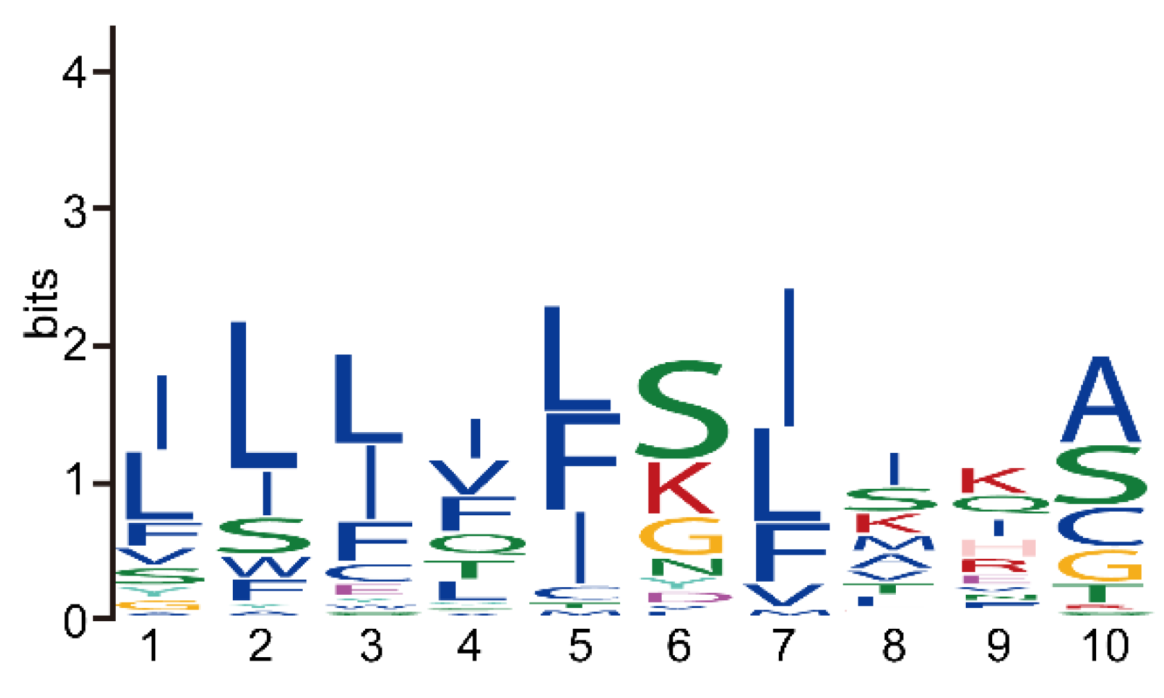
3.4. The Reduction of Key Motifs in NbRBL

The Ricin B lectin domain has been referred to as the (QxW)3 domain and the three homologous regions as the QxW repeats. Through multiple sequence alignment of NbRBL family, it is found that family 1 is relatively conservative in all subfamilies. Compared with RTB of castor, subfamily 1 also has three distinct subdomains: α, β, and γ. However, there is no obvious QxW motif in the α subdomain, even the QxW motif in α subdomains turns into QxF motif, which is found in three other three families (Figure 3 and Figures S1–S3).
We also analyzed the sequence feature of the SPs in NbRBL and found that their lengths ranged from 12 to 24 amino acids. A conserved amino acid motif [ILF][LI][LIF][IV][LFI][SK][IL]IK[ASC] was predicted, demonstrating that NbRBLs have similar secretion pathways (Figure 4). In addition, some researchers have found that there is a conserved amino acid sequence PEXEL/VTS/HT at the N-terminus of most secreted proteins of Plasmodium, which enable secreted proteins to pass through the vacuole [1,31]. We found a conserved amino acid sequence in the SP of NbRBL family members. It is speculated that they have similar secretory pathway and are secreted into host cells to play a regulatory role.

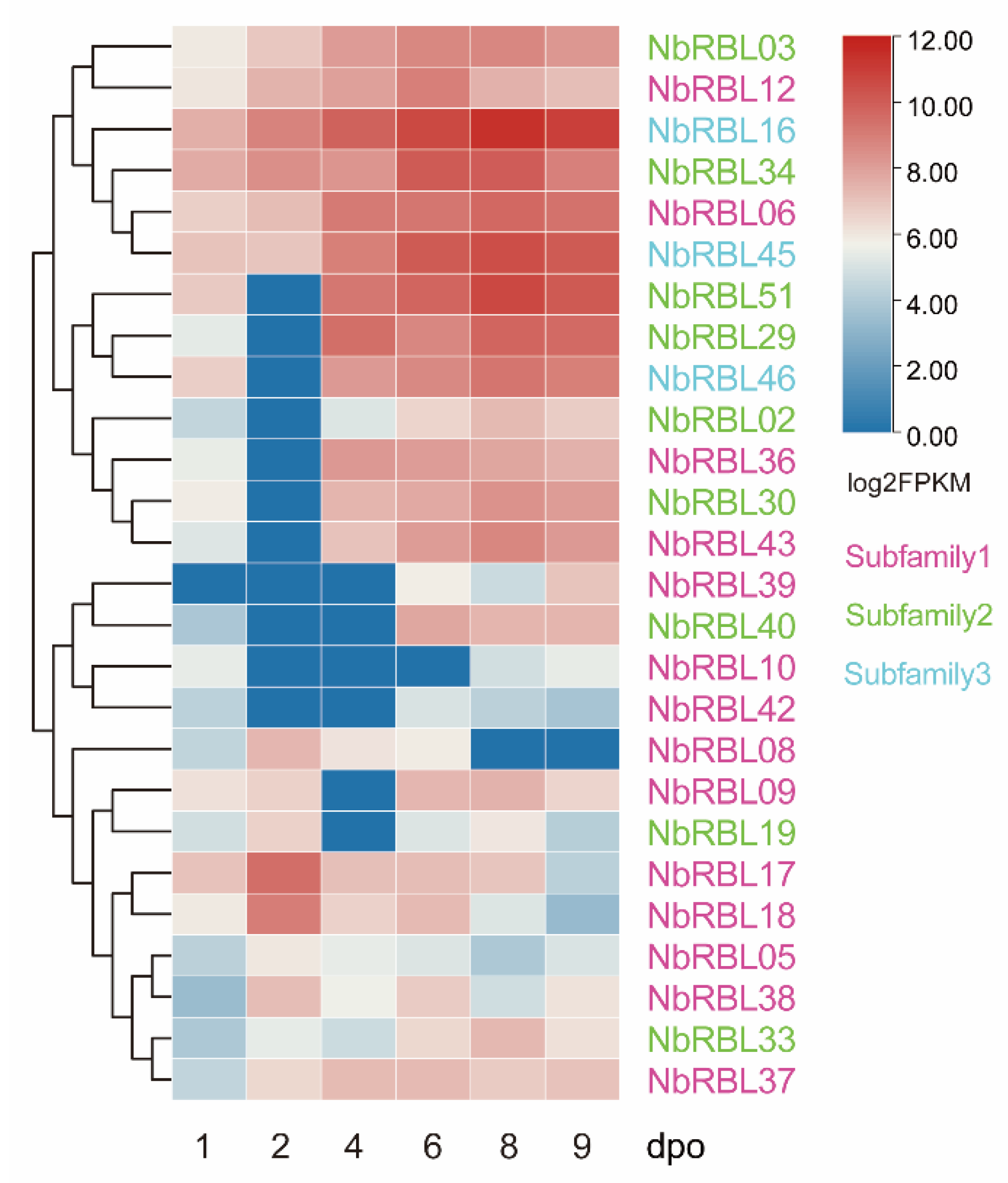
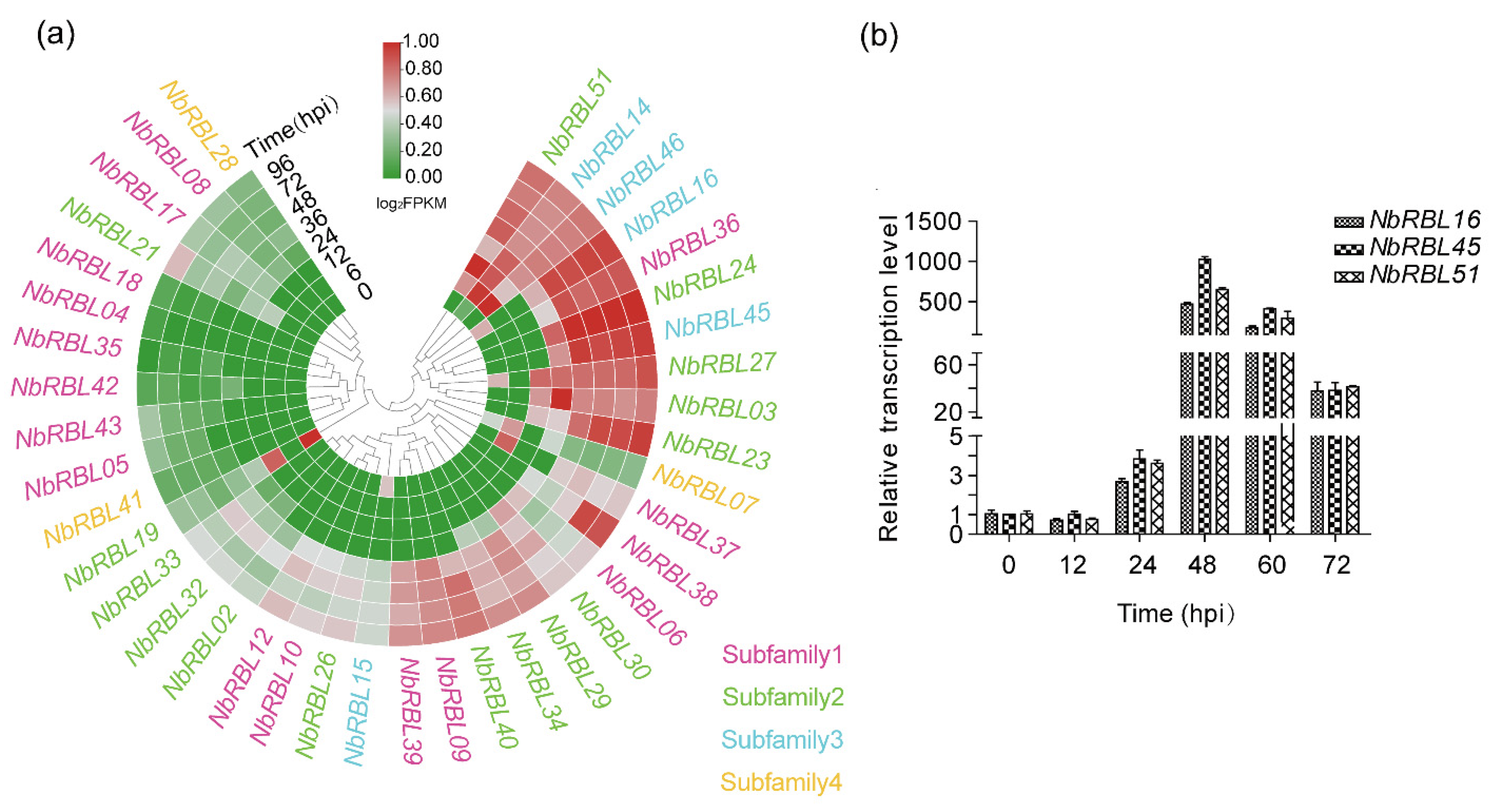
3.5. Expressions of Nbrbls during Infection

N. bombycis can be transmitted vertically from infected females to eggs, resulting in congenital infections in embryos. Based on the RNA-seq data from articles published in the scientific literature [30], we analyzed the expression patterns of Nbrbls in B. mori embryos infected with N. bombycis and found that 26 of the identified Nbrbls were expressed during infection in N. bombycis. No expression of NbRBL subfamily 4 was detectable, Members of all other subfamilies showed expression during infection. Among them, 14 of 17 members in subfamily 1 were expressed. In addition, we found that three NbRBLs from subfamily 2 (Nbrbl51) and subfamily 3 (Nbrbl16 and Nbrbl45) were highly expressed. Apart from Nbrbl06, most of the members of subfamily 1 showed a lower level of expression. Five genes from subfamily 1 (Nbrbl05, Nbrbl08, Nbrbl09 Nbrbl17, and Nbrbl18) were highly expressed early on and down-regulated during embryos development (Figure 5). We further analyzed the expression patterns of Nbrbls in the infected BmE-SWU1 cells. The results showed that the overwhelming majority of Nbrbls were expressed and Nbrbl16, Nbrbl45, and Nbrbl51 were highly expressed (Figure 6a). Then, we selected three genes (Nbrbl16, Nbrbl45, and Nbrbl51), highly expressed to examine their expression profile in the BmE-SWU1 cells after N. bombycis infection (Figure 6b). The data showed that the expression of these three genes were up-regulated at 48 hpi.

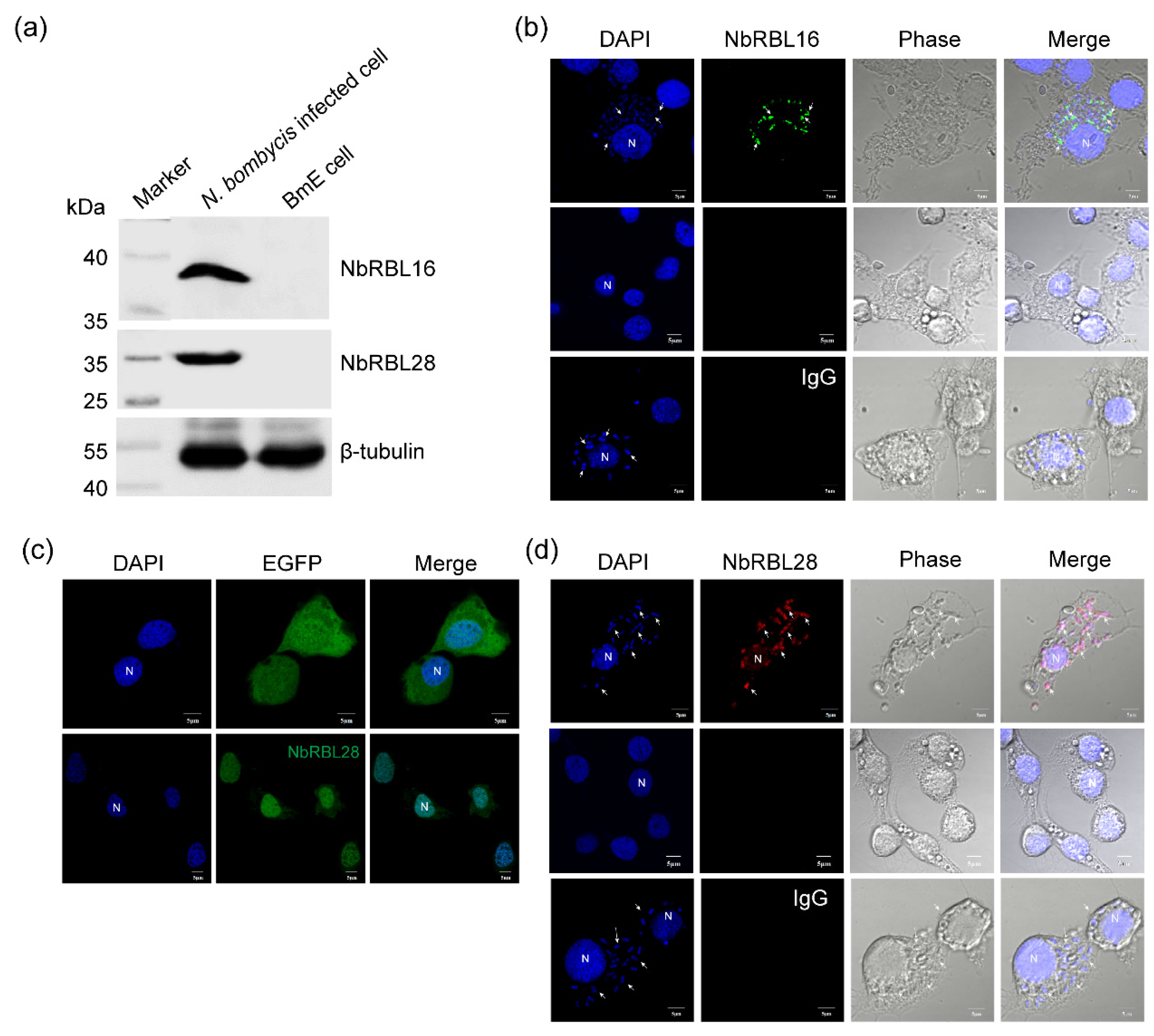
3.6. Subcellular Localization of NbRBL16 and NbRBL28

First, we verified the specificity of the antibody using Western blotting, which revealed that the antibody of NbRBL16 and NbRBL28 distinguishes these endogenous proteins from the total proteins in N. bombycis infected cell (Figure 7a). The NbRBL16 protein is located in the cytoplasm of schizont in the proliferating stage while the mature spores gave no fluorescent signal (Figure 7b), similar to that of Cyto-NbHsp70 [28]. NbRBL28 contains an N-terminal signal peptide and nuclear localization signal (NLS) sequences (Figure 1), which was co-expressed with EGFP in BmE-SWU1 to assess whether it could be secreted into the host nucleus. As expected, we found that four NbRBL28 proteins could be located in the BmE-SWU1 cell nucleus (Figure 7c). Although NbRBL28-EGFP fusion protein was located in the host nucleus, NbRBL28 could be secreted into the host nucleus during N. bombycis infection. The result showed that NbRBL28 was not only located in the cytoplasm of the schizont, but was also detected in the host cell nucleus (Figure 7d), which demonstrated that NbRBL28 was a secreted protein targeted to the host cell nucleus.

3.7. NbRBL28 Regulates Gene Expressions Involved in Host Cell Cycle

Since NbRBL28 was detected in the infected host cell nucleus, it was likely to account for the gene expression changes triggered by infection. To verify this hypothesis, we performed a transcriptomic analysis of the BmE-SWU1 cells transfected with Nbrbl28::egfp- and egfp-containing plasmids. Filtered data were presented in Table S3. To identify which pathways were differently changed, we performed KOG and KEGG pathway analysis. KOG analysis revealed that a number of cell cycle, cell division processes, as well as replication transcription processes, were enriched (Figure 8a). KAGG pathway analysis revealed an enrichment of 11 pathways with p < 0.05 in BmE-SWU1 expressing Nbrbl28::egfp. Genes involved in transcriptional regulation (E2F1 and SDS3) were up-regulated, while a gene (Rad51) functioning in DNA repair was down-regulated (Figure 8b,c). The E2F1 is a transcription factor involved in transformation of cell cycle from G1 phase to S phase [32,33]. The Rad51 participates in the cell cycle, replication and repair [34,35]. In summary, these data indicated that NbRBL28 was positively regulating the expression of host cell genes involved in controlling the cell cycle progression.

4. Discussion

Ricin B-lectin domain proteins have been identified in bacteria, fungi, plants, invertebrates, and higher animals. Examples include Xylanase in Streptomyces, Ricin in the Castor bean, lactose-binding lectin in earthworms, the mannose-receptor in macrophages and RsA in Rhizoctonia solani [36]. RBL has been identified in most genera of microsporidia, including Anncaliia, Encephalitozoon, Nematocida, and Spraguea [14,37]. Because of its broad distribution and presence in the microsporidia, it is speculated that this gene family predates microsporidia evolution. Encephalitozoon, which has a highly reduced genome, still retains this gene family [38]. We have here identified 52 Nbrbls, which is the largest gene family in N. bombycis. Further study of RBL is helpful to understand the evolution of microsporidia gene and the relationship between microsporidia and its host.
Members of this family form clusters in the genome of microsporidia. In E. cuniculi, four rbls are located on a single syntenic block, and six of the eight rbls of N. ceranae are found in NCER_1015 [14]. In N. bombycis, NBO_0006 was at the core of the gene family, and rbl was most likely the first to appear in this region. In addition, most of the genes appeared in pairs, and there were a large number of duplicate genes. The animal-derived “piggyBac” DNA transposons were found the in genome of N. bombycis. These mobile genetic elements encode functional transposases that are capable of recognizing a specific TTAA motif, which it cleaves to insert itself across different regions of the genome [39]. These results indicated that transposable elements most likely played an important role in mediating the duplication of Nbrbls. It is speculated that N. bombycis has a small number of rbls on a single syntenic block as E. cuniculi in the early stage of evolution, and then gene amplification events such as tandem repeat and fragment repeat in the process of evolution occurred. Gene duplication is very important in the evolution of organisms, and gene duplication and differentiation have been considered as the driving force for a gene to produce new functions. These may suggest that the family would obtain new functions through gene replication and non-synonymous mutation to adapt to the changing living environment.
Our work also showed that evolutionary divergence has also occurred among microsporidia RBL protein genes. First of all, the number of rbl in different microsporidia varies tremendously. There are only 4 rbls in E. cuniculi and 52 rbls in N. bombycis. However, rbl has not been identified in N. parisii genome [11]. Secondly, some members of RBL protein family of microsporidia lost their RBLD. The 52 NbRBLs can be classified into 4 subfamilies with phylogenetic analysis. Subfamily 1 is relatively more conserved as all members have a RBLD and most proteins have SP, indicating that the NbRBLs of subfamily 1 retain an original galactose-binding function. There is big difference among subfamily 1 and other subfamilies, in which some members lost the RBLD. It showed that the sequences of NbRBL varied greatly, so that likely became a multi-functional family. Compared with Ricin B lectin, the motif of NbRBL turned into QxF from QxW, which was also found in the RBLs of Anncaliia algerae [16]. Phenylalanine was replaced by tryptophan, both of which were hydrophobic amino acids. Compared with tryptophan, the molecular weight of phenylalanine is smaller, and the structure becomes simpler. It is suggested that this was a kind of reduction that happened at the amino acid level in microsporidia. Besides, this motif substitution may alter the selectivity of RBLs for specific glycoproteins on host cytoplasm membrane, which are important for the parasite infection.
Previous studies have shown that Nbrbl (identified as Nbrbl03 in our study) was highly transcribed after 42 hpi [15], Our results showed that Nbrbl03 was highly expressed after 12 hpi in BmE-SWU1 cells. We also found that Nbrbl14, Nbrbl46, and Nbrbl51 were also highly expressed at 6 hpi, but some Nbrbls (such as Nbrbl04, Nbrbl05, Nbrbl17, and Nbrbl18, etc.) were expressed at low levels in the infected BmE-SWU1 cells. Interestingly, Nbrbl17 and Nbrbl18 were high level expressed in the infected embryos, which suggested that different NbRBLs may have different biological functions.
Interestingly, NbRBL28 was the first RBL member that was found to be secreted into the host nucleus and likely to modulate the host cell cycle. This modulation model was also reported in other intracellular pathogens [40,41], for instance, T. gondii secrete GRA16, GRA24, ROP16, and TgIST into the host nucleus to interfere with gene expressions [42,43,44,45,46,47,48]. Besides, there are 13 NbRBLs without a SP, of which 9 were predicted to be located in the nucleus (Table 2), suggesting that these members may regulate the gene expressions of the parasites themselves. Furthermore, it has been reported that lectins have diverse roles in parasites, and can mediate adhesion of the parasite to the host cell [49]. For example, the NbRBL03 was reported to enhance spore adhesion to the host cells [15], and that NbRBL51 is an only member, containing a transmembrane domain, indicating that it is a membrane protein and most likely promotes adhesion too. Similar to the RTB, secreted and transmembrane NbRBLs likely bind to glycoproteins on the host cytoplasm membrane to mediate the adhesion. Therefore, NbRBLs play important and multiple roles during infection and pathogen development.
In summary, we primarily discussed identification, phylogenetic classification, molecular evolution, and gene expression analyses of the NbRBL gene family. The increase of NbRBL genes suggested that certain members have evolved to carry out a larger number of functions to adapt to intracellular life. Therefore, RBL, which is an ideal target, holds significance to the study of microsporidium gene evolution and the analysis of the mechanism of interaction between microsporidium and host.

Supplementary Materials

The following supporting information can be downloaded at: https://0-www-mdpi-com.brum.beds.ac.uk/article/10.3390/jof8060551/s1, Figure S1: Multiple sequence alignment of NbRBL subfamily 2; Figure S2: Multiple sequence alignment of NbRBL subfamily 3; Figure S3: Multiple sequence alignment of NbRBL subfamily 4; Table S1: Data of NbRBLs in the B. mori embryo infected by N. bombycis; Table S2: Data of NbRBLs in the BmE-SWU1 infected by N. bombycis; Table S3: RNA-Seq of NbRBL28 expressed in BmE-SWU1.

Author Contributions

T.L. and Z.Z. contributed to conception and design of the study; J.X. and J.L. contributed to experimental analysis; J.X., J.C., and T.L. contributed to data analysis; J.X., T.L., and C.R.V. wrote the first draft of the manuscript. All authors have read and agreed to the published version of the manuscript.

Funding

This work was supported by grants from the National Natural Science Foundation of China (31772678 and 31472151) and the Natural Science Foundation of Chongqing, China (cstc2019yszx-jcyjX0010 and cstc2021jcyj-msxmX1003).

Institutional Review Board Statement

Not applicable.

Informed Consent Statement

Not applicable.

Data Availability Statement

The raw data was deposited in GenBank under the BioProject ID PRJNA808047 and BioSample accession SAMN26022686.

Acknowledgments

The authors would like to thank all the authors who published the manuscripts included in this work.

Conflicts of Interest

The authors declare no conflict of interest.

References

  1. Hiller, N.L.; Bhattacharjee, S.; van Ooij, C.; Liolios, K.; Harrison, T.; Lopez-Estraño, C.; Haldar, K. A host-targeting signal in virulence proteins reveals a secretome in malarial infection. Science 2004, 306, 1934–1937. [Google Scholar] [CrossRef] [PubMed]
  2. Didier, E.S.; Didier, P.J.; Snowden, K.F.; Shadduck, J.A. Microsporidiosis in mammals. Microbes Infect. 2000, 2, 709–720. [Google Scholar] [CrossRef]
  3. Franzen, C. Microsporidia: A review of 150 years of research. Open Parasitol. J. 2008, 2, 709–720. [Google Scholar] [CrossRef]
  4. Ruan, Y.; Xu, X.; He, Q.; Li, L.; Guo, J.; Bao, J.; Pan, G.; Li, T.; Zhou, Z. The largest meta-analysis on the global prevalence of microsporidia in mammals, avian and water provides insights into the epidemic features of these ubiquitous pathogens. Parasites Vectors 2021, 14, 186. [Google Scholar] [CrossRef] [PubMed]
  5. Aydin, L.; Güleğen, E.; Girişgin, O.; Kurtaraner, L. Occurrence of Nosema bombycis (Naegeli, 1857) in silkworms in Turkey. Turk. J. Parasitol. 2007, 31, 72–74. [Google Scholar]
  6. Cuomo, C.A.; Desjardins, C.A.; Bakowski, M.A.; Goldberg, J.; Ma, A.T.; Becnel, J.J.; Didier, E.S.; Fan, L.; Heiman, D.I.; Levin, J.Z.; et al. Microsporidian genome analysis reveals evolutionary strategies for obligate intracellular growth. Genome Res. 2012, 22, 2478–2488. [Google Scholar] [CrossRef] [Green Version]
  7. Xu, J.; He, Q.; Ma, Z.; Li, T.; Zhang, X.; Debrunner-Vossbrinck, B.A.; Zhou, Z.; Vossbrinck, C.R. The Genome of Nosema sp. Isolate YNPr: A Comparative Analysis of Genome Evolution within the Nosema/Vairimorpha Clade. PLoS ONE 2016, 11, e0162336. [Google Scholar] [CrossRef]
  8. Audi, J.; Belson, M.; Patel, M.; Schier, J.; Osterloh, J. Ricin poisoning: A comprehensive review. JAMA 2005, 294, 2342–2351. [Google Scholar] [CrossRef]
  9. Rutenber, E.; Ready, M.; Robertus, J.D. Structure and evolution of ricin B chain. Nature 1987, 326, 624–626. [Google Scholar] [CrossRef]
  10. Rutenber, E.; Robertus, J.D. Structure of ricin B-chain at 2.5 A resolution. Proteins 1991, 10, 260–269. [Google Scholar] [CrossRef]
  11. Munishkin, A.; Wool, I.G. Systematic deletion analysis of ricin A-chain function. Single amino acid deletions. J. Biol. Chem. 1995, 270, 30581–30587. [Google Scholar] [CrossRef] [PubMed] [Green Version]
  12. Choi, N.W.; Estes, M.K.; Langridge, W.H. Ricin toxin B subunit enhancement of rotavirus NSP4 immunogenicity in mice. Viral Immunol. 2006, 19, 54–63. [Google Scholar] [CrossRef] [PubMed]
  13. Vijayan, M.; Chandra, N. Lectins. Curr. Opin. Struct. Biol. 1999, 9, 707–714. [Google Scholar] [CrossRef]
  14. Campbell, S.E.; Williams, T.A.; Yousuf, A.; Soanes, D.M.; Paszkiewicz, K.H.; Williams, B.A. The genome of Spraguea lophii and the basis of host-microsporidian interactions. PLoS Genet. 2013, 9, e1003676. [Google Scholar] [CrossRef] [Green Version]
  15. Liu, H.; Li, M.; Cai, S.; He, X.; Shao, Y.; Lu, X. Ricin-B-lectin enhances microsporidia Nosema bombycis infection in BmN cells from silkworm Bombyx mori. Acta Biochim. Biophys. Sin. 2016, 48, 1050–1057. [Google Scholar] [CrossRef] [Green Version]
  16. Prybylski, N.; Fayet, M.; Dubuffet, A.; Delbac, F.; Kocer, A.; Gardarin, C.; Michaud, P.; El Alaoui, H.; Dubessay, P. Ricin B lectin-like proteins of the microsporidian Encephalitozoon cuniculi and Anncaliia algerae are involved in host-cell invasion. Parasitol. Int. 2022, 87, 102518. [Google Scholar] [CrossRef]
  17. Li, T.; Pan, G.Q.; Vossbrinck, C.R.; Xu, J.S.; Li, C.F.; Chen, J.; Long, M.X.; Yang, M.; Xu, X.F.; Xu, C.; et al. SilkPathDB: A comprehensive resource for the study of silkworm pathogens. Database (Oxford) 2017, 1, bax001. [Google Scholar] [CrossRef]
  18. Mistry, J.; Chuguransky, S.; Williams, L.; Qureshi, M.; Salazar, G.A.; Sonnhammer, E.L.L.; Tosatto, S.C.E.; Paladin, L.; Raj, S.; Richardson, L.J.; et al. Pfam: The protein families database in 2021. Nucleic Acids Res. 2021, 49, D412–D419. [Google Scholar] [CrossRef]
  19. Camacho, C.; Coulouris, G.; Avagyan, V.; Ma, N.; Papadopoulos, J.; Bealer, K.; Madden, T.L. BLAST+: Architecture and applications. BMC Bioinform. 2009, 10, 421. [Google Scholar] [CrossRef] [Green Version]
  20. Wilkins, M.R.; Gasteiger, E.; Bairoch, A.; Sanchez, J.C.; Williams, K.L.; Appel, R.D.; Hochstrasser, D.F. Protein identification and analysis tools in the ExPASy server. Methods Mol. Biol. 1999, 112, 531–552. [Google Scholar] [CrossRef]
  21. Horton, P.; Park, K.J.; Obayashi, T.; Fujita, N.; Harada, H.; Adams-Collier, C.J.; Nakai, K. WoLF PSORT: Protein localization predictor. Nucleic Acids Res. 2007, 35, W585–W587. [Google Scholar] [CrossRef] [Green Version]
  22. Almagro Armenteros, J.J.; Tsirigos, K.D.; Sønderby, C.K.; Petersen, T.N.; Winther, O.; Brunak, S.; von Heijne, G.; Nielsen, H. SignalP 5.0 improves signal peptide predictions using deep neural networks. Nat. Biotechnol. 2019, 37, 420–423. [Google Scholar] [CrossRef] [PubMed]
  23. Bailey, T.L.; Boden, M.; Buske, F.A.; Frith, M.; Grant, C.E.; Clementi, L.; Ren, J.; Li, W.W.; Noble, W.S. MEME SUITE: Tools for motif discovery and searching. Nucleic Acids Res. 2009, 37, W202–W208. [Google Scholar] [CrossRef] [PubMed]
  24. Stamatakis, A. RAxML-VI-HPC: Maximum likelihood-based phylogenetic analyses with thousands of taxa and mixed models. Bioinformatics 2006, 22, 2688–2690. [Google Scholar] [CrossRef] [PubMed]
  25. Letunic, I.; Bork, P. Interactive Tree Of Life (iTOL) v4: Recent updates and new developments. Nucleic Acids Res. 2019, 47, W256–W259. [Google Scholar] [CrossRef] [PubMed] [Green Version]
  26. Stamatakis, A. RAxML version 8: A tool for phylogenetic analysis and post-analysis of large phylogenies. Bioinformatics 2014, 30, 1312–1313. [Google Scholar] [CrossRef]
  27. Wu, Z.; Li, Y.; Pan, G.; Tan, X.; Hu, J.; Zhou, Z.; Xiang, Z. Proteomic analysis of spore wall proteins and identification of two spore wall proteins from Nosema bombycis (Microsporidia). Proteomics 2008, 8, 2447–2461. [Google Scholar] [CrossRef]
  28. He, Q.; Luo, J.; Xu, J.Z.; Meng, X.Z.; Pan, G.Q.; Li, T.; Zhou, Z.Y. Characterization of Hsp70 gene family provides insight into its functions related to microsporidian proliferation. J. Invertebr. Pathol. 2020, 174, 107394. [Google Scholar] [CrossRef]
  29. He, Q.; Vossbrinck, C.R.; Yang, Q.; Meng, X.Z.; Luo, J.; Pan, G.Q.; Zhou, Z.Y.; Li, T. Evolutionary and functional studies on microsporidian ATP-binding cassettes: Insights into the adaptation of microsporidia to obligated intracellular parasitism. Infect. Genet. Evol. J. Mol. Epidemiol. Evol. Genet. Infect. Dis. 2019, 68, 136–144. [Google Scholar] [CrossRef]
  30. Song, Y.; Tang, Y.; Yang, Q.; Li, T.; He, Z.; Wu, Y.; He, Q.; Li, T.; Li, C.; Long, M.; et al. Proliferation characteristics of the intracellular microsporidian pathogen Nosema bombycis in congenitally infected embryos. J. Invertebr. Pathol. 2020, 169, 107310. [Google Scholar] [CrossRef]
  31. Marti, M.; Good, R.T.; Rug, M.; Knuepfer, E.; Cowman, A.F. Targeting malaria virulence and remodeling proteins to the host erythrocyte. Science 2004, 306, 1930–1933. [Google Scholar] [CrossRef] [PubMed]
  32. DeGregori, J.; Johnson, D.G. Distinct and Overlapping Roles for E2F Family Members in Transcription, Proliferation and Apoptosis. Curr. Mol. Med. 2006, 6, 739–748. [Google Scholar] [CrossRef] [PubMed]
  33. Vélez-Cruz, R.; Manickavinayaham, S.; Biswas, A.K.; Clary, R.W.; Premkumar, T.; Cole, F.; Johnson, D.G. RB localizes to DNA double-strand breaks and promotes DNA end resection and homologous recombination through the recruitment of BRG1. Genes Dev. 2016, 30, 2500–2512. [Google Scholar] [CrossRef] [PubMed] [Green Version]
  34. Sun, Y.; McCorvie, T.J.; Yates, L.A.; Zhang, X. Structural basis of homologous recombination. Cell. Mol. Life Sci. 2020, 77, 3–18. [Google Scholar] [CrossRef] [PubMed] [Green Version]
  35. Krejci, L.; Altmannova, V.; Spirek, M.; Zhao, X. Homologous recombination and its regulation. Nucleic Acids Res. 2012, 40, 5795–5818. [Google Scholar] [CrossRef] [PubMed]
  36. Candy, L.; Peumans, W.J.; Menu-Bouaouiche, L.; Astoul, C.H.; Van Damme, J.; Van Damme, E.J.; Erard, M.; Rouge, P. The Gal/GalNAc-specific lectin from the plant pathogenic basidiomycete Rhizoctonia solani is a member of the ricin-B family. BioChem. Biophys. Res. Commun. 2001, 282, 655–661. [Google Scholar] [CrossRef]
  37. Aurrecoechea, C.; Barreto, A.; Brestelli, J.; Brunk, B.P.; Caler, E.V.; Fischer, S.; Gajria, B.; Gao, X.; Gingle, A.; Grant, G.; et al. AmoebaDB and MicrosporidiaDB: Functional genomic resources for Amoebozoa and Microsporidia species. Nucleic Acids Res. 2011, 39, D612–D619. [Google Scholar] [CrossRef] [Green Version]
  38. Katinka, M.D.; Duprat, S.; Cornillot, E.; Méténier, G.; Thomarat, F.; Prensier, G.; Barbe, V.; Peyretaillade, E.; Brottier, P.; Wincker, P.; et al. Genome sequence and gene compaction of the eukaryote parasite Encephalitozoon cuniculi. Nature 2001, 414, 450–453. [Google Scholar] [CrossRef] [Green Version]
  39. Pan, G.; Xu, J.; Li, T.; Xia, Q.; Liu, S.L.; Zhang, G.; Li, S.; Li, C.; Liu, H.; Yang, L.; et al. Comparative genomics of parasitic silkworm microsporidia reveal an association between genome expansion and host adaptation. BMC Genom. 2013, 14, 186. [Google Scholar] [CrossRef] [Green Version]
  40. Radoshevich, L.; Cossart, P. Listeria monocytogenes: Towards a complete picture of its physiology and pathogenesis. Nat. Rev. MicroBiol. 2018, 16, 32–46. [Google Scholar] [CrossRef]
  41. Villares, M.; Berthelet, J.; Weitzman, J.B. The clever strategies used by intracellular parasites to hijack host gene expression. Semin. Immunopathol. 2020, 42, 215–226. [Google Scholar] [CrossRef] [PubMed]
  42. Bougdour, A.; Durandau, E.; Brenier-Pinchart, M.P.; Ortet, P.; Barakat, M.; Kieffer, S.; Curt-Varesano, A.; Curt-Bertini, R.L.; Bastien, O.; Coute, Y.; et al. Host cell subversion by Toxoplasma GRA16, an exported dense granule protein that targets the host cell nucleus and alters gene expression. Cell Host Microbe 2013, 13, 489–500. [Google Scholar] [CrossRef] [PubMed]
  43. Braun, L.; Brenier-Pinchart, M.P.; Hammoudi, P.M.; Cannella, D.; Kieffer-Jaquinod, S.; Vollaire, J.; Josserand, V.; Touquet, B.; Coute, Y.; Tardieux, I.; et al. The Toxoplasma effector TEEGR promotes parasite persistence by modulating NF-kappaB signalling via EZH2. Nat. MicroBiol. 2019, 4, 1208–1220. [Google Scholar] [CrossRef] [PubMed]
  44. Franco, M.; Panas, M.W.; Marino, N.D.; Lee, M.C.; Buchholz, K.R.; Kelly, F.D.; Bednarski, J.J.; Sleckman, B.P.; Pourmand, N.; Boothroyd, J.C. A Novel Secreted Protein, MYR1, Is Central to Toxoplasma’s Manipulation of Host Cells. mBio 2016, 7, e02231-15. [Google Scholar] [CrossRef] [Green Version]
  45. Gay, G.; Braun, L.; Brenier-Pinchart, M.P.; Vollaire, J.; Josserand, V.; Bertini, R.L.; Varesano, A.; Touquet, B.; De Bock, P.J.; Coute, Y.; et al. Toxoplasma gondii TgIST co-opts host chromatin repressors dampening STAT1-dependent gene regulation and IFN-gamma-mediated host defenses. J. Exp. Med. 2016, 213, 1779–1798. [Google Scholar] [CrossRef]
  46. Gilbert, L.A.; Ravindran, S.; Turetzky, J.M.; Boothroyd, J.C.; Bradley, P.J. Toxoplasma gondii targets a protein phosphatase 2C to the nuclei of infected host cells. Eukaryot. Cell 2007, 6, 73–83. [Google Scholar] [CrossRef] [Green Version]
  47. Olias, P.; Etheridge, R.D.; Zhang, Y.; Holtzman, M.J.; Sibley, L.D. Toxoplasma Effector Recruits the Mi-2/NuRD Complex to Repress STAT1 Transcription and Block IFN-gamma-Dependent Gene Expression. Cell Host Microbe 2016, 20, 72–82. [Google Scholar] [CrossRef] [Green Version]
  48. Sabou, M.; Doderer-Lang, C.; Leyer, C.; Konjic, A.; Kubina, S.; Lennon, S.; Rohr, O.; Viville, S.; Cianferani, S.; Candolfi, E.; et al. Toxoplasma gondii ROP16 kinase silences the cyclin B1 gene promoter by hijacking host cell UHRF1-dependent epigenetic pathways. Cell. Mol. Life Sci. 2020, 77, 2141–2156. [Google Scholar] [CrossRef] [Green Version]
  49. Loukas, A.; Maizels, R.M. Helminth C-type lectins and host-parasite interactions. Parasitol. Today 2000, 16, 333–339. [Google Scholar] [CrossRef]
Figure 1. Phylogenetic analysis of NbRBLs. The phylogenetic tree was constructed using RaxML [24] with the Maximum Likelihood model from multiple sequence alignment of NbRBLs and visualized using the iTOL (http://itol.embl.de/, accessed on 15 September 2021). The NbRBL family was divided into four subfamilies. Branches in same background color indicate members in a subfamily. RICOM RBL, the castor ricin B chain (GenBank accession no, ACY38598.1), was used as the out group.
Jof 08 00551 g001
Figure 2. Chromosomal distribution of Nbrbls. (a) The 52 Nbrbls were localized to specific scaffolds based on the whole-genome sequences of the N. bombycis CQ1. A different color represents a different NbRBL subfamily. On the NBO_6 Scaffold, each NbRBL subfamily members were distributed. (b) Syntenic distributions of Nbrbls between scaffolds NB0_6, NB0_462, and NB0_463.
Jof 08 00551 g002
Figure 3. Multiple sequence alignment of NbRBL subfamily 1. (a) NbRBL subfamily 1 has three distinct subdomains: α, β, and γ, which have same structure as the RTB of castor. However, there is no obvious QxW motif in α subdomain, and the QxW motif in α subdomains turns into QxF motif (the green box). (b) Amino acid molecular structure of tryptophan and phenylalanine. Compared with tryptophan, the molecular weight of phenylalanine is smaller, and the structure becomes simpler.
Jof 08 00551 g003
Figure 4. The SP motif of NbRBL family. Conserved motifs of NbRBL SPs analyzed using MEME tools. The conserved amino acid sequence [ILF][LI][LIF][IV][LFI][SK][IL]IK[ASC] in the SP of NbRBL protein, demonstrated that NbRBL proteins had similar secretion pathways.
Jof 08 00551 g004
Figure 5. Expression patterns of Nbrbls in the B. mori embryo infected by N. bombycis. The expression level of Nbrbls in N. bombycis-infected embryo of B. mori after 1–9 days post oviposition (dpo) was calculated from the RNA-Seq data we published before [30]. Each column represents a time-point, each row represents a gene. For detailed FPKM of Nbrbls, see Table S1.
Jof 08 00551 g005
Figure 6. Expression of the Nbrbls in the BmE-SWU1 cells infected with N. bombycis. (a) RNA-seq analysis of Nbrbls expression in N. bombycis- infected the BmE-SWU1 cells. Each column represents a time-point, each row represents a gene. The quantification of Nbrbl expressions was shown in Table S2. (b) RT-PCR examination of expression profiles of the Nbrbls in BmE-SWU1 infected with N. bombycis.
Jof 08 00551 g006
Figure 7. Immunoblot and IFA detecting the subcellular localization of NbRBL16 and NbRBL28 in an infected cell. (a) Specific detection of the NbRBL16 and NbRBL28 antibody. Proteins extracted from N. bombycis-infected cells were subjected to Western blot using polyclonal antibody against NbRBL16 and NbRBL28. (b) BmE-SWU1 cells were infected with N. bombycis at 48 h. Cells were fixed and stained with DAPI (DNA-specific dye, blue), anti-NbRBL16 (green) antibodies, and the mouse IgG as control. The uninfected BmE-SWU1 cells were incubated with anti-NbRBL16 (green) antibodies as the control. (c) The subcellular localization of NbRBL28::EGFP in BmE-SWU1 cell. (d) BmE-SWU1 cells were infected with N. bombycis at 48 h. Cells were fixed and stained with DAPI DNA-specific dye (blue), anti- NbRBL28 (red) antibodies and the mouse IgG as control. The uninfected BmE-SWU1 cells were incubated with anti-NbRBL28 (red) antibodies as control. The white arrowheads indicate N. bombycis. Bars, 5 μm.
Jof 08 00551 g007
Figure 8. NbRBL28 alters the host cell transcriptome. (a) KOG function classification of the differentially expressed genes when comparing RBL28 versus EGFP-transfected cells. (b) Heatmap representation of the genes identified by KEGG analysis of the different data sets. (c,d) RT-qPCR analysis for differently expressed genes were coincidental with those of RNA-Seq. *, p < 0.05; **, p < 0.01.
Jof 08 00551 g008
Table 1. PCR primers.
Gene NameForward Primers (5′ to 3′)Reverse Primers (5′ to 3′)
Nbrbl16GTTCTGTCAATCCAAGTGTTCCACTGTGCTTAGAAAGACGATCA
Nbrbl45TCCTGTTGATCAAAACGTTGTCTGAGTGTGGTGTATATCGTCAG
Nbrbl51TGTGTCTACGTGTGTCGATAAATCAAGAGAACCAGCAGTAAGAC
NbtubulinCTGGGGATAGTATGATCGCAAGACACAGCATCCATTGGAAACG
E2FIGAAATCTTCACAGAACGGAGTGAGAACGTTCGTGATGTCGTATA
SDS3AAACATCTCAACTCTGGCAGTACCTTATTCGTTCCACATTCGTC
Rad51ACAGTCGTCCCACAGCAACGATGAGGCAGTGTAGGT
SW22934TTCGTACTGGCTCTTCTCGTCAAAGTTGATAGCAATTCCCT
Table 2. The NbRBLs identified in N. bombycis genome.
NbRBLsLocus IDGenBank Accession No.Amino Acid
Residues (Aa)
Molecular Weight
(Da)
pISignal PeptideSubcellular
Localization
Domain
NbRBL01NBO_6:56729..57397:+ON21141822225,337.447.561-13cytosolNA
NbRBL02NBO_6g0014ON21141931536,999.707.961-17nucleusNA
NbRBL03NBO_6g0015ON21142051457,702.246.001-15nucleusRicin B-lectin
NbRBL04NBO_6g0041ON21142120523,554.409.091-14nucleusRicin B-lectin
NbRBL05NBO_6g0043ON21142227031,416.477.741-16nucleusRicin B-lectin
NbRBL06NBO_6g0045ON21142320422,808.197.261-14mitochondriaRicin B-lectin
NbRBL07NBO_6g0046ON21142428532,927.329.001-20nucleusNA
NbRBL08NBO_6g0047ON21142520522,803.268.991-14extracellularRicin B-lectin
NbRBL09NBO_6g0048ON21142615317,934.294.70NonucleusRicin B-lectin
NbRBL10NBO_6g0049ON21142719422,864.246.251-14nucleusRicin B-lectin
NbRBL11NBO_6:123114..123683:−ON21142818921,566.638.761-16extracellularRicin B-lectin
NbRBL12NBO_6g0050ON21142911513,064.908.63NocytoskeletonRicin B-lectin
NbRBL13NBO_6:131790..132149:−ON21143011913,888.146.32NonucleusNA
NbRBL14NBO_6g0058ON21143112314,260.486.39NonucleusNA
NbRBL15NBO_6g0060ON21143220523,463.038.991-15nucleusNA
NbRBL16NBO_6gi003ON21143337442,355.076.351-15nucleusRicin B-lectin
NbRBL17NBO_6g0061ON21143418922,357.925.741-17nucleusRicin B-lectin
NbRBL18NBO_6g0062ON21143522425,993.406.831-14Golgi complexRicin B-lectin
NbRBL19NBO_6g0108ON21143624526,625.598.65NonucleusRicin B-lectin
NbRBL20NBO_26:48175..49206:+ON21143734337,408.148.461-18extracellularNA
NbRBL21NBO_26g0023ON21143823024,843.756.49NonucleusNA
NbRBL22NBO_27:44489..45466:+ON21143932535,631.085.671-14extracellularNA
NbRBL23NBO_27g0016ON21144034237,076.996.181-15extracellularNA
NbRBL24NBO_27g0018ON21144125928,920.218.681-12extracellularRicin B-lectin
NbRBL25NBO_27:50577..51263:+ON21144222826,353.805.90NomitochondriaNA
NbRBL26NBO_27g0019ON21144327830,457.439.041-23extracellularNA
NbRBL27NBO_27g0020ON21144432235,330.538.641-12extracellularNA
NbRBL28NBO_163g0001ON21144528532,984.509.131-20nucleusNA
NbRBL29NBO_32g0011ON21144623326,695.268.921-27nucleusNA
NbRBL30NBO_179g0001ON21144721124,218.895.121-15nucleusNA
NbRBL31NBO_264:2462..3085:−ON21144820723,435.108.811-12nucleusRicin B-lectin
NbRBL32NBO_416g0002ON21144923225,229.277.74NonucleusNA
NbRBL33NBO_462g0008ON21145031536,938.627.961-17nucleusNA
NbRBL34NBO_462g0009ON21145117318,373.247.84NonucleusNA
NbRBL35NBO_463g0001ON21145220522,747.168.991-14extracellularRicin B-lectin
NbRBL36NBO_463g0002ON21145319422,561.945.711-22endoplasmic reticulumRicin B-lectin
NbRBL37NBO_463g0004ON21145419422,912.326.301-14nucleusRicin B-lectin
NbRBL38NBO_463g0005ON21145518921,464.438.761-14extracellularRicin B-lectin
NbRBL39NBO_463g0006ON21145619021,675.999.061-23extracellularRicin B-lectin
NbRBL40NBO_508g0001ON21145716218,502.319.071-20extracellularRicin B-lectin
NbRBL41NBO_721g0001ON21145825529,309.349.101-20nucleusRicin B-lectin
NbRBL42NBO_981g0001ON21145924928,931.568.28NonucleusRicin B-lectin
NbRBL43NBO_1133g0001ON21146020422,731.157.231-14extracellularRicin B-lectin
NbRBL44NBO_1134:2994..3426:+ON21146114416,418.606.811-24extracellularRicin B-lectin
NbRBL45NBO_1135g0001ON21146224728,315.536.741-24extracellularRicin B-lectin
NbRBL46NBO_1136g0001ON21146323226,654.788.781-12mitochondriaRicin B-lectin
NbRBL47NBO_1196:1..366:−ON21146412212,946.284.55NonucleusNA
NbRBL48NBO_1196:1677..2495:−ON21146527229,582.358.931-17mitochondriaNA
NbRBL49NBO_1196:2983..3561:−ON21146619222,211.527.631-15mitochondriaNA
NbRBL50NBO_1196:4918..5391:−ON21146715717,024.655.00NocytosolRicin B-lectin
NbRBL51NBO_1214g0002ON21146827031,043.068.89Noendoplasmic reticulumRicin B-lectin
Transmembrane
NbRBL52NBO_1263:1090..1494:+ON21146913514,919.375.24NocytosolRicin B-lectin
Publisher’s Note: MDPI stays neutral with regard to jurisdictional claims in published maps and institutional affiliations.

