
Marine Gel Interactions with Hydrophilic and Hydrophobic Pollutants
by
 , , , ,
Peter H. Santschi
1,* ,
,
Wei-Chun Chin
2,
Antonietta Quigg
3,
Chen Xu
1,
Manoj Kamalanathan
3,
Peng Lin
1 and
Ruei-Feng Shiu
4,5 1
Department of Marine and Coastal Environmental Science, Texas A&M University at Galveston, Galveston, TX 77554, USA
2
Department of Bioengineering, University of California, Merced, CA 95343, USA
3
Department of Marine Biology, Texas A&M University at Galveston, Galveston, TX 77554, USA
4
Institute of Marine Environment and Ecology, National Taiwan Ocean University, Keelung 20224, Taiwan
5
Center of Excellence for the Oceans, National Taiwan Ocean University, Keelung 20224, Taiwan
*
Author to whom correspondence should be addressed.
Gels 2021, 7(3), 83; https://0-doi-org.brum.beds.ac.uk/10.3390/gels7030083
Submission received: 13 May 2021
/
Revised: 16 June 2021
/
Accepted: 27 June 2021
/
Published: 6 July 2021
(This article belongs to the Special Issue Marine Gels)
Abstract
:Microgels play critical roles in a variety of processes in the ocean, including element cycling, particle interactions, microbial ecology, food web dynamics, air–sea exchange, and pollutant distribution and transport. Exopolymeric substances (EPS) from various marine microbes are one of the major sources for marine microgels. Due to their amphiphilic nature, many types of pollutants, especially hydrophobic ones, have been found to preferentially associate with marine microgels. The interactions between pollutants and microgels can significantly impact the transport, sedimentation, distribution, and the ultimate fate of these pollutants in the ocean. This review on marine gels focuses on the discussion of the interactions between gel-forming EPS and pollutants, such as oil and other hydrophobic pollutants, nanoparticles, and metal ions.
1. Introduction
Pollutants in the environment encompass many extraneous substances that, when interacting with natural organic matter (NOM), change their properties as they then become parts of a new, macromolecular, complex. Pollutants are mostly human-made and include hydrophilic metal ions, hydrophobic or amphiphilic low-molecular-weight organic molecules, and nanoparticles, including micro- and nano-plastics. Very often then, these pollutants are ‘hitching’ a ride with the natural organic molecules, which are composed of terrestrially derived humic and fulvic substances, and microbially secreted EPS. While most of the literature on interactions between metal ions and NOM is devoted to understanding the binding strength, and the extent and kinetics of binding, there is much less known on the nonspecific interactions of metal ions with gel-forming EPS that can modify its gel properties. In this paper, we focus on reviewing the recent literature on interactions between gel-forming EPS and pollutants such as oil and other hydrophobic pollutants, nanoparticles, and metal ions.
EPS are mainly composed of proteins and polysaccharides, as well as smaller amounts of nucleic acids, lipids, and humic substances. EPS make up an important part of NOM in the ocean, in its particulate, colloidal, and macromolecular forms [1]. The plankton–EPS system is a dynamic system, whereby phytoplankton and bacteria form a synergistic relationship in the phycosphere. Phytoplankton secrete photosynthesized carbohydrates and polysaccharides, and associated bacteria degrade some of this material and make available other compounds such as vitamin B12 [2] and hydroxamate siderophores [3,4] to phytoplankton.
The microbial community can regulate the physico-chemical properties of the released EPS in response to changing conditions by secreting [5] polysaccharide-rich EPS (mostly phytoplankton) and protein-rich EPS (mostly bacteria [6]). These biopolymers can interact and bond with each other via ionic forces, van der Waals forces, electrostatic forces, hydrophobic interactions, hydrogen linkages, and crosslinking through chemical bonds. In EPS gels, all these forces can be active, depending on the chemical composition, e.g., proteins vs. polysaccharides. In Table 1, the terminology used in this paper is summarized.
Transparent exopolymeric particles, TEP, are commonly considered precursors of EPS [7,8,9]. They are ubiquitously present in marine and fresh water systems yet ‘nonvisible’ under the microscope unless they are stained (e.g., Alcian blue; [10]). TEP are primarily assessed as acidic polysaccharides [7]. EPS and TEP do not refer to exactly the same materials: TEP are exopolymers, but not all exopolymeric substances occur as TEP or can form TEP ([7]). EPS forming microbial biofilms have shown to be gels [11,12,13,14,15,16,17,18,19,20,21]. However, TEP are not strictly considered to be gels, as their formation relies on coagulation theory, not on intermolecular energies and assembly processes as for gel formation. Nevertheless, these terms (EPS, TEP, and gels) are often used interchangeably, as in the case of biofilm formation and biofouling [12].
Gels are conceptually considered a type of soft matter [13] and are well-defined [14]. However, microgel concentrations are operationally determined using flow cytometry, after staining with chlortetracycline, expressed as total organic carbon concentration [15], with the kinetics of gel formation determined using dynamic light scattering over hours to days [14]. Gels can also be visualized using environmental electron microscopy [14,16] and/or confocal laser scanning microscopy (e.g., [16]). Coomassie stainable particles (CSP), which are protein-containing particles and can be stained with Coomassie Brilliant Blue, are another type of gel-like particles, proposed by Long and Azam [17], that have been identified in seawater, freshwater, and phytoplankton cultures. TEP and CSP could be discrete particles, or subunits of the same particles [18].
As stated above, EPS are not a defined chemical compound, and their size is in the nano- to micro-size range. TEP are commonly assessed operationally by assaying using the Alcian blue staining of particles collected on a 0.7 µm filter [13,14,15,20,21]. As has been demonstrated by Hung et al. [22], this method can be biased, but it is nonetheless widely used. EPS are commonly assessed by the sum of the major components, proteins, and polysaccharides of a colloidal or particulate sample [23,24,25]. Gels, on the other hand, are assessed by flow cytometry, electron microscopy, or dynamic light scattering (DLS) in the filter-passing fraction [14]. Xu et al. [26] were the first to inter-calibrate the three methods, and they found reasonable agreement between them. Before proteins and carbohydrates can be assessed in the filter-passing fraction, EPS have to be pre-concentrated using ultrafiltration, dialysis, or similar techniques. Analytical methods that have been used to determine the major components of EPS include spectrophotometric assays, FTIR, Raman, GC-MS, HPLC, electron microscopy, and NMR. Although proteins and polysaccharides are determined separately, they mostly co-exist in the same macromolecules such as proteoglycans or glycoproteins [9]. Carbohydrates and proteins are determined spectrophotochemically as monomers produced in a sample after a hydrolysis step, and are calibrated against standards, while individual monosaccharides or amino acids can also be determined by HPLC [27]. On the other hand, both polysaccharides and proteins can be more quantitatively determined by NMR and FTIR, as no digestion step is needed [26].
The physico-chemical behavior of EPS (e.g., attachment and aggregation) is mostly determined by the relative hydrophobicity of EPS. Proteins, because of their amphiphilic nature, are considered to contribute most to the relative hydrophobicity of EPS. Their net charge, and thus, their relative hydrophilicity, is dependent on the ambient pH. Amino acids that have hydrophobic side chains are glycine (Gly), alanine (Ala), valine (Val), leucine (Leu), isoleucine (Ile), proline (Pro), phenylalanine (Phe), methionine (Met), and tryptophan (Trp). Individual sugars have different relative hydrophilicities, e.g., pentoses are usually less hydrophilic than hexoses, which is related to the CH-surface area of sugar molecules accessible to water molecules [28].
Proteins are important for the initial attachment process to surfaces [29]. Proteinaceous components of the biofilm matrix include secreted extracellular proteins, cell surface adhesins, and protein subunits of cell appendages such as flagella and pili [30]. Proteins also stabilize the biofilm matrix and three-dimensional biofilm architecture, while proteinaceous enzymes are involved in the degradation of the biofilm components.
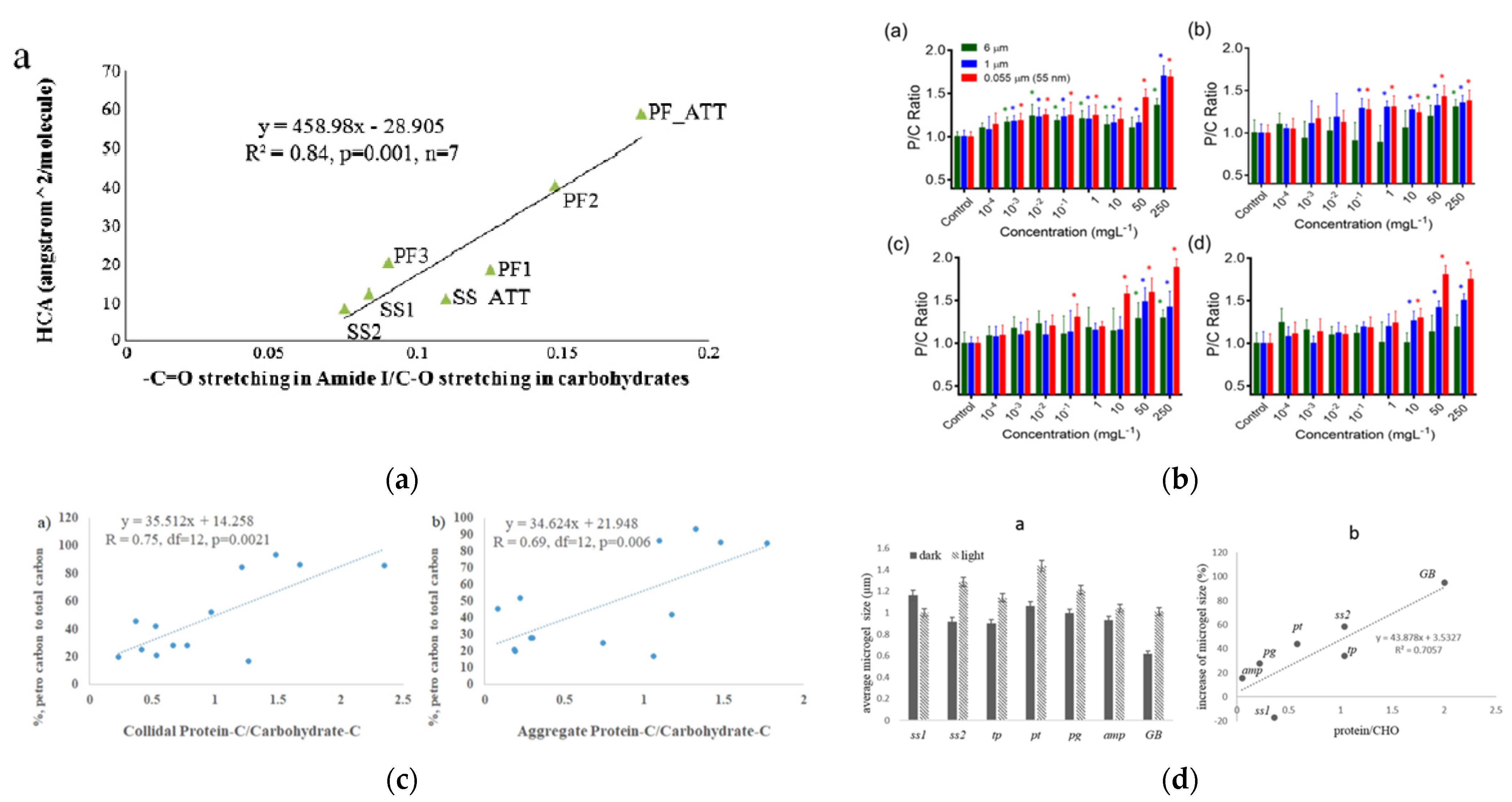
The ratio of proteins to carbohydrates of EPS (P/C) has been found to be closely related to the ‘stickiness’ of EPS and their relative hydrophobicity. For example, the P/C ratio is related to aggregation propensity, e.g., [20], surface tension [21], presence of nano-plastics or oil in microbial cultures [19,31], light-induced chemical crosslinking [23], and, when mineral matter is present, the sedimentation efficiency of marine snow [24]. Figure 1 shows some examples of how these properties can be related to the P/C ratio. Furthermore, the hydraulic residence time or sedimentation efficiency in wastewater treatment systems is also related to the P/C ratio [25]. Protein/carbohydrate ratios of EPS aggregates are thus an indicator of attachment propensity, i.e., its ‘stickiness’ [32], which can also be directly assessed by magnetic tweezers [33]. Compared with the laborious chemical techniques needed to directly measure protein and carbohydrate content, the P/C ratio can also be expediently obtained with simple fluorescence measurements [31]. The P/C ratio can be a more convenient and informative parameter for the assessment of EPS aggregation behaviors.
All these physical properties depend on various physical and chemical factors, such as cross-linker density, cross-liner types, polymer length, pH, types of polymers, temperature, degree of swelling, or temperature. Unfortunately, to the best of our knowledge, there is no available direct measurement for these properties for natural EPS gels in the literature. However, several studies on alginate (or other purified EPS) are available in the literature that might provide some rough assessments. Mechanical and rheological (viscoelastic) properties of alginate gels were shown to be dependent on the cross-linker type, density, ionic conditions, gelling temperatures, or EPS concentrations [36,37,38,39]. The specific gravity of a typical synthetic hydrogel (PVA (polyvinyl alcohol) gels) is around 1.05 [40]. For EPS sludges, the density has been reported to be around 1.004–1.048 (g/mL) [41]. For alginate gels depending on the gelling conditions, the value can vary from 1.03 to 1.12 (g/mL) [36]. However, these specific gravity measurements were conducted in non-seawater conditions (in lower salt conditions). Please note that the specific gravity of seawater is around 1.025. The specific gravity of EPS gels in seawater might shift from the measurements in non-seawater environments. The appearance and sizes of marine EPS gels are highly heterogeneous. No typical or characteristic morphology or shape has been found or concluded. The size of EPS gels in seawater can range from sub-micrometers to millimeters, even several centimeters.
EPS are highly heterogeneous mixtures of biopolymers from various microbes in seawater and are generally associated with different types of particles (anthropogenic, minerals, or biological debris), and these physical properties (rheology, morphology, or specific gravity) of natural EPS gels are usually complex and highly variable to determine or generalize.
2. Relative Hydrophobicity of EPS
Exudates from different aquatic organisms can have hydrophilic and hydrophobic moieties. Mostly hydrophilic exudates include the so-called hydrocolloids that are secreted by macro-algae, e.g., seaweeds, and harvested for their distinct chemical properties valued in the food industry as thickening and gelling agents (e.g., [42]). They include acid polysaccharides such as alginates, carrageenans, pectins, gums, and more neutral polysaccharides such as agar and similar substances, extracted from seaweeds, bacteria, and other organisms [42]. Most of these, but not all of them, form gels in the presence of metal ions such as Ca2+. The kind and location of acid functional group determines their food or physiological properties, e.g., alginates are blood coagulants, while carrageenans are anti-coagulants [43].
Due to the solubility limitation of water, hydrophobic moieties of EPS are normally not exposed to the water but, rather, are found in the interior of the structure or proteins or humic substances. As a consequence, EPS and humic substances become amphiphilic. The relative hydrophobicity of these biomolecules, represented by the hydrophobic contact area (HCA), is an important parameter that regulates the kinetics and extent of particle aggregation and dis-aggregation reactions in the water column, and thus influences the removal of associated radionuclides (e.g., Thorium-234) and organic pollutants (e.g., petroleum hydrocarbons). Xu et al. [20] found that the P/C ratio of EPS, determined by FTIR, is linearly related to the HCA, determined by HPLC. This implies that the P/C ratio can be used as an indicator for the relative hydrophobicity of macromolecules. This then also implies that the relative hydrophobicity of the carrier biopolymers of pollutants is mainly controlled by their relative protein contents [34]. High protein content in EPS has also been found to greatly accelerate the formation of marine gels that are not subject to disaggregation after EDTA addition that complexes the Ca2+ that are bridging hydrophilic components of EPS, thus rendering gel formation through irreversible hydrophobic interactions [28,44,45].
3. Stability of Microgels upon the Addition of Amphiphiles, e.g., Dispersants
Contrary to the results of [46], which showed the instability of marine gels when irradiated by UV, there is now ample evidence that sunlight irradiation causes reactive oxygen species (ROS)-mediated chemical crosslinking reactions, leading to the photoflocculation of NOM [47]. This was shown through increases in the concentrations of molecular weight, particle size, and mass [23]. On the other hand, global change-induced increases in temperature and hydrogen ion concentrations will have the tendency to decrease the stability of gels [48].
In addition, the aggregation and dispersion of marine gels can be affected by heterogeneous particles and agents in surrounding environments. For example, nano-carbonaceous particles were shown to reduce marine gel formation, due to their negative surface charges interfering with Ca2+ bridge cross-linking [49]. This observation is consistent with Zhang et al. [50], which suggests that quantum dots with negative charges have a stronger capability to stabilize EPS gel than positively charged ones. In addition, the microgel size significantly decreased when in the presence of surfactants, especially in the anionic type. Furthermore, negatively charged surfactants such as sodium dodecyl sulfate (SDS) can disrupt existing native microgels, converting larger aggregates into smaller particles. Notably, in addition to human-made pollutants, the input of natural substances can also cause changes in the dynamics of microgels. Shiu et al. [51] indicated that the self-assembly of marine gels would be decreased in the presence of NOM such as Suwannee River humic acid, fulvic acid, and natural organic matter at low concentrations (0.1–10 mg L−1). As mentioned above, a reduction in marine microgel size induced by various specific conditions could lead to a decrease in the downward flux of nutrients and organic carbon, thereby disturbing the organic carbon cycle and biological pump.
Chiu et al. [52] demonstrated that the application of the dispersant Corexit (used to disrupt oil spills) can inhibit EPS aggregation and/or disperse pre-existing microgels in laboratory studies. To represent potential situations during oil spills, a water-accommodated fraction (WAF) of oil and a chemical enhanced WAF (CEWAF) were prepared by mixing oil and dispersant in artificial seawater. It was found that CEWAF can enhance EPS aggregation, with more aggregates accumulating at the air–water interface. While more hydrophobic EPS forms (higher P/C ratio) showed a high resistance to Corexit dispersion, hydrophilic EPS (lower P/C ratio) dispersed more readily when the dispersant Corexit was added, thereby suggesting that P/C ratio plays an important role in determining the stability of microgels in the presence of dispersants. In addition, Shiu et al. [31] showed a negative correlation between P/C ratio and the relative amount of extracellular DNA in EPS, indicating that a higher cellular stress level when exposed to pollutants (WAF and CEWAF) is associated with EPS of higher P/C ratios, resulting in a lower concentration of DNA. This suggests that marine microbes can actively modify their EPS release and composition in response to oil and Corexit treatments.
4. Incorporation of Oil and Other Hydrophobic Pollutants into Gel-Forming EPS and Marine Aggregates
Much has been written on the role of EPS-containing aggregates (‘Marine Snow’) in accomodating oil and forming ‘Marine Oil Snow’, MOS [53,54,55,56,57]. Even though EPS gels are normally hydrophilic on the outside, and they hide hydrophobic entities of mostly proteins in their interiors, hydrophobic pollutants such as oil can be accommodated well within gels upon unfolding of the proteins. EPS were found to be crucial to the formation of marine oil snow (MOS), which can form in the presence and absence of Corexit [27]. Using a radiocarbon mass balance or 13C-NMR quantification after a dichloromethane extraction, it was found that the presence of dispersants enhanced the amounts of protein and oil being incorporated into oil-carrying aggregates, yet slowed the sedimentation efficiency of the MOS [24]. EPS with higher P/C ratios (i.e., greater hydrophobicity) tended to facilitate the incorporation of oil and/or Corexit, and the formation of oil-carrying aggregates. When not enough mineral matter is present, colloidal aggregates can become less able to sink due to the lowered density caused by petroleum components. These observations and assessments were confirmed in subsequent mesocosm experiments that simulated both near-shore and off-shore conditions, resulting in significant relationships between the P/C ratio in aggregates/colloids and the percentage of petrocarbon incorporation into these phases regardless of conditions [34]. The P/C ratio of EPS in both the aggregate and the colloidal fraction was thus a key factor for regulating the oil contribution to the sinking aggregates. These studies also pointed out the necessity to consider more closely the presence of a mineral phase, as ballast, to overcome the buoyancy effects of oil in the oil-carrying EPS aggregates.
EPS (as the sum of individually determined proteins and carbohydrates), extracted by EDTA from the surface-attached fraction of particles in mesocosm experiments (with and without oil), correlated well with TEP [26], supporting the use of EPS as a surrogate for TEP measurements in experiments in the presence of Corexit, where TEP cannot be determined, due to analytical interference.
The water solubility of other hydrophobic pollutants such as dioxins, and PAHs, which are normally only sparingly soluble, can be greatly enhanced by their association with ‘dissolved’ organic carbon (operationally defined as passing through a 0.7 or 0.5 µm filter), which contains natural colloidal, macromolecular organic matter (operationally defined as the fraction retained by an ultrafilter of 1 or 10 kDa pore size, and passing through a 0.5 or 0.7 µm filter) composed of EPS and humic substances ([58]). For example, empirical relationships describing the binding of hydrophobic organic compounds to sedimentary (Kd) and colloidal matter (Kc) have been proposed and experimentally verified. The reader is referred to numerous reviews on this subject, e.g., Schwarzenbach et al. [44]. While this solubility enhancement is important for transport in more turbulent aquatic systems, in water-submerged waste disposal sites, it has been found, using state-of-the art techniques, that the truly dissolved (≤1 kDa fraction) concentration of dioxins in a waste pit was even lower than predicted from Kow and BC values [45].
5. Specific and Nonspecific Interactions of Marine Gels with Metal Ions
The various interactions of metal ions with natural macromolecular organic molecules were reviewed in Buffle [58], Guo et al. [59], Doucet et al. [60], and Santschi et al. [32,61,62]. There are some main differences between trace metal complexation to a low-molecular-weight (LMW) ligand (e.g., citric acid) and to a high-molecular-weight (HMW) polyelectrolyte complexant, whereby the same functional group is attached to either a simple molecule or a macromolecular backbone (e.g., acid polysaccharides, macromolecular thiols, carboxylates, and proteins). LMW ligands have a small number of metal-specific functional groups, while multiple HMW ligands can be attached to different locations in the macromolecule, from where they can chelate trace metals in different ways. The nature of those ligands is relatively well recognized. The interaction between ligands and cations is generally divided into two categories depending on the hardness and softness of acids and bases. Hard acids and bases are characterized by small size, high electronegativity, and low polarizability, including A-type (e.g. Al3+) metals and F, O, and N. They are readily hydrated and tend to form outer-sphere complexes by ionic bonds. Soft acids and bases are characterized by relatively large size, low electronegativity, and high polarizability, including B-type metals (e.g., Ag+ and Hg2+) and S, I, and Br. They usually exist dehydrated and tend to form inner-sphere complexes by covalent bonds, which are far more stable than outer-sphere complexes.
High molecular (HMW) ligands are thus macromolecules that have a large number of surface functional groups (SFGs). They are composed of humic substances, polysaccharides, amino acids and peptides, and hydrocarbons. SFGs would be present on the outside of the biopolymer as they present themselves to the water. Due to the more complicated architecture of these biopolymers, the actual architecture can change depending on conditions (e.g., pH, redox, and salinity), and micelles could form at higher concentrations of colloidal forms of NOM. Advanced reverse osmosis/electrodialysis that consistently recovers 68 ± 2% of DOC allowed the molecular-level characterization of this macromolecular fraction via various spectroscopic (including advanced NMR) techniques [63]. It was found that condensed aromatic and quaternary anomeric carbons contribute to this deep refractory DOC pool, the quaternary anomeric carbons being a newly identified and potentially important component of bio-refractory carbohydrate-like carbon. Their results support the multi-pool (e.g. 3-pool: labile, semi-labile, and refractory) conceptual model of marine DOM biogeochemistry. Therefore, the average values of chemical (stability constants for complexation, acid-base, etc.) or physical properties (e.g., residence times) are sometimes not very meaningful and are subject to biases. The secondary effects that make up such biases can be categorized into several major groups or categories.
- (1)
- Polyfunctional properties: They have various kinds of SFGs (R-COOH, R-OH, R-SH, R-NH2, etc.). Those different SFGs also have different affinities to hard and soft cations ([44]. Sometimes, a metal ion is bound to more than two SFGs. There may be competition for cations between different SFGs. For example, B-type metals have stronger affinities to (S, S) > (S, N) > (N, O) > (O, O). The same SFG can have different properties depending on the types of backbone (aliphatic or aromatic) to which they bind. Finally, the geometry, such as cavities formed near SFGs, and flexibility of the organic molecules can make a significant difference to the stability of the complexation. These kinds of steric factors are controlled by ionic strength and pH in bulk solution.
- (2)
- Conformational changes: Depending on the hydration/dehydration processes, hydrogen bonds between hydrated cations and SFGs, or metallic bridges, and the conformation of the macromolecules can form aggregates or gels. The hydration water has a different structure from that of water in the bulk solution, and it makes the stability different [58,64]. The nature of a SFG in both LMW ligand and HMW macromolecules is similar. However, the fate of the same SFG may differ depending on the fate of particles and dissolved solutes.
- (3)
- Polyelectric properties: HMW macromolecules have SFGs (e.g., R-COOH) that protonate at low pH and deprotonate at high pH. When they deprotonate under basic conditions, negatively charged SFGs repulse one another. This process creates an electric field and causes more energy needed to remove protons from SFGs, eventually increasing the pKa. The formation of electric fields depends on the proportion of protonated sites. This indicates that the degree of protonation or deprotonation is not solely controlled by pH in bulk solution, but also by the near-field interactions between potential ligands.
- (4)
- Binding heterogeneity effects, with binding constants becoming a function of the metal ion-to-surface site ratio [58], occur because the strongest ligands are present at the lowest concentrations, while weaker ligands are present at higher concentrations. This necessitates experimental assessments at ambient concentrations of metals and ligands, or at least use the proper ratios.
- (5)
- ‘Particle concentration effects’ on kinetic constants (ki) and particle–water partition coefficients (Kd) are a consequence of incomplete separation between particles and solution and colloids, as there often are strong metal complexing macromolecular ligands in the 0.45 µm filter-passing fraction. This effect causes experimentally determined Kd and ki values to become a function of particle (Cp) concentration. This ‘particle concentration effect’ on kinetic constants (ki) and particle–water partition coefficients (Kd) was demonstrated using, as an example, thorium ions in the ocean, that is, the Brownian pumping model of Honeyman and Santschi [65].
Both humic substances [66] and EPS [67] can be considered a heterogeneous Donnan gel phase, similar to the situation in mucus [68]. Donnan equilibria can dominate the exchange of cations and anions across EPS gels surrounding microbial cells. For example, the Donnan mechanism affects mucin release [68] and mucus hydration [69], the swelling of exocytosed polymer-gels in Phaeocystis pouchetii [67], and the cation exchange membrane behavior of EPS in salt-adapted granular sludge [70]. Furthermore, toxic effects in saline environments on microbial consortia can be alleviated by the selective binding of cations to negatively charged EPS surrounding their cells, which prevents their diffusion into the deeper parts of the biofilm. The toxic effects of metal cations have been explained by various mechanisms, i.e., their ability to replace metallic enzyme cofactors, thereby disrupting the biological function of these cofactors, and the induction of redox reactions with cellular thiols, provoking Fenton-type reactions that produce reactive oxygen species and by interference with membrane transport processes [70].
When macromolecules form gels, as in the case of EPS, there will be other nonspecific interactions during crosslinking. For example, the crosslinking ability of counter-ions and the stability of the resulting networks increase with the second power of the valence electrons [71]. As an example, Fe3+ or Al3+ should be able to cross-link dissolved organic matter (DOM) at a fraction of the concentration that Ca2+ does. Furthermore, the degree of interaction for trivalent metal ions is higher as compared to that for divalent metal ions at physiological pH (pH ∼ 7.0) [72]). Moreover, organic polycations including spermine or spermidine, the condensing peptides of nucleic acids released from dead cells, have four cationic sites and are found in seawater at nanomolar concentrations [73]. They could be very powerful DOM crosslinkers even at submicromolar concentrations. Other polycations such as porins released by bacteria could also be interesting candidates to evaluate.
Felz et al. [74] recently reviewed evidence of how metal ions impact structural EPS hydrogels from aerobic granular sludge. They reported that structural EPS contain alginate hydrogels, but the two are not the same. Structural EPS are more protein-rich, and their gel forming ability, stiffness determined by the Young’s modulus, and binding ability are better in the presence of transition metals (rather than alkaline earth metals) than for alginates. They also showed that structural EPS are highly complex, and they have different gelling mechanisms than the acidic polysaccharides alginate, polygalacturonic acid, and kappa carrageenan. In addition, the structural EPS hydrogels show strong integrity toward the chelating reagent EDTA.
It was clearly demonstrated that riverine and marine DOM polymers have the capacity for scavenging selected heavy metals via aggregation processes [75]. The presence of many anionic functional groups on the surface of polymers may provide cation exchange sites for complexing heavy or trace metals [76]. The highest binding affinity in three selected metals was for Cu ions, followed by Ni and Mn ions. The affinity trend is in agreement with the Irving–Williams Series (Cu > Ni > Mn) and other marine colloidal studies. For example, a higher level of colloidal Cu than colloidal Ni was found in coastal areas such as the Danshuei River estuary, Taiwan [77]; the San Francisco Bay estuary, USA [78]; and Narragansett Bay, Rhode Island [79]. This may indicate that gels binding with metals are generally affected by ligand interaction and types of polymers [75].
6. Gel Interactions with Nanoparticles
Studies have shown that the secretion of EPS from microbial cells is significantly affected by surrounding environmental conditions. For example, EPS with higher protein-to-carbohydrate (P/C) ratios are induced by unfavorable growth conditions, including nutrient limitation, toxins (nanoparticles, oil, and dispersant), and light exposure [16,35]. EPS with high P/C ratios are more hydrophobic and sticky, and are thus able to physically block or chemically ‘quench’ the hazardous agents [80], resulting in the lower effective concentration of toxins entering microbial cells. Furthermore, protein-rich EPS can potentially facilitate faster assembly rates of marine aggregates and alter their aggregation sizesv. The interactions between microbe growth/survival and critical characteristics of EPS (P/C ratio) in the presence of micro- and nano-plastics have received little attention. Therefore, understanding the complex biochemical interactions between three key components (microbes, nanoparticles, and EPS) during nanoparticle (NP) exposure is important to elucidate the fate of NPs, e.g., plastics, especially in their aggregation and scavenging processes in marine environments.
7. Gel Interactions with Micro- and Nano-Plastics
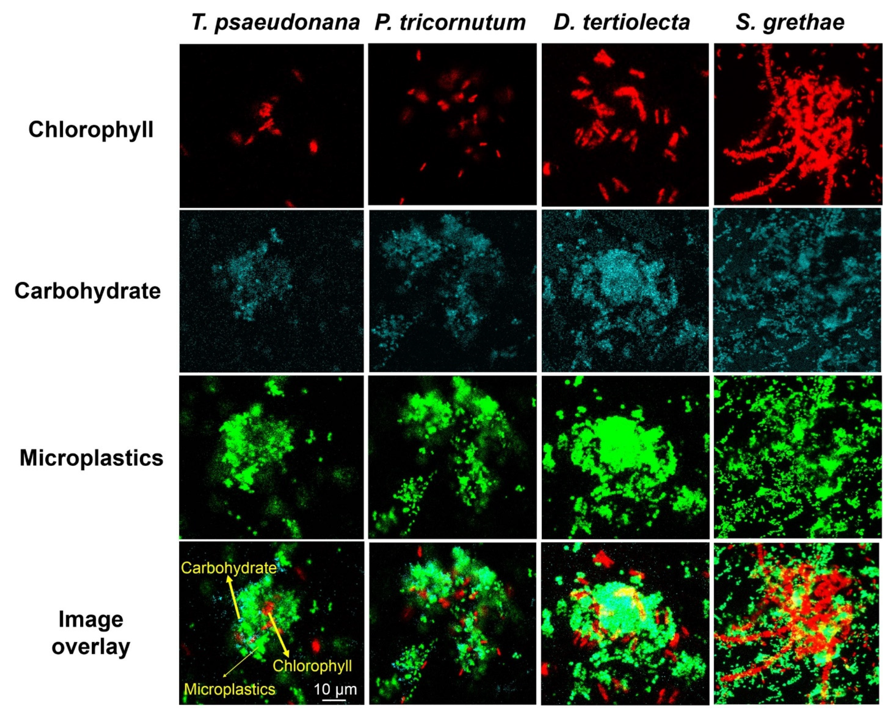
Microplastics, as with many other micro- and nanoparticles, are rapidly covered by biofilms that then further interact with marine biogenic particles [81] to marine plastic snow (MPS). An example of nano-plastic particles enmeshed in EPS and phytoplankton cells, i.e., MPS, is given in Figure 2.
The first step is the formation of microgels. In the study by Ding et al. [15], it was shown that EPS microgel formation in seawater was greatly accelerated by small amounts of amphiphilic EPS or nano-plastics. Later, Chen et al. [16] showed that phytoplankton EPS microgel formation is also accelerated by nano-plastics in seawater, but to a lesser extent. In both cases, it was demonstrated that hydrophobic interactions dominated, which were not affected by EDTA additions, in contrast to hydrophilic interactions. Phytoplankton EPS microgel formation is greatly accelerated by nano-plastics in seawater, likely due to a higher protein content of the EPS produced [19,20]. Furthermore, EPS microgel formation from DOM was greatly accelerated by nano-plastics in different river and lake waters, as well as seawater [19]. Patches of algal cells with 1 μm polystyrene micro-particles encased in an EPS matrix have been observed [19], supporting the hypothesis that plastics would be incoporated into the phytoplankton EPS matrix to form aggregates (marine plastic snow). The sinking route of marine aggregates can scavenge micro- and nano-plastics, which may explain why the negative mass balance of entering vs. measured marine plastics in the surface waters is still lower than expected [15]. EPS effects should be considered in models for predicting and understanding the fate and transport of marine plastic debris.
8. Gel Interactions with Organisms
Gel interactions with marine organisms are quite diverse. For example, Haye et al. [15] showed for the first time that filter-feeding organisms effectively filter suspended particles, as well as negatively charged colloidal-sized nanoplastics (0.04 µm), at ambient (1 mg/L) concentrations, most likely as gel-like marine plastic snow. In addition, these authors showed that the chemical composition of EPS controls the uptake of metal ions from the water by oysters. For example, alginate gels greatly ameliorated the metal ion uptake, while carrageenans did not, when compared to ambient colloidal-sized EPS from Galveston Bay. Finally, biofilms composed of EPS gels growing on plastics are reservoirs for antibiotic and metal resistance genes in marine environments. Additionally, marine organisms would be unable to discriminate between target food sources and organic aggregates. These self-assembled microgels concentrate organic and inorganic materials (as above sections), and thus, the accumulation may affect the exposure of higher-trophic-level predators such as zooplankton, invertebrates, filter-feeding fish, and even humans [82,83].
9. Conclusions
Marine gels (analytically determined as gels, EPS or TEP) are ubiquitous, as they form from amphiphilic microbial exudates of macromolecular exopolymeric substances in aquatic systems, and they provide a physical barrier surrounding the microbial cells, mediating the transport of hydrophilic and hydrophobic compounds to and from the cells. They are mostly composed of proteins and polysaccharides, whose relative contribution can change in response to environmental conditions, e.g., the presence of pollutants. These gels, because of their amphiphilic nature, can strongly interact with ionic and nonionic constituents in various ways, e.g., through both hydrophilic and hydrophobic interactions that facilitate certain physical, chemical, or biological pathways.
10. Open Questions
How do marine snow and macrogels of millimeter and centimeter size form from microgels of 5 µm size?—A good start to answer this question has been made by Buffle et al. [84] in their three-colloidal component approach. In their model, they propose that large aggregates form through floc formation by crosslinking many of the individual polysaccharide-rich fibrils. However, more work needs to be carried out to understand the complex physical, chemical, and biological interactions that lead to floc and aggregate formation.
How do marine gels made from EPS respond to changes in environmental conditions, e.g., temperature, pH, redox, nutrient, ionic composition, and strength?—While global-change-related increases in temperature and hydrogen ion concentrations had been addressed by Chen et al. [48], other relevant changes in environmental conditions had not been properly addressed.
Author Contributions
All authors contributed equally to the article. All authors have read and agreed to the published version of the manuscript.
Funding
This research received no external funding.
Conflicts of Interest
The authors declare no conflict of interest.
References
- Santschi, P.; Balnois, E.; Wilkinson, K.; Zhang, J.; Buffle, J.; Guo, L. Fibrillar polysaccharides in marine macromolecular organic matter as imaged by atomic force microscopy and transmission electron microscopy. Limnol. Oceanogr. 1998, 43, 896–908. [Google Scholar] [CrossRef]
- Droop, M.R. Vitamins, phytoplankton and bacteria: Symbiosis or scavenging? J. Plankton Res. 2007, 29, 107–113. [Google Scholar] [CrossRef] [Green Version]
- Kazamia, E.; Sutak, R.; Paz-Yepes, J.; Dorrell, R.G.; Vieira, F.R.J.; Mach, J.; Morrissey, J.; Leon, S.; Lam, F.; Pelletier, E.; et al. Endocytosis-mediated siderophore uptake as a strategy for Fe acquisition in diatoms. Sci. Adv. 2018, 4, eaar4536. [Google Scholar] [CrossRef] [PubMed] [Green Version]
- Butler, A. Marine Siderophores and Microbial Iron Mobilization. Biometals 2005, 18, 369–374. [Google Scholar] [CrossRef] [PubMed]
- Chin, W.-C.; Orellana, M.V.; Quesada, I.; Verdugo, P. Secretion in Unicellular Marine Phytoplankton: Demonstration of Regulated Exocytosis in Phaeocystis globosa. Plant Cell Physiol. 2004, 45, 535–542. [Google Scholar] [CrossRef] [PubMed] [Green Version]
- Rabin, N.; Zheng, Y.; Opoku-Temeng, C.; Du, Y.; Bonsu, E.; Sintim, H.O. Biofilm formation mechanisms and targets for developing antibiofilm agents. Future Med. Chem. 2015, 7, 493–512. [Google Scholar] [CrossRef] [PubMed]
- Passow, U. Transparent exopolymer particles (TEP) in aquatic environments. Prog. Oceanogr. 2002, 55, 287–333. [Google Scholar] [CrossRef] [Green Version]
- Wurl, O.; Miller, L.; Vagle, S. Production and fate of transparent exopolymer particles in the ocean. J. Geophys. Res. 2011, 116, C00H13. [Google Scholar] [CrossRef] [Green Version]
- Decho, A.W.; Gutierrez, T. Microbial Extracellular Polymeric Substances (EPSs) in Ocean Systems. Front. Microbiol. 2017, 8, 922. [Google Scholar] [CrossRef]
- Passow, U.; Alldredge, A.L. A dye-binding assay for the spectrophotometric measurement of transparent exopolymer particles (TEP). Limnol. Oceanogr. 1995, 40, 1326–1335. [Google Scholar] [CrossRef]
- Bar-Zeev, E.; Berman-Frank, I.; Girshevitz, O.; Berman, T. Revised paradigm of aquatic biofilm formation facilitated by microgel transparent exopolymer particles. Proc. Natl. Acad. Sci. USA 2012, 109, 9119–9124. [Google Scholar] [CrossRef] [PubMed] [Green Version]
- Berman, T. TEP, an Ubiquitous Constituent of NOM is an important factor in Membrane Biofouling. In Proceedings of the 2011 IWA Specialty Conference on Natural Organic Matter, Costa Mesa, CA, USA, 27–29 July 2011. [Google Scholar]
- De Gennes, P.G. Soft matter. Rev. Mod. Phys. 1992, 64, 645–648. [Google Scholar] [CrossRef]
- Chin, W.-C.; Orellana, M.V.; Verdugo, P. Spontaneous assembly of marine dissolved organic matter into polymer gels. Nature 1998, 391, 568–572. [Google Scholar] [CrossRef]
- Ding, Y.X.; Chin, W.C.; Rodriguez, A.; Hung, C.C.; Santschi, P.H.; Verdugo, P. Amphiphilic exopolymers from Sagittula stellata induce DOM self-assembly and formation of marine microgels. Mar. Chem. 2008, 112, 11–19. [Google Scholar] [CrossRef]
- Chen, C.-S.; Anaya, J.M.; Zhang, S.; Spurgin, J.; Chuang, C.-Y.; Xu, C.; Miao, A.-J.; Chen, E.Y.T.; Schwehr, K.A.; Jiang, Y.; et al. Effects of Engineered Nanoparticles on the Assembly of Exopolymeric Substances from Phytoplankton. PLoS ONE 2011, 6, e21865. [Google Scholar] [CrossRef] [PubMed] [Green Version]
- Long, R.A.; Azam, F. Abundant protein-containing particles in the sea. Aquat. Microb. Ecol. 1996, 10, 213–221. [Google Scholar] [CrossRef] [Green Version]
- Thornton, D.C.O. Coomassie Stainable Particles (CSP): Protein Containing Exopolymer Particles in the Ocean. Front. Mar. Sci. 2018, 5. [Google Scholar] [CrossRef]
- Shiu, R.-F.; Vazquez, C.I.; Tsai, Y.-T.; Torres, G.V.; Chen, C.-S.; Santschi, P.H.; Quigg, A.; Chin, W.-C. Nano-plastics induce aquatic particulate organic matter (microgels) formation. Sci. Total Environ. 2020. [Google Scholar] [CrossRef]
- Xu, C.; Zhang, S.; Chuang, C.-y.; Miller, E.J.; Schwehr, K.A.; Santschi, P.H. Chemical composition and relative hydrophobicity of microbial exopolymeric substances (EPS) isolated by anion exchange chromatography and their actinide-binding affinities. Mar. Chem. 2011, 126, 27–36. [Google Scholar] [CrossRef]
- Schwehr, K.A.; Xu, C.; Chiu, M.-H.; Zhang, S.; Sun, L.; Lin, P.; Beaver, M.; Jackson, C.; Agueda, O.; Bergen, C.; et al. Protein: Polysaccharide ratio in exopolymeric substances controlling the surface tension of seawater in the presence or absence of surrogate Macondo oil with and without Corexit. Mar. Chem. 2018, 206, 84–92. [Google Scholar] [CrossRef]
- Hung, C.; Guo, L.; Santschi, P.; Alvarado-Quiroz, N.; Haye, J. Distributions of carbohydrate species in the Gulf of Mexico. Mar. Chem. 2003, 81, 119–135. [Google Scholar] [CrossRef]
- Sun, L.; Xu, C.; Chin, W.C.; Zhang, S.; Lin, P.; Schwehr, K.A.; Quigg, A.; Chiu, M.-H.; Chin, W.-C.; Santschi, P.H. Light-induced aggregation of microbial exopolymeric substances. Chemosphere 2017, 181, 675–681. [Google Scholar] [CrossRef] [PubMed]
- Xu, C.; Zhang, S.; Beaver, M.; Wozniak, A.; Obeid, W.; Lin, Y.; Wade, T.L.; Schwehr, K.A.; Lin, P.; Sun, L.; et al. Decreased sedimentation efficiency of petro- and non-petro-carbon caused by a dispersant for Macondo surrogate oil in a mesocosm simulating a coastal microbial community. Mar. Chem. 2018, 206, 34–43. [Google Scholar] [CrossRef]
- Ren, B.; Young, B.; Variola, F.; Delatolla, R. Protein to polysaccharide ratio in EPS as an indicator of non-optimized operation of tertiary nitrifying MBBR. Water Qual. Res. J. 2016, 5, 297–306. [Google Scholar] [CrossRef] [Green Version]
- Xu, C.; Chin, W.-C.; Lin, P.; Chen, H.M.; Lin, P.; Chiu, M.-C.; Waggoner, D.C.; Xing, W.; Sun, L.; Schwehr, K.A.; et al. Marine Gels, Extracellular Polymeric Substances (EPS) and Transparent Exopolymeric Particles (TEP) in natural seawater and seawater contaminated with a water accommodated fraction of Macondo oil surrogate. Mar. Chem. 2019, 215, 103667. [Google Scholar] [CrossRef]
- Xu, C.; Zhang, S.; Beaver, M.; Lin, P.; Sun, L.; Doyle, S.M.; Sylvan, J.B.; Wozniak, A.; Hatcher, P.G.; Kaiser, K.; et al. The role of microbially-mediated exopolymeric substances (EPS) in regulating Macondo oil transport in a mesocosm experiment. Mar. Chem. 2018, 206, 52–61. [Google Scholar] [CrossRef]
- Janado, M.; Yano, Y. Hydrophobic nature of sugars as evidenced by their differential affinity for polystyrene gel in aqueous media. J. Solut. Chem. 1985, 14, 891–902. [Google Scholar] [CrossRef]
- Petrova, O.E.; Sauer, K. Sticky situations: Key components that control bacterial surface attachment. J Bacteriol. 2016, 104, 2413–2425. [Google Scholar] [CrossRef] [Green Version]
- Fong, J.N.C.; Yildiz, F.H. Biofilm Matrix Proteins. Microbiol. Spectr. 2015, 3. [Google Scholar] [CrossRef] [Green Version]
- Shiu, R.-F.; Chiu, M.-H.; Vazquez, C.I.; Tsai, Y.-Y.; Le, A.; Kagiri, A.; Xu, C.; Kamalanathan, M.; Bacosa, H.P.; Doyle, S.M.; et al. Protein to carbohydrate (P/C) ratio changes in microbial extracellular polymeric substances induced by oil and Corexit. Mar. Chem. 2020, 223, 103789. [Google Scholar] [CrossRef]
- Santschi, P.H.; Xu, C.; Schwehr, K.A.; Lin, P.; Sun, L.; Chin, W.C.; Kamalanathan, M.; Bacosa, H.P.; Quigg, A. Can the protein/carbohydrate (P/C) ratio of exopolymeric substances (EPS) be used as a proxy for their ‘stickiness’ and aggregation propensity? Mar. Chem. 2020, 218, 103734. [Google Scholar] [CrossRef]
- Chen, C.-S.; Shiu, R.-F.; Hsieh, Y.-Y.; Xu, C.; Vazquez, C.I.; Cui, Y.; Hsu, I.C.; Quigg, A.; Santschi, P.H.; Chin, W.-C. Stickiness of Extracellular Polymeric Substances on different surfaces via Magnetic Tweezers. Sci. Total Environ. 2021. [Google Scholar] [CrossRef]
- Xu, C.; Lin, P.; Zhang, S.; Sun, L.; Xing, W.; Schwehr, K.A.; Chin, W.-C.; Wade, T.L.; Knap, A.H.; Hatcher, P.G.; et al. The interplay of extracellular polymeric substances and oil/Corexit to affect the petroleum incorporation into sinking marine oil snow in four mesocosms. Sci. Total Environ. 2019, 693, 133626. [Google Scholar] [CrossRef]
- Sun, L.; Chin, W.-C.; Chiu, M.-H.; Xu, C.; Lin, P.; Schwehr, K.A.; Quigg, A.; Santschi, P.H. Sunlight induced aggregation of dissolved organic matter: Role of proteins in linking organic carbon and nitrogen cycling in seawater. Sci. Total Environ. 2019, 654, 872–877. [Google Scholar] [CrossRef] [PubMed]
- Jeong, C.; Kim, S.; Lee, C.; Cho, S.; Kim, S.-B. Changes in the physical properties of calcium alginate gel beads under a wide range of gelation temperature conditions. Foods 2020, 9, 180. [Google Scholar] [CrossRef] [PubMed] [Green Version]
- Lee, K.Y.; Rowley, J.A.; Eiselt, P.; Moy, E.M.; Bouhadir, K.H.; Mooney, D.J. Controlling mechanical and swelling properties of alginate hydrogels independently by cross-linker type and cross-linking density. Macromolecules 2000, 33, 4291–4294. [Google Scholar] [CrossRef]
- Lotti, T.; Carretti, E.; Berti, D.; Montis, C.; Del Buffa, S.; Lubello, C.; Feng, C.; Malpei, F. Hydrogels formed by anammox extracellular polymeric substances: Structural and mechanical insights. Sci. Rep. 2019, 9, 1–9. [Google Scholar]
- Kakita, H.; Kamishima, H. Some properties of alginate gels derived from algal sodium alginate. In Proceedings of the Nineteenth International Seaweed Symposium, Kobe, Japan, 26–31 March 2007; pp. 93–99. [Google Scholar]
- Sajjan, A.; Banapurmath, N.; Tapaskar, R.; Patil, S.; Kalahal, P.; Shettar, A. Preparation of polymer electrolyte hydrogels using poly (vinyl alcohol) and tetraethylorthosilicate for battery applications. In Proceedings of the IOP Conference Series: Materials Science and Engineering, Karnataka, India, 2–3 March 2018; p. 012078. [Google Scholar]
- Feng, C.; Lotti, T.; Canziani, R.; Lin, Y.; Tagliabue, C.; Malpei, F. Extracellular biopolymers recovered as raw biomaterials from waste granular sludge and potential applications: A critical review. Sci. Total Environ. 2020, 753, 142051. [Google Scholar] [CrossRef]
- Saha, D.; Bhattacharya, S. Hydrocolloids as thickening and gelling agents in food: A critical review. J. Food Sci. Technol. 2010, 47, 587–597. [Google Scholar] [CrossRef] [Green Version]
- Kuznetsova, T.A.; Andryukov, B.G.; Besednova, N.N.; Zaporozhets, T.S.; Kalinin, A.V. Marine Algae Polysaccharides as Basis for Wound Dressings, Drug Delivery, and Tissue Engineering: A Review. J. Mar. Sci. Eng. 2020, 8, 481. [Google Scholar] [CrossRef]
- Schwarzenbach, R.P.; Gschwend, P.M.; Imboden, D.M. Environmental Organic Chemistry, 2nd ed.; Wiley-Interscience, John Wiley and Sons, Inc.: Hoboken, NJ, USA, 2003. [Google Scholar]
- Louchouarn, P.; Seward, S.; Cornelissen, G.; Arp, H.P.H.; Yeager, K.M.; Brinkmeyer, R.; Santschi, P.H. Limited mobility of dioxins near San Jacinto Super Fund site (waste pit) in the Houston Ship Channel, Texas due to high amorphous organic carbon. Environ. Pollut. 2018, 238, 988–998. [Google Scholar] [CrossRef]
- Orellana, M.V.; Verdugo, P. Ultraviolet radiation blocks the organic carbon exchange between the dissolved phase and the gel phase in the ocean. Limnol. Oceanogr. 2003, 48, 1618–1623. [Google Scholar]
- Chen, H.; Abdulla, H.A.N.; Sanders, R.L.; Myneni, S.C.B.; Mopper, K.; Hatcher, P.G. Production of Black Carbon-like and Aliphatic Molecules from Terrestrial Dissolved Organic Matter in the Presence of Sunlight and Iron. Environ. Sci. Technol. Lett. 2014, 1, 399–404. [Google Scholar] [CrossRef]
- Chen, C.-S.; Anaya, J.M.; Chen, E.Y.-T.; Farr, E.; Chin, W.-C. Ocean Warming—Acidification Synergism Undermines Dissolved Organic Matter Assembly. PLoS ONE 2015. [Google Scholar] [CrossRef]
- Shiu, R.-F.; Chin, W.-C.; Lee, C.-L. Carbonaceous particles reduce marine microgel formation. Sci. Rep. 2014, 4, 5856. [Google Scholar] [CrossRef] [Green Version]
- Zhang, S.; Jiang, Y.; Chen, C.-S.; Spurgin, J.; Schwehr, K.A.; Quigg, A.; Chin, W.-C.; Santschi, P.H. Aggregation, Dissolution, and Stability of Quantum Dots in Marine Environments: Importance of Extracellular Polymeric Substances. Environ. Sci. Technol. 2012, 46, 8764–8772. [Google Scholar] [CrossRef] [PubMed]
- Shiu, R.-F.; Lee, C.-L.; Chin, W.-C. Reduction in the exchange of coastal dissolved organic matter and microgels by inputs of extra riverine organic matter. Water Res. 2018, 131, 161–166. [Google Scholar] [CrossRef]
- Chiu, M.H.; Vazquez, C.I.; Shiu, R.F.; Le, C.; Sanchez, N.R.; Kagiri, A.; Garcia, C.A.; Nguyen, C.H.; Tsai, S.M.; Zhang, S.; et al. Impact of exposure of crude oil and dispersant (Corexit) on aggregation of extracellular polymeric substances. Sci. Total Environ. 2019, 657, 1535–1542. [Google Scholar] [CrossRef] [PubMed]
- Passow, U.; Ziervogel, K.; Asper, V.; Diercks, A. Marine snow formation in the aftermath of the Deepwater Horizon oil spill in the Gulf of Mexico. Environ. Res. Lett. 2012, 7, 035301. [Google Scholar] [CrossRef] [Green Version]
- Passow, U.; Ziervogel, K. Marine Snow Sedimented Oil Released During the Deepwater Horizon Spill. Oceanography 2016, 29, 118–125. [Google Scholar] [CrossRef] [Green Version]
- Passow, U.; Sweet, J.; Francis, S.; Xu, C.; Dissanayake, A.L.; Lin, Y.Y.; Santschi, P.H.; Quigg, A. Incorporation of oil into diatom aggregates. Mar. Ecol. Prog. Ser. 2019, 612, 65–86. [Google Scholar] [CrossRef] [Green Version]
- Passow, U.; Hetland, R.D. What Happened to All of the Oil? Oceanography 2016, 29, 88–95. [Google Scholar] [CrossRef] [Green Version]
- Engel, A.; Endres, S.; Galgani, L.; Schartau, M. Marvelous Marine Microgels: On the Distribution and Impact of Gel-Like Particles in the Oceanic Water-Column. Front. Mar. Sci. 2020, 7. [Google Scholar] [CrossRef]
- Buffle, J. Complexation Reactions in Aquatic Systems. An Analytical Approach; Ellis Horwood Pub.: Chichester, UK, 1988. [Google Scholar]
- Guo, L.; Sanschi, P.H. Ultrafiltration and its applications to sampling and characterization of aquatic colloids. In International Union of Pure and Applied Chemistry (IUPAC) Series on Analytical and Physical Chemistry of Environmental Systems; John Wiley: Hoboken, NJ, USA, 2007. [Google Scholar]
- Doucet, F.J.; Lead, J.R.; Santschi, P.H. Colloid-Trace Element Interactions in Aquatic Systems. In International Union of Pure and Applied Chemistry (IUPAC) Series on Analytical and Physical Chemistry of Environmental Systems; John Wiley: Hoboken, NJ, USA, 2007. [Google Scholar]
- Santschi, P.H. Marine colloids, agents of the self-cleansing capacity of aquatic systems: Historical perspective and new discoveries. Mar. Chem. 2018, 207, 124–135. [Google Scholar] [CrossRef]
- Santschi, P.; Lenhart, J.; Honeyman, B. Heterogeneous processes affecting trace contaminant distribution in estuaries: The role of natural organic matter. Mar. Chem. 1997, 58, 99–125. [Google Scholar] [CrossRef]
- Helms, J.R.; Mao, J.D.; Chen, H.M.; Perdue, E.M.; Green, N.W.; Hatcher, P.G.; Mopper, K.; Stubbins, A. Spectroscopic characterization of oceanic dissolved organic matter isolated by reverse osmosis coupled with electrodialysis. Mar. Chem. 2015, 177, 278–287. [Google Scholar] [CrossRef] [Green Version]
- Doucet, F.J.; Lead, J.R.; Santschi, P.H. Colloid-Trace Element Interactions in Aquatic Systems; John Wiley & Son, Inc.: Hoboken, NJ. USA, 2007. [Google Scholar]
- Honeyman, B.; Santschi, P. A Brownian-Pumping Model for Oceanic Trace-Metal Scavenging—Evidence from TH-Isotopes. J. Mar. Res. 1989, 47, 951–992. [Google Scholar] [CrossRef]
- Benedetti, M.F.; van Riemsdijk, W.H.; Koopal, L.K. Humic substances considered as a heterogeneous Donnan gel phase. Environ. Sci. Tech. 30: 1805-1813. Humic substances considered as a heterogeneous Donnan gel phase. Environ. Sci. Techol. 1996, 30, 1805–1813. [Google Scholar] [CrossRef]
- Chin, W.; Orellanam, M.V.; Verdugo, P. Exocytosis in Phaeocystis pouchetii: Donnan mechanism of swelling of exocytosed polymer-gels. EOS Trans. Am. Geophys. Un. 1996, 76, 75. [Google Scholar]
- Aitken, L.M.; Verdugo, P. Donnan mechanism of mucin release and conditioning in goblet cells: The role of polygons. J. Exp. Biol. 1989, 53, 73–79. [Google Scholar]
- Tam, P.Y.; Verdugo, P. Control of mucus hydration as a Donnan equilibrium process. Nature 1981, 292, 340–342. [Google Scholar] [CrossRef]
- Sudmalisa, D.; Mubita, T.M.; Gagliano, M.C.; Dinis, E.; Zeeman, G.; Rijnaarts, H.H.M.; Temmink, H. Cation exchange membrane behaviour of extracellular polymericsubstances (EPS) in salt adapted granular sludge. Water Res. 2020, 178, 115855. [Google Scholar] [CrossRef]
- Tanaka, T. Gels. Sci. Am. 1981, 244, 124–138. [Google Scholar] [CrossRef]
- Kanti De, S.; Kanwa, N.; Chakraborty, A. Influence of Trivalent Metal Ions on Lipid Vesicles: Gelation and Fusion Phenomena. Langmuir 2019, 35, 6429–6440. [Google Scholar] [CrossRef]
- Nishibori, N.; Yuasa, A.; Sakai, M.; Fujihara, S.; Nishio, S. Free polyamine concentrations in coastal seawater during phytoplankton bloom. Fish. Sci. 2001, 67, 79–83. [Google Scholar] [CrossRef]
- Felz, S.; Kleikamp, H.; Zlopasa, J.; van Loosdrecht, M.C.M.; Lin, Y. Impact of metal ions on structural EPS hydrogels from aerobic granular sludge. Biofilm 2020, 2, 100011. [Google Scholar] [CrossRef] [PubMed]
- Shiu, R.-F.; Lee, C.-L. Role of microgel formation in scavenging of chromophoric dissolved organic matter and heavy metals in a river-sea system. J. Hazard. Mater. 2017, 328, 12–20. [Google Scholar] [CrossRef] [Green Version]
- Joshi, P.M.; Juwarkar, A.A. In Vivo Studies to Elucidate the Role of Extracellular Polymeric Substances from Azotobacter in Immobilization of Heavy Metals. Environ. Sci. Technol. 2009, 43, 5884–5889. [Google Scholar] [CrossRef] [PubMed]
- Jiann, K.-T.; Wen, L.-S.; Santschi, P.H. Trace metal (Cd, Cu, Ni and Pb) partitioning, affinities and removal in the Danshuei River estuary, a macro-tidal, temporally anoxic estuary in Taiwan. Mar. Chem. 2005, 96, 293–313. [Google Scholar] [CrossRef]
- San˜udo-Wilhelmy, S.A.; Rivera-Duarte, I.; Russell Flegal, A. Distribution of colloidal trace metals in the San Francisco Bay estuary. Geochim. Cosmochim. Acta 1996, 60, 4933–4944. [Google Scholar] [CrossRef]
- Wells, M.L.; Smith, G.J.; Bruland, K.W. The distribution of colloidal and particulate bioactive metals in Narragansett Bay, RI. Mar. Chem. 2000, 71, 143–163. [Google Scholar] [CrossRef]
- Nichols, C.A.M.; Guezennec, J.; Bowman, J.P. Bacterial Exopolysaccharides from Extreme Marine Environments with Special Consideration of the Southern Ocean, Sea Ice, and Deep-Sea Hydrothermal Vents: A Review. Mar. Biotechnol. 2005, 7, 253–271. [Google Scholar] [CrossRef] [PubMed] [Green Version]
- Michels, J.; Stippkugel, A.; Lenz, M.; Wirtz, K.; Engel, A. Rapid aggregation of biofilm-covered microplastics with marine biogenic particles. Proc. R. Soc. B 2018, 285. [Google Scholar] [CrossRef] [PubMed] [Green Version]
- Lusher, A.L.; Hernandez-Milian, G.; O’Brien, J.; Berrow, S.; O’Connor, I.; Officer, R. Microplastic and macroplastic ingestion by a deep diving, oceanic cetacean: The True’s beaked whale Mesoplodon mirus. Environ. Pollut. 2015, 199, 185–191. [Google Scholar] [CrossRef]
- Cole, M.; Lindeque, P.; Fileman, E.; Halsband, C.; Goodhead, R.; Moger, J.; Galloway, T.S. Microplastic Ingestion by Zooplankton. Environ. Sci. Technol. 2013, 47, 6646–6655. [Google Scholar] [CrossRef]
- Buffle, J.; Wilkinson, K.J.; Stoll, S.; Filella, M.; Zhang, J. A Generalized Description of Aquatic Colloidal Interactions: The Three-colloidal Component Approach. Environ. Sci. Technol. 1998, 32, 2887–2899. [Google Scholar] [CrossRef]
Figure 1.
(a) Examples for the relative hydrophobicity of EPS that increases with P/C ratio ([20], with permission of the publisher), (b) the relationship between nanoplastics concentration and the size-dependent induction of EPS with higher P/C ratio ([19], with permission of the publisher), (c) the relationships of % petro-carbon to total carbon in colloidal or sinking aggregates that increase with the P/C ratio of EPS ([34], with permission of the publisher), and (d) the microgel size increase due to light-induced ROS chemical crosslinking of proteins in EPS that scale with their P/C ratio ([35], with permission of the publisher).

Figure 2.
Marine Plastic Snow ([19], with permission from the publisher).
Figure 2.
Marine Plastic Snow ([19], with permission from the publisher).


Table 1.
Terminology used in this paper.
| NOM | natural organic matter |
| DOM | dissolved organic matter (i.e., passing a filter of about 0.5 µm pore size) |
| DOC | dissolved organic carbon (i.e., passing a filter of about 0.5 µm pore size) |
| EPS | exopolymeric substances, found in the colloidal or particulate fraction |
| TEP | transparent exoplymeric particles, operationally determined |
| Gels | a type of soft matter that is operationally determined in aquatic systems |
| HMW | high molecular weight (relative term, usually more than 1 kDa) |
| LMW | low molecular weight (relative term, usually less than 1 kDa |
| SFG | surface functional group |
| DLS | dynamic light scattering |
| FTIR | fourier transform infrared spectroscopy |
Publisher’s Note: MDPI stays neutral with regard to jurisdictional claims in published maps and institutional affiliations. |
© 2021 by the authors. Licensee MDPI, Basel, Switzerland. This article is an open access article distributed under the terms and conditions of the Creative Commons Attribution (CC BY) license (https://creativecommons.org/licenses/by/4.0/).
Share and Cite
MDPI and ACS Style
Santschi, P.H.; Chin, W.-C.; Quigg, A.; Xu, C.; Kamalanathan, M.; Lin, P.; Shiu, R.-F. Marine Gel Interactions with Hydrophilic and Hydrophobic Pollutants. Gels 2021, 7, 83. https://0-doi-org.brum.beds.ac.uk/10.3390/gels7030083
AMA Style
Santschi PH, Chin W-C, Quigg A, Xu C, Kamalanathan M, Lin P, Shiu R-F. Marine Gel Interactions with Hydrophilic and Hydrophobic Pollutants. Gels. 2021; 7(3):83. https://0-doi-org.brum.beds.ac.uk/10.3390/gels7030083
Chicago/Turabian StyleSantschi, Peter H., Wei-Chun Chin, Antonietta Quigg, Chen Xu, Manoj Kamalanathan, Peng Lin, and Ruei-Feng Shiu. 2021. "Marine Gel Interactions with Hydrophilic and Hydrophobic Pollutants" Gels 7, no. 3: 83. https://0-doi-org.brum.beds.ac.uk/10.3390/gels7030083
Note that from the first issue of 2016, this journal uses article numbers instead of page numbers. See further details here.