Potential miRNAs for miRNA-Based Therapeutics in Breast Cancer


1
Department of Biomedical Science, Faculty of Medicine and Health Sciences, Universiti Putra Malaysia, Selangor 43400, Malaysia
2
School of Biological Sciences, Nanyang Technological University, 60 Nanyang Drive, Singapore 637551, Singapore
*
Author to whom correspondence should be addressed.
Non-Coding RNA 2020, 6(3), 29; https://0-doi-org.brum.beds.ac.uk/10.3390/ncrna6030029
Submission received: 13 June 2020
/
Revised: 4 July 2020
/
Accepted: 7 July 2020
/
Published: 13 July 2020
(This article belongs to the Special Issue Non-Coding RNAs as Therapeutic Targets)
Abstract
:MicroRNAs (miRNAs) are small non-coding RNAs that can post-transcriptionally regulate the genes involved in critical cellular processes. The aberrant expressions of oncogenic or tumor suppressor miRNAs have been associated with cancer progression and malignancies. This resulted in the dysregulation of signaling pathways involved in cell proliferation, apoptosis and survival, metastasis, cancer recurrence and chemoresistance. In this review, we will first (i) provide an overview of the miRNA biogenesis pathways, and in vitro and in vivo models for research, (ii) summarize the most recent findings on the roles of microRNAs (miRNAs) that could potentially be used for miRNA-based therapy in the treatment of breast cancer and (iii) discuss the various therapeutic applications.
1. Introduction
According to GLOBOCAN, breast cancer is ranked the second most common cancer in the world. It is the most frequently occurring (24.2%) with the highest cancer fatality among women (15%). Asia (49.6%) has the highest mortality cases for both genders, followed by Europe (22%), Africa (11.8%), Latin America and the Caribbean (8.4%), North America (7.5%) and Oceania (0.77%) [1]. Based on the intrinsic molecular subtypes, breast cancer can be classified into various forms: Luminal A, Luminal B, Basal-like, Her2-enriched, and Normal-like. Particularly, triple negative breast cancer (TNBC) constitutes about 15–20% of all breast cancers [2]. Using transcriptomic profiling, it was identified that 49% of TNBC cases are Basal-like subtypes and 30% are claudin-low subtype [3]. TNBC indicates the deficit expressions of the estrogen receptor (ER), progesterone receptor (PR) and human epidermal growth factor receptor-2 (Her2), which thereby render endocrine or targeted therapies ineffective [2,3]. TNBC is highly heterogeneous and aggressive, with no standard recommended therapy for treatment. On the other hand, these therapies can effectively treat cancer subtypes Luminal A (triple positive), Luminal B (ER+, PR+ and Her2-) and Her2 enriched (ER-, PR-, Her2+). Nonetheless, regardless of all subtypes, hormone resistance and tumor relapse remain the major factors to hamper the effectiveness of therapy for breast cancer.
MicroRNAs (miRNA) are a class of short single stranded, non-coding RNAs (approximately ~22 nucleotides) that play an important role in regulating cancer development and progression [4]. They function by binding to the partial complementary mRNA seed sequence at the 3’ untranslated region (UTR) of mRNA targets [5]. Ultimately, the translation is repressed, or the cytoplasmic mRNA is degraded. Aberrations in miRNA expressions can affect gene expressions that are essential in cellular development, differentiation, proliferation, survival, apoptosis, resistance, and motility. As miRNAs can silence a wide variety of these genes simultaneously, and that the dysregulation of miRNAs are associated with different breast cancer subtypes and tumor malignancy, this makes them an attractive candidate for drug development. This review will summarize the recent findings on the emerging roles of miRNAs and discuss their potential in miRNA-based therapy for breast cancer.
2. miRNA Biogenesis and Mode of Action
miRNA synthesis is carried out through canonical and non-canonical pathways (Figure 1). Most miRNAs are processed by the canonical pathway. The first step of the canonical biogenesis pathway is the transcription of genomic DNA by RNA polymerase II to generate primary miRNA (pri-miRNA). Pri-mRNA consists of a stem-loop structure, a methylguanosine cap and may or may not necessarily have a poly-(A) tail [6,7]. It is cleaved by the Drosha–DiGeorge Syndrome Critical Region 8 (Drosha–DGCR8) complex to form a 70–100 nt precursor miRNA (pre-miRNA). Pre-miRNA is then transported to the cytoplasm by the association with Exportin-5/RanGTP complex. Subsequently, RNase III endonuclease Dicer cleaves the terminal loop to form a mature miRNA duplex. This duplex is loaded into the Argonaute family of proteins (AGO1–4). Based on the thermodynamic properties and the degree of complementarity between the miRNA and the AGO protein, one of two miRNA strands will be selected as the guide strand. The guide strand complexes with the Ago protein to form the miRNA-induced silencing complex (miRISC), while the other passenger strand from the duplex is degraded [6,8].
The non-canonical miRNA biogenesis pathway is an alternative for generating miRNAs (Figure 1). It can be classified into Drosha-DGCR8-independent or Dicer-independent systems. These pathways have been thoroughly reviewed by Stavast et al., 2019 and Abdelfattah et al., 2015 [7,9]. Briefly, pri-miRNAs that are encoded in the intron of coding genes, also known as miRtrons, are being processed by nuclear splicing machinery to form shorter hairpin loops that are shorter than the canonical pri-miRNAs. These miRtrons are Drosha–DGCR8-independent and they undergo hydrolysis by debranching enzyme 1 (DBR1) protein. The mirtron-derived pre-miRNAs are then directly translocated to the cytoplasm mediated by Exportin-5 (XPO-5) and bypassed the cleavage by endoribonuclease Dicer. For Dicer-independent systems, short hairpin RNAs (shRNA), small nucleolar RNAs (snoRNAs) and unique miRNAs such as pre-miR-451 are processed and transported similarly to the canonical pathway. They bypass the activity of Dicer and are further processed [5,7,8,9]. Both pathways will eventually lead to the formation of an miRISC complex, consisting of guide miRNA strand and the AGO protein. The guide miRNA is able to bind to the 5′ UTR promotor and coding regions of the corresponding mRNA via complementary base-pairing, known as miRNA response elements (MREs) [5]. Perfect complementarity between the miRNA of the miRISC complex and the MRE of the mRNA target promotes the AGO-dependent mRNA cleavage. Partial complementarity with only 2–8 base pairing, called the seed region, is sufficient for the interaction [10]. The miRISC complex, in this case, promotes the dissociate of eIF4A from the target mRNAs [11]. Furthermore, the miRISC complex together with TNRC6 protein, recruits the PAN2–PAN3 and CCR4–CAF1–NOT deadenylase complexes that remove the 3′ poly-A tail of the mRNA targets and thereby, inhibiting translation [12].
3. Cell Lines as In Vitro Model
There are more than 100 breast cancer cell lines that are commercially available. A study by Dai et al., 2017 characterized 84 of these cell lines based on their molecular profiles and genetic compositions [13]. These cell lines were classified into Luminal A, Luminal B, Basal-like, Her2 enriched and Normal-like (Figure 2). Each subtype has distinctive features. Some of the common Luminal A subtype cell lines with ER and PR positive but Her2 negative profile are MCF-7, T47D and MDA-MB-415. The cell lines for the Luminal B subtype with ER and Her2 positive but PR negative are MDA-MB-330 and MDA-MB-361. The HCC1008 cell line is a Her2 subtype which has a status of Her2 positive and a hormone negative profile. Basal-like subtype cell lines are triple negative and can be further classified into A and B, with Basal B being more aggressive than Basal A. For example, MDA-MB-468 and BT-20 are classified as Basal A and SKBR-7, BT-549 and MDA-MB-231 cell lines are Basal B, characterized as a claudin-low subtype [13,14,15]. Notably, each of these subtypes derives from difference sources. For example, MDA-MB-231 and BT-549 are Basal A subtype. The MDA-MB-231 cell line originates from adenocarcinoma while BT-549 from invasive ductal carcinoma [13,14,15]. In addition, the cell lines of the same subtype may also have a different genetic profile, leading to different responses to the same treatment. Thus, it is important to choose the suitable cell lines for an appropriate research study. In addition, the long-term serial passaging of cell lines are also known to alter the key functions of the original cell lines. Therefore, cell-based studies often are accompanied by in vivo studies to determine the functional roles of miRNA in breast cancer [16].
Most of the cellular assays utilize a two-dimensional (2D) monolayer cell culture system by growing cells on coated flat dishes. One of the major drawbacks of this system is the failure to mimic the cellular microenvironment in vivo, such as the lack of cross-talking between the different types of cells within the system, and the absence of extracellular matrix and growth factors [17]. To overcome these limitations, the 3D cell culture was developed. The cells can be grown into 3D spheroids in a suspension medium or embedded within or on the surface of a scaffold or matrix. The spheroid core cells are hidden from the environment and thus, receive less oxygen and nutrients from the medium. This resembles the tumor microenvironment occurring in vivo [18]. To resolve the dissimilarity between the genetic and biological responses of the cell lines and human subjects, animal models were utilized to evaluate the therapeutic efficacy and toxicity prior to clinical trials. Murine is the most extensively employed in vivo model due to the ability to breed rapidly, its susceptibility to multiple human diseases and the fact it is easy to handle. In general, mouse xenograft models can be classified into three main categories: 1) cell-derived xenografts (CDX), 2) patient-derived xenografts (PDX), and 3) the syngenic model (SM). Some of the mice stains that are utilized in CDX and PDX xenograft models are NOD/SCID and nude mice. The CDX transplantation model involves the subcutaneous or orthotropic transplantations of tumor cells such as MDA-MB-231 and MCF-7 into immunocompromised mice to investigate the metastatic potential and cancer progression [19,20]. This model is commonly used for gene function validation and to evaluate the effectness of anti-cancer drugs. For a PDX transplantation model, primary human breast carcinoma derived from patients are characterized by institutions or organisations and can be transplanted into immunodeficient mice [21]. Compared to the CDX model which uses cancer cell lines, the PDX model maintains the characteristic and genomic signature of the human tumors and therefore, providing a more accurate reflection of drug tolerance and the sensitivity of a patient during drug screening. The SM model transplants murine cancer cells such as Py2T and 4T1 into immune-competent mice (e.g., FVB/N and BALB/c). This animal model is primarily used for studying the interaction between tumors and immune cells, and the effects of treatment involved in the immune system such as immunotherapies. In addition, transgenic mice can be modified by knocking out certain genes to unravel the underlying molecular mechanisms. Toru et al., 2019 also developed a novel C57BL/6 mouse model of the bone metastases of breast cancer by using Py8119 and PyMT-BO1 (bone metastatic) cells. These mice allow the researchers to study the microenvironment and the immune system of the bone metastases of breast cancer [22]. Taken together, these well established in vitro and in vivo models are important for the validation and evaluation of the safety and the efficacy of drugs, and understanding the underlying mechanisms regulating cancer progression.
4. Roles of miRNAs in Cancer
The dysregulation in miRNA expressions is associated with the development of cancer and resistance to cancer therapy. Although miRNA is a promising alternate therapeutic for breast cancer, literature on the roles of miRNAs as tumor suppressor miRNAs (ts-miRNA) and oncogenic miRNAs (onco-miR) have been inconsistent. Recent studies have also shown that certain miRNAs that have previously been known to be solely oncogenic or tumor suppressive, are now having a dual function in breast cancer. For example, miR-125b has been a well recognized ts-miRNA that can target ENPEP [23], CK2-α [23], CCNJ [23], MEGF9 [23], MMP11 [24] and KIAA1522 [25] genes and inhibit cancer progression in MDA-MB-231 cells. However, a recent study by Nie et al., 2019 demonstrated that miR-125b can also act as an oncogenic onco-miRNA through the Adenomatous polyposis coli (APC)-mediated Wnt/β-catenin pathway [26]. In addition, miRNAs can influence the expressions of multiple genes and therefore, diversify the effects on cancer. Hence, the selection of miRNAs for therapeutic approaches should be handled with caution. In this section, the emerging roles of existing miRNAs and potential miRNAs for miRNA therapeutics will be discussed. The well established miRNAs such as miR-34 [27], miR-155 [28], miR-221/222 [29] will be excluded from discussion. The potential miRNAs selected for discussion are based on the following criteria (Figure 3):
- Sole function as onco-miR or ts-miR;
- miRNA signatures (miRNA profiles in tumor stages and subtypes);
- Validation of miRNA functions (loss/gain of function);
- Multiple reports supporting its functions (same cell types/subtypes);
- Pharmacological studies—non-toxic (in vitro and in vivo studies);
- Sensitizing cancer cells to standard therapy (optional).
4.1. Tumor-Suppressive miRNAs (ts-miRs)
ts-miRs are underexpressed in breast cancers and can directly or indirectly suppress the expression of oncogenic genes, preventing cancer development and malignancy (Figure 3 and Figure 4).
miR-124 expression negatively correlates to pathological breast cancer tissues. miR-124 level in malignant tissues (Grade 3) was significantly lower than that of the less severe (Grade 2) [87]. Another three studies also showed a further reduction of the miR-124 expression in distant metastases of the lymph node and bones compared to the non-metastatic ones [88,89,90]. Breast cancer patients who have a higher miR-124 expression have a better overall survival rate than the patients with low miR-124 expression [91]. In addition, the investigation of miR-124 expression in nine breast cancer cell lines showed that the miR-124 level was the lowest in the highly metastatic MDA-MB-231 (TNBC) amongst the others [87]. Slug is a master regulatory transcription factor for the epithelial–mesenchymal transition (EMT) and is responsible for downregulating E-cadherin and upregulating vimentin. The miR-124 direct repression of Slug resulted in the reduction of the metastatic potential of MDA-MB-231 cells. Shi et al., 2019 and Ji et al., 2019 also showed that miR-124 could negatively regulate the ZEB2 [92] and STAT3 [93] expression via 3’ UTR interaction and inhibit the viability and invasion of breast cancer on TNBC cell lines. PDCD6 is another direct target that is found to regulate EMT and cell motility. Reconstituting PDCD6 expression impaired the tumor suppressive function of miR-124 [91]. Li et al., 2013 revealed that flotillin-1 (FLOT1) is also suppressed by miR-124 and the inverse correction between miR-124 and FLOT1 levels was associated with tumor stage and progression [88]. Zhang et al., 2016 and Wang et al., 2016 also confirmed that Beclin-1 (autography-related protein and Cbl (uiquitin protein ligase) were controlled by miR-124 and the negatively regulated progression of breast cancer [94,95]. Metastasis can also be due to the change in modulating factors specific to distant sites. Cai et al., 2018 demonstrated that miR-124 can bind to IL-11 and negatively modulate its expression in vivo and in vitro. The reduction of active IL-11 by miR-124 regulates osteoclastogenesis and suppresses metastasis to the bone [89]. These findings proved that miR-124 is important in regulation the invasion and metastatic potential of malignant breast cancer. Apart from this, miR-124 was also investigated on their role in chemoresistant breast cancer. As aforementioned, miR-124 could interact with STAT3 and another study by Liu et al., 2019 demonstrated that miR-124 could sensitize doxorubicin-resistant (DOX-R) cells by the regulation of the STAT3 and HIF-1 signaling pathway [96]. STAT3 expression was upregulated in DOX-R breast cancer stem cells as shown by Western blot. The overexpression of miR-124 resulted in the suppressed invasion and proliferation of DOX-R MCF-7 while co-transfection with STAT3 reversed the effect. miR-124 mediated and downregulated STAT3-induced HIF-1 expression in MCF-7 using Western blot and sensitized DOX-R MCF-7 to doxorubicin. Another study by Hu et al., 2019 also showed that miR-124 targets ABCC4 and sensitizes chemoresistance MCF-7 to DOX [97]. Large tumor size and metastatic tumor also correlate with the increased ABCC4 protein level and are reduced in miR-124 level. By overexpressing miR-124 or inhibiting active ABCC4, this resulted in a decreased in cell proliferation, invasion and migration of DOX-resistant MCF-7. In addition, the combined effects of ABCC4 and miR-124 synergistically inhibit these effects. These studies demonstrate that miR-124 could be used as a single agent or potentially, in combination with other therapies to synergize the anti-cancer effects on chemoresistant breast cancer.
miR-125a belongs to one of the three homologs of the miR-125 family with another two—hsa-miR-125b-1 and hsa-miR-125-2. It acts as a tumor suppressor and its expression is significantly downregulated in breast cancer tissues and cell lines. The lower expression of miR-125a is associated with lower overall free survival (OFS) and progression-free survival (PRS) [99]. Patients with metastasized miR-125-positive lymph nodes have a worse survival rate compared to miR-125 negative lymph nodes. HuR is an RNA binding protein (RBP) that regulates the transcription of oncogenes. miR-125’s direct repression of HuR through the 3′ UTR resulted in the inhibition of cell growth in breast cancer cell lines [100]. Fyn is a tyrosine protein that can induce Ras/PI3K/Akt signaling and promote cancer pathogenesis and drug resistance. miR-125a binds Fyn and regulates its expression and activity. The inhibition of Fyn induced cell cycle arrest and reduced migration in breast cancer [101,102]. BAP-1 is also another target of miR-125 and BAP-1 suppression resulted in the cell apoptosis of MCF-7 and MDA-MB-468 cells [103]. miR-125 also helps to regulate gene expression involving cancer progression. The overexpression of miR-125a-5p increases PTEN expression and significantly reduces phosphorylated MEK and ERK expressions and suppression in cell proliferation and migration in MCF-7. Using Hoechst staining, flow cytometry and Western blot, miR-125a-induced apoptosis was shown to be mediated by caspase-3 cleavage and decreased in the Bcl-2 level [104]. Two recent papers also demonstrated the role of miR-125 as a drug-resistant sensitizer. Zheng et al. found that miR-125a can directly interacts with CDK3 and facilitates CDK3-dependent inhibition of the transcriptional activity of ERα by decreasing the ERα Ser118 phosphorylation. This resulted in suppressed cell proliferation and colony formation in ER-positive breast cancer [105]. miR-125a is downregulated in tamoxifen (TAM)-resistant MCF-7 and miR-125 overexpression re-sensitized the cells to TAM in vitro and in vivo. The other study by Ninio-Many et al., 2020 revealed that combination treatment with miR-125a-3p and Trastuzumab (TRA) could synergistically suppress cancer progression [106]. miR-125a induced an increase in cell surface ErbB2 and re-sensitized MDA-MB-231 (Her2 (-)) to TRA, which targets the ERBB2 (Her2) receptor. Co-treatment with miR-125 and TRA synergistically suppressed cell migration and tumor growth in vitro and in vivo. These studies indicate that miR-125a not only targets Her2 (+) breast cancer and could also overcome resistant cancer when used in combination with chemotherapeutic drugs.
miR-137 is significantly downregulated, specifically in the TNBC subtype of breast cancer. miR-137 binds with the 3′-UTR of BCL-11a and downregulates its expression and inhibits the BCL11a-DNMT-1 mediated proliferation in MDA-MB-231 cells but not in MCF-7 [122]. miR-137 also represses the expression of ERRα through the downstream effect of cyclinE1 and WNT11 proteins which are involved in proliferation and migration [123]. Another four studies also showed that DUSP4 [124], Del-1 [125], Tac1 [126], KDM5 [127] histone demethylases were direct targets of miR-137 and were overexpressed in TNBC cells. The suppression of these targets by miR-137 promote cancer progression. In addition, the increase in miR-137 level alleviates resistant to cisplatin (Cis) and DOX significantly by inhibiting the expression of DUSP4 [124] and follistatin-like 1 (FSTL1) [128] via integrin β3/Wnt signaling in TNBC. On the contrary, mir-137’s direct inhibition of BMP7 could enhance the epithelial–mesenchymal transition (EMT) potentials in MCF-7 [129]. The cell lines mainly used in these studies are the MCF-7 of Luminal A subtype and MDA-MB-231 of the Basal subtype. These studies suggest that miR-137 consistency and specifically inhibit TNBC progression and susceptibility to chemotherapeutic drugs.
miR-139 is significantly underexpressed in the tumor. Amongst the subtypes, TNBC patients show a higher downregulation of miR-139 compared to ER/PR or Her2-expressing ones [133]. miR-139 is associated with tumor aggression. A recent report indicated that miR-139 was dramatically downregulated in an aggressive grade 3 tumor with a hormonal negative (especially, TBNC) status, with poor clinical outcome [134]. An interesting target by miR-139 is the TOP2a gene that encodes for the Topoisomerase 2a protein, which is important in transcription and cell replication. A high TOP2a level was observed in Luminal A and B subtypes but not in Her2-positive or Basal-like patients. The direct suppression of TOP2A by miR-139 in MCF-7 and T47D suppresses cell proliferation [135]. These studies indicated their functional role in development and metastasis. The miR-139 suppression of RAB1A, which belongs to the Ras oncogene family, is associated with cell growth, migration, and invasion [136]. Another miR-139 target is the OIP5 oncogene. The inhibition of OIP5 resulted in the retardation of cell proliferation through miR-139-5p/Notch1 [137]. Recently, a study by Pajic et al., 2018 showed that miR-139-5p could sensitize breast cancer to radiotherapy (RT). MicroRNA microarray profiling showed that miR-139 was overexpressed in the non-relapse breast cancer patients compared to patients with relapsed breast cancer (surgery + RT) [138]. Introducing the miR-139 mimic into MCF-7, followed by radiation exposure significantly enhanced cell death. A luciferase reporter assay demonstrated that miR-139 could negatively regulate MAT2A, POLQ, TOP1, and TOP2A genes which are critical in DNA repair and antioxidant. Moreover, combination treatment using RT with miR-139-5p mimics significantly suppressed proliferation (low Ki-67 cells) and DNA repair (high Phospho-H2AX) and sensitizing the cells to radiotherapy in vivo. Moreover, miR-139-5p can also mediate Notch1-induced chemosensitivity to docetaxel in vitro [139]. These studies indicate that miR-139 as a potential therapeutic agent could be used in single or combination regime with other therapies in treating breast cancer.
miR-145 is significantly downregulated in the metastatic breast cancer specimens and cell lines compared with normal breast tissues. The expression of miR-145 was lower in patients with metastatic cancer as compared to non-metastatic cancers. miR-145 is epigenetically downregulated by hypermethylation in metastatic breast cancer [147,148]. In the same study, the researchers also showed that miR-145 can directly target angiogenic factor ANGPT2 and suppress tumor metastasis. TP-53 activation can stimulate miR-145 expression via the interaction between the p53 response element and the promoter region of miR-145 [149]. The upregulation of miR-145 directly silences oncogene c-Myc and c-Myc downstream target genes, eIF4E, and CDK4, resulting in the suppression of tumor growth. Increasing evidences have shown that miR-145 suppresses the metastatic potential of early stage cancer. The ectopic expression of miR-145 on metastatic cell lines has no significant effect on cell proliferation. The silencing of mucin 1 (MUC1) gene, which in turn downregulates β-catenin, cyclin D1 and cadherin 11, causes the invasiveness and metastasis of these cells to be suppressed [150]. In contrast, other studies showed that miR-145 can directly bind SOX-2 [151], TGFβR2 [152] and TGF-β1 [153] and inhibit cancer progression in metastatic MDA-MB-231 and MCF-7 cells. García-García et al., 2019 reported that miR-145-5p restoration induced cell death and sensitized BT-20, MDA-MB-231, MCF7 and SKBR3 cells to Cis treatment [152]. In addition, after treatment with the chemotherapy regimen, patients with low level of miR-145 have a higher disease-free survival rate as compared to patients with a high level of miR-145. Götte et al., 2010 also demonstrated that miR-145 modulates the actin cytoskeleton remodeling and homogenizes cortical actin distribution, nuclear rotation, and migration via the direct interaction with 3′UTR of JAM-A and fascin in MDA-MB-231 cells [154]. In a similar context, the expression of SMAD3 [148], DR5 [148], BRCA2 [148], HBXIP [155] and RTKN [156] directly silenced by miR-145 inhibits tumor metastasis. Angiogenesis is one of the hallmarks of epithelial–mesenchymal transition (EMT). Zou et al., 2012 showed that miR-145 can also negatively regulate N-RAS and VEGF-A post translationally and inhibit tumor growth and angiogenesis in vivo [157]. Meanwhile, the miR-145-induced expression of the Ago2 protein inhibits the migration of MDA-MB-231 in an Ago2-dependent manner [158]. The gene expression profiling of 1538 transcripts also revealed that 698 of the transcripts are downregulated and 840 are upregulated in the presence of miR-145-5p and Ago1 expression. Of which, the genes regulating cell proliferation, chemoresistance and migration were downregulated, suggesting its crucial role in regulating anti-cancer gene expression. miR-145 is one of the most promising candidates for developing miRNA-based therapy targeting chemoresistance TNBC.
miR-671-5p is among the most recently researched miRNAs that was observed to be downregulated in breast cancer patients with invasive ductal carcinoma and ductal hyperplasia [261]. microRNA profiling of interstitial fluid of breast tumors showed that the expression of miR-671 was downregulated in Luminal A, Luminal B and TNBC subtypes, but upregulated in the Her2 subtype [262]. It has the sole function as tumor suppressor by targeting FOXM1 and suppresses the VEGF/TGFB-mediated of proliferation and invasion, and BRIP1/Rad51-mediated repair mechanism [263,264]. Tan et al., 2019 also demonstrated that miR-671-5p sensitizes breast cancer cells to UV, Cis, paclitaxel (PAX) and epirubicin (Epi), but not 5-Fu [263]. In addition, the miRNA profiling of chemoresistant subline showed that the expression of miR-671-5p was up by 2-fold in docetaxel-resistant cells but not in epiadriamycin and vinorelbine-resistant cells [266,267].
4.2. Oncogenic miRNAs (onco-miRs)
Onco-miRs are overexpressed in breast cancers and can directly or indirectly suppress the expression of tumor-suppressive genes, leading to malignancy (Figure 3 and Figure 4).
miR-96 expression is elevated in non-malignant and malignant breast cancer cell lines and patients [347,402,407,408,410,411,412,413,414]. Particularly, the miR-96 level was dramatically higher in breast cancers with Her2-enriched subtype, followed by Basal, Luminal A and Luminal B subtypes [347]. This miRNA has shown to be involved in promoting cell proliferation, tumor invasion and epithelial–mesenchymal transition (EMT). FOXO1 and FOXO3a are a tumor suppressive transcriptional factor that can bind to mitogen such as estrogen receptor α and cyclin-dependent kinase inhibitors (CDKi) and regulate cell growth and survival. Guttilla et al., 2009 and Lin et al., 2010 showed that the miR-96 direct suppression of FOXO1 [402] and FOXO3a [408] suppressed the expression of downstream CDKi targets, p27(Kip1) and p21(Cip1), which in turn, upregulates cyclin D1. Notably, the study by Hong et al., 2016 also showed that miR-96 overexpression leads to an increase in the CDK6 and CDK4 mRNA levels [409]. These resulted in the decrease in the cell proliferation of breast cancer cell lines including MCF-7, T47D and MDA-MB-231 [402,408,409]. Following up with these studies, Shi et al., 2017 further demonstrated that miR-96-induced the inhibition of FOXO1 and was also able to suppress autophagy and apoptosis, and promote the proliferation, migration, and invasion in MCF-7 and MDA-MB-231 cells [410]. Since then, there were five papers published to identify its targets and to support its role in EMT. These researchers showed that miR-96 was able to interact with protein tyrosine phosphatase PTPN9 [409], metastasis suppressor-1 (MTSS1) [412], breast cancer metastasis suppressor 1-like (BRMS1L) [413] and growth hormone receptor (GHR) [413] and ZEB1 [414]. The overexpression of miR-96 downregulates these targets and enhances metastatic potentials in breast cancer cells. In addition, the xenograft of MCF-7 harboring pri-miR-96 into the mammary fat pad of mice displayed local infiltration into the muscle and distant spreading into the blood and lymph vessels [413]. One study by Moazzeni performed target screening and showed that ATP-binding cassette transporter A1 (ABCA1) was a target of miR-96. ABCA1 protein is involved in suppressing apoptosis and drug resistance [39]. This indicates that miR-96 may play a regulatory role in the chemosensitization of the breast cancer.
miR-370 is an oncogenic miRNA that is upregulated in patients with breast cancer. High miR-370 expression is associated with metastasis of lymph nodes, perineural invasion and the advanced stage of the breast cancer [437,438]. Recent studies have slowly revealed that miR-370 has a sole function as a ts-miRNA. Huang et al., 2019 showed that with the overexpression of miR-370, the proliferation and colony formation ability of low miR-370 expressing MCF-10A cells were being enhanced [436]. The knockdown of miR-370 reduced these effects in MDA-MB-231 in vitro and in vivo. WNK3, a key regulator of cellular physiology, was also identified as a target of miR-370 using a luciferase report assay. The overexpression of WNK2 suppressed cell proliferation and colony formation in miR-370 transfected MCF-10A cells [437]. Another study by Xiao et al., 2019 demonstrated that the miR-370 binds and negatively regulates the glutamate receptor GRM4 expression using the dual luciferase report assay [377]. The activity of Firefly/Renilla driven by the GRM4 promoter was reduced while the mutated miR-370 vector rescued the inhibitory effect of the luciferase activity. When miR-370 was transfected into stable MDA-MB-231 expressing GRM4, the invasive capability was enhanced with the increased number of colonies formed. Notably, although both studies showed that miR-370 was upregulated in MDA-MB-231 cells in comparison to the MCF-10A cells, the expression of miR-370 in MCF-7 and SK-BR3 was inconsistent. Apart from looking into the functionality of miR-370 in tumor progression, a previous study by Lv et al., 2014 showed that miR-370 expression was downregulated in MCF-7/DOX cells and chemoresistance specimens from patients with breast cancer [438]. Unfortunately, no studies to date have focused on the role of miR-370 in regulating resistance to neoadjuvant chemotherapy.
Apart from the above eight miRNAs, there are another five emerging miRNAs with therapeutic potential, namely miR-26a, miR-193a/b, miR216a/b, miR223 and miR-1246. Their interacting partners and effects on breast cancer are summarized in Table 1.
Briefly, these four ts-miRNAs could interact with multiple protein partners and suppress the oncogenic properties of breast cancers; 1) miR-26a binds to RNF6 [53], CHD1 [54], GREB1 [54], KPNA2 [54], metadherin [55] and MCL-1 [56], 2) miR-193a/b to DDAH1 [172], PTP1B [173], MORC4 [174], WT1 [175], RAB22A [176], uPA [178], 3) miR-216a/b to TLR4 [194], PKC-α [195], PAK2 [196], SDCBP [197], P2X7R [198] and HDAC8 [199] and 4) miR223 to caprin-1 [203] and STAT5A [206]. Particularly, miR-26a, miR-193a/b and miR-223 could re-sensitize breast cancer cell lines to chemotherapeutic drugs. Tormo et al., 2017 showed that the co-treatment with Tra and siRNA targeting miR-26a suppressed CCNE2 expression and re-sensitized the induced resistant BT-474 cells (Luminal B subtype) [57]. miR-26a/b could also negatively regulate ERBB2 expression post transcriptionally. The forced expression of miR-26a/b decreased the cell viability of TAM-resistant MCF-7 [58]. To investigate the role of miR-193b on drug resistance, Long et al., 2015 first established a DOX-resistant (DOXR) MCF-7 cell line and they showed that the miR-193b expression was significantly downregulated. Using Annexin V/PI staining, the apoptotic effect induced by DOX was greatly enhanced after transfecting miR-193b mimic into DOXR MCF-7 [177]. One of the interesting features of miR-223 is the re-sensitizing effect of broad-spectrum chemotherapeutic drugs on breast cancer cell lines. Two studies by Pinatel et al., 2014 and Sun et al., 2016 showed that the overexpression of miR-223 enhanced the HAX-1-induced anti-cancer effect of DOX and Cis [205], as well as the STAT-5A-induced susceptibility to PTX in MDA-MB-231 cells [206].
On the other hand, the onco-miR-1246 is significantly upregulated in metastatic and drug-resistant breast cancer [266,456]. A recent study by Li et al. indicated that miR-1246 enhances CyclinG2-mediated proliferation, migration and chemoresistance to DOX, Epi and gemcitabine by targeting the expression of Cyclin G2 in breast cancer [456]. However, more studies would have to be conducted to confirm the roles of miR-1246 in drug resistance and cancer progression.
These findings indicate that the inhibition of miR-1246 and the reconstitution of miR-26a, miR-193a/b, miR-216a/b and miR-223 are promising strategies for chemoresistant and metastatic breast cancer.
5. miRNA-Based Therapeutics
Currently, there are two strategies to miRNA therapeutics [463,464]; 1) directly, by inhibiting onco-miRs using miRNA antagonists, or indirectly, by utilizing miRNAs or non-miRNA targets that are known to downregulate the specific onco-miRs, and 2) to restore the loss-of-function of ts-miRNAs using ts-miR mimetics. Table 2 shows a summary of the different types of miRNA-based therapies for breast cancer treatment.
5.1. miRNA Suppression (Synthetic miRNA-Induced Inhibition)
Because miRNA is a single stranded mRNA and these are exposed to a harsh environment within the cells, the use of synthetic oligonucleotides has been modified to enhance stability, target affinity, and promote cellular uptake. miRNA inhibition focuses on suppressing the overly expressed onco-miR in breast cancer treatment. Synthetic oligonucleotides that are commonly used include locked nucleic acid (LNA), antisense anti-miR oligonucleotides (AMOs) and miRNA sponges [464]. These modifications are often used in inhibition studies to elucidate the roles of miRNAs in cancer.
The logic behind AMOs is to use a sequence that is antisense to their target miRNA, which could result in an efficient and irreversible silencing of the targeted miRNA. They are chemically modified at the C2 carbon of the sugar molecule with a methylated hydroxyl group (2′-OMe RNAs). A new generation of AMOs adds N, N-diethyl-4-(4-nitronaphthalen-1-ylazo)-phenylamine (ZEN) at the 5′- and 3′ ends of the 2′-OMe oligonucleotide to enhance its efficiency and protect itself from nuclease and reduced toxicity. Other modifications include the following five:
- Addition of methoxyethyl group at the RNA 2′-OH (2-MOE);
- Addition of fluorine 2′-hydroxyl group at C2 carbon of the sugar group (2′-F);
- Substitution of oxygen of the phosphate backbone to sulfur to form phosphonothioate linkage;
- Substitution of phosphate with the uncharged phosphonodiamidite group to form phosphorothioate linkage, known as phosphorodiamidate morpholino oligomers (PMOs);
- Substitution of phosphate backbone with a pseudo-peptide polymer (N-(2-aminoethyl) glycine) to form an uncharged synthetic DNA, known as peptide nucleic acid (PNA).
Commercial companies utilize the combination of several modifications to generate ts-miR inhibitory oligos. For example, the antagomir (inhibitor) from GenePharma was modified with cholesterol at the 3′ end, and the addition of 2-OMe modified bases and four thiol modifications at the 3′ end [473]. Wang et al., 2017 showed that transfecting with the modified ts-miR-451 antagomir from the GenePharma company rescued the miR-451 suppressive effect in cancer progression and metastasis in vivo and in vitro [443]. Apart from this modification system, miRNA sponges are exogenous competitive inhibitors with multiple tandem binding sites that have strong affinity to the miRNA of interest. This would abolish the miRNA/mRNA interaction. Chemically modified AMOs are generally expensive and have a more off-target effect, albeit being effective as silencers in in vitro studies. Several studies have combined several modification systems together to enhance the anti-cancer effect by the mean of increasing the structure stability and prolong the half-life of the miRNA, with the aim to reduce off-target effects within the cells. One study by Gao et al., 2015 compared the anti-cancer effect of PEI-PLL/miR21-Sponge and PEI-PLL/miR-21-AMO in MCF-7 cells [465]. Both methods induced a significant reduction in cell viability via upregulating the PDCD4 expression, which in turn activated a caspase-3-dependent apoptosis pathway. Notably, PEI-PLL/miR21-Sponge displayed a higher anti-cancer effect when compared to the AMO group. This enhanced effect was due to the prolonged transfection effect by PEI-PLL and that sponge-miR21 plasmid may have a more stable structure than the AMO oligonucleotide. One of the major downsides of miRNA antagonists is the incomplete and temporal knockdown of target miRNAs. Recently, the CRISPR/Cas9 system was developed to effectively overcome these limitations by permanently inducing the gene knockdown of miRNAs in cell lines. This system comprises of a Cas9 nuclease that cleaves a specific DNA site next to a protospacer adjacent motif (PAM) and a guide RNA (gRNA) that facilitates the Cas9 to the specific region, leading to gene-knockout. In a recent study, Hannafon et al. showed that CRISPR/Cas9-induced knockout specifically repressed the targeted miR-23b/27b expression, with minimal disruption to adjacent miRNA precursors in MCF-7 cells [314]. This genetic depletion of the oncogenic miRNAs effectively suppressed tumor growth in vitro and in vivo.
5.2. miRNA Replenishment (Delivery Systems)
miRNAs are single-stranded small RNAs that are highly unstable, thus naked RNAs are prone to nuclease degradation before reaching their destination. Safe and specific miRNA carriers have been developed to overcome these problems [463]. The criteria for an ideal delivery system include the protection of miRNAs from degradation; the facilitation of cellular uptake; and being bioinert, biocompatible and non-immunogenic. miRNA delivery systems can be classified into viral and non-viral systems (Table 2). A viral system uses viral vectors such as lentivirus, adenovirus, retrovirus, and adeno-associated virus (AAV). Each of these viral vectors possesses unique characteristics and properties that are meant for different purposes as a delivery vehicle. The miRNA cassette can be introduced into a viral vector and transfected into the host cells transiently or permanently. For instance, the lentiviral vector can be engineered with interferon-α and Tie2 enhancer/promoter, co-expressing with miR-126/miR-130a to generate a hematopoietic stem/progenitor cells (HSC) stable cell line for cell-based therapy, in suppressing tumor growth and lung metastasis in vivo [466]. On the other hand, Trepel et al. modified miR-1d expressing AAV by changing tumor-targeted AAV capsid variant (ESGLSQS) and a non-specific cytomegaly virus (CMV) promoter to reduce cardiotoxicity and enhance the specificity to cancer [467]. The intravenous administering of modified vectors harboring the herpes simplex virus thymidine kinase (HSVtk) gene to PyMT mice, which is characterized by metastatic cancer growth, resulting in the suppression of multifocal breast tumors. Another recent study by Shu et al. also developed a retroviral system using a pSEBR-CimiR-vector consisting of a human elongation factor 1a-HIV enhancer hybrid promoter (hEFH) and the substitution of CimR with ts-miR to form a circular anti-miR-21 sponge [468]. This circularized anti-miR-21 sponge effectivity inhibits the oncogenic function of miR-231 and suppresses the tumor growth of the breast using the xenograft tumor model.
Non-viral systems can be further classified into three main sub-groups: polymeric, lipid- and inorganic-based carriers. The typical size of nanoparticles ranges from 1–150 nm and therefore, it can facilitate the efficient uptake of desired ligands by the cancer cells [474]. It also possesses tunable properties that allow the nanoparticles to be synthesized to variable coordination geometries that are of desire. In addition, the unique physiochemical properties of the nanoparticles exhibit a high stabilization in a reduced environment, as well as biocompatibility and resistance to endonuclease activity. Recent studies have demonstrated extensive efforts in modifying drugs to improve pharmacokinetics and enhancing delivery efficiency to target tissues. Among these, gold nanoparticle (AuNP) is of interest. AuNP is non-toxic in nature and with further modifications, it enables a higher dosage tolerance to target tissues [474]. It also possesses anti-cancer effects [475]. A study by Ramchandani et al., 2020 developed a tumor suppressive miR-708 mimetic conjugated with AuNP, which was layered with positively charged poly-L-lysine (PLL) followed by polyelectrolytes [469]. Using a mice model, miR-708-AuNP delivery led to a markedly fewer number of lung metastases. Histopathological analyses showed no toxicity to the heart, liver, kidneys, spleen, or bone marrow. Similarly, using serum biochemistry and immunogenic profiling, the study indicated the absence of renal, hepatic or muscle toxicity with no immunogenic effect. Another system is the use of the nano-lipid delivery system. The three disadvantages are the prolonged stability of the active pharmaceutical ingredients, low drug loading and drug release from liposomal delivery. However, the recent development of the lipid-based delivery system has overcome these limitations. For instance, Lujan et al., 2019 formulated a stable nanometer-sized liposome for miRNA encapsulation and facilitated its efficient transfer to breast cancer [470]. The nano-liposome was able to retain the small particle size under the condition of PBS, 10% FBS and ultrapure water. The delivery of the liposome enhanced the expression of miR-203 up to 40-fold. Another recent study by Yan et al. formulated a miRNA liposome by combining the functional liposomes (constituting of egg phosphatidylcholine (EPC), cholesterol, stearamide, and DSPE-PEG2000-tLyp-1) and miRNA complex consisting of tumor suppressor miRNA-203 and calf thymus DNA together [471]. Not only were the miRNA liposomes effectively internalized by MDA-MB-231 cells compared to the control, they also inhibited the invasion and migration by silencing the slug-mediated TGF-β/Smad pathway. The cytotoxicity profile also showed that the miRNA liposomal treatment did not affect the weight and no pathological abnormalities were observed in the major organs of the cancer-bearing mice. Another multifunctional nanocarrier is the polymer-based nanocarrier. A creatine-based nano-polymer POEG-PCre was synthesized by the reversible addition-fragmentation chain transfer (RAFT) co-polymerization of an OEG500 monomer and a vinylbenzyl chloride (VBC) monomer and creatine [472]. The co-delivery of DOX and tRNA-mir-34a using POEG-PCre nanocarrier was effectively transferred into the tumor. Treatment with DOX with tRNA-miR-34a synergized the anticancer effect by inducing apoptosis, necrosis and enhanced immune cell infiltration to the tumor bearing site in vitro and in vivo. Like the cytotoxic profile from previous studies, the co-delivery of DOX and tRNA-miR-34a did not induce much cytotoxic effect. These carriers have shown to be well tolerated by healthy normal cells while inducing anti-cancer effects.
6. Conclusions
The increasing evidences constantly reveal novel roles of miRNAs in regulating the hallmarks of breast cancers. Here, we described some of the potential miRNAs for miRNA-based therapeutics. Dysregulated miRNA signatures in different subtypes and cancer stages imply the importance of personalized therapy for the management of breast cancer. Recent advancement in developing delivery systems have been demonstrated to be non-invasive, effective, and as safe methods for miRNA-based therapies. Further studies are required to explore these miRNA potential candidates for therapeutic purposes and to unveil the critical underlying mechanisms regulated by these miRNAs to ensure its functional role in suppressing or enhancing tumor progression in breast cancer. With this, miRNA-based therapeutic strategies could be combined with conventional therapies, such as chemotherapy, endocrine therapy or targeted therapy, with the aim of enhancing or synergizing anti-cancer effects with reduced toxicity, and the improved overall survival rate of breast cancer patients.
Funding
This study was supported by Trans-Disciplinary Research Grant Scheme (5535000).
Conflicts of Interest
Authors declare that they have no conflict of interests.
References
- Bray, F.; Ferlay, J.; Soerjomataram, I.; Siegel, R.L.; Torre, L.A.; Jemal, A. Global Cancer Statistics 2018: GLOBOCAN Estimates of Incidence and Mortality Worldwide for 36 Cancers in 185 Countries. CA. Cancer J. Clin. 2018, 68, 394–424. [Google Scholar] [CrossRef] [Green Version]
- Hudis, C.A.; Gianni, L. Triple-Negative Breast Cancer: An Unmet Medical Need. Oncologist 2011, 16, 1–11. [Google Scholar] [CrossRef] [PubMed] [Green Version]
- Prat, A.; Perou, C.M. Deconstructing the molecular portraits of breast cancer. Mol. Oncol. 2011, 5, 5–23. [Google Scholar] [CrossRef] [PubMed]
- Bhat, S.A.; Majid, S.; Hassan, T. MicroRNAs and Its Emerging Role as Breast Cancer Diagnostic Marker—A Review. Adv. Biomark. Sci. Technol. 2019, 1, 1–8. [Google Scholar] [CrossRef]
- O’Brien, J.; Hayder, H.; Zayed, Y.; Peng, C. Overview of MicroRNA Biogenesis, Mechanisms of Actions, and Circulation. Front. Endocrinol. 2018, 9, 402. [Google Scholar] [CrossRef] [Green Version]
- Jiang, S.; Yan, W. Current view of microRNA processing. Signal Transduct. Insights 2016, 5, STI.S12317. [Google Scholar] [CrossRef] [Green Version]
- Stavast, C.J.; Erkeland, S.J. The Non-Canonical Aspects of MicroRNAs: Many Roads to Gene Regulation. Cells 2019, 8, 1465. [Google Scholar] [CrossRef] [Green Version]
- Iorio, M.V.; Casalini, P.; Piovan, C.; Braccioli, L.; Tagliabue, E. Breast Cancer and MicroRNAs: Therapeutic Impact. Breast 2011, 20, S63–S70. [Google Scholar] [CrossRef]
- Abdelfattah, A.M.; Park, C.; Choi, M.Y. Update on Non-Canonical MicroRNAs. Biomol. Concepts 2014, 5, 275–287. [Google Scholar] [CrossRef] [Green Version]
- Pratt, A.J.; MacRae, I.J. The RNA-Induced Silencing Complex: A Versatile Gene-Silencing Machine. J. Boil. Chem. 2009, 284, 17897–17901. [Google Scholar] [CrossRef] [Green Version]
- Fukao, A.; Mishima, Y.; Takizawa, N.; Oka, S.; Imataka, H.; Pelletier, J.; Sonenberg, N.; Thoma, C.; Fujiwara, T. MicroRNAs Trigger Dissociation of EIF4AI and EIF4AII from Target MRNAs in Humans. Mol. Cell 2014, 56, 79–89. [Google Scholar] [CrossRef] [PubMed] [Green Version]
- Braun, J.E.; Huntzinger, E.; Fauser, M.; Izaurralde, E. GW182 Proteins Directly Recruit Cytoplasmic Deadenylase Complexes to MiRNA Targets. Mol. Cell 2011, 44, 120–133. [Google Scholar] [CrossRef] [PubMed]
- Dai, X.; Cheng, H.; Bai, Z.; Li, J. Breast Cancer Cell Line Classification and Its Relevance with Breast Tumor Subtyping. J. Cancer 2017, 8, 3131–3141. [Google Scholar] [CrossRef] [PubMed] [Green Version]
- Prat, A.; Parker, J.S.; Karginova, O.; Fan, C.; Livasy, C.; Herschkowitz, J.I.; He, X.; Perou, C.M. Phenotypic and Molecular Characterization of the Claudin-Low Intrinsic Subtype of Breast Cancer. Breast Cancer Res. 2010, 12, R68. [Google Scholar] [CrossRef] [Green Version]
- Neve, R.M.; Chin, K.; Fridlyand, J.; Yeh, J.; Baehner, F.L.; Fevr, T.; Clark, L.; Bayani, N.; Coppé, J.-P.; Tong, F.; et al. A Collection of Breast Cancer Cell Lines for the Study of Functionally Distinct Cancer Subtypes. Cancer Cell 2006, 10, 515–527. [Google Scholar] [CrossRef] [Green Version]
- Hughes, P.; Marshall, D.; Reid, Y.; Parkes, H.; Gelber, C. The Costs of Using Unauthenticated, over-Passaged Cell Lines: How Much More Data Do We Need? BioTechniques. 2007, 43, 575–586. [Google Scholar] [CrossRef]
- Duval, K.; Grover, H.; Han, L.H.; Mou, Y.; Pegoraro, A.F.; Fredberg, J.; Chen, Z. Modeling Physiological Events in 2D vs. 3D Cell Culture. Physiology 2017, 32, 266–277. [Google Scholar] [CrossRef]
- Gurski, L.A.; Petrelli, N.J.; Jia, X.; Farach-Carson, M.C. 3D Matrices for Anti-cancer Drug Testing and Development. Oncol. Issues 2010, 25, 20–25. [Google Scholar] [CrossRef]
- Park, M.K.; Lee, C.H.; Lee, H. Mouse Models of Breast Cancer in Preclinical Research. Lab. Anim. Res. 2018, 34, 160–165. [Google Scholar] [CrossRef] [Green Version]
- Fantozzi, A.; Christofori, G. Mouse Models of Breast Cancer Metastasis. Breast Cancer Res. 2006, 8, 1–11. [Google Scholar]
- Whittle, J.R.; Lewis, M.T.; Lindeman, G.J.; Visvader, J.E. Patient-derived Xenograft Models of Breast Cancer and Their Predictive Power. Breast Cancer Res. 2015, 17, 1–13. [Google Scholar]
- Hiraga, T.; Ninomiya, T. Establishment and Characterization of a C57BL/6 Mouse Model of Bone Metastasis of Breast Cancer. J. Bone Miner. Metab. 2019, 37, 235–242. [Google Scholar] [CrossRef] [PubMed]
- Feliciano, A.; Castellví, J.; Artero-Castro, A.; Leal, J.A.; Romagosa, C.; Hernández-Losa, J.; Peg, V.; Fabra, A.; Vidal, F.; Kondoh, H.; et al. MiR-125b Acts as a Tumor Suppressor in Breast Tumorigenesis via Its Novel Direct Targets ENPEP, CK2-α, CCNJ, and MEGF9. PLoS ONE 2013, 8, e76247. [Google Scholar] [CrossRef] [Green Version]
- Wang, Y.; Wei, Y.; Fan, X.; Zhang, P.; Wang, P.; Cheng, S.; Zhang, J. MicroRNA-125b as a Tumor Suppressor by Targeting MMP11 in Breast Cancer. Thorac. Cancer 2020. [Google Scholar] [CrossRef] [Green Version]
- Li, Y.; Wang, Y.; Fan, H.; Zhang, Z.; Li, N. MiR-125b-5p Inhibits Breast Cancer Cell Proliferation, Migration and Invasion by Targeting KIAA1522. Biochem. Biophys. Res. Commun. 2018, 504, 277–282. [Google Scholar] [CrossRef] [PubMed]
- Nie, J.; Jiang, H.C.; Zhou, Y.C.; Jiang, B.; He, W.J.; Wang, Y.F.; Dong, J. MiR-125b Regulates the Proliferation and Metastasis of Triple Negative Breast Cancer Cells via the Wnt/β-Catenin Pathway and EMT. Biosci. Biotechnol. Biochem. 2019, 83, 1062–1071. [Google Scholar] [CrossRef]
- Imani, S.; Wu, R.C.; Fu, J. MicroRNA-34 Family in Breast Cancer: From Research to Therapeutic Potential. J. Cancer 2018, 9, 3765–3775. [Google Scholar] [CrossRef] [Green Version]
- Mattiske, S.; Suetani, R.J.; Neilsen, P.M.; Callen, D.F. The Oncogenic Role of MiR-155 in Breast Cancer. Cancer Epidemiology Biomarkers Prev. 2012, 21, 1236–1243. [Google Scholar] [CrossRef] [Green Version]
- Chen, W.X.; Hu, Q.; Qiu, M.T.; Zhong, S.L.; Xu, J.J.; Tang, J.H.; Zhao, J.H. MiR-221/222: Promising Biomarkers for Breast Cancer. Tumor Biol. 2013, 34, 1361–1370. [Google Scholar] [CrossRef]
- Mayr, C.; Hemann, M.T.; Bartel, D.P. Disrupting the Pairing between Let-7 and Hmga2 Enhances Oncogenic Transformation. Science 2007, 315, 1576–1579. [Google Scholar] [CrossRef] [Green Version]
- Kim, S.J.; Shin, J.Y.; Lee, K.D.; Bae, Y.K.; Sung, K.W.; Nam, S.J.; Chun, K.H. MicroRNA Let-7a Suppresses Breast Cancer Cell Migration and Invasion through Downregulation of C-C Chemokine Receptor Type 7. Breast Cancer Res. 2012, 14, R14. [Google Scholar] [CrossRef] [Green Version]
- Mi, Y.Z.; Liu, F.; Liang, X.L.; Liu, S.N.; Huang, X.C.; Sang, M.X.; Geng, C.Z. Tumor Suppressor Let-7a Inhibits Breast Cancer Cell Proliferation, Migration and Invasion by Targeting MAGE-A1. Neoplasma 2019, 66, 54–62. [Google Scholar] [CrossRef] [PubMed]
- Wei, Y.; Liu, G.; Wu, B.; Yuan, Y.; Pan, Y. Let-7d Inhibits Growth and Metastasis in Breast Cancer by Targeting Jab1/Cops5. Cell. Physiol. Biochem. 2018, 47, 2126–2135. [Google Scholar] [CrossRef]
- Liu, R.; Li, J.; Lai, Y.; Liao, Y.; Liu, R.; Qiu, W. Hsa-MiR-1 Suppresses Breast Cancer Development by down-Regulating K-Ras and Long Non-Coding RNA MALAT1. Int. J. Biol. Macromol. 2015, 81, 491–497. [Google Scholar] [CrossRef]
- Li, Q.; Zhu, F.; Chen, P. MiR-7 and MiR-218 Epigenetically Control Tumor Suppressor Genes RASSF1A and Claudin-6 by Targeting HoxB3 in Breast Cancer. Biochem. Biophys. Res. Commun. 2012, 424, 28–33. [Google Scholar] [CrossRef]
- Reddy, S.D.N.; Ohshiro, K.; Rayala, S.K.; Kumar, R. MicroRNA-7, a Homeobox D10 Target, Inhibits P21-Activated Kinase 1 and Regulates Its Functions. Cancer Res. 2008, 68, 8195–8200. [Google Scholar] [CrossRef] [PubMed] [Green Version]
- Webster, R.J.; Giles, K.M.; Price, K.J.; Zhang, P.M.; Mattick, J.S.; Leedman, P.J. Regulation of Epidermal Growth Factor Receptor Signaling in Human Cancer Cells by MicroRNA-7. J. Biol. Chem. 2009, 284, 5731–5741. [Google Scholar] [CrossRef] [Green Version]
- Okuda, H.; Xing, F.; Pandey, P.R.; Sharma, S.; Watabe, M.; Pai, S.K.; Mo, Y.-Y.; Iiizumi-Gairani, M.; Hirota, S.; Liu, Y.; et al. MiR-7 Suppresses Brain Metastasis of Breast Cancer Stem-like Cells by Modulating KLF4. Cancer Res. 2013, 73, 1434–1444. [Google Scholar] [CrossRef] [PubMed] [Green Version]
- Moazzeni, H.; Najafi, A.; Khani, M. Identification of Direct Target Genes of MiR-7, MiR-9, MiR-96, and MiR-182 in the Human Breast Cancer Cell Lines MCF-7 and MDA-MB-231. Mol. Cell. Probes 2017, 34, 45–52. [Google Scholar] [CrossRef]
- Shi, Y.; Luo, X.; Li, P.; Tan, J.; Wang, X.; Xiang, T.; Ren, G. MiR-7-5p Suppresses Cell Proliferation and Induces Apoptosis of Breast Cancer Cells Mainly by Targeting REGγ. Cancer Lett. 2015, 358, 27–36. [Google Scholar] [CrossRef]
- Cui, Y.X.; Bradbury, R.; Flamini, V.; Wu, B.; Jordan, N.; Jiang, W.G. MicroRNA-7 Suppresses the Homing and Migration Potential of Human Endothelial Cells to Highly Metastatic Human Breast Cancer Cells. Br. J. Cancer 2017, 117, 89–101. [Google Scholar] [CrossRef] [Green Version]
- Hong, T.; Ding, J.; Li, W. Mir-7 Reverses Breast Cancer Resistance to Chemotherapy by Targeting MRP1 and BCL2. OncoTargets Ther. 2019, 12, 11097–11105. [Google Scholar] [CrossRef] [PubMed] [Green Version]
- Patel, N.; Garikapati, K.R.; Ramaiah, M.J.; Polavarapu, K.K.; Bhadra, U.; Bhadra, M.P. MiR-15a/MiR-16 Induces Mitochondrial Dependent Apoptosis in Breast Cancer Cells by Suppressing Oncogene BMI1. Life Sci. 2016, 164, 60–70. [Google Scholar] [CrossRef] [PubMed]
- Janaki Ramaiah, M.; Lavanya, A.; Honarpisheh, M.; Zarea, M.; Bhadra, U.; Bhadra, M.P. MiR-15/16 Complex Targets P70S6 Kinase1 and Controls Cell Proliferation in MDA-MB-231 Breast Cancer Cells. Gene 2014, 552, 255–264. [Google Scholar] [CrossRef] [PubMed]
- Haghi, M.; Taha, M.F.; Javeri, A. Suppressive Effect of Exogenous MiR-16 and MiR-34a on Tumorigenesis of Breast Cancer Cells. J. Cell. Biochem. 2019, 120, 13342–13353. [Google Scholar] [CrossRef]
- Ruan, L.; Qian, X. MIR-16-5p Inhibits Breast Cancer by Reducing AKT3 to Restrain NF-ΚB Pathway. Biosci. Rep. 2019, 39. [Google Scholar] [CrossRef] [Green Version]
- Fan, M.; Sethuraman, A.; Brown, M.; Sun, W.; Pfeffer, L.M. Systematic Analysis of Metastasis-Associated Genes Identifies MiR-17-5p as a Metastatic Suppressor of Basal-like Breast Cancer. Breast Cancer Res. Treat. 2014, 146, 487–502. [Google Scholar] [CrossRef] [PubMed] [Green Version]
- Yu, Z.; Wang, C.; Wang, M.; Li, Z.; Casimiro, M.C.; Liu, M.; Wu, K.; Whittle, J.; Ju, X.; Hyslop, T.; et al. A Cyclin D1/MicroRNA 17/20 Regulatory Feedback Loop in Control of Breast Cancer Cell Proliferation. J. Cell Biol. 2008, 182, 509–517. [Google Scholar] [CrossRef]
- Jiang, H.; Wang, P.; Li, X.; Wang, Q.; Deng, Z.-B.; Zhuang, X.; Mu, J.; Zhang, L.; Wang, B.; Yan, J.; et al. Restoration of MiR17/20a in Solid Tumor Cells Enhances the Natural Killer Cell Antitumor Activity by Targeting Mekk2. Cancer Immunol. Res. 2014, 2, 789–799. [Google Scholar] [CrossRef] [PubMed] [Green Version]
- Krutilina, R.; Sun, W.; Sethuraman, A.; Brown, M.; Seagroves, T.N.; Pfeffer, L.M.; Ignatova, T.; Fan, M. MicroRNA-18a Inhibits Hypoxia-Inducible Factor 1α Activity and Lung Metastasis in Basal Breast Cancers. Breast Cancer Res. 2014, 16, R78. [Google Scholar] [CrossRef] [Green Version]
- Zhang, N.; Zhang, H.; Liu, Y.; Su, P.; Zhang, J.; Wang, X.; Sun, M.; Chen, B.; Zhao, W.; Wang, L.; et al. SREBP1, Targeted by MiR-18a-5p, Modulates Epithelial-Mesenchymal Transition in Breast Cancer via Forming a Co-Repressor Complex with Snail and HDAC1/2. Cell Death Differ. 2019, 26, 843–859. [Google Scholar] [CrossRef] [PubMed]
- Yan, L.-X.; Liu, Y.-H.; Xiang, J.-W.; Wu, Q.-N.; Xu, L.-B.; Luo, X.-L.; Zhu, X.-L.; Liu, C.; Xu, F.-P.; Luo, D.-L.; et al. PIK3R1 Targeting by MiR-21 Suppresses Tumor Cell Migration and Invasion by Reducing PI3K/AKT Signaling and Reversing EMT, and Predicts Clinical Outcome of Breast Cancer. Int. J. Oncol. 2016, 48, 471–484. [Google Scholar] [CrossRef] [Green Version]
- Huang, Z.M.; Ge, H.F.; Yang, C.C.; Cai, Y.; Chen, Z.; Tian, W.Z.; Tao, J.L. MicroRNA-26a-5p inhibits breast cancer cell growth by suppressing RNF6 expression. Kaohsiung J. Med Sci. 2019, 35, 467–473. [Google Scholar] [CrossRef] [PubMed] [Green Version]
- Tan, S.; Ding, K.; Li, R.; Zhang, W.; Li, G.; Kong, X.; Qian, P.; Lobie, P.E.; Zhu, T. Identification of miR-26 as a key mediator of estrogen stimulated cell proliferation by targeting CHD1, GREB1 and KPNA2. Breast Cancer Res. 2014, 16, R40. [Google Scholar] [CrossRef] [PubMed] [Green Version]
- Liu, P.; Tang, H.; Chen, B.; He, Z.; Deng, M.; Wu, M.; Liu, X.; Yang, L.; Ye, F.; Xie, X. MiR-26a Suppresses Tumour Proliferation and Metastasis by Targeting Metadherin in Triple Negative Breast Cancer. Cancer Lett. 2015, 357, 384–392. [Google Scholar] [CrossRef] [PubMed]
- Gao, J.; Li, L.; Wu, M.; Liu, M.; Xie, X.; Guo, J.; Tang, H.; Xie, X. MiR-26a inhibits proliferation and migration of breast cancer through repression of MCL-1. PLoS ONE 2013, 8, 41309. [Google Scholar] [CrossRef]
- Tormo, E.; Adam-Artigues, A.; Ballester, S.; Pineda, B.; Zazo, S.; González-Alonso, P.; Albanell, J.; Rovira, A.; Rojo, F.; Lluch, A.; et al. The role of miR-26a and miR-30b in HER2+ breast cancer trastuzumab resistance and regulation of the CCNE2 gene. Scientific Rep. 2017, 7, 41309. [Google Scholar] [CrossRef] [PubMed]
- Tan, S.; Ding, K.; Chong, Q.Y.; Zhao, J.; Liu, Y.; Shao, Y.; Zhang, Y.; Yu, Q.; Xiong, Z.; Zhang, W.; et al. Decreased miR26a/b and increased HuR expression post-transcriptionally upregulates ERBB2 to mediate acquired tamoxifen resistance in ER+ breast cancer cells. J. Biol. Chem. 2017, 292, 13551–13564. [Google Scholar] [CrossRef] [Green Version]
- Li, J.; Kong, X.; Zhang, J.; Luo, Q.; Li, X.; Fang, L. MiRNA-26b Inhibits Proliferation by Targeting PTGS2 in Breast Cancer. Cancer Cell Int. 2013, 13, 7. [Google Scholar] [CrossRef] [Green Version]
- Zhang, L.; Du, Y.; Xu, S.; Jiang, Y.; Yuan, C.; Zhou, L.; Ma, X.; Bai, Y.; Lu, J.; Ma, J. DEPDC1, Negatively Regulated by MiR-26b, Facilitates Cell Proliferation via the up-Regulation of FOXM1 Expression in TNBC. Cancer Lett. 2019, 442, 242–251. [Google Scholar] [CrossRef]
- Xiong, J.; Wei, B.; Ye, Q.; Liu, W. MiR-30a-5p/UBE3C Axis Regulates Breast Cancer Cell Proliferation and Migration. Biochem. Biophys. Res. Commun. 2019, 516, 1013–1018. [Google Scholar] [CrossRef]
- Li, L.; Kang, L.; Zhao, W.; Feng, Y.; Liu, W.; Wang, T.; Mai, H.; Huang, J.; Chen, S.; Liang, Y.; et al. MiR-30a-5p Suppresses Breast Tumor Growth and Metastasis through Inhibition of LDHA-Mediated Warburg Effect. Cancer Lett. 2017, 400, 89–98. [Google Scholar] [CrossRef] [PubMed]
- Zhang, H.D.; Jiang, L.H.; Sun, D.W.; Li, J.; Tang, J.H. MIR.30a Inhibits the Biological Function of Breast Cancer Cells by Targeting Notch1. Int. J. Mol. Med. 2017, 40, 1235–1242. [Google Scholar] [CrossRef] [PubMed] [Green Version]
- Fu, J.; Xu, X.; Kang, L.; Zhou, L.; Wang, S.; Lu, J.; Cheng, L.; Fan, Z.; Yuan, B.; Tian, P.; et al. MiR-30a Suppresses Breast Cancer Cell Proliferation and Migration by Targeting Eya2. Biochem. Biophys. Res. Commun. 2014, 445, 314–319. [Google Scholar] [CrossRef] [PubMed]
- Ouzounova, M.; Vuong, T.; Ancey, P.B.; Ferrand, M.; Durand, G.; Le-Calvez Kelm, F.; Croce, C.; Matar, C.; Herceg, Z.; Hernandez-Vargas, H. MicroRNA MiR-30 Family Regulates Non-Attachment Growth of Breast Cancer Cells. BMC Genom. 2013, 14, 139. [Google Scholar] [CrossRef] [Green Version]
- Xiao, B.; Shi, X.; Bai, J. MiR-30a Regulates the Proliferation and Invasion of Breast Cancer Cells by Targeting Snail. Oncol. Lett. 2019, 17, 406–413. [Google Scholar] [CrossRef] [Green Version]
- Wang, X.; Qiu, H.; Tang, R.; Song, H.; Pan, H.; Feng, Z.; Chen, L. MiR-30a Inhibits Epithelial-Mesenchymal Transition and Metastasis in Triple-Negative Breast Cancer by Targeting ROR1. Oncol. Rep. 2018, 39, 2635–2643. [Google Scholar] [CrossRef] [Green Version]
- Croset, M.; Pantano, F.; Kan, C.W.S.; Bonnelye, E.; Descotes, F.; Alix-Panabieres, C.; Lecellier, C.H.; Bachelier, R.; Allioli, N.; Hong, S.S.; et al. MiRNA-30 Family Members Inhibit Breast Cancer Invasion, Osteomimicry, and Bone Destruction by Directly Targeting Multiple Bone Metastasis–Associated Genes. Cancer Res. 2018, 78, 5259–5273. [Google Scholar] [CrossRef] [Green Version]
- Bao, S.; Wang, X.; Wang, Z.; Yang, J.; Liu, F.; Yin, C. MicroRNA-30 Mediates Cell Invasion and Metastasis in Breast Cancer. Biochem. Cell Biol. 2018, 96, 825–831. [Google Scholar] [CrossRef]
- Sossey-Alaoui, K.; Downs-Kelly, E.; Das, M.; Izem, L.; Tubbs, R.; Plow, E.F. WAVE3, an Actin Remodeling Protein, Is Regulated by the Metastasis Suppressor MicroRNA, MiR-31, during the Invasion-Metastasis Cascade. Int. J. Cancer 2011, 129, 1331–1343. [Google Scholar] [CrossRef] [Green Version]
- Luo, L.J.; Yang, F.; Ding, J.J.; Yan, D.L.; Wang, D.D.; Yang, S.J.; Ding, L.; Li, J.; Chen, D.; Ma, R.; et al. MiR-31 Inhibits Migration and Invasion by Targeting SATB2 in Triple Negative Breast Cancer. Gene 2016, 594, 47–58. [Google Scholar] [CrossRef] [PubMed]
- Rasheed, S.A.K.; Teo, C.R.; Beillard, E.J.; Voorhoeve, P.M.; Zhou, W.; Ghosh, S.; Casey, P.J. MicroRNA-31 Controls G Protein Alpha-13 (GNA13) Expression and Cell Invasion in Breast Cancer Cells. Mol. Cancer 2015, 14, 67. [Google Scholar] [CrossRef] [PubMed] [Green Version]
- Weihua, Z.; Guorong, Z.; Xiaolong, C.; Weizhan, L. MiR-33a Functions as a Tumor Suppressor in Triple-Negative Breast Cancer by Targeting EZH2. Cancer Cell Int. 2020, 20, 1–12. [Google Scholar] [CrossRef] [PubMed] [Green Version]
- Yang, S.; Li, Y.; Gao, J.; Zhang, T.; Li, S.; Luo, A.; Chen, H.; Ding, F.; Wang, X.; Liu, Z. MicroRNA-34 Suppresses Breast Cancer Invasion and Metastasis by Directly Targeting Fra-1. Oncogene 2013, 32, 4294–4303. [Google Scholar] [CrossRef]
- Wu, J.; Li, W.-Z.; Huang, M.-L.; Wei, H.-L.; Wang, T.; Fan, J.; Li, N.-L.; Ling, R. Regulation of Cancerous Progression and Epithelial-Mesenchymal Transition by MiR-34c-3p via Modulation of MAP3K2 Signaling in Triple-Negative Breast Cancer Cells. Biochem. Biophys. Res. Commun. 2017, 483, 10–16. [Google Scholar] [CrossRef]
- Si, W.; Li, Y.; Shao, H.; Hu, R.; Wang, W.; Zhang, K.; Yang, Q. MiR-34a Inhibits Breast Cancer Proliferation and Progression by Targeting Wnt1 in Wnt/β-Catenin Signaling Pathway. Am. J. Med. Sci. 2016, 352, 191–199. [Google Scholar] [CrossRef]
- Wu, M.Y.; Fu, J.; Xiao, X.; Wu, J.; Wu, R.C. MiR-34a Regulates Therapy Resistance by Targeting HDAC1 and HDAC7 in Breast Cancer. Cancer Lett. 2014, 354, 311–319. [Google Scholar] [CrossRef]
- Kim, N.H.; Kim, H.S.; Li, X.Y.; Lee, I.; Choi, H.S.; Kang, S.E.; Cha, S.Y.; Ryu, J.K.; Yoon, D.; Fearon, E.R.; et al. A P53/MiRNA-34 Axis Regulates Snail1-Dependent Cancer Cell Epithelial-Mesenchymal Transition. J. Cell Biol. 2011, 195, 417–433. [Google Scholar] [CrossRef] [Green Version]
- Lee, Y.M.; Lee, J.Y.; Ho, C.C.; Hong, Q.S.; Yu, S.L.; Tzeng, C.R.; Yang, P.C.; Chen, H.W. MiRNA-34b as a Tumor Suppressor in Estrogen-Dependent Growth of Breast Cancer Cells. Breast Cancer Res. 2011, 13, R116. [Google Scholar] [CrossRef] [Green Version]
- Achari, C.; Winslow, S.; Ceder, Y.; Larsson, C. Expression of MiR-34c Induces G2/M Cell Cycle Arrest in Breast Cancer Cells. BMC Cancer 2014, 14, 538. [Google Scholar] [CrossRef] [Green Version]
- Huang, X.; Xie, X.; Wang, H.; Xiao, X.; Yang, L.; Tian, Z.; Guo, X.; Zhang, L.; Tang, H.; Xie, X. PDL1 and LDHA Act as CeRNAs in Triple Negative Breast Cancer by Regulating MiR-34a. J. Exp. Clin. Cancer Res. 2017, 36, 129. [Google Scholar] [CrossRef] [PubMed] [Green Version]
- Rui, X.; Zhao, H.; Xiao, X.; Wang, L.; Mo, L.; Yao, Y. Microrna-34a Suppresses Breast Cancer Cell Proliferation and Invasion by Targeting Notch1. Exp. Ther. Med. 2018, 16, 4387–4392. [Google Scholar] [CrossRef] [PubMed] [Green Version]
- Zhang, L.; Wang, L.; Dong, D.; Wang, Z.; Ji, W.; Yu, M.; Zhang, F.; Niu, R.; Zhou, Y. MiR-34b/c-5p and the Neurokinin-1 Receptor Regulate Breast Cancer Cell Proliferation and Apoptosis. Cell Prolif. 2019, 52, e12527. [Google Scholar] [CrossRef]
- Xu, M.; Li, D.; Yang, C.; Ji, J.S. MicroRNA-34a Inhibition of the TLR Signaling Pathway Via CXCL10 Suppresses Breast Cancer Cell Invasion and Migration. Cell. Physiol. Biochem. 2018, 46, 1286–1304. [Google Scholar] [CrossRef] [PubMed] [Green Version]
- Wang, B.; Li, D.; Kovalchuk, I.; Apel, I.J.; Chinnaiyan, A.M.; Wóycicki, R.K.; Cantor, C.R.; Kovalchuk, O. MiR-34a Directly Targets TRNAiMet Precursors and Affects Cellular Proliferation, Cell Cycle, and Apoptosis. Proc. Natl. Acad. Sci. USA 2018, 115, 7392–7397. [Google Scholar] [CrossRef] [PubMed] [Green Version]
- Liu, F.; Sang, M.; Meng, L.; Gu, L.; Liu, S.; Li, J.; Geng, C. MiR-92b Promotes Autophagy and Suppresses Viability and Invasion in Breast Cancer by Targeting EZH2. Int. J. Oncol. 2018, 53, 1505–1515. [Google Scholar] [CrossRef] [PubMed] [Green Version]
- Liang, Y.J.; Wang, Q.Y.; Zhou, C.X.; Yin, Q.Q.; He, M.; Yu, X.T.; Cao, D.X.; Chen, G.Q.; He, J.R.; Zhao, Q. MiR-124 targets Slug to regulate epithelial–mesenchymal transition and metastasis of breast cancer. Carcinogenesis 2013, 34, 713–722. [Google Scholar] [CrossRef] [PubMed]
- Li, L.; Luo, J.; Wang, B.; Wang, D.; Xie, X.; Yuan, L.; Guo, J.; Xi, S.; Gao, J.; Lin, X.; et al. Microrna-124 Targets Flotillin-1 to Regulate Proliferation and Migration in Breast Cancer. Mol. Cancer 2013, 12, 163. [Google Scholar] [CrossRef] [Green Version]
- Cai, W.L.; Huang, W.D.; Li, B.; Chen, T.R.; Li, Z.X.; Zhao, C.L.; Li, H.Y.; Wu, Y.M.; Yan, W.J.; Xiao, J.R. MicroRNA-124 Inhibits Bone Metastasis of Breast Cancer by Repressing Interleukin-11. Mol. Cancer 2018, 17, 9. [Google Scholar] [CrossRef] [Green Version]
- Dong, L.L.; Chen, L.M.; Wang, W.M.; Zhang, L.M. Decreased expression of microRNA-124 is an independent unfavorable prognostic factor for patients with breast cancer. Diagnostic Pathol. 2015, 10, 45. [Google Scholar] [CrossRef] [Green Version]
- Zhang, L.; Chen, X.; Liu, B.; Han, J. MicroRNA-124-3p Directly Targets PDCD6 to Inhibit Metastasis in Breast Cancer. Oncol. Lett. 2018, 15, 984–990. [Google Scholar] [CrossRef] [Green Version]
- Ji, H.; Sang, M.; Liu, F.; Ai, N.; Geng, C. MiR-124 Regulates EMT Based on ZEB2 Target to Inhibit Invasion and Metastasis in Triple-Negative Breast Cancer. Pathol. Res. Pr. 2019, 215, 697–704. [Google Scholar] [CrossRef]
- Shi, P.; Chen, C.; Li, X.; Wei, Z.; Liu, Z.; Liu, Y. MicroRNA-124 Suppresses Cell Proliferation and Invasion of Triple Negative Breast Cancer Cells by Targeting STAT3. Mol. Med. Rep. 2019, 49, 3667–3675. [Google Scholar] [CrossRef] [Green Version]
- Wang, Y.; Chen, L.; Wu, Z.; Wang, M.; Jin, F.; Wang, N.; Hu, X.; Liu, Z.; Zhang, C.-Y.; Zen, K.; et al. MiR-124-3p Functions as a Tumor Suppressor in Breast Cancer by Targeting CBL. BMC Cancer 2016, 16, 826. [Google Scholar] [CrossRef] [Green Version]
- Zhang, F.; Wang, B.; Long, H.; Yu, J.; Li, F.; Hou, H.; Yang, Q. Decreased miR-124-3p Expression Prompted Breast Cancer Cell Progression Mainly by Targeting Beclin-1. Clinical Lab. 2016, 62, 1139–1145. [Google Scholar] [CrossRef] [PubMed]
- Liu, C.; Xing, H.; Guo, C.; Yang, Z.; Wang, Y.; Wang, Y. MiR-124 Reversed the Doxorubicin Resistance of Breast Cancer Stem Cells through STAT3/HIF-1 Signaling Pathways. Cell Cycle 2019, 18, 2215–2227. [Google Scholar] [CrossRef] [PubMed]
- Hu, D.; Li, M.; Su, J.; Miao, K.; Qiu, X. Dual-Targeting of MiR-124-3p and ABCC4 Promotes Sensitivity to Adriamycin in Breast Cancer Cells. Genet. Test. Mol. Biomark. 2019, 23, 156–165. [Google Scholar] [CrossRef]
- Lv, X.B.; Jiao, Y.; Qing, Y.; Hu, H.; Cui, X.; Lin, T.; Song, E.; Yu, F. MiR-124 Suppresses Multiple Steps of Breast Cancer Metastasis by Targeting A Cohort of Pro-metastatic Genes In Vitro. Chin. J. Cancer 2011, 30, 821–830. [Google Scholar] [CrossRef] [PubMed] [Green Version]
- Hsieh, T.H.; Hsu, C.Y.; Tsai, C.F.; Long, C.Y.; Chai, C.Y.; Hou, M.F.; Lee, J.N.; Wu, D.C.; Wang, S.C.; Tsai, E.M. MiR-125a-5p is a Prognostic Biomarker That Targets HDAC4 to Suppress Breast Tumorigenesis. Oncotarget 2014, 6, 494–509. [Google Scholar] [CrossRef] [Green Version]
- Guo, X.; Wu, Y.; Hartley, R.S. MicroRNA-125a Represses Cell Growth by Targeting HuR in Breast Cancer. RNA Biol. 2009, 6, 575–583. [Google Scholar] [CrossRef] [Green Version]
- Yadav, V.; Denning, M.F. Fyn is Induced by Ras/PI3K/Akt Signaling and is Required for Enhanced Invasion/migration. Mol. Carcinog. 2011, 50, 346–352. [Google Scholar] [CrossRef] [Green Version]
- Ninio-Many, L.; Grossman, H.; Shomron, N.; Chuderland, D.; Shalgi, R. MicroRNA-125a-3p Reduces Cell Proliferation and Migration by Targeting Fyn. J. Cell Sci. 2013, 126, 2867–2876. [Google Scholar] [CrossRef] [PubMed] [Green Version]
- Yan, L.; Yu, M.C.; Gao, G.L.; Liang, H.W.; Zhou, X.Y.; Zhu, Z.T.; Zhang, C.Y.; Wang, Y.B.; Chen, X. MiR-125a-5p Functions as a Tumour Suppressor in Breast Cancer by Downregulating BAP1. J. Cell. Biochem. 2018, 119, 8773–8783. [Google Scholar] [CrossRef] [PubMed]
- Liang, Z.; Pan, Q.; Zhang, Z.; Huang, C.; Yan, Z.; Zhang, Y.; Li, J. MicroRNA-125a-5p Controls the Proliferation, Apoptosis, Migration and PTEN/MEK1/2/ERK1/2 Signaling Pathway in MCF-7 Breast Cancer Cells. Mol. Med. Rep. 2019, 20, 4507–4514. [Google Scholar] [CrossRef] [PubMed] [Green Version]
- Zheng, L.; Meng, X.; Li, X.; Zhang, Y.; Li, C.; Xiang, C.; Xing, Y.; Xia, Y.; Xi, T. MIR-125a-3p Inhibits ERα Transactivation and Overrides Tamoxifen Resistance by Targeting CDK3 in Estrogen Receptor-Positive Breast Cancer. Faseb J. 2018, 32, 588–600. [Google Scholar] [CrossRef] [Green Version]
- Ninio-Many, L.; Hikri, E.; Burg Golani, T.; Stemmer, S.M.; Shalgi, R.; Ben-Aharon, I. miR-125a induces HER2 expression and sensitivity to trastuzumab in triple negative breast cancer lines. Front. Oncol. 2020, 10, 191. [Google Scholar] [CrossRef] [PubMed]
- Hsieh, T.H.; Hsu, C.Y.; Tsai, C.F.; Long, C.Y.; Wu, C.H.; Wu, D.C.; Lee, J.N.; Chang, W.C.; Tsai, E.M. HDAC Inhibitors Target HDAC5, Upregulate MicroRNA-125a-5p, and Induce Apoptosis in Breast Cancer Cells. Mol. Ther. 2015, 23, 656–666. [Google Scholar] [CrossRef] [Green Version]
- Png, K.J.; Halberg, N.; Yoshida, M.; Tavazoie, S.F. A MicroRNA Regulon That Mediates Endothelial Recruitment and Metastasis by Cancer Cells. Nature 2012, 481, 190–196. [Google Scholar] [CrossRef]
- Tavazoie, S.F.; Alarcón, C.; Oskarsson, T.; Padua, D.; Wang, Q.; Bos, P.D.; Gerald, W.L.; Massagué, J. Endogenous Human MicroRNAs That Suppress Breast Cancer Metastasis. Nature 2008, 451, 147–152. [Google Scholar] [CrossRef] [Green Version]
- Alhasan, L. MiR-126 modulates angiogenesis in breast cancer by targeting VEGF-A -mRNA Asian Pacific. J. Cancer Prev. 2019, 20, 193–197. [Google Scholar]
- Hong, Z.; Hong, C.; Ma, B.; Wang, Q.; Zhang, X.; Li, L.; Wang, C.; Chen, D. MicroRNA-126-3p Inhibits the Proliferation, Migration, Invasion, and Angiogenesis of Triple-Negative Breast Cancer Cells by Targeting RGS3. Oncol. Rep. 2019, 42, 1569–1579. [Google Scholar] [CrossRef] [PubMed]
- Zhu, Y.; Yu, F.; Jiao, Y.; Feng, J.; Tang, W.; Yao, H.; Gong, C.; Chen, J.; Su, F.; Zhang, Y.; et al. Reduced MiR-128 in Breast Tumor-Initiating Cells Induces Chemotherapeutic Resistance via Bmi-1 and ABCC5. Clin. Cancer Res. 2011, 17, 7105–7115. [Google Scholar] [CrossRef] [PubMed] [Green Version]
- Zhao, J.; Li, D.; Fang, L. MiR-128-3p Suppresses Breast Cancer Cellular Progression via Targeting LIMK1. Biomed. Pharmacother. 2019, 115, 108947. [Google Scholar] [CrossRef]
- Xiao, M.; Lou, C.; Xiao, H.; Yang, Y.; Cai, X.; Li, C.; Jia, S.; Huang, Y. MiR-128 Regulation of Glucose Metabolism and Cell Proliferation in Triple-Negative Breast Cancer. Br. J. Surg. 2018, 105, 75–85. [Google Scholar] [CrossRef] [PubMed]
- Zeng, H.; Wang, L.; Wang, J.; Chen, T.; Li, H.; Zhang, K.; Chen, J.; Zhen, S.; Tuluhong, D.; Li, J.; et al. MicroRNA-129-5p Suppresses Adriamycin Resistance in Breast Cancer by Targeting SOX2. Arch. Biochem. Biophys. 2018, 651, 52–60. [Google Scholar] [CrossRef] [PubMed]
- Yu, Y.; Zhao, Y.; Sun, X.H.; Ge, J.; Zhang, B.; Wang, X.; Cao, X.C. Down-Regulation of MiR-129-5p via the Twist1-Snail Feedback Loop Stimulates the Epithelial-Mesenchymal Transition and Is Associated with Poor Prognosis in Breast Cancer. Oncotarget 2015, 6, 34423–34436. [Google Scholar] [CrossRef] [PubMed] [Green Version]
- Shui, Y.; Yu, X.; Duan, R.; Bao, Q.; Wu, J.; Yuan, H.; Ma, C. MiR-130b-3p Inhibits Cell Invasion and Migration by Targeting the Notch Ligand Delta-like 1 in Breast Carcinoma. Gene 2017, 609, 80–87. [Google Scholar] [CrossRef]
- Huang, J.; Zhao, M.; Hu, H.; Wang, J.; Ang, L.; Zheng, L. MicroRNA-130a Reduces Drug Resistance in Breast Cancer. Int. J. Clin. Exp. Pathol. 2019, 12, 2699–2705. [Google Scholar]
- Chen, X.; Zhao, M.; Huang, J.; Li, Y.; Wang, S.; Harrington, C.A.; Qian, D.Z.; Sun, X.X.; Dai, M.S. MicroRNA-130a Suppresses Breast Cancer Cell Migration and Invasion by Targeting FOSL1 and Upregulating ZO-1. J. Cell. Biochem. 2018, 119, 4945–4956. [Google Scholar] [CrossRef]
- Kong, X.; Zhang, J.; Li, J.; Shao, J.; Fang, L. MiR-130a-3p Inhibits Migration and Invasion by Regulating RAB5B in Human Breast Cancer Stem Cell-like Cells. Biochem. Biophys. Res. Commun. 2018, 501, 486–493. [Google Scholar] [CrossRef]
- Leivonen, S.K.; Sahlberg, K.K.; Mäkelä, R.; Due, E.U.; Kallioniemi, O.; Børresen-Dale, A.L.; Perälä, M. High-Throughput Screens Identify MicroRNAs Essential for HER2 Positive Breast Cancer Cell Growth. Mol. Oncol. 2014, 8, 93–104. [Google Scholar] [CrossRef] [PubMed]
- Chen, F.; Luo, N.; Hu, Y.; Li, X.; Zhang, K. MiR-137 Suppresses Triple-Negative Breast Cancer Stemness and Tumorigenesis by Perturbing BCL11A-DNMT1 Interaction. Cell. Physiol. Biochem. 2018, 47, 2147–2158. [Google Scholar] [CrossRef]
- Zhao, Y.; Li, Y.; Lou, G.; Zhao, L.; Xu, Z.; Zhang, Y.; He, F. MiR-137 Targets Estrogen-related Receptor Alpha and Impairs the Proliferative and Migratory Capacity of Breast Cancer Cells. PLoS ONE 2012, 7, e39102. [Google Scholar] [CrossRef] [PubMed]
- Du, F.; Yu, L.; Wu, Y.; Wang, S.; Yao, J.; Zheng, X.; Xie, S.; Zhang, S.; Lu, X.; Liu, Y.; et al. MiR-137 Alleviates Doxorubicin Resistance in Breast Cancer through Inhibition of Epithelial-Mesenchymal Transition by Targeting DUSP4. Cell Death Dis. 2019, 10, 922. [Google Scholar] [CrossRef]
- Kim, H.; Lee, S.H.; Lee, M.N.; Oh, G.T.; Choi, K.C.; Choi, E.Y. P53 Regulates the Transcription of the Anti-inflammatory Molecule Developmental Endothelial Locus-1 (Del-1). Oncotarget 2013, 4, 1976. [Google Scholar] [CrossRef] [PubMed] [Green Version]
- Lee, J.M.; Cho, K.W.; Kim, E.J.; Tang, Q.; Kim, K.S.; Tickle, C.; Jung, H.S. A Contrasting Function for MiR-137 in Embryonic Mammogenesis and Adult Breast Carcinogenesis. Oncotarget 2015, 6, 22048. [Google Scholar] [CrossRef] [Green Version]
- Denis, H.; Van Grembergen, O.; Delatte, B.; Dedeurwaerder, S.; Putmans, P.; Calonne, E.; Rothé, F.; Sotiriou, C.; Fuks, F.; Deplus, R. MicroRNAs Regulate KDM5 Histone Demethylases in Breast Cancer Cells. Mol. BioSyst. 2016, 12, 404–413. [Google Scholar] [CrossRef]
- Cheng, S.; Huang, Y.; Lou, C.; He, Y.; Zhang, Y.; Zhang, Q. FSTL1 Enhances Chemoresistance and Maintains Stemness in Breast Cancer Cells via Integrin β3/Wnt Signaling Under MiR-137 Regulation. Cancer Biol. Ther. 2019, 20, 328–337. [Google Scholar] [CrossRef] [Green Version]
- Ying, X.; Sun, Y.; He, P. MicroRNA-137 Inhibits BMP7 to Enhance the Epithelialmesenchymal Transition of Breast Cancer Cells. Oncotarget 2017, 8, 18348–18358. [Google Scholar] [CrossRef] [Green Version]
- Liang, Z.; Feng, Q.; Xu, L.; Li, S.; Zhou, L. CREPT Regulated by MiR-138 Promotes Breast Cancer Progression. Biochem. Biophys. Res. Commun. 2017, 493, 263–269. [Google Scholar] [CrossRef]
- Zhang, J.; Liu, D.; Feng, Z.; Mao, J.; Zhang, C.; Lu, Y.; Li, J.; Zhang, Q.; Li, Q.; Li, L. MicroRNA-138 Modulates Metastasis and EMT in Breast Cancer Cells by Targeting Vimentin. Biomed. Pharmacother. 2016, 77, 135–141. [Google Scholar] [CrossRef] [PubMed]
- Zhao, C.; Ling, X.; Li, X.; Hou, X.; Zhao, D. MicroRNA-138-5p Inhibits Cell Migration, Invasion and EMT in Breast Cancer by Directly Targeting RHBDD1. Breast Cancer 2019, 26, 817–825. [Google Scholar] [CrossRef] [PubMed]
- Krishnan, K.; Steptoe, A.L.; Martin, H.C.; Pattabiraman, D.R.; Nones, K.; Waddell, N.; Mariasegaram, M.; Simpson, P.T.; Lakhani, S.R.; Vlassov, A.; et al. MiR-139-5p Is A Regulator of Metastatic Pathways in Breast Cancer. RNA 2013, 19, 1767–1780. [Google Scholar] [CrossRef] [PubMed] [Green Version]
- Dai, H.; Gallagher, D.; Schmitt, S.; Pessetto, Z.Y.; Fan, F.; Godwin, A.K.; Tawfik, O. Role of MiR-139 as a Surrogate Marker for Tumor Aggression in Breast Cancer. Hum. Pathol. 2017, 61, 68–77. [Google Scholar] [CrossRef]
- Hua, W.; Sa, K.D.; Zhang, X.; Jia, L.T.; Zhao, J.; Yang, A.G.; Zhang, R.; Fan, J.; Bian, K. MicroRNA-139 Suppresses Proliferation in Luminal Type Breast Cancer Cells by Targeting Topoisomerase II Alpha. Biochem. Biophys. Res. Commun. 2015, 463, 1077–1083. [Google Scholar] [CrossRef] [PubMed]
- Zhang, W.; Xu, J.; Wang, K.; Tang, X.; He, J. MIR-139-3p Suppresses the Invasion and Migration Properties of Breast Cancer Cells by Targeting RAB1A. Oncol. Rep. 2019, 42, 1699–1708. [Google Scholar] [CrossRef]
- Li, H.C.; Chen, Y.F.; Feng, W.; Cai, H.; Mei, Y.; Jiang, Y.M.; Chen, T.; Xu, K.; Feng, D.X. Loss of the Opa Interacting Protein 5 Inhibits Breast Cancer Proliferation through MiR-139-5p/NOTCH1 Pathway. Gene 2017, 603, 1–8. [Google Scholar] [CrossRef]
- Pajic, M.; Froio, D.; Daly, S.; Doculara, L.; Millar, E.; Graham, P.H.; Drury, A.; Steinmann, A.; de Bock, C.E.; Boulghourjian, A.; et al. miR-139-5p Modulates Radiotherapy Resistance in Breast Cancer by Repressing Multiple Gene Networks of DNA Repair and ROS Defense. Cancer Res. 2018, 78, 501–515. [Google Scholar] [CrossRef] [Green Version]
- Zhang, H.D.; Sun, D.W.; Mao, L.; Zhang, J.; Jiang, L.H.; Li, J.; Wu, Y.; Ji, H.; Chen, W.; Wang, J.; et al. MiR-139-5p Inhibits the Biological Function of Breast Cancer Cells by Targeting Notch1 and Mediates Chemosensitivity to Docetaxel. Biochem. Biophys. Res. Commun. 2015, 465, 702–713. [Google Scholar] [CrossRef]
- Salem, O.; Erdem, N.; Jung, J.; Münstermann, E.; Wörner, A.; Wilhelm, H.; Wiemann, S.; Körner, C. The Highly Expressed 5′isomiR of Hsa-MiR-140-3p Contributes to the Tumor-Suppressive Effects of MiR-140 by Reducing Breast Cancer Proliferation and Migration. BMC Genom. 2016, 17, 566. [Google Scholar] [CrossRef] [Green Version]
- Wu, D.; Zhang, J.; Lu, Y.; Bo, S.; Li, L.; Wang, L.; Zhang, Q.; Mao, J. MiR-140-5p Inhibits the Proliferation and Enhances the Efficacy of Doxorubicin to Breast Cancer Stem Cells by Targeting Wnt1. Cancer Gene Ther. 2019, 26, 74–82. [Google Scholar] [CrossRef] [PubMed]
- Mansoori, B.; Mohammadi, A.; Ghasabi, M.; Shirjang, S.; Dehghan, R.; Montazeri, V.; Holmskov, U.; Kazemi, T.; Duijf, P.; Gjerstorff, M.; et al. MiR-142-3p as Tumor Suppressor MiRNA in the Regulation of Tumorigenicity, Invasion and Migration of Human Breast Cancer by Targeting Bach-1 Expression. J. Cell. Physiol. 2019, 234, 9816–9825. [Google Scholar] [CrossRef] [PubMed]
- Xu, W.; Wang, W. MicroRNA-142-5p Modulates Breast Cancer Cell Proliferation and Apoptosis by Targeting Phosphatase and Tensin Homolog. Mol. Med. Rep. 2018, 17, 7529–7536. [Google Scholar] [CrossRef] [Green Version]
- Tavanafar, F.; Safaralizadeh, R.; Hosseinpour-Feizi, M.A.; Mansoori, B.; Shanehbandi, D.; Mohammadi, A.; Baradaran, B. Restoration of MiR-143 Expression Could Inhibit Migration and Growth of MDA-MB-468 Cells through down-Regulating the Expression of Invasion-Related Factors. Biomed. Pharmacother. 2017, 91, 920–924. [Google Scholar] [CrossRef]
- Yan, X.; Chen, X.; Liang, H.; Deng, T.; Chen, W.; Zhang, S.; Liu, M.; Gao, X.; Liu, Y.; Zhao, C.; et al. MiR-143 and MiR-145 Synergistically Regulate ERBB3 to Suppress Cell Proliferation and Invasion in Breast Cancer. Mol. Cancer 2014, 13, 220. [Google Scholar] [CrossRef] [Green Version]
- Yin, Y.; Cai, J.; Meng, F.; Sui, C.; Jiang, Y. MiR-144 Suppresses Proliferation, Invasion, and Migration of Breast Cancer Cells through Inhibiting CEP55. Cancer Biol. Ther. 2018, 19, 306–315. [Google Scholar] [CrossRef] [Green Version]
- Liu, S.Y.; Li, X.Y.; Chen, W.Q.; Hu, H.; Luo, B.; Shi, Y.X.; Wu, T.W.; Li, Y.; Kong, Q.Z.; Lu, H.D.; et al. Demethylation of the MIR145 Promoter Suppresses Migration and Invasion in Breast Cancer. Oncotarget. 2017, 8, 61731. [Google Scholar] [CrossRef] [Green Version]
- Manvati, S.; Mangalhara, K.C.; Kalaiarasan, P.; Chopra, R.; Agarwal, G.; Kumar, R.; Saini, S.K.; Kaushik, M.; Arora, A.; Kumari, U.; et al. MiR-145 Supports Cancer Cell Survival and Shows Association with DDR Genes, methylation pattern, and epithelial to mesenchymal transition. Cancer Cell Int. 2019, 19, 1–12. [Google Scholar] [CrossRef] [Green Version]
- Sachdeva, M.; Zhu, S.; Wu, F.; Wu, H.; Walia, V.; Kumar, S.; Elble, R.; Watabe, K.; Mo, Y.Y. P53 Represses C-Myc Through Induction of the Tumor Suppressor miR-145. Proc. Natl. Acad. Sci. USA 2009, 106, 3207–3212. [Google Scholar] [CrossRef] [PubMed] [Green Version]
- Sachdeva, M.; Mo, Y.Y. MicroRNA-145 Suppresses Cell Invasion and Metastasis by Directly Targeting Mucin 1. Cancer Res. 2010, 70, 378–387. [Google Scholar] [CrossRef] [Green Version]
- Tang, W.; Zhang, X.; Tan, W.; Gao, J.; Pan, L.; Ye, X.; Chen, L.; Zheng, W. MiR-145-5p Suppresses Breast Cancer Progression by Inhibiting SOX2. J. Surg. Res. 2019, 236, 278–287. [Google Scholar] [CrossRef] [PubMed]
- García-García, F.; Salinas-Vera, Y.M.; García-Vázquez, R.; Marchat, L.A.; Rodríguez-Cuevas, S.; López-González, J.S.; Carlos-Reyes, Á.; Ramos-Payán, R.; Aguilar-Medina, M.; Pérez-Plasencia, C.; et al. MiR-145-5p Is Associated with Pathological Complete Response to Neoadjuvant Chemotherapy and Impairs Cell Proliferation by Targeting TGFβR2 in Breast Cancer. Oncol. Rep. 2019, 41, 3527–3534. [Google Scholar] [CrossRef]
- Ding, Y.; Zhang, C.; Zhang, J.; Zhang, N.; Li, T.; Fang, J.; Zhang, Y.; Zuo, F.; Tao, Z.; Tang, S.; et al. miR-145 inhibits proliferation and migration of breast cancer cells by directly or indirectly regulating TGF-β1 expression. Int. J. Oncol. 2017, 50, 1701–1710. [Google Scholar] [CrossRef] [Green Version]
- Götte, M.; Mohr, C.; Koo, C.Y.; Stock, C.; Vaske, A.K.; Viola, M.; Ibrahim, S.A.; Peddibhotla, S.; Teng, Y.H.F.; Low, J.Y.; et al. MiR-145-Dependent Targeting of Junctional Adhesion Molecule A and Modulation of Fascin Expression Are Associated with Reduced Breast Cancer Cell Motility and Invasiveness. Oncogene 2010, 29, 6569–6580. [Google Scholar] [CrossRef] [Green Version]
- Jiang, Y.; Wang, D.; Ren, H.; Shi, Y.; Gao, Y. MiR-145-Targeted HBXIP Modulates Human Breast Cancer Cell Proliferation. Thorac. Cancer 2019, 10, 71–77. [Google Scholar] [CrossRef] [PubMed] [Green Version]
- Wang, S.; Bian, C.; Yang, Z.; Bo, Y.; Li, J.; Zeng, L.; Zhou, H.; Zhao, R.C. MiR-145 Inhibits Breast Cancer Cell Growth through RTKN. Int. J. Oncol. 2009, 34, 1461–1466. [Google Scholar] [CrossRef]
- Zou, C.; Xu, Q.; Mao, F.; Li, D.; Bian, C.; Liu, L.Z.; Jiang, Y.; Chen, X.; Qi, Y.; Zhang, X.; et al. MiR-145 inhibits tumor angiogenesis and growth by N-RAS and VEGF. Cell Cycle 2012, 11, 2137–2145. [Google Scholar] [CrossRef] [Green Version]
- Bellissimo, T.; Tito, C.; Ganci, F.; Sacconi, A.; Masciarelli, S.; Di Martino, G.; Porta, N.; Cirenza, M.; Sorci, M.; De Angelis, L.; et al. Argonaute 2 drives miR-145-5p-dependent gene expression program in breast cancer cells. Cell Death Dis. 2019, 10, 1–12. [Google Scholar] [CrossRef] [Green Version]
- Hu, J.; Guo, H.; Li, H.; Liu, Y.; Liu, J.; Chen, L.; Zhang, J.; Zhang, N. MiR-145 Regulates Epithelial to Mesenchymal Transition of Breast Cancer Cells by Targeting Oct4. PLoS ONE 2012, 7, e45965. [Google Scholar] [CrossRef] [Green Version]
- Dong, Y.; Chang, C.; Liu, J.; Qiang, J. Targeting of GIT1 by MiR-149* in Breast Cancer Suppresses Cell Proliferation and Metastasis in Vitro and Tumor Growth in Vivo. Onco. Targets. Ther. 2017, 10, 5873–5882. [Google Scholar] [CrossRef] [Green Version]
- Liu, C.; Li, W.; Zhang, L.; Song, C.; Yu, H. Tumor-Suppressor MicroRNA-151-5p Regulates the Growth, Migration and Invasion of Human Breast Cancer Cells by Inhibiting SCOS5. Am. J. Transl. Res. 2019, 11, 7376–7384. [Google Scholar]
- Sengupta, D.; Deb, M.; Rath, S.K.; Kar, S.; Parbin, S.; Pradhan, N.; Patra, S.K. DNA Methylation and Not H3K4 Trimethylation Dictates the Expression Status of MiR-152 Gene Which Inhibits Migration of Breast Cancer Cells via DNMT1/CDH1 Loop. Exp. Cell Res. 2016, 346, 176–187. [Google Scholar] [CrossRef] [PubMed]
- Zuo, Z.; Ye, F.; Liu, Z.; Huang, J.; Gong, Y. MicroRNA-153 Inhibits Cell Proliferation, Migration, Invasion and Epithelial-mesenchymal Transition in Breast Cancer via Direct Targeting of RUNX2. Exp. Ther. Med. 2019, 17, 4693. [Google Scholar] [CrossRef]
- Shi, D.; Li, Y.; Fan, L.; Zhao, Q.; Tan, B.; Cui, G. Upregulation of Mir-153 Inhibits Triple-Negative Breast Cancer Progression by Targeting Zeb2-Mediated Emt and Contributes to Better Prognosis. Onco. Targets Ther. 2019, 12, 9611–9625. [Google Scholar] [CrossRef] [PubMed] [Green Version]
- Liang, H.; Ge, F.; Xu, Y.; Xiao, J.; Zhou, Z.; Liu, R.; Chen, C. MiR-153 Inhibits the Migration and the Tube Formation of Endothelial Cells by Blocking the Paracrine of Angiopoietin 1 in Breast Cancer Cells. Angiogenesis 2018, 21, 849–860. [Google Scholar] [CrossRef] [Green Version]
- Xu, H.; Fei, D.; Zong, S.; Fan, Z. MicroRNA-154 Inhibits Growth and Invasion of Breast Cancer Cells through Targeting E2F5. Am. J. Transl. Res. 2016, 8, 2620. [Google Scholar] [PubMed]
- Zhu, X.; Qiu, J.; Zhang, T.; Yang, Y.; Guo, S.; Li, T.; Jiang, K.; Zahoor, A.; Deng, G.; Qiu, C. MicroRNA-188-5p Promotes Apoptosis and Inhibits Cell Proliferation of Breast Cancer Cells via the MAPK Signaling Pathway by Targeting Rap2c. J. Cell. Physiol. 2020, 235, 2389–2402. [Google Scholar] [CrossRef]
- Yu, Y.; Yin, W.; Yu, Z.H.; Zhou, Y.J.; Chi, J.R.; Ge, J.; Cao, X.C. MiR-190 Enhances Endocrine Therapy Sensitivity by Regulating SOX9 Expression in Breast Cancer. J. Exp. Clin. Cancer Res. 2019, 38. [Google Scholar] [CrossRef] [PubMed]
- Yu, Y.; Luo, W.; Yang, Z.J.; Chi, J.R.; Li, Y.R.; Ding, Y.; Ge, J.; Wang, X.; Cao, X.C. MiR-190 Suppresses Breast Cancer Metastasis by Regulation of TGF-β-Induced Epithelial-Mesenchymal Transition. Mol. Cancer 2018, 17. [Google Scholar] [CrossRef] [Green Version]
- Hu, F.; Meng, X.; Tong, Q.; Liang, L.; Xiang, R.; Zhu, T.; Yang, S. BMP-6 Inhibits Cell Proliferation by Targeting MicroRNA-192 in Breast Cancer. Biochim. Biophys. Acta Mol. Basis Dis. 2013, 1832, 2379–2390. [Google Scholar] [CrossRef] [Green Version]
- Chen, P.; Feng, Y.; Zhang, H.; Shi, X.; Li, B.; Ju, W.; Yu, X.; Zhang, N.; Luo, X. MicroRNA-192 Inhibits Cell Proliferation and Induces Apoptosis in Human Breast Cancer by Targeting Caveolin 1. Oncol. Rep. 2019, 42, 1667–1676. [Google Scholar] [CrossRef] [PubMed] [Green Version]
- Hulin, J.A.; Tommasi, S.; Elliot, D.; Hu, D.G.; Lewis, B.C.; Mangoni, A.A. MiR-193b regulates breast cancer cell migration and vasculogenic mimicry by targeting dimethylarginine dimethylaminohydrolase 1. Sci. Rep. 2017, 7, 13996. [Google Scholar] [CrossRef] [PubMed]
- Yu, M.; Liu, Z.; Liu, Y.; Zhou, X.; Sun, F.; Liu, Y.; Li, L.; Hua, S.; Zhao, Y.; Gao, H.; et al. PTP1B Markedly Promotes Breast Cancer Progression and Is Regulated by MiR-193a-3p. FEBS J. 2019, 286, 1136–1153. [Google Scholar] [CrossRef]
- Yang, Z.; Zhuang, Q.; Hu, G.; Geng, S. MORC4 Is a Novel Breast Cancer Oncogene Regulated by MiR-193b-3p. J. Cell. Biochem. 2019, 120, 4634–4643. [Google Scholar] [CrossRef]
- Xie, F.Y.; Hosany, S.; Zhong, S.; Jiang, Y.; Zhang, F.; Lin, L.L.; Wang, X.B.; Gao, S.M.; Hu, X.Q. MicroRNA-193a Inhibits Breast Cancer Proliferation and Metastasis by Downregulating WT1. PLoS ONE 2017, 12, e0185565. [Google Scholar] [CrossRef] [Green Version]
- Sun, L.; He, M.; Xu, N.; Xu, D.H.; Ben-David, Y.; Yang, Z.Y.; Li, Y.J. Regulation of RAB22A by MiR-193b Inhibits Breast Cancer Growth and Metastasis Mediated by Exosomes. Int. J. Oncol. 2018, 53, 2705–2714. [Google Scholar] [CrossRef] [PubMed] [Green Version]
- Long, J.; Ji, Z.; Jiang, K.; Wang, Z.; Meng, G. miR-193b Modulates Resistance to Doxorubicin in Human Breast Cancer Cells by Downregulating MCL-1. BioMed Res. Int. 2015, 2015, 373574. [Google Scholar] [CrossRef]
- Li, X.F.; Yan, P.J.; Shao, Z.M. Downregulation of miR-193b Contributes to Enhance Urokinase-Type Plasminogen Activator (uPA) Expression and Tumor Progression and Invasion in Human Breast Cancer. Oncogene. 2009, 28, 3937–3948. [Google Scholar] [CrossRef] [Green Version]
- Wang, Y.; Zhang, X.; Zou, C.; Kung, H.F.; Lin, M.C.; Dress, A.; Wardle, F.; Jiang, B.H.; Lai, L. MiR-195 Inhibits Tumor Growth and Angiogenesis through Modulating IRS1 in Breast Cancer. Biomed. Pharmacother. 2016, 80, 95–101. [Google Scholar] [CrossRef]
- Yu, W.; Liang, X.; Li, X.; Zhang, Y.; Sun, Z.; Liu, Y.; Wang, J. MicroRNA-195: A Review of Its Role in Cancers. OncoTargets Ther. 2018, 11, 7109–7123. [Google Scholar] [CrossRef] [Green Version]
- Zhu, X.; Rao, X.; Yao, W.; Zou, X. Downregulation of MiR-196b-5p Impedes Cell Proliferation and Metastasis in Breast Cancer through Regulating COL1A1. Am. J. Transl. Res. 2018, 10, 3122–3132. [Google Scholar] [PubMed]
- Lin, X.; Qiu, W.; Xiao, Y.; Ma, J.; Xu, F.; Zhang, K.; Gao, Y.; Chen, Q.; Li, Y.; Li, H.; et al. MiR-199b-5p Suppresses Tumor Angiogenesis Mediated by Vascular Endothelial Cells in Breast Cancer by Targeting ALK1. Front. Genet. 2020, 10, 1397. [Google Scholar] [CrossRef] [PubMed] [Green Version]
- Wu, A.; Chen, Y.; Liu, Y.; Lai, Y.; Liu, D. MiR-199b-5p Inhibits Triple Negative Breast Cancer Cell Proliferation, Migration and Invasion by Targeting DDR1. Oncol. Lett. 2018, 16, 4889–4896. [Google Scholar] [CrossRef] [PubMed] [Green Version]
- Zhang, H.-F.; Xu, L.-Y.; Li, E.-M. A Family of Pleiotropically Acting MicroRNAs in Cancer Progression, MiR-200: Potential Cancer Therapeutic Targets. Curr. Pharm. Des. 2014, 20, 1896–1903. [Google Scholar] [CrossRef] [PubMed]
- Castilla, M.Á.; Díaz-Martín, J.; Sarrió, D.; Romero-Pérez, L.; López-García, M.Á.; Vieites, B.; Biscuola, M.; Ramiro-Fuentes, S.; Isacke, C.M.; Palacios, J. MicroRNA-200 Family Modulation in Distinct Breast Cancer Phenotypes. PLoS ONE 2012, 7, e47709. [Google Scholar] [CrossRef] [Green Version]
- Gregory, P.A.; Bert, A.G.; Paterson, E.L.; Barry, S.C.; Tsykin, A.; Farshid, G.; Vadas, M.A.; Khew-Goodall, Y.; Goodall, G.J. The MiR-200 Family and MiR-205 Regulate Epithelial to Mesenchymal Transition by Targeting ZEB1 and SIP1. Nat. Cell Biol. 2008, 10, 593–601. [Google Scholar] [CrossRef]
- Duhachek-Muggy, S.; Zolkiewska, A. ADAM12-L Is a Direct Target of the MiR-29 and MiR-200 Families in Breast Cancer. BMC Cancer 2015, 15, 93. [Google Scholar] [CrossRef] [Green Version]
- Lu, Z.; Jiao, D.; Qiao, J.; Yang, S.; Yan, M.; Cui, S.; Liu, Z. Restin Suppressed Epithelial-Mesenchymal Transition and Tumor Metastasis in Breast Cancer Cells through Upregulating Mir-200a/b Expression via Association with P73. Mol. Cancer 2015, 14, 102. [Google Scholar] [CrossRef] [Green Version]
- Sossey-Alaoui, K.; Pluskota, E.; Szpak, D.; Schiemann, W.P.; Plow, E.F. The Kindlin-2 Regulation of Epithelial-to-Mesenchymal Transition in Breast Cancer Metastasis Is Mediated through MiR-200b. Sci. Rep. 2018, 8, 7360. [Google Scholar] [CrossRef] [Green Version]
- Wang, C.; Zheng, X.; Shen, C.; Shi, Y. MicroRNA-203 Suppresses Cell Proliferation and Migration by Targeting BIRC5 and LASP1 in Human Triple-Negative Breast Cancer Cells. J. Exp. Clin. Cancer Res. 2012, 31, 58. [Google Scholar] [CrossRef] [Green Version]
- Liu, J.; Li, Y. Trichostatin A and Tamoxifen Inhibit Breast Cancer Cell Growth by MIR-204 and ERα Reducing AKT/MTOR Pathway. Biochem. Biophys. Res. Commun. 2015, 467, 242–247. [Google Scholar] [CrossRef]
- Hong, B.S.; Ryu, H.S.; Kim, N.; Kim, J.; Lee, E.; Moon, H.; Kim, K.H.; Jin, M.S.; Kwon, N.H.; Kim, S.; et al. Tumor Suppressor MiRNA-204-5p Regulates Growth, Metastasis, and Immune Microenvironment Remodeling in Breast Cancer. Cancer Res. 2019, 79, 1520–1534. [Google Scholar] [CrossRef]
- Gao, J.B.; Zhu, M.N.; Zhu, X.L. MiRNA-215-5p Suppresses the Aggressiveness of Breast Cancer Cells by Targeting Sox9. FEBS Open Bio 2019, 9, 1957–1967. [Google Scholar] [CrossRef] [Green Version]
- Roscigno, G.; Cirella, A.; Affinito, A.; Quintavalle, C.; Scognamiglio, I.; Palma, F.; Ingenito, F.; Nuzzo, S.; De Micco, F.; Cuccuru, A.; et al. miR-216a Acts as a Negative Regulator of Breast Cancer by Modulating Stemness Properties and Tumor Microenvironment. Int. J. Mol. Sci. 2020, 21, 2313. [Google Scholar] [CrossRef] [Green Version]
- Cui, Y.; Wang, J.; Liu, S.; Qu, D.; Jin, H.; Zhu, L.; Yang, J.; Zhang, J.; Li, Q.; Zhang, Y.; et al. MiR-216a Promotes Breast Cancer Cell Apoptosis by Targeting PKCα. Fundam. Clin. Pharmacol. 2019, 33, 397–404. [Google Scholar] [CrossRef]
- Zhang, Y.; Lin, P.; Zou, J.Y.; Zou, G.; Wang, W.Z.; Liu, Y.L.; Zhao, H.W.; Fang, A.P. MiR-216a-5p act as a tumor suppressor, regulating the cell proliferation and metastasis by targeting PAK2 in breast cancer. Eur. Rev. Med. Pharmacol. Sci. 2019, 23, 2469–2475. [Google Scholar]
- Jana, S.; Sengupta, S.; Biswas, S.; Chatterjee, A.; Roy, H.; Bhattacharyya, A. MiR-216b Suppresses Breast Cancer Growth and Metastasis by Targeting SDCBP. Biochem. Biophys. Res. Commun. 2017, 482, 126–133. [Google Scholar] [CrossRef] [PubMed]
- Zheng, L.; Zhang, X.; Yang, F.; Zhu, J.; Zhou, P.; Yu, F.; Hou, L.; Xiao, L.; He, Q.; Wang, B. Regulation of the P2 × 7R by MicroRNA-216b in Human Breast Cancer. Biochem. Biophys. Res. Commun. 2014, 452, 197–204. [Google Scholar] [CrossRef]
- Menbari, M.N.; Rahimi, K.; Ahmadi, A.; Elyasi, A.; Darvishi, N.; Hosseini, V.; Mohammadi-Yeganeh, S.; Abdi, M. MiR-216b-5p Inhibits Cell Proliferation in Human Breast Cancer by down-Regulating HDAC8 Expression. Life Sci. 2019, 237, 116945. [Google Scholar] [CrossRef] [PubMed]
- Xie, Q.; Wang, S.; Zhao, Y.; Zhang, Z.; Qin, C.; Yang, X. MicroRNA-216a Suppresses the Proliferation and Migration of Human Breast Cancer Cells via the Wnt/β-Catenin Signaling Pathway. Oncol. Rep. 2019, 41, 2647–2656. [Google Scholar] [CrossRef] [PubMed]
- Setijono, S.R.; Park, M.; Kim, G.; Kim, Y.; Cho, K.W.; Song, S.J. MiR-218 and MiR-129 Regulate Breast Cancer Progression by Targeting Lamins. Biochem. Biophys. Res. Commun. 2018, 496, 826–833. [Google Scholar] [CrossRef] [PubMed]
- Yang, L.; Li, Q.; Wang, Q.; Jiang, Z.; Zhang, L. Silencing of MiRNA-218 Promotes Migration and Invasion of Breast Cancer via Slit2-Robo1 Pathway. Biomed. Pharmacother. 2012, 66, 535–540. [Google Scholar] [CrossRef]
- Gong, B.; Hu, H.; Chen, J.; Cao, S.; Yu, J.; Xue, J.; Chen, F.; Cai, Y.; He, H.; Zhang, L. Caprin-1 Is a Novel MicroRNA-223 Target for Regulating the Proliferation and Invasion of Human Breast Cancer Cells. Biomed. Pharmacother. 2013, 67, 629–636. [Google Scholar] [CrossRef] [PubMed]
- Yang, Y.; Jiang, Z.; Ma, N.; Wang, B.; Liu, J.; Zhang, L.; Gu, L. MicroRNA-223 Targeting STIM1 Inhibits the Biological Behavior of Breast Cancer. Cell. Physiol. Biochem. 2018, 45, 856–866. [Google Scholar] [CrossRef] [PubMed]
- Sun, X.; Li, Y.; Zheng, M.; Zuo, W.; Zheng, W. MicroRNA-223 Increases the Sensitivity of Triple-negative Breast Cancer Stem Cells to TRAIL-induced Apoptosis by Targeting HAX-1. PLoS ONE 2016, 11, e0162754. [Google Scholar] [CrossRef] [PubMed] [Green Version]
- Pinatel, E.M.; Orso, F.; Penna, E.; Cimino, D.; Elia, A.R.; Circosta, P.; Dentelli, P.; Brizzi, M.F.; Provero, P.; Taverna, D. MiR-223 Is A Coordinator of Breast Cancer Progression As Revealed by Bioinformatics Predictions. PLoS ONE 2014, 9, e84859. [Google Scholar] [CrossRef] [PubMed] [Green Version]
- Fabris, L.; Berton, S.; Citron, F.; D’Andrea, S.; Segatto, I.; Nicoloso, M.S.; Massarut, S.; Armenia, J.; Zafarana, G.; Rossi, S.; et al. Radiotherapy-induced MiR-223 Prevents Relapse of Breast Cancer by Targeting The EGF Pathway. Oncogene 2016, 35, 4914–4926. [Google Scholar] [CrossRef]
- Li, C.; Wang, A.; Chen, Y.; Liu, Y.; Zhang, H.; Zhou, J. MicroRNA-299-5p Inhibits Cell Metastasis in Breast Cancer by Directly Targeting Serine/Threonine Kinase 39. Oncol. Rep. 2020, 43, 1221–1233. [Google Scholar] [CrossRef] [Green Version]
- Guan, J.; Zhou, Y.; Mao, F.; Lin, Y.; Shen, S.; Zhang, Y.; Sun, Q. MicroRNA-320a Suppresses Tumor Cell Growth and Invasion of Human Breast Cancer by Targeting Insulin-like Growth Factor 1 Receptor. Oncol. Rep. 2018, 40, 849–858. [Google Scholar] [CrossRef] [PubMed] [Green Version]
- Luo, L.; Yang, R.; Zhao, S.; Chen, Y.; Hong, S.; Wang, K.; Wang, T.; Cheng, J.; Zhang, T.; Chen, D. Decreased MiR-320 Expression Is Associated with Breast Cancer Progression, Cell Migration, and Invasiveness via Targeting Aquaporin 1. Acta Biochim. Biophys. Sin. 2018, 50, 473–480. [Google Scholar] [CrossRef] [PubMed] [Green Version]
- Ghaemi, Z.; Soltani, B.M.; Mowla, S.J. MicroRNA-326 Functions as a Tumor Suppressor in Breast Cancer by Targeting ErbB/PI3K Signaling Pathway. Front. Oncol. 2019, 9, 653. [Google Scholar] [CrossRef] [PubMed] [Green Version]
- Zhao, M.; Zhang, M.; Tao, Z.; Cao, J.; Wang, L.; Hu, X. MiR-331-3p Suppresses Cell Proliferation in TNBC Cells by Downregulating NRP2. Technol. Cancer Res. Treat. 2020, 19. [Google Scholar] [CrossRef] [PubMed] [Green Version]
- Zhang, S.; Kim, K.H.; Jin, U.H.; Pfent, C.; Cao, H.; Amendt, B.; Liu, X.; Wilson-Robles, H.; Safe, S. Aryl Hydrocarbon Receptor Agonists Induce MicroRNA-335 Expression and Inhibit Lung Metastasis of Estrogen Receptor Negative Breast Cancer Cells. Mol. Cancer Ther. 2012, 11, 108–118. [Google Scholar] [CrossRef] [Green Version]
- Wu, Z.S.; Wu, Q.; Wang, C.Q.; Wang, X.N.; Wang, Y.; Zhao, J.J.; Mao, S.S.; Zhang, G.H.; Zhang, N.; Xu, X.C. MiR-339-5p Inhibits Breast Cancer Cell Migration and Invasion in Vitro and May Be a Potential Biomarker for Breast Cancer Prognosis. BMC Cancer 2010, 10, 542. [Google Scholar] [CrossRef] [PubMed] [Green Version]
- Shi, S.; Chen, X.; Liu, H.; Yu, K.; Bao, Y.; Chai, J.; Gao, H.; Zou, L. LGR5 Acts as a Target of MiR-340-5p in the Suppression of Cell Progression and Drug Resistance in Breast Cancer via Wnt/β-Catenin Pathway. Gene 2019, 683, 47–53. [Google Scholar] [CrossRef] [PubMed]
- Assiri, A.A.; Mourad, N.; Shao, M.; Kiel, P.; Liu, W.; Skaar, T.C.; Overholser, B.R. MicroRNA 362-3p Reduces HERG-Related Current and Inhibits Breast Cancer Cells Proliferation. Cancer Genom. Proteom. 2019, 16, 433–442. [Google Scholar] [CrossRef]
- Hao, S.; Tian, W.; Chen, Y.; Wang, L.; Jiang, Y.; Gao, B.; Luo, D. MicroRNA-374c-5p Inhibits the Development of Breast Cancer through TATA-Box Binding Protein Associated Factor 7-Mediated Transcriptional Regulation of DEP Domain Containing 1. J. Cell. Biochem. 2019, 120, 15360–15368. [Google Scholar] [CrossRef] [Green Version]
- Wang, Y.; Zhang, Z.; Wang, J. MicroRNA-384 Inhibits the Progression of Breast Cancer by Targeting ACVR1. Oncol. Rep. 2018, 39, 2563–2574. [Google Scholar] [CrossRef] [Green Version]
- Zhang, G.; Liu, Z.; Xu, H.; Yang, Q. MIR-409-3p Suppresses Breast Cancer Cell Growth and Invasion by Targeting Akt1. Biochem. Biophys. Res. Commun. 2016, 469, 189–195. [Google Scholar] [CrossRef]
- Wu, H.; Li, J.; Guo, E.; Luo, S.; Wang, G. MiR-410 Acts as a Tumor Suppressor in Estrogen Receptor-Positive Breast Cancer Cells by Directly Targeting ERLIN2 via the ERS Pathway. Cell. Physiol. Biochem. 2018, 48, 461–474. [Google Scholar] [CrossRef]
- Zhang, X.; Gao, D.; Fang, K.; Guo, Z.; Li, L. Med19 Is Targeted by MiR-101–3p/MiR-422a and Promotes Breast Cancer Progression by Regulating the EGFR/MEK/ERK Signaling Pathway. Cancer Lett. 2019, 444, 105–115. [Google Scholar] [CrossRef] [PubMed]
- Zou, Y.; Chen, Y.; Yao, S.; Deng, G.; Liu, D.; Yuan, X.; Liu, S.; Rao, J.; Xiong, H.; Yuan, X.; et al. MiR-422a Weakened Breast Cancer Stem Cells Properties by Targeting PLP2. Cancer Biol. Ther. 2018, 19, 436–444. [Google Scholar] [CrossRef] [PubMed] [Green Version]
- Hu, X.; Wang, J.; He, W.; Zhao, P.; Ye, C. MicroRNA-433 Targets AKT3 and Inhibits Cell Proliferation and Viability in Breast Cancer. Oncol. Lett. 2018, 15, 3998–4004. [Google Scholar] [CrossRef] [Green Version]
- Zhang, T.; Jiang, K.; Zhu, X.; Zhao, G.; Wu, H.; Deng, G.; Qiu, C. MiR-433 Inhibits Breast Cancer Cell Growth via the MAPK Signaling Pathway by Targeting Rap1a. Int. J. Biol. Sci. 2018, 14, 622–632. [Google Scholar] [CrossRef] [PubMed] [Green Version]
- Xie, D.; Song, H.; Wu, T.; Li, D.; Hua, K.; Xu, H.; Zhao, B.; Wu, C.; Hu, J.; Ji, C.; et al. MicroRNA-424 Serves an Anti-Oncogenic Role by Targeting Cyclin-Dependent Kinase 1 in Breast Cancer Cells. Oncol. Rep. 2018, 40, 3416–3426. [Google Scholar] [CrossRef] [PubMed]
- Wang, J.; Wang, S.; Zhou, J.; Qian, Q. MiR-424-5p Regulates Cell Proliferation, Migration and Invasion by Targeting Doublecortin-like Kinase 1 in Basal-like Breast Cancer. Biomed. Pharmacother. 2018, 102, 147–152. [Google Scholar] [CrossRef]
- Zhang, X.; Wu, M.; Chong, Q.-Y.; Zhang, W.; Qian, P.; Yan, H.; Qian, W.; Zhang, M.; Lobie, P.E.; Zhu, T. Amplification of Hsa-MiR-191/425 Locus Promotes Breast Cancer Proliferation and Metastasis by Targeting DICER1. Carcinogenesis 2018, 39, 1506–1516. [Google Scholar] [CrossRef]
- Ma, P.; Ni, K.; Ke, J.; Zhang, W.; Feng, Y.; Mao, Q. MiR-448 Inhibits the Epithelial-Mesenchymal Transition in Breast Cancer Cells by Directly Targeting the E-Cadherin Repressor ZEB1/2. Exp. Biol. Med. 2018, 243, 473–480. [Google Scholar] [CrossRef]
- Jiang, X.; Zhou, Y.; Sun, A.J.; Xue, J.L. NEAT1 Contributes to Breast Cancer Progression through Modulating MiR-448 and ZEB1. J. Cell. Physiol. 2018, 233, 8558–8566. [Google Scholar] [CrossRef]
- Xu, B.; Zhang, X.; Wang, S.; Shi, B. MiR-449a Suppresses Cell Migration and Invasion by Targeting PLAGL2 in Breast Cancer. Pathol. Res. Pr. 2018, 214, 790–795. [Google Scholar] [CrossRef]
- Jiang, J.; Yang, X.; He, X.; Ma, W.; Wang, J.; Zhou, Q.; Li, M.; Yu, S. MicroRNA-449b-5p Suppresses the Growth and Invasion of Breast Cancer Cells via Inhibiting CREPT-Mediated Wnt/β-Catenin Signaling. Chem. Biol. Interact. 2019, 302, 74–82. [Google Scholar] [CrossRef] [PubMed]
- Huang, X.; Lyu, J. Tumor Suppressor Function of MiR-483-3p on Breast Cancer via Targeting of the Cyclin E1 Gene. Exp. Ther. Med. 2018, 16, 2615–2620. [Google Scholar] [CrossRef] [PubMed] [Green Version]
- Menbari, M.N.; Rahimi, K.; Ahmadi, A.; Mohammadi-Yeganeh, S.; Elyasi, A.; Darvishi, N.; Hosseini, V.; Abdi, M. MiR-483-3p Suppresses the Proliferation and Progression of Human Triple Negative Breast Cancer Cells by Targeting the HDAC8>oncogene. J. Cell. Physiol. 2020, 235, 2631–2642. [Google Scholar] [CrossRef] [PubMed]
- Jiang, L.; He, D.; Yang, D.; Chen, Z.; Pan, Q.; Mao, A.; Cai, Y.; Li, X.; Xing, H.; Shi, M.; et al. MiR-489 Regulates Chemoresistance in Breast Cancer via Epithelial Mesenchymal Transition Pathway. Febs Lett. 2014, 588. [Google Scholar] [CrossRef] [PubMed]
- Soni, M.; Patel, Y.; Markoutsa, E.; Jie, C.; Liu, S.; Xu, P.; Chen, H. Autophagy, Cell Viability, and Chemoresistance Are Regulated by MiR-489 in Breast Cancer. Mol. Cancer Res. 2018, 16, 1348–1360. [Google Scholar] [CrossRef] [Green Version]
- Jia, Z.; Liu, Y.; Gao, Q.; Han, Y.; Zhang, G.; Xu, S.; Cheng, K.; Zou, W. MiR-490-3p Inhibits the Growth and Invasiveness in Triple-Negative Breast Cancer by Repressing the Expression of TNKS2. Gene 2016, 593, 41–47. [Google Scholar] [CrossRef]
- Hui, Z.; Yiling, C.; Wenting, Y.; Xuqun, H.; Chuanyi, Z.; Hui, L. MiR-491-5p Functions as a Tumor Suppressor by Targeting JMJD2B in ERα-Positive Breast Cancer. Febs Lett. 2015, 589, 812–821. [Google Scholar] [CrossRef]
- Tu, Y.; Liu, L.; Zhao, D.; Liu, Y.; Ma, X.; Fan, Y.; Wan, L.; Huang, T.; Cheng, Z.; Shen, B. Overexpression of MiRNA-497 Inhibits Tumor Angiogenesis by Targeting VEGFR2. Sci. Rep. 2015, 5, 13827. [Google Scholar] [CrossRef] [Green Version]
- Luo, Q.; Li, X.; Gao, Y.; Long, Y.; Chen, L.; Huang, Y.; Fang, L. MiRNA-497 Regulates Cell Growth and Invasion by Targeting Cyclin E1 in Breast Cancer. Cancer Cell Int. 2013, 13, 95. [Google Scholar] [CrossRef] [Green Version]
- Chai, C.; Wu, H.; Wang, B.; Eisenstat, D.D.; Leng, R.P. MicroRNA-498 Promotes Proliferation and Migration by Targeting the Tumor Suppressor PTEN in Breast Cancer Cells. Carcinogenesis 2018, 39, 1185–1196. [Google Scholar] [CrossRef]
- Wang, J.; Liu, H.; Li, M. Downregulation of MiR-505 Promotes Cell Proliferation, Migration and Invasion, and Predicts Poor Prognosis in Breast Cancer. Oncol. Lett. 2019, 18, 247–254. [Google Scholar] [CrossRef] [Green Version]
- Zhao, Y.; Pang, W.; Yang, N.; Hao, L.; Wang, L. MicroRNA-511 Inhibits Malignant Behaviors of Breast Cancer by Directly Targeting SOX9 and Regulating the PI3K/Akt Pathway. Int. J. Oncol. 2018, 53, 2715–2726. [Google Scholar] [CrossRef] [Green Version]
- Li, D.; Song, H.; Wu, T.; Xie, D.; Hu, J.; Zhao, J.; Shen, Q.; Fang, L. MiR-519d-3p Suppresses Breast Cancer Cell Growth and Motility via Targeting LIM Domain Kinase 1. Mol. Cell. Biochem. 2018, 444, 169–178. [Google Scholar] [CrossRef] [PubMed]
- Chu, C.; Liu, X.; Bai, X.; Zhao, T.; Wang, M.; Xu, R.; Li, M.; Hu, Y.; Li, W.; Yang, L.; et al. MiR-519d Suppresses Breast Cancer Tumorigenesis and Metastasis via Targeting MMP3. Int. J. Biol. Sci. 2018, 14, 228–236. [Google Scholar] [CrossRef] [Green Version]
- Ren, Z.; Yang, T.; Ding, J.; Liu, W.; Meng, X.; Zhang, P.; Liu, K.; Wang, P. MiR-520d-3p Antitumor Activity in Human Breast Cancer via Post-Transcriptional Regulation of Spindle and Kinetochore Associated 2 Expression. Am. J. Transl. Res. 2018, 10, 1097–1108.108. [Google Scholar] [PubMed]
- Guo, J.; Gong, G.; Zhang, B. MiR-539 Acts as a Tumor Suppressor by Targeting Epidermal Growth Factor Receptor in Breast Cancer. Sci. Rep. 2018, 8, 2073. [Google Scholar] [CrossRef] [PubMed] [Green Version]
- Lyu, H.; Wang, S.; Huang, J.; Wang, B.; He, Z.; Liu, B. Survivin-Targeting MiR-542-3p Overcomes HER3 Signaling-Induced Chemoresistance and Enhances the Antitumor Activity of Paclitaxel against HER2-Overexpressing Breast Cancer. Cancer Lett. 2018, 420, 97–108. [Google Scholar] [CrossRef] [PubMed]
- Yin, C.; Mou, Q.; Pan, X.; Zhang, G.; Li, H.; Sun, Y. MiR-577 Suppresses Epithelial-Mesenchymal Transition and Metastasis of Breast Cancer by Targeting Rab25. Thorac. Cancer 2018, 9, 472–479. [Google Scholar] [CrossRef]
- Chu, J. MicroRNA-589 Serves as a Tumor Suppressor MicroRNA through Directly Targeting Metastasis-Associated Protein 2 in Breast Cancer. Oncol. Lett. 2019, 18, 2232–2239. [Google Scholar] [CrossRef]
- Abdolvahabi, Z.; Nourbakhsh, M.; Hosseinkhani, S.; Hesari, Z.; Alipour, M.; Jafarzadeh, M.; Ghorbanhosseini, S.S.; Seiri, P.; Yousefi, Z.; yarahmadi, S.; et al. MicroRNA-590-3P Suppresses Cell Survival and Triggers Breast Cancer Cell Apoptosis via Targeting Sirtuin-1 and Deacetylation of P53. J. Cell. Biochem. 2019, 120, 9356–9368. [Google Scholar] [CrossRef]
- Rohini, M.; Gokulnath, M.; Miranda, P.J.; Selvamurugan, N. MiR-590–3p Inhibits Proliferation and Promotes Apoptosis by Targeting Activating Transcription Factor 3 in Human Breast Cancer Cells. Biochimie 2018, 154, 10–18. [Google Scholar] [CrossRef] [PubMed]
- Huang, X.; Tang, F.; Weng, Z.; Zhou, M.; Zhang, Q. MiR-591 Functions as Tumor Suppressor in Breast Cancer by Targeting TCF4 and Inhibits Hippo-YAP/TAZ Signaling Pathway. Cancer Cell Int. 2019, 19, 108. [Google Scholar] [CrossRef] [PubMed]
- El Helou, R.; Pinna, G.; Cabaud, O.; Wicinski, J.; Bhajun, R.; Guyon, L.; Rioualen, C.; Finetti, P.; Gros, A.; Mari, B.; et al. MiR-600 Acts as a Bimodal Switch That Regulates Breast Cancer Stem Cell Fate through WNT Signaling. Cell Rep. 2017, 18, 2256–2268. [Google Scholar] [CrossRef] [PubMed] [Green Version]
- Hu, J.Y.; Yi, W.; Wei, X.; Zhang, M.Y.; Xu, R.; Zeng, L.S.; Huang, Z.J.; Chen, J.S. MiR-601 Is a Prognostic Marker and Suppresses Cell Growth and Invasion by Targeting PTP4A1 in Breast Cancer. Biomed. Pharmacother. 2016, 79, 247–253. [Google Scholar] [CrossRef]
- Wu, J.; Yuan, P.; Mao, Q.; Lu, P.; Xie, T.; Yang, H.; Wang, C. MiR-613 Inhibits Proliferation and Invasion of Breast Cancer Cell via VEGFA. Biochem. Biophys. Res. Commun. 2016, 478, 274–278. [Google Scholar] [CrossRef]
- Xiong, H.; Yan, T.; Zhang, W.; Shi, F.; Jiang, X.; Wang, X.; Li, S.; Chen, Y.; Chen, C.; Zhu, Y. MiR-613 Inhibits Cell Migration and Invasion by Downregulating Daam1 in Triple-Negative Breast Cancer. Cell. Signal. 2018, 44, 33–42. [Google Scholar] [CrossRef]
- Zhou, W.B.; Zhong, C.N.; Luo, X.P.; Zhang, Y.Y.; Zhang, G.Y.; Zhou, D.X.; Liu, L.P. MIR-625 Suppresses Cell Proliferation and Migration by Targeting HMGA1 in Breast Cancer. Biochem. Biophys. Res. Commun. 2016, 470, 838–844. [Google Scholar] [CrossRef]
- Lin, C.; Gao, B.; Yan, X.; Lei, Z.; Chen, K.; Li, Y.; Zeng, Q.; Chen, Z.; Li, H. MicroRNA 628 Suppresses Migration and Invasion of Breast Cancer Stem Cells through Targeting SOS1. Onco. Targets. Ther. 2018, 11, 5419–5428. [Google Scholar] [CrossRef] [Green Version]
- Gong, X.F.; Yu, A.L.; Tang, J.; Wang, C.L.; He, J.R.; Chen, G.Q.; Zhao, Q.; He, M.; Zhou, C.X. MicroRNA-630 Inhibits Breast Cancer Progression by Directly Targeting BMI1. Exp. Cell Res. 2018, 362, 378–385. [Google Scholar] [CrossRef]
- Meng, D.; Lei, M.; Han, Y.; Zhao, D.; Zhang, X.; Yang, Y.; Liu, R. MicroRNA-645 Targets Urokinase Plasminogen Activator and Decreases the Invasive Growth of MDA-MB-231 Triple-Negative Breast Cancer Cells. Onco. Targets. Ther. 2018, 11, 7733–7743. [Google Scholar] [CrossRef] [Green Version]
- Chen, L.; Li, Y.; Fu, Y.; Peng, J.; Mo, M.H.; Stamatakos, M.; Teal, C.B.; Brem, R.F.; Stojadinovic, A.; Grinkemeyer, M.; et al. Role of Deregulated MicroRNAs in Breast Cancer Progression Using FFPE Tissue. PLoS ONE. 2013, 8, e54213. [Google Scholar] [CrossRef]
- Halvorsen, A.R.; Helland, Å.; Gromov, P.; Wielenga, V.T.; Talman, M.L.M.; Brunner, N.; Sandhu, V.; Børresen-Dale, A.L.; Gromova, I.; Haakensen, V.D. Profiling of Micro RNA s in Tumor Interstitial Fluid of Breast Tumors–A Novel Resource to Identify Biomarkers for Prognostic Classification and Detection of Cancer. Mol. Oncol. 2017, 11, 220–234. [Google Scholar] [CrossRef] [PubMed] [Green Version]
- Tan, X.; Li, Z.; Ren, S.; Rezaei, K.; Pan, Q.; Goldstein, A.T.; Macri, C.J.; Cao, D.; Brem, R.F.; Fu, S.W. Dynamically Decreased MiR-671-5p Expression Is Associated with Oncogenic Transformation and Radiochemoresistance in Breast Cancer. Breast Cancer Res. 2019, 21, 89–114. [Google Scholar] [CrossRef] [PubMed]
- Tan, X.; Fu, Y.; Chen, L.; Lee, W.; Lai, Y.; Rezaei, K.; Tabbara, S.; Latham, P.; Teal, C.B.; Man, Y.G.; et al. MiR-671-5p Inhibits Epithelial-to-mesenchymal Transition by Downregulating FOXM1 Expression in Breast Cancer. Oncotarget 2016, 7, 293. [Google Scholar] [CrossRef] [PubMed]
- Xiong, D.D.; Chen, H.; He, R.Q.; Lan, A.H.; Zhong, J.C.; Chen, G.; Feng, Z.B.; Wei, K.L. MicroRNA-671-3p Inhibits the Development of Breast Cancer: A Study Based on In Vitro Experiments, In-house Quantitative Polymerase Chain Reaction and Bioinformatics Analysis. Int. J. Oncol. 2018, 52, 1801–1814. [Google Scholar] [CrossRef] [Green Version]
- Chen, X.; Lu, P.; Wang, D.D.; Yang, S.J.; Wu, Y.; Shen, H.Y.; Zhong, S.L.; Zhao, J.H.; Tang, J.H. The Role of MiRNAs in Drug Resistance and Prognosis of Breast Cancer Formalin-fixed paraffin-embedded Tissues. Gene 2016, 595, 221–226. [Google Scholar] [CrossRef]
- Zhong, S.; Chen, X.; Wang, D.; Zhang, X.; Shen, H.; Yang, S.; Lv, M.; Tang, J.; Zhao, J. MicroRNA Expression Profiles of Drug-resistance Breast Cancer Cells and Their Exosomes. Oncotarget 2016, 7, 19601. [Google Scholar] [CrossRef]
- Ryu, S.; McDonnell, K.; Choi, H.; Gao, D.; Hahn, M.; Joshi, N.; Park, S.M.; Catena, R.; Do, Y.; Brazin, J.; et al. Suppression of MiRNA-708 by Polycomb Group Promotes Metastases by Calcium-Induced Cell Migration. Cancer Cell 2013, 23, 63–76. [Google Scholar] [CrossRef] [Green Version]
- Lee, J.W.; Guan, W.; Han, S.; Hong, D.K.; Kim, L.S.; Kim, H. MicroRNA-708-3p Mediates Metastasis and Chemoresistance through Inhibition of Epithelial-to-Mesenchymal Transition in Breast Cancer. Cancer Sci. 2018, 109, 1404–1413. [Google Scholar] [CrossRef] [Green Version]
- Han, M.L.; Wang, F.; Gu, Y.T.; Pei, X.H.; Ge, X.; Guo, G.C.; Li, L.; Duan, X.; Zhu, M.Z.; Wang, Y.M. MicroR-760 Suppresses Cancer Stem Cell Subpopulation and Breast Cancer Cell Proliferation and Metastasis: By down-Regulating NANOG. Biomed. Pharmacother. 2016, 80, 304–310. [Google Scholar] [CrossRef]
- Li, Y.; Liang, Y.; Sang, Y.; Song, X.; Zhang, H.; Liu, Y.; Jiang, L.; Yang, Q. MIR-770 Suppresses the Chemo-Resistance and Metastasis of Triple Negative Breast Cancer via Direct Targeting of STMN1 Article. Cell Death Dis. 2018, 9, 14. [Google Scholar] [CrossRef] [PubMed]
- Wang, G.; Dong, Y.; Liu, H.; Ji, N.; Cao, J.; Liu, A.; Tang, X.; Ren, Y. Loss of MiR-873 Contributes to Gemcitabine Resistance in Triple-Negative Breast Cancer via Targeting ZEB1. Oncol. Lett. 2019, 18, 3837–3844. [Google Scholar] [CrossRef] [PubMed] [Green Version]
- Wang, L.; Gao, W.; Hu, F.; Xu, Z.; Wang, F. MicroRNA-874 Inhibits Cell Proliferation and Induces Apoptosis in Human Breast Cancer by Targeting CDK9. FEBS Lett. 2014, 588, 4527–4535. [Google Scholar] [CrossRef] [PubMed] [Green Version]
- Xu, J.; Zheng, J.; Wang, J.; Shao, J. MiR-876-5p Suppresses Breast Cancer Progression through Targeting TFAP2A. Exp. Ther. Med. 2019, 18, 1458–1464. [Google Scholar] [CrossRef] [Green Version]
- Huang, S.; Chen, L. MiR-888 Regulates Side Population Properties and Cancer Metastasis in Breast Cancer Cells. Biochem. Biophys. Res. Commun. 2014, 450, 1234–1240. [Google Scholar] [CrossRef] [PubMed]
- Wang, C.; Xu, C.; Niu, R.; Hu, G.; Gu, Z.; Zhuang, Z. MiR-890 Inhibits Proliferation and Invasion and Induces Apoptosis in Triple-Negative Breast Cancer Cells by Targeting CD147. BMC Cancer 2019, 19, 577. [Google Scholar] [CrossRef]
- Li, W.J.; Xie, X.X.; Bai, J.; Wang, C.; Zhao, L.; Jiang, D.Q. Increased Expression of MiR-1179 Inhibits Breast Cancer Cell Metastasis by Modulating Notch Signaling Pathway and Correlates with Favorable Prognosis. Eur. Rev. Med. Pharmacol. Sci. 2018, 22, 8374–8382. [Google Scholar] [CrossRef]
- Tang, D.; Zhang, Q.; Zhao, S.; Wang, J.; LU, K.; Song, Y.; Zhao, L.; Kang, X.; Wang, J.; Xu, S.; et al. The Expression and Clinical Significance of MicroRNA-1258 and Heparanase in Human Breast Cancer. Clin. Biochem. 2013, 46, 926–932. [Google Scholar] [CrossRef]
- Zhang, P.; Yang, F.; Luo, Q.; Yan, D.; Sun, S. MiR-1284 Inhibits the Growth and Invasion of Breast Cancer Cells by Targeting ZIC2. Oncol. Res. 2019, 27, 253–260. [Google Scholar] [CrossRef]
- Hironaka-Mitsuhashi, A.; Otsuka, K.; Gailhouste, L.; Sanchez Calle, A.; Kumazaki, M.; Yamamoto, Y.; Fujiwara, Y.; Ochiya, T. MiR-1285-5p/TMEM194A Axis Affects Cell Proliferation in Breast Cancer. Cancer Sci. 2020, 111, 395–405. [Google Scholar] [CrossRef] [Green Version]
- Schwarzenbacher, D.; Klec, C.; Pasculli, B.; Cerk, S.; Rinner, B.; Karbiener, M.; Ivan, C.; Barbano, R.; Ling, H.; Wulf-Goldenberg, A.; et al. MiR-1287-5p Inhibits Triple Negative Breast Cancer Growth by Interaction with Phosphoinositide 3-Kinase CB, Thereby Sensitizing Cells for PI3Kinase Inhibitors. Breast Cancer Res. 2019, 21, 20. [Google Scholar] [CrossRef] [Green Version]
- Wang, B.; Wu, H.; Chai, C.; Lewis, J.; Pichiorri, F.; Eisenstat, D.D.; Pomeroy, S.L.; Leng, R.P. MicroRNA-1301 Suppresses Tumor Cell Migration and Invasion by Targeting the P53/UBE4B Pathway in Multiple Human Cancer Cells. Cancer Lett. 2017, 401, 20–32. [Google Scholar] [CrossRef]
- Peng, X.; Yan, B.; Shen, Y. MiR-1301-3p Inhibits Human Breast Cancer Cell Proliferation by Regulating Cell Cycle Progression and Apoptosis through Directly Targeting ICT1. Breast Cancer 2018, 25, 742–752. [Google Scholar] [CrossRef] [PubMed]
- Xu, Y.; Liu, M. MicroRNA-1323 Downregulation Promotes Migration and Invasion of Breast Cancer Cells by Targeting Tumor Protein D52. J. Biochem. 2020. [Google Scholar] [CrossRef]
- Zhang, Y.; Fang, J.; Zhao, H.; Yu, Y.; Cao, X.; Zhang, B. Downregulation of MicroRNA-1469 Promotes the Development of Breast Cancer via Targeting HOXA1 and Activating PTEN/PI3K/AKT and Wnt/β-Catenin Pathways. J. Cell. Biochem. 2019, 120, 5097–5107. [Google Scholar] [CrossRef]
- Kong, P.; Chen, L.; Yu, M.; Tao, J.; Liu, J.; Wang, Y.; Pan, H.; Zhou, W.; Wang, S. MiR-3178 Inhibits Cell Proliferation and Metastasis by Targeting Notch1 in Triple-Negative Breast Cancer. Cell Death Dis. 2018, 9, 1059. [Google Scholar] [CrossRef] [PubMed]
- Ji, Z.C.; Han, S.H.; Xing, Y.F. Overexpression of MiR-3196 Suppresses Cell Proliferation and Induces Cell Apoptosis through Targeting ERBB3 in Breast Cancer. Eur. Rev. Med. Pharm. Pharmacol. Sci. 2018, 22, 8383–8390. [Google Scholar] [CrossRef]
- Wang, Z.; Tong, D.; Han, C.; Zhao, Z.; Wang, X.; Jiang, T.; Li, Q.; Liu, S.; Chen, L.; Chen, Y.; et al. Blockade of MiR-3614 Maturation by IGF2BP3 Increases TRIM25 Expression and Promotes Breast Cancer Cell Proliferation. EBioMedicine 2019, 41, 357–369. [Google Scholar] [CrossRef] [Green Version]
- Zhao, J.; Jiang, G.Q. MiR-4282 Inhibits Proliferation, Invasion and Metastasis of Human Breast Cancer by Targeting Myc. Eur. Rev. Med. Pharmacol. Sci. 2018, 22, 8763–8771. [Google Scholar] [CrossRef]
- Gholipour, N.; Ohradanova-Repic, A.; Ahangari, G. A Novel Report of MiR-4301 Induces Cell Apoptosis by Negatively Regulating DRD2 Expression in Human Breast Cancer Cells. J. Cell. Biochem. 2018, 119, 6408–6417. [Google Scholar] [CrossRef] [PubMed]
- Sheng, Y.; Hu, R.; Zhang, Y.; Luo, W. MicroRNA-4317 Predicts the Prognosis of Breast Cancer and Inhibits Tumor Cell Proliferation, Migration, and Invasion. Clin. Exp. Med. 2020, 1–9. [Google Scholar] [CrossRef] [PubMed]
- Chu, J.; Li, Y.; Fan, X.; Ma, J.; Li, J.; Lu, G.; Zhang, Y.; Huang, Y.; Li, W.; Huang, X.; et al. MiR-4319 Suppress the Malignancy of Triple-Negative Breast Cancer by Regulating Self-Renewal and Tumorigenesis of Stem Cells. Cell. Physiol. Biochem. 2018, 48, 593–604. [Google Scholar] [CrossRef] [PubMed]
- Liu, X.; Wang, J.; Zhang, G. MiR-4458 Regulates Cell Proliferation and Apoptosis through Targeting SOCS1 in Triple-Negative Breast Cancer. J. Cell. Biochem. 2019, 120, 12943–12948. [Google Scholar] [CrossRef] [PubMed]
- Liu, G.; Wang, P.; Zhang, H. MiR-6838-5p Suppresses Cell Metastasis and the EMT Process in Triple-Negative Breast Cancer by Targeting WNT3A to Inhibit the Wnt Pathway. J. Gene Med. 2019, 21, e3129. [Google Scholar] [CrossRef]
- Selcuklu, S.D.; Donoghue, M.T.A.; Rehmet, K.; De Gomes, M.S.; Fort, A.; Kovvuru, P.; Muniyappa, M.K.; Kerin, M.J.; Enright, A.J.; Spillane, C. MicroRNA-9 Inhibition of Cell Proliferation and Identification of Novel MiR-9 Targets by Transcriptome Profiling in Breast Cancer Cells. J. Biol. Chem. 2012, 287, 29516–29528. [Google Scholar] [CrossRef] [Green Version]
- Ma, L.; Young, J.; Prabhala, H.; Pan, E.; Mestdagh, P.; Muth, D.; Teruya-Feldstein, J.; Reinhardt, F.; Onder, T.T.; Valastyan, S.; et al. MiR-9, a MYC/MYCN-Activated MicroRNA, Regulates E-Cadherin and Cancer Metastasis. Nat. Cell Biol. 2010, 12, 247–256. [Google Scholar] [CrossRef] [Green Version]
- Yang, J.; Li, T.; Gao, C.; Lv, X.; Liu, K.; Song, H.; Xing, Y.; Xi, T. FOXO1 3′UTR Functions as a CeRNA in Repressing the Metastases of Breast Cancer Cells via Regulating MiRNA Activity. FEBS Lett. 2014, 588, 3218–3224. [Google Scholar] [CrossRef] [Green Version]
- Zhao, L.; Zhao, Y.; He, Y.; Mao, Y. MiR-19b Promotes Breast Cancer Metastasis through Targeting MYLIP and Its Related Cell Adhesion Molecules. Oncotarget 2017, 8, 64330–64343. [Google Scholar] [CrossRef]
- Jin, J.; Sun, Z.; Yang, F.; Tang, L.; Chen, W.; Guan, X. MiR-19b-3p Inhibits Breast Cancer Cell Proliferation and Reverses Saracatinib-Resistance by Regulating PI3K/Akt Pathway. Arch. Biochem. Biophys. 2018, 645, 54–60. [Google Scholar] [CrossRef]
- Yin, R.; Guo, L.; Gu, J.; Li, C.; Zhang, W. Over Expressing MiR-19b-1 Suppress Breast Cancer Growth by Inhibiting Tumor Microenvironment Induced Angiogenesis. Int. J. Biochem. Cell Biol. 2018, 97, 43–51. [Google Scholar] [CrossRef]
- Bai, X.; Han, G.; Liu, Y.; Jiang, H.; He, Q. MiRNA-20a-5p Promotes the Growth of Triple-Negative Breast Cancer Cells through Targeting RUNX3. Biomed. Pharmacother. 2018, 103, 1482–1489. [Google Scholar] [CrossRef]
- Li, S.; Qiang, Q.; Shan, H.; Shi, M.; Gan, G.; Ma, F.; Chen, B. MiR-20a and MiR-20b Negatively Regulate Autophagy by Targeting RB1CC1/FIP200 in Breast Cancer Cells. Life Sci. 2016, 147, 143–152. [Google Scholar] [CrossRef] [PubMed]
- Zhou, W.; Shi, G.; Zhang, Q.; Wu, Q.; Li, B.; Zhang, Z. MicroRNA-20b Promotes Cell Growth of Breast Cancer Cells Partly via Targeting Phosphatase and Tensin Homologue (PTEN). Cell Biosci. 2014, 4, 62. [Google Scholar] [CrossRef] [PubMed] [Green Version]
- De, S.; Das, S.; Mukherjee, S.; Das, S.; Sengupta (Bandyopadhyay), S. Establishment of Twist-1 and TGFBR2 as Direct Targets of MicroRNA-20a in Mesenchymal to Epithelial Transition of Breast Cancer Cell-Line MDA-MB-231. Exp. Cell Res. 2017, 361, 85–92. [Google Scholar] [CrossRef] [PubMed]
- Zou, Q.; Tang, Q.; Pan, Y.; Wang, X.; Dong, X.; Liang, Z.; Huang, D. MicroRNA-22 Inhibits Cell Growth and Metastasis in Breast Cancer via Targeting of SIRT1. Exp. Ther. Med. 2017, 14, 1009–1016. [Google Scholar] [CrossRef] [PubMed] [Green Version]
- Chen, B.; Tang, H.; Liu, X.; Liu, P.; Yang, L.; Xie, X.; Ye, F.; Song, C.; Xie, X.; Wei, W. MiR-22 as a Prognostic Factor Targets Glucose Transporter Protein Type 1 in Breast Cancer. Cancer Lett. 2015, 356, 410–417. [Google Scholar] [CrossRef]
- Zhang, X.; Li, Y.; Wang, D.; Wei, X. MiR-22 Suppresses Tumorigenesis and Improves Radiosensitivity of Breast Cancer Cells by Targeting Sirt1. Biol. Res. 2017, 50, 27. [Google Scholar] [CrossRef] [Green Version]
- Song, Y.K.; Wang, Y.; Wen, Y.Y.; Zhao, P.; Bian, Z.J. MicroRNA-22 Suppresses Breast Cancer Cell Growth and Increases Paclitaxel Sensitivity by Targeting NRAS. Technol. Cancer Res. Treat. 2018, 17, 1–8. [Google Scholar] [CrossRef]
- Liu, X.; Zhang, L.; Tong, Y.; Yu, M.; Wang, M.; Dong, D.; Shao, J.; Zhang, F.; Niu, R.; Zhou, Y. MicroRNA-22 Inhibits Proliferation, Invasion and Metastasis of Breast Cancer Cells through Targeting Truncated Neurokinin-1 Receptor and ERα. Life Sci. 2019, 217, 57–69. [Google Scholar] [CrossRef]
- Song, S.J.; Poliseno, L.; Song, M.S.; Ala, U.; Webster, K.; Ng, C.; Beringer, G.; Brikbak, N.J.; Yuan, X.; Cantley, L.C.; et al. XMicroRNA-Antagonism Regulates Breast Cancer Stemness and Metastasis via TET-Family-Dependent Chromatin Remodeling. Cell 2013, 154, 311. [Google Scholar] [CrossRef] [Green Version]
- Liu, Y.; Huang, H.; Cao, Y.; Wu, Q.; Li, W.; Zhang, J. Suppression of OGT by MicroRNA24 Reduces FOXA1 Stability and Prevents Breast Cancer Cells Invasion. Biochem. Biophys. Res. Commun. 2017, 487, 755–762. [Google Scholar] [CrossRef] [PubMed]
- Cui, S.; Liao, X.; Ye, C.; Yin, X.; Liu, M.; Hong, Y.; Yu, M.; Liu, Y.; Liang, H.; Zhang, C.Y.; et al. ING5 Suppresses Breast Cancer Progression and Is Regulated by MiR-24. Mol. Cancer 2017, 16, 89. [Google Scholar] [CrossRef] [PubMed]
- Eastlack, S.C.; Dong, S.; Ivan, C.; Alahari, S.K. Suppression of PDHX by MicroRNA-27b Deregulates Cell Metabolism and Promotes Growth in Breast Cancer. Mol. Cancer 2018, 17, 100. [Google Scholar] [CrossRef] [PubMed]
- Hannafon, B.N.; Cai, A.; Calloway, C.L.; Xu, Y.F.; Zhang, R.; Fung, K.M.; Ding, W.Q. MiR-23b and MiR-27b Are Oncogenic MicroRNAs in Breast Cancer: Evidence from a CRISPR/Cas9 Deletion Study. BMC Cancer 2019, 19, 642. [Google Scholar] [CrossRef]
- Takahashi, R.U.; Miyazaki, H.; Takeshita, F.; Yamamoto, Y.; Minoura, K.; Ono, M.; Kodaira, M.; Tamura, K.; Mori, M.; Ochiya, T. Loss of MicroRNA-27b Contributes to Breast Cancer Stem Cell Generation by Activating ENPP1. Nat. Commun. 2015, 6, 7318. [Google Scholar] [CrossRef]
- Tsuchiya, Y.; Nakajima, M.; Takagi, S.; Taniya, T.; Yokoi, T. MicroRNA Regulates the Expression of Human Cytochrome P450 1B1. Cancer Res. 2006, 66, 9090–9098. [Google Scholar] [CrossRef] [Green Version]
- Chen, D.; Si, W.; Shen, J.; Du, C.; Lou, W.; Bao, C.; Zheng, H.; Pan, J.; Zhong, G.; Xu, L.; et al. MiR-27b-3p Inhibits Proliferation and Potentially Reverses Multi-Chemoresistance by Targeting CBLB/GRB2 in Breast Cancer Cells. Cell Death Dis. 2018, 9, 188. [Google Scholar] [CrossRef]
- Tang, W.; Yu, F.; Yao, H.; Cui, X.; Jiao, Y.; Lin, L.; Chen, J.; Yin, D.; Song, E.; Liu, Q. MiR-27a Regulates Endothelial Differentiation of Breast Cancer Stem like Cells. Oncogene 2014, 33, 2629–2638. [Google Scholar] [CrossRef] [Green Version]
- Zhang, B.; Shetti, D.; Fan, C.; Wei, K. MiR-29b-3p Promotes Progression of MDA-MB-231 Triple-Negative Breast Cancer Cells through Downregulating TRAF3. Biol. Res. 2019, 52, 38. [Google Scholar] [CrossRef] [Green Version]
- Li, Y.; Cai, B.; Shen, L.; Dong, Y.; Lu, Q.; Sun, S.; Liu, S.; Ma, S.; Ma, P.X.; Chen, J. MiRNA-29b Suppresses Tumor Growth through Simultaneously Inhibiting Angiogenesis and Tumorigenesis by Targeting Akt3. Cancer Lett. 2017, 397, 111–119. [Google Scholar] [CrossRef]
- Rostas, J.W.; Pruitt, H.C.; Metge, B.J.; Mitra, A.; Bailey, S.K.; Bae, S.; Singh, K.P.; Devine, D.J.; Dyess, D.L.; Richards, W.O.; et al. MicroRNA-29 Negatively Regulates EMT Regulator N-Myc Interactor in Breast Cancer. Mol. Cancer 2014, 13, 200. [Google Scholar] [CrossRef] [PubMed] [Green Version]
- Dobson, J.R.; Taipaleenmäki, H.; Hu, Y.J.; Hong, D.; van Wijnen, A.J.; Stein, J.L.; Stein, G.S.; Lian, J.B.; Pratap, J. Hsa-Mir-30c Promotes the Invasive Phenotype of Metastatic Breast Cancer Cells by Targeting NOV/CCN3. Cancer Cell Int. 2014, 14, 73. [Google Scholar] [CrossRef] [PubMed] [Green Version]
- Shukla, K.; Sharma, A.K.; Ward, A.; Will, R.; Hielscher, T.; Balwierz, A.; Breunig, C.; Münstermann, E.; König, R.; Keklikoglou, I.; et al. MicroRNA-30c-2-3p Negatively Regulates NF-ΚB Signaling and Cell Cycle Progression through Downregulation of TRADD and CCNE1 in Breast Cancer. Mol. Oncol. 2015, 9, 1106–1119. [Google Scholar] [CrossRef] [PubMed] [Green Version]
- Lai, Y.H.; Chen, J.; Wang, X.P.; Wu, Y.Q.; Peng, H.T.; Lin, X.H.; Wang, W.J. Collagen Triple Helix Repeat Containing-1 Negatively Regulated by MicroRNA-30c Promotes Cell Proliferation and Metastasis and Indicates Poor Prognosis in Breast Cancer. J. Exp. Clin. Cancer Res. 2017, 36, 92. [Google Scholar] [CrossRef] [PubMed]
- Ai, H.; Zhou, W.; Wang, Z.; Qiong, G.; Chen, Z.; Deng, S. MicroRNAs-107 Inhibited Autophagy, Proliferation, and Migration of Breast Cancer Cells by Targeting HMGB1. J. Cell. Biochem. 2019, 120, 8696–8705. [Google Scholar] [CrossRef]
- Wang, G.; Ma, C.; Shi, X.; Guo, W.; Niu, J. MiR-107 Enhances the Sensitivity of Breast Cancer Cells to Paclitaxel. Open Med. 2019, 14, 456–466. [Google Scholar] [CrossRef] [Green Version]
- Martello, G.; Rosato, A.; Ferrari, F.; Manfrin, A.; Cordenonsi, M.; Dupont, S.; Enzo, E.; Guzzardo, V.; Rondina, M.; Spruce, T.; et al. A MicroRNA Targeting Dicer for Metastasis Control. Cell 2010, 141, 1195–1207. [Google Scholar] [CrossRef] [Green Version]
- Ferracin, M.; Bassi, C.; Pedriali, M.; Pagotto, S.; D’Abundo, L.; Zagatti, B.; Corrà, F.; Musa, G.; Callegari, E.; Lupini, L.; et al. MiR-125b Targets Erythropoietin and Its Receptor and Their Expression Correlates with Metastatic Potential and ERBB2/HER2 Expression. Mol. Cancer 2013, 12, 130. [Google Scholar] [CrossRef] [Green Version]
- Wang, S.; Huang, J.; Lyu, H.; Lee, C.K.; Tan, J.; Wang, J.; Liu, B. Functional Cooperation of MiR-125a, MiR-125b, and MiR-205 in Entinostat-Induced Downregulation of ErbB2/ErbB3 and Apoptosis in Breast Cancer Cells. Cell Death Dis. 2013, 4, e556. [Google Scholar] [CrossRef] [Green Version]
- Wang, Y.; Zhang, X.; Ding, J.; Chao, Z.; Dress, A.; Lai, L. MKNK2 Is a Valid Target of MiR-125b in Breast Cancer. Gene Rep. 2016, 5, 92–97. [Google Scholar] [CrossRef]
- Chen, Y.; Zhang, J.; Wang, H.; Zhao, J.; Xu, C.; Du, Y.; Luo, X.; Zheng, F.; Liu, R.; Zhang, H.; et al. MiRNA-135a Promotes Breast Cancer Cell Migration and Invasion by Targeting HOXA10. BMC Cancer 2012, 12, 111. [Google Scholar] [CrossRef] [PubMed] [Green Version]
- Ahmad, A.; Zhang, W.; Wu, M.; Tan, S.; Zhu, T. Tumor-Suppressive MiRNA-135a Inhibits Breast Cancer Cell Proliferation by Targeting ELK1 and ELK3 Oncogenes. Genes Genom. 2018, 40, 243–251. [Google Scholar] [CrossRef] [PubMed]
- Liu, B.; Du, R.; Zhou, L.; Xu, J.; Chen, S.; Chen, J.; Yang, X.; Liu, D.X.; Shao, Z.M.; Zhang, L.; et al. MiR-200c/141 Regulates Breast Cancer Stem Cell Heterogeneity via Targeting HIPK1/β-Catenin Axis. Theranostics 2018, 8, 5801–5813. [Google Scholar] [CrossRef]
- Liu, Q.; Wang, W.; Yang, X.; Zhao, D.; Li, F.; Wang, H. MicroRNA-146a Inhibits Cell Migration and Invasion by Targeting RhoA in Breast Cancer. Oncol. Rep. 2016, 36, 189–196. [Google Scholar] [CrossRef] [PubMed] [Green Version]
- Chen, J.; Jiang, Q.; Jiang, X.-Q.; Li, D.-Q.; Jiang, X.-C.; Wu, X.-B.; Cao, Y.-L. MiR-146a Promoted Breast Cancer Proliferation and Invasion by Regulating NM23-H1. J. Biochem. 2020, 167, 41–48. [Google Scholar] [CrossRef]
- Zhang, Y.; Ding, S.; Yang, J.; Chen, X.; Huang, W. Identification of MiR-146a Is Associated with the Aggressiveness and Suppresses Proliferation via Targeting CDKN2A in Breast Cancer. Pathol. Oncol. Res. 2018, 26, 1–7. [Google Scholar] [CrossRef]
- Liang, R.; Li, Y.; Wang, M.; Tang, S.C.; Xiao, G.; Sun, X.; Li, G.; Du, N.; Liu, D.; Ren, H. MiR-146a Promotes the Asymmetric Division and Inhibits the Self-Renewal Ability of Breast Cancer Stem-like Cells via Indirect Upregulation of Let-7. Cell Cycle 2018, 17, 1445–1456. [Google Scholar] [CrossRef] [PubMed] [Green Version]
- Gu, M.; Wang, L.; Yang, C.; Li, X.; Jia, C.; Croteau, S.; Ruan, X.; Hardy, P. Micro-RNA-181a Suppresses Progestin-Promoted Breast Cancer Cell Growth. Maturitas 2018, 114, 60–66. [Google Scholar] [CrossRef] [PubMed]
- Tian, Y.; Fu, X.; Li, Q.; Wang, Y.; Fan, D.; Zhou, Q.; Kuang, W.; Shen, L. MicroRNA-181 Serves an Oncogenic Role in Breast Cancer via the Inhibition of SPRY4. Mol. Med. Rep. 2018, 18, 5603–5613. [Google Scholar] [CrossRef]
- Yang, C.; Tabatabaei, S.N.; Ruan, X.; Hardy, P. The dual regulatory role of MiR-181a in breast cancer. Cell. Physiol. Biochem. 2017, 44, 843–856. [Google Scholar] [CrossRef] [PubMed]
- Liu, K.; Xie, F.; Gao, A.; Zhang, R.; Zhang, L.; Xiao, Z.; Hu, Q.; Huang, W.; Huang, Q.; Lin, B.; et al. SOX2 Regulates Multiple Malignant Processes of Breast Cancer Development through the SOX2/MiR-181a-5p, MiR-30e-5p/TUSC3 Axis. Mol. Cancer 2017, 16, 62. [Google Scholar] [CrossRef] [PubMed]
- Niu, J.; Xue, A.; Chi, Y.; Xue, J.; Wang, W.; Zhao, Z.; Fan, M.; Yang, C.H.; Shao, Z.M.; Pfeffer, L.M.; et al. Induction of MiRNA-181a by Genotoxic Treatments Promotes Chemotherapeutic Resistance and Metastasis in Breast Cancer. Oncogene 2016, 35, 1302–1313. [Google Scholar] [CrossRef] [PubMed] [Green Version]
- Kronski, E.; Fiori, M.E.; Barbieri, O.; Astigiano, S.; Mirisola, V.; Killian, P.H.; Bruno, A.; Pagani, A.; Rovera, F.; Pfeffer, U.; et al. MiR181b Is Induced by the Chemopreventive Polyphenol Curcumin and Inhibits Breast Cancer Metastasis via Down-Regulation of the Inflammatory Cytokines CXCL1 and -2. Mol. Oncol. 2014, 8, 581–595. [Google Scholar] [CrossRef] [Green Version]
- Yoo, J.O.; Kwak, S.Y.; An, H.J.; Bae, I.H.; Park, M.J.; Han, Y.H. MiR-181b-3p Promotes Epithelial-Mesenchymal Transition in Breast Cancer Cells through Snail Stabilization by Directly Targeting YWHAG. Biochim. Biophys. Acta Mol. Cell Res. 2016, 1863, 1601–1611. [Google Scholar] [CrossRef] [PubMed]
- Taylor, M.A.; Sossey-Alaoui, K.; Thompson, C.L.; Danielpour, D.; Schiemann, W.P. TGF-β Upregulates MiR-181a Expression to Promote Breast Cancer Metastasis. J. Clin. Invest. 2013, 123, 150–163. [Google Scholar] [CrossRef] [Green Version]
- Langer, E.M.; Kendsersky, N.D.; Daniel, C.J.; Kuziel, G.M.; Pelz, C.; Murphy, K.M.; Capecchi, M.R.; Sears, R.C. ZEB1-repressed microRNAs inhibit autocrine signaling that promotes vascular mimicry of breast cancer cells. Oncogene 2018, 37, 1005–1019. [Google Scholar] [CrossRef] [PubMed] [Green Version]
- Li, P.; Sheng, C.; Huang, L.; Zhang, H.; Huang, L.; Cheng, Z.; Zhu, Q. MiR-183/-96/-182 Cluster Is up-Regulated in Most Breast Cancers and Increases Cell Proliferation and Migration. Breast Cancer Res. 2014, 16, 473. [Google Scholar] [CrossRef] [Green Version]
- Lowery, A.J.; Miller, N.; Dwyer, R.M.; Kerin, M.J. Dysregulated MiR-183 Inhibits Migration in Breast Cancer Cells. Bmc Cancer 2010, 10, 502. [Google Scholar] [CrossRef] [Green Version]
- Cuiffo, B.G.; Campagne, A.; Bell, G.W.; Lembo, A.; Orso, F.; Lien, E.C.; Bhasin, M.K.; Raimo, M.; Hanson, S.E.; Marusyk, A.; et al. MSC-Regulated MicroRNAs Converge on the Transcription Factor FOXP2 and Promote Breast Cancer Metastasis. Cell Stem Cell 2014, 15, 762–774. [Google Scholar] [CrossRef] [Green Version]
- Chen, J.; Shin, V.Y.; Siu, M.T.; Ho, J.C.W.; Cheuk, I.; Kwong, A. MiR-199a-5p Confers Tumor-Suppressive Role in Triple-Negative Breast Cancer. BMC Cancer 2016, 16, 887. [Google Scholar] [CrossRef] [Green Version]
- Huang, R.; Li, J.; Pan, F.; Zhang, B.; Yao, Y. The Activation of GPER Inhibits Cells Proliferation, Invasion and EMT of Triple-Negative Breast Cancer via CD151/MiR-199a-3p Bio-Axis. Am. J. Transl. Res. 2020, 12, 32–44. [Google Scholar] [PubMed]
- Yuan, X.; Liu, J.; Ye, X. Effect of MiR-200c on the Proliferation, Migration and Invasion of Breast Cancer Cells and Relevant Mechanisms. J. BUON. 2019, 24, 61–67. [Google Scholar] [PubMed]
- Bian, X.; Liang, Z.; Feng, A.; Salgado, E.; Shim, H. HDAC Inhibitor Suppresses Proliferation and Invasion of Breast Cancer Cells through Regulation of MiR-200c Targeting CRKL. Biochem. Pharmacol.. 2018, 147, 30–37. [Google Scholar] [CrossRef] [PubMed]
- Zhang, D.D.; Li, Y.; Xu, Y.; Kim, J.; Huang, S. Phosphodiesterase 7B/MicroRNA-200c Relationship Regulates Triple-Negative Breast Cancer Cell Growth. Oncogene 2019, 38, 1106–1120. [Google Scholar] [CrossRef] [PubMed]
- Feng, Z.M.; Qiu, J.; Chen, X.W.; Liao, R.X.; Liao, X.Y.; Zhang, L.P.; Chen, X.; Li, Y.; Chen, Z.T.; Sun, J.G. Essential Role of MiR-200c in Regulating Self-Renewal of Breast Cancer Stem Cells and Their Counterparts of Mammary Epithelium. BMC Cancer 2015, 15, 1–12. [Google Scholar] [CrossRef] [Green Version]
- Shimono, Y.; Zabala, M.; Cho, R.W.; Lobo, N.; Dalerba, P.; Qian, D.; Diehn, M.; Liu, H.; Panula, S.P.; Chiao, E.; et al. Downregulation of MiRNA-200c Links Breast Cancer Stem Cells with Normal Stem Cells. Cell 2009, 138, 592–603. [Google Scholar] [CrossRef] [Green Version]
- Chen, J.; Tian, W.; He, H.; Chen, F.; Huang, J.; Wang, X.; Chen, Z. Downregulation of MiR-200c-3p Contributes to the Resistance of Breast Cancer Cells to Paclitaxel by Targeting SOX2. Oncol. Rep. 2018, 40, 3821–3829. [Google Scholar] [CrossRef] [Green Version]
- Elgamal, O.A.; Park, J.K.; Gusev, Y.; Azevedo-Pouly, A.C.P.; Jiang, J.; Roopra, A.; Schmittgen, T.D. Tumor Suppressive Function of Mir-205 in Breast Cancer Is Linked to HMGB3 Regulation. PLoS ONE 2013, 8, e76402. [Google Scholar] [CrossRef]
- Wang, Z.; Liao, H.; Deng, Z.; Yang, P.; Du, N.; Zhanng, Y.; Ren, H. MiRNA-205 Affects Infiltration and Metastasis of Breast Cancer. Biochem. Biophys. Res. Commun. 2013, 441, 139–143. [Google Scholar] [CrossRef]
- Piovan, C.; Palmieri, D.; Di Leva, G.; Braccioli, L.; Casalini, P.; Nuovo, G.; Tortoreto, M.; Sasso, M.; Plantamura, I.; Triulzi, T.; et al. Oncosuppressive Role of P53-Induced MiR-205 in Triple Negative Breast Cancer. Mol. Oncol. 2012, 6, 458–472. [Google Scholar] [CrossRef]
- Adachi, R.; Horiuchi, S.; Sakurazawa, Y.; Hasegawa, T.; Sato, K.; Sakamaki, T. ErbB2 Down-Regulates MicroRNA-205 in Breast Cancer. Biochem. Biophys. Res. Commun. 2011, 411, 804–808. [Google Scholar] [CrossRef]
- Ma, C.; Shi, X.; Guo, W.; Feng, F.; Wang, G. MiR-205-5p Downregulation Decreases Gemcitabine Sensitivity of Breast Cancer Cells via ERp29 Upregulation. Exp. Ther. Med. 2019, 18, 3525–3533. [Google Scholar] [CrossRef] [PubMed] [Green Version]
- Qiu, C.; Huang, F.; Zhang, Q.; Chen, W.; Zhang, H. MiR-205-3p Promotes Proliferation and Reduces Apoptosis of Breast Cancer MCF-7 Cells and Is Associated with Poor Prognosis of Breast Cancer Patients. J. Clin. Lab. Anal. 2019, 33, e22966. [Google Scholar] [CrossRef] [Green Version]
- Fu, Y.; Shao, Z.M.; He, Q.Z.; Jiang, B.Q.; Wu, Y.; Zhuang, Z.G. Hsa-MiR-206 Represses the Proliferation and Invasion of Breast Cancer Cells by Targeting Cx43. Eur. Rev. Med. Pharm. Sci. 2015, 19, 2091–2104. [Google Scholar]
- Liang, Z.; Bian, X.; Shim, H. Downregulation of MicroRNA-206 Promotes Invasion and Angiogenesis of Triple Negative Breast Cancer. Biochem. Biophys. Res. Commun. 2016, 477, 461–466. [Google Scholar] [CrossRef] [PubMed] [Green Version]
- Ge, X.; Lyu, P.; Cao, Z.; Li, J.; Guo, G.; Xia, W.; Gu, Y. Overexpression of MiR-206 Suppresses Glycolysis, Proliferation and Migration in Breast Cancer Cells via PFKFB3 Targeting. Biochem. Biophys. Res. Commun. 2015, 463, 1115–1121. [Google Scholar] [CrossRef] [PubMed]
- Amir, S.; Simion, C.; Umeh-Garcia, M.; Krig, S.; Moss, T.; Carraway, K.L.; Sweeney, C. Regulation of the T-Box Transcription Factor Tbx3 by the Tumour Suppressor MicroRNA-206 in Breast Cancer. Br. J. Cancer 2016, 114, 1125–1134. [Google Scholar] [CrossRef] [PubMed] [Green Version]
- Wang, J.; Tsouko, E.; Jonsson, P.; Bergh, J.; Hartman, J.; Aydogdu, E.; Williams, C. MiR-206 Inhibits Cell Migration through Direct Targeting of the Actin-Binding Protein Coronin 1C in Triple-Negative Breast Cancer. Mol. Oncol. 2014, 8, 1690–1702. [Google Scholar] [CrossRef]
- Zhou, J.; Tian, Y.; Li, J.; Lu, B.; Sun, M.; Zou, Y.; Kong, R.; Luo, Y.; Shi, Y.; Wang, K.; et al. MiR-206 Is down-Regulated in Breast Cancer and Inhibits Cell Proliferation through the up-Regulation of CyclinD2. Biochem. Biophys. Res. Commun. 2013, 433, 207–212. [Google Scholar] [CrossRef]
- Huang, L.; Dai, T.; Lin, X.; Zhao, X.; Chen, X.; Wang, C.; Li, X.; Shen, H.; Wang, X. MicroRNA-224 Targets RKIP to Control Cell Invasion and Expression of Metastasis Genes in Human Breast Cancer Cells. Biochem. Biophys. Res. Commun. 2012, 425, 127–133. [Google Scholar] [CrossRef] [PubMed]
- Cheng, Y.; Li, Z.; Xie, J.; Wang, P.; Zhu, J.; Li, Y.; Wang, Y. MiRNA-224-5p Inhibits Autophagy in Breast Cancer Cells via Targeting Smad4. Biochem. Biophys. Res. Commun. 2018, 506, 793–798. [Google Scholar] [CrossRef] [PubMed]
- Zhang, L.; Zhang, X.; Wang, X.; He, M.; Qiao, S. MicroRNA-224 Promotes Tumorigenesis through Downregulation of Caspase-9 in Triple-Negative Breast Cancer. Dis. Markers 2019, 7378967. [Google Scholar] [CrossRef] [PubMed]
- Liu, F.; Liu, Y.; Shen, J.; Zhang, G.; Han, J. MicroRNA-224 Inhibits Proliferation and Migration of Breast Cancer Cells by down-Regulating Fizzled 5 Expression. Oncotarget 2016, 7, 49130–49142. [Google Scholar] [CrossRef] [PubMed] [Green Version]
- Lettlova, S.; Brynychova, V.; Blecha, J.; Vrana, D.; Vondrusova, M.; Soucek, P.; Truksa, J. MiR-301a-3p Suppresses Estrogen Signaling by Directly Inhibiting ESR1 in ERα Positive Breast Cancer. Cell. Physiol. Biochem. 2018, 46, 2601–2615. [Google Scholar] [CrossRef] [PubMed]
- Ma, F.; Zhang, J.; Zhong, L.; Wang, L.; Liu, Y.; Wang, Y.; Peng, L.; Guo, B. Upregulated MicroRNA-301a in Breast Cancer Promotes Tumor Metastasis by Targeting PTEN and Activating Wnt/β-Catenin Signaling. Gene 2014, 535, 191–197. [Google Scholar] [CrossRef] [PubMed]
- Luo, T.; Yan, Y.; He, Q.; Ma, X.; Wang, W. MiR-328-5p Inhibits MDA-MB-231 Breast Cancer Cell Proliferation by Targeting RAGE. Oncol. Rep. 2018, 39, 2906–2914. [Google Scholar] [CrossRef] [Green Version]
- Xiao, B.; Chen, D.; Zhou, Q.; Hang, J.; Zhang, W.; Kuang, Z.; Sun, Z.; Li, L. Glutamate Metabotropic Receptor 4 (GRM4) Inhibits Cell Proliferation, Migration and Invasion in Breast Cancer and Is Regulated by MiR-328-3p and MiR-370-3p. BMC Cancer 2019, 19. [Google Scholar] [CrossRef] [Green Version]
- Hua, B.; Li, Y.; Yang, X.; Niu, X.; Zhao, Y.; Zhu, X. MicroRNA-361-3p Promotes Human Breast Cancer Cell Viability by Inhibiting the E2F1/P73 Signalling Pathway. Biomed. Pharm.. 2020, 125, 109994. [Google Scholar] [CrossRef]
- Han, J.; Yu, J.; Dai, Y.; Li, J.; Guo, M.; Song, J.; Zhou, X. Overexpression of MiR-361-5p in Triple-Negative Breast Cancer (TNBC) Inhibits Migration and Invasion by Targeting RQCD1 and Inhibiting the EGFR/PI3K/Akt Pathway. Bosn. J. Basic Med. Sci. 2019, 19, 52–59. [Google Scholar] [CrossRef]
- Huang, Q.; Gumireddy, K.; Schrier, M.; le Sage, C.; Nagel, R.; Nair, S.; Egan, D.A.; Li, A.; Huang, G.; Klein-Szanto, A.J.; et al. The MicroRNAs MiR-373 and MiR-520c Promote Tumour Invasion and Metastasis. Nat. Cell Biol. 2008, 10, 202–210. [Google Scholar] [CrossRef]
- Keklikoglou, I.; Koerner, C.; Schmidt, C.; Zhang, J.D.; Heckmann, D.; Shavinskaya, A.; Allgayer, H.; Gückel, B.; Fehm, T.; Schneeweiss, A.; et al. MicroRNA-520/373 Family Functions as a Tumor Suppressor in Estrogen Receptor Negative Breast Cancer by Targeting NF-B and TGF-Β Signaling Pathways. Oncogene 2012, 31, 4150–4163. [Google Scholar] [CrossRef] [PubMed]
- Rocha Simonini, P.D.S.; Breiling, A.; Gupta, N.; Malekpour, M.; Youns, M.; Omranipour, R.; Malekpour, F.; Volinia, S.; Croce, C.M.; Najmabadi, H.; et al. Epigenetically Deregulated MicroRNA-375 Is Involved in a Positive Feedback Loop with Estrogen Receptor α in Breast Cancer Cells. Cancer Res. 2010, 70, 9175–9184. [Google Scholar] [CrossRef] [PubMed] [Green Version]
- Hong, S.; Noh, H.; Teng, Y.; Shao, J.; Rehmani, H.; Ding, H.F.; Dong, Z.; Su, S.B.; Shi, H.; Kim, J.; et al. SHOX2 Is a Direct MiR-375 Target and a Novel Epithelial-to-Mesenchymal Transition Inducer in Breast Cancer Cells. Neoplasia 2014, 16, 279–290.e5. [Google Scholar] [CrossRef] [Green Version]
- Pan, Y.; Jiao, G.; Wang, C.; Yang, J.; Yang, W. MicroRNA-421 Inhibits Breast Cancer Metastasis by Targeting Metastasis Associated 1. Biomed. Pharmacother. 2016, 83, 1398–1406. [Google Scholar] [CrossRef] [PubMed]
- Wang, Y.; Liu, Z.; Shen, J. MicroRNA-421-Targeted PDCD4 Regulates Breast Cancer Cell Proliferation. Int. J. Mol. Med. 2019, 43, 267–275. [Google Scholar] [CrossRef] [PubMed] [Green Version]
- Aili, T.; Paizula, X.; Ayoufu, A. MiR-455-5p Promotes Cell Invasion and Migration in Breast Cancer. Mol. Med. Rep. 2018, 17, 1825–1832. [Google Scholar] [CrossRef] [PubMed]
- Wang, B.; Zou, A.; Ma, L.; Chen, X.; Wang, L.; Zeng, X.; Tan, T. MiR-455 Inhibits Breast Cancer Cell Proliferation through Targeting CDK14. Eur. J. Pharmacol. 2017, 807, 138–143. [Google Scholar] [CrossRef]
- Ma, L.; Teruya-Feldstein, J.; Weinberg, R.A. Tumour Invasion and Metastasis Initiated by MicroRNA-10b in Breast Cancer. Nature 2007, 449, 682–688. [Google Scholar] [CrossRef]
- Wu, B.; Liu, G.; Jin, Y.; Yang, T.; Zhang, D.; Ding, L.; Zhou, F.; Pan, Y.; Wei, Y. MiR-15b-5p Promotes Growth and Metastasis in Breast Cancer by Targeting HPSE2. Front. Oncol. 2020, 10, 108. [Google Scholar] [CrossRef] [Green Version]
- Han, M.; Liu, M.; Wang, Y.; Chen, X.; Xu, J.; Sun, Y.; Zhao, L.; Qu, H.; Fan, Y.; Wu, C. Antagonism of MiR-21 Reverses Epithelial-Mesenchymal Transition and Cancer Stem Cell Phenotype through AKT/ERK1/2 Inactivation by Targeting PTEN. PLoS ONE 2012, 7, e39520. [Google Scholar] [CrossRef] [Green Version]
- Xiao-mei, W.; Jing, X.; Zhi-qiang, C.; Quan-zhou, P.; Jin-tao, H.; Li-kun, G.; Shi-fen, Z.; Jin-zhong, C. Programmed Cell Death 4 (PDCD4) Is an Important Functional Target of MicroRNA-21 in Cervical Cancer Cell. Chin. J. Diagn. Pathology 2011, 18, 199–202. [Google Scholar]
- Li, J.; Zhang, Y.; Zhang, W.; Jia, S.; Tian, R.; Kang, Y.; Ma, Y.; Li, D. Genetic Heterogeneity of Breast Cancer Metastasis May Be Related to MiR-21 Regulation of TIMP-3 in Translation. Int. J. Surg. Oncol. 2013, 2013, 875078. [Google Scholar] [CrossRef] [PubMed] [Green Version]
- Mackenzie, T.A.; Schwartz, G.N.; Calderone, H.M.; Graveel, C.R.; Winn, M.E.; Hostetter, G.; Wells, W.A.; Sempere, L.F. Stromal Expression of MiR-21 Identifies High-Risk Group in Triple-Negative Breast Cancer. Am. J. Pathol. 2014, 184, 3217–3225. [Google Scholar] [CrossRef] [Green Version]
- Song, B.; Wang, C.; Liu, J.; Wang, X.; Lv, L.; Wei, L.; Xie, L.; Zheng, Y.; Song, X. MicroRNA-21 Regulates Breast Cancer Invasion Partly by Targeting Tissue Inhibitor of Metalloproteinase 3 Expression. J. Exp. Clin. Cancer Res. 2010, 29, 29. [Google Scholar] [CrossRef] [Green Version]
- Yan, L.X.; Wu, Q.N.; Zhang, Y.; Li, Y.Y.; Liao, D.Z.; Hou, J.H.; Fu, J.; Zeng, M.S.; Yun, J.P.; Wu, Q.L.; et al. Knockdown of MiR-21 in Human Breast Cancer Cell Lines Inhibits Proliferation, in Vitro Migration and in Vivo Tumor Growth. Breast Cancer Res. 2011, 13. [Google Scholar] [CrossRef] [PubMed] [Green Version]
- Xie, Y.; Liu, Y.; Fan, X.; Zhang, L.; Li, Q.; Li, S.; Wang, H.; Xiao, Y. MicroRNA-21 Promotes Progression of Breast Cancer via Inhibition of Mitogen-Activated Protein Kinase10 (MAPK10). Biosci. Rep. 2020. [Google Scholar] [CrossRef] [Green Version]
- Wang, H.; Tan, Z.; Hu, H.; Liu, H.; Wu, T.; Zheng, C.; Wang, X.; Luo, Z.; Wang, J.; Liu, S.; et al. MicroRNA-21 Promotes Breast Cancer Proliferation and Metastasis by Targeting LZTFL1. BMC Cancer 2019, 19, 738. [Google Scholar] [CrossRef] [Green Version]
- Ma, F.; Li, W.; Liu, C.; Li, W.; Yu, H.; Lei, B.; Ren, Y.; Li, Z.; Pang, D.; Qian, C. MiR-23a Promotes TGF-Β1-Induced EMT and Tumor Metastasis in Breast Cancer Cells by Directly Targeting CDH1 and Activating Wnt/β-Catenin Signaling. Oncotarget 2017, 8, 69538–69550. [Google Scholar] [CrossRef] [Green Version]
- Hesari, A.; Azizian, M.; Darabi, H.; Nesaei, A.; Hosseini, S.A.; Salarinia, R.; Motaghi, A.A.; Ghasemi, F. Expression of circulating miR-17, miR-25, and miR-133 in breast cancer patients. J. Cell. Biochem. 2019, 120, 7109–7114. [Google Scholar] [CrossRef]
- Chen, H.; Pan, H.; Qian, Y.; Zhou, W.; Liu, X. MiR-25-3p Promotes the Proliferation of Triple Negative Breast Cancer by Targeting BTG2. Mol. Cancer 2018, 17, 4. [Google Scholar] [CrossRef]
- Wu, J.; Sun, Z.; Sun, H.; Li, Y. MicroRNA-27a Promotes Tumorigenesis via Targeting AKT in Triple Negative Breast Cancer. Mol. Med. Rep. 2018, 17, 562–570. [Google Scholar] [CrossRef] [Green Version]
- Guttilla, I.K.; White, B.A. Coordinate Regulation of FOXO1 by MiR-27a, MiR-96, and MiR-182 in Breast Cancer Cells. J. Biol. Chem. 2009, 284, 23204–23216. [Google Scholar] [CrossRef] [PubMed] [Green Version]
- Pei, Y.; Lei, Y.; Liu, X. MiR-29a Promotes Cell Proliferation and EMT in Breast Cancer by Targeting Ten Eleven Translocation 1. Biochim. Biophys. Acta Mol. Basis Dis. 2016, 1862, 2177–2185. [Google Scholar] [CrossRef]
- Wu, Y.; Shi, W.; Tang, T.; Wang, Y.; Yin, X.; Chen, Y.; Zhang, Y.; Xing, Y.; Shen, Y.; Xia, T.; et al. MiR-29a Contributes to Breast Cancer Cells Epithelial–Mesenchymal Transition, Migration, and Invasion via down-Regulating Histone H4K20 Trimethylation through Directly Targeting SUV420H2. Cell Death Dis. 2019, 10, 176. [Google Scholar] [CrossRef]
- Han, M.; Wang, Y.; Guo, G.; Li, L.; Dou, D.; Ge, X.; Lv, P.; Wang, F.; Gu, Y. MicroRNA-30d Mediated Breast Cancer Invasion, Migration, and EMT by Targeting KLF11 and Activating STAT3 Pathway. J. Cell. Biochem. 2018, 119, 8138–8145. [Google Scholar] [CrossRef]
- Xia, W.; Zhou, J.Y.; Luo, H.B.; Liu, Y.Z.; Peng, C.C.; Zheng, W.L.; Ma, W.L. MicroRNA-32 Promotes Cell Proliferation, Migration and Suppresses Apoptosis in Breast Cancer Cells by Targeting FBXW7. Cancer Cell Int. 2017, 17, 14. [Google Scholar] [CrossRef] [Green Version]
- Xia, H.; Long, J.; Zhang, R.; Yang, X.; Ma, Z. MiR-32 Contributed to Cell Proliferation of Human Breast Cancer Cells by Suppressing of PHLPP2 Expression. Biomed. Pharmacother. 2015, 75, 105–110. [Google Scholar] [CrossRef]
- Lin, H.; Dai, T.; Xiong, H.; Zhao, X.; Chen, X.; Yu, C.; Li, J.; Wang, X.; Song, L. Unregulated miR-96 Induces Cell Proliferation in Human Breast Cancer by Downregulating Transcriptional Factor FOXO3a. PLoS ONE. 2010, 5, e15797. [Google Scholar] [CrossRef] [PubMed]
- Hong, Y.; Liang, H.; Wang, Y.; Zhang, W.; Zhou, Y.; Yu, M.; Cui, S.; Liu, M.; Wang, N.; Ye, C.; et al. MiR-96 Promotes Cell Proliferation, Migration and Invasion by Targeting PTPN9 in Breast Cancer. Sci. Rep. 2016, 6, 37421. [Google Scholar] [CrossRef] [PubMed] [Green Version]
- Shi, Y.; Zhao, Y.; Shao, N.; Ye, R.; Lin, Y.; Zhang, N.; Li, W.; Zhang, Y.; Wang, S. Overexpression of MicroRNA-96-5p Inhibits Autophagy and Apoptosis and Enhances the Proliferation, Migration and Invasiveness of Human Breast Cancer Cells. Oncology Lett. 2017, 13, 4402–4412. [Google Scholar] [CrossRef] [PubMed]
- Banks, S.A.; Pierce, M.L.; Soukup, G.A. Sensational MicroRNAs: Neurosensory Roles of the MicroRNA-183 Family. Mol. Neurobiol. 2019, 57, 358–371. [Google Scholar] [CrossRef] [PubMed]
- Xie, W.; Sun, F.; Chen, L.; Cao, X. MiR-96 Promotes Breast Cancer Metastasis by Suppressing MTSS1. Oncology Lett. 2018, 15, 3464–3471. [Google Scholar] [CrossRef] [PubMed] [Green Version]
- Zhang, W.; Qian, P.; Zhang, X.; Zhang, M.; Wang, H.; Wu, M.; Kong, X.; Tan, S.; Ding, K.; Perry, J.K.; et al. Autocrine/Paracrine Human Growth Hormone-Stimulated MicroRNA 96-182-183 Cluster Promotes Epithelial-Mesenchymal Transition and Invasion in Breast Cancer. J. Biol. Chem. 2015, 290, 13812–13829. [Google Scholar] [CrossRef] [PubMed] [Green Version]
- Anderson, O.; Guttilla Reed, I.K. Regulation of cell growth and migration by miR-96 and miR-183 in a breast cancer model of epithelial-mesenchymal transition. PLoS ONE. 2020, 15, e0233187. [Google Scholar] [CrossRef]
- Zhou, W.; Fong, M.Y.; Min, Y.; Somlo, G.; Liu, L.; Palomares, M.R.; Yu, Y.; Chow, A.; O’Connor, S.T.F.; Chin, A.R.; et al. Cancer-Secreted MiR-105 Destroys Vascular Endothelial Barriers to Promote Metastasis. Cancer Cell 2014, 25, 501–515. [Google Scholar] [CrossRef] [PubMed] [Green Version]
- Li, H.Y.; Liang, J.L.; Kuo, Y.L.; Lee, H.H.; Calkins, M.J.; Chang, H.T.; Lin, F.C.; Chen, Y.C.; Hsu, T.I.; Hsiao, M.; et al. MiR-105/93-3p Promotes Chemoresistance and Circulating MiR-105/93-3p Acts as a Diagnostic Biomarker for Triple Negative Breast Cancer. Breast Cancer Res. 2017, 19, 133. [Google Scholar] [CrossRef]
- Wang, Z.; Li, T.E.; Chen, M.; Pan, J.J.; Shen, K.W. MiR-106b-5p Contributes to the Lung Metastasis of Breast Cancer via Targeting CNN1 and Regulating Rho/ROCK1 Pathway. Aging 2020, 12, 1867–1887. [Google Scholar] [CrossRef]
- You, F.; Luan, H.; Sun, D.; Cui, T.; Ding, P.; Tang, H.; Sun, D. MiRNA-106a Promotes Breast Cancer Cell Proliferation, Clonogenicity, Migration, and Invasion Through Inhibiting Apoptosis and Chemosensitivity. DNA Cell Biol. 2019, 38, 198–207. [Google Scholar] [CrossRef]
- Manne, R.K.; Agrawal, Y.; Bargale, A.; Patel, A.; Paul, D.; Gupta, N.A.; Rapole, S.; Seshadri, V.; Subramanyam, D.; Shetty, P.; et al. A MicroRNA/Ubiquitin Ligase Feedback Loop Regulates Slug-Mediated Invasion in Breast Cancer. Neoplasia 2017, 19, 483–495. [Google Scholar] [CrossRef]
- Ivanovska, I.; Ball, A.S.; Diaz, R.L.; Magnus, J.F.; Kibukawa, M.; Schelter, J.M.; Kobayashi, S.V.; Lim, L.; Burchard, J.; Jackson, A.L.; et al. MicroRNAs in the MiR-106b Family Regulate P21/CDKN1A and Promote Cell Cycle Progression. Mol. Cell. Biol. 2008, 28, 2167–2174. [Google Scholar] [CrossRef] [Green Version]
- Zhang, C.M.; Zhao, J.; Deng, H.Y. MiR-155 Promotes Proliferation of Human Breast Cancer MCF-7 Cells through Targeting Tumor Protein 53-Induced Nuclear Protein 1. J. Biomed. Sci. 2013, 20, 79. [Google Scholar] [CrossRef] [Green Version]
- Kong, W.; He, L.; Coppola, M.; Guo, J.; Esposito, N.N.; Coppola, D.; Cheng, J.Q. MicroRNA-155 Regulates Cell Survival, Growth, and Chemosensitivity by Targeting FOXO3a in Breast Cancer. J. Biol. Chem. 2010, 285, 17869–17879. [Google Scholar] [CrossRef] [Green Version]
- Jiang, S.; Zhang, H.W.; Lu, M.H.; He, X.H.; Li, Y.; Gu, H.; Liu, M.F.; Wang, E.D. MicroRNA-155 Functions as an OncomiR in Breast Cancer by Targeting the Suppressor of Cytokine Signaling 1 Gene. Cancer Res. 2010, 70, 3119–3127. [Google Scholar] [CrossRef] [Green Version]
- Zhang, W.; Chen, C.-J.; Guo, G.-L. MiR-155 Promotes the Proliferation and Migration of Breast Cancer Cells via Targeting SOCS1 and MMP16. Eur. Rev. Med. Pharmacol. Sci. 2018, 22, 7323–7332. [Google Scholar] [CrossRef]
- Zhang, L.; Chen, T.; Yan, L.; Xu, H.; Wang, Y.; Li, Y.; Wang, H.; Chen, S.; Wang, W.; Chen, C.; et al. MiR-155-3p Acts as a Tumor Suppressor and Reverses Paclitaxel Resistance via Negative Regulation of MYD88 in Human Breast Cancer. Gene 2019, 700, 85–95. [Google Scholar] [CrossRef]
- Liu, J.-H.; Yang, Y.; Song, Q.; Li, J.B. MicroRNA-155 regulates the Proliferation and Metastasis of Human Breast Cancers by Targeting MAPK7. J. Buon. 2019, 24, 1075–1080. [Google Scholar] [PubMed]
- Chen, Y.; Wei, H.; Liu, Y.; Zheng, S. Promotional Effect of MicroRNA-194 on Breast Cancer Cells via Targeting F-Box/WD Repeat-Containing Protein 7. Oncol. Lett. 2018, 15, 4439–4444. [Google Scholar] [CrossRef] [Green Version]
- Yang, F.; Xiao, Z.; Zhang, S. Knockdown of MiR-194-5p Inhibits Cell Proliferation, Migration and Invasion in Breast Cancer by Regulating the Wnt/β-Catenin Signaling Pathway. Int. J. Mol. Med. 2018, 42, 3355–3363. [Google Scholar] [CrossRef] [Green Version]
- Zhang, Y.; Zhao, Z.; Li, S.; Dong, L.; Li, Y.; Mao, Y.; Liang, Y.; Tao, Y.; Ma, J. Inhibition of MiR-214 Attenuates the Migration and Invasion of Triple-negative Breast Cancer Cells. Mol. Med. Rep. 2019, 49, 4035–4042. [Google Scholar] [CrossRef]
- Zhang, S.; Liu, X.; Liu, J.; Guo, H.; Xu, H.; Zhang, G. PGC-1 Alpha Interacts with MicroRNA-217 to Functionally Regulate Breast Cancer Cell Proliferation. Biomed. Pharmacother. 2017, 85, 541–548. [Google Scholar] [CrossRef]
- Zhang, Q.; Yuan, Y.; Cui, J.; Xiao, T.; Jiang, D. MiR-217 Promotes Tumor Proliferation in Breast Cancer via Targeting DACH1. J. Cancer 2015, 6, 184–191. [Google Scholar] [CrossRef] [PubMed] [Green Version]
- Zong, Y.; Zhang, Y.; Sun, X.; Xu, T.; Cheng, X.; Qin, Y. MiR-221/222 Promote Tumor Growth and Suppress Apoptosis by Targeting LncRNA GAS5 in Breast Cancer. Biosci. Rep. 2019, 39, 39. [Google Scholar] [CrossRef] [PubMed] [Green Version]
- Li, B.; Lu, Y.; Wang, H.; Han, X.; Mao, J.; Li, J.; Yu, L.; Wang, B.; Fan, S.; Yu, X.; et al. MiR-221/222 Enhance the Tumorigenicity of Human Breast Cancer Stem Cells via Modulation of PTEN/Akt Pathway. Biomed. Pharmacother. 2016, 79, 93–101. [Google Scholar] [CrossRef] [PubMed]
- Li, B.; Lu, Y.; Yu, L.; Han, X.; Wang, H.; Mao, J.; Shen, J.; Wang, B.; Tang, J.; Li, C.; et al. MiR-221/222 Promote Cancer Stem-like Cell Properties and Tumor Growth of Breast Cancer via Targeting PTEN and Sustained Akt/NF-ΚB/COX-2 Activation. Chem. Biol. Interact. 2017, 277, 33–42. [Google Scholar] [CrossRef] [PubMed]
- Song, H.; Li, D.; Wu, T.; Xie, D.; Hua, K.; Hu, J.; Deng, X.; Ji, C.; Deng, Y.; Fang, L. MicroRNA-301b Promotes Cell Proliferation and Apoptosis Resistance in Triple-Negative Breast Cancer by Targeting CYLD. BMB Rep. 2018, 51, 602–607. [Google Scholar] [CrossRef] [Green Version]
- Chang, Y.Y.; Kuo, W.H.; Hung, J.H.; Lee, C.Y.; Lee, Y.H.; Chang, Y.C.; Lin, W.C.; Shen, C.Y.; Huang, C.S.; Hsieh, F.J.; et al. Deregulated microRNAs in triple-negative breast cancer revealed by deep sequencing. Mol. Cancer 2015, 14, 36. [Google Scholar] [CrossRef] [Green Version]
- Huang, L.; Liu, X. MicroRNA-370 Promotes Cell Growth by Targeting WNK2 in Breast Cancer. DNA Cell Biol. 2019, 38, 501–509. [Google Scholar] [CrossRef] [PubMed]
- Lv, J.; Xia, K.; Xu, P.; Sun, E.; Ma, J.; Gao, S.; Zhou, Q.; Zhang, M.; Wang, F.; Chen, F.; et al. miRNA expression patterns in chemoresistant breast cancer tissues. Biomed. Pharmacother. 2014, 68, 935–942. [Google Scholar] [CrossRef] [PubMed]
- Cheng, X.; Chen, J.; Huang, Z. MiR-372 Promotes Breast Cancer Cell Proliferation by Directly Targeting LATS2. Exp. Ther. Med. 2018, 15, 2812–2817. [Google Scholar] [CrossRef] [PubMed]
- Fan, X.; Huang, X.; Li, Z.; Ma, X. MicroRNA-372-3p Promotes the Epithelial-Mesenchymal Transition in Breast Carcinoma by Activating the Wnt Pathway. J. Buon. 2018, 23, 1309–1315. [Google Scholar]
- Son, D.; Kim, Y.; Lim, S.; Kang, H.G.; Kim, D.H.; Park, J.W.; Cheong, W.; Kong, H.K.; Han, W.; Park, W.Y.; et al. MiR-374a-5p Promotes Tumor Progression by Targeting ARRB1 in Triple Negative Breast Cancer. Cancer Lett. 2019, 454, 224–233. [Google Scholar] [CrossRef]
- Cai, J.; Guan, H.; Fang, L.; Yang, Y.; Zhu, X.; Yuan, J.; Wu, J.; Li, M. MicroRNA-374a Activates Wnt/β-Catenin Signaling to Promote Breast Cancer Metastasis. J. Clin. Invest. 2013, 123, 566–579. [Google Scholar] [CrossRef] [PubMed] [Green Version]
- Wang, W.; Zhang, L.; Wang, Y.; Ding, Y.; Chen, T.; Wang, Y.; Wang, H.; Li, Y.; Duan, K.; Chen, S.; et al. Involvement of MiR-451 in Resistance to Paclitaxel by Regulating YWHAZ in Breast Cancer. Cell Death Dis. 2017, 8, e3071. [Google Scholar] [CrossRef] [Green Version]
- Li, Q.; Liu, J.; Meng, X.; Pang, R.; Li, J. MicroRNA-454 May Function as an Oncogene via Targeting AKT in Triple Negative Breast Cancer. J. Biol. Res. 2017, 24. [Google Scholar] [CrossRef] [PubMed] [Green Version]
- Ren, L.; Chen, H.; Song, J.; Chen, X.; Lin, C.; Zhang, X.; Hou, N.; Pan, J.; Zhou, Z.; Wang, L.; et al. MiR-454-3p-Mediated Wnt/β-Catenin Signaling Antagonists Suppression Promotes Breast Cancer Metastasis. Theranostics 2019, 9, 449–465. [Google Scholar] [CrossRef]
- Guo, Q.J.; Mills, J.N.; Bandurraga, S.G.; Nogueira, L.M.; Mason, N.J.; Camp, E.R.; Larue, A.C.; Turner, D.P.; Findlay, V.J. MicroRNA-510 Promotes Cell and Tumor Growth by Targeting Peroxiredoxin1 in Breast Cancer. Breast Cancer Res. 2013, 15, R70. [Google Scholar] [CrossRef] [Green Version]
- Navarro, P.; Ramkissoon, S.H.; Shah, S.; Park, J.M.; Murthy, R.G.; Patel, S.A.; Greco, S.J.; Rameshwar, P. An Indirect Role for the Oncomir-519b in the Expression of Truncated Neurokinin-1 in Breast Cancer Cells. Exp. Cell Res. 2012, 318, 2604–2615. [Google Scholar] [CrossRef] [Green Version]
- Hunter, S.; Nault, B.; Ugwuagbo, K.C.; Maiti, S.; Majumder, M. Mir526b and Mir655 Promote Tumour Associated Angiogenesis and Lymphangiogenesis in Breast Cancer. Cancers 2019, 11, 938. [Google Scholar] [CrossRef] [Green Version]
- Huang, L.; Tang, X.; Shi, X.; Su, L. MiR-532-5p Promotes Breast Cancer Proliferation and Migration by Targeting RERG. Exp. Ther. Med. 2019, 19, 400–408. [Google Scholar] [CrossRef] [PubMed] [Green Version]
- Zhan, Y.; Liang, X.; Li, L.; Wang, B.; Ding, F.; Li, Y.; Wang, X.; Zhan, Q.; Liu, Z. MicroRNA-548j Functions as a Metastasis Promoter in Human Breast Cancer by Targeting Tensin1. Mol. Oncol. 2016, 10, 838–849. [Google Scholar] [CrossRef] [Green Version]
- Mitra, A.; Rostas, J.W.; Dyess, D.L.; Shevde, L.A.; Samant, R.S. Micro-RNA-632 Downregulates DNAJB6 in Breast Cancer. Lab. Investig. 2012, 92, 1310–1317. [Google Scholar] [CrossRef] [Green Version]
- Li, L.; Qiu, X.G.; Lv, P.W.; Wang, F. MiR-639 Promotes the Proliferation and Invasion of Breast Cancer Cell in Vitro. Cancer Cell Int. 2014, 14, 39. [Google Scholar] [CrossRef] [Green Version]
- Zhao, X.G.; Hu, J.Y.; Tang, J.; Yi, W.; Zhang, M.Y.; Deng, R.; Mai, S.J.; Weng, N.Q.; Wang, R.Q.; Liu, J.; et al. MiR-665 Expression Predicts Poor Survival and Promotes Tumor Metastasis by Targeting NR4A3 in Breast Cancer. Cell Death Dis. 2019, 10, 479. [Google Scholar] [CrossRef] [PubMed]
- Di Modica, M.; Regondi, V.; Sandri, M.; Iorio, M.V.; Zanetti, A.; Tagliabue, E.; Casalini, P.; Triulzi, T. Breast Cancer-Secreted MiR-939 Downregulates VE-Cadherin and Destroys the Barrier Function of Endothelial Monolayers. Cancer Lett. 2017, 384, 94–100. [Google Scholar] [CrossRef] [Green Version]
- Liu, X.; Bi, L.; Wang, Q.; Wen, M.; Li, C.; Ren, Y.; Jiao, Q.; Mao, J.H.; Wang, C.; Wei, G.; et al. MiR-1204 Targets VDR to Promotes Epithelial-Mesenchymal Transition and Metastasis in Breast Cancer. Oncogene 2018, 37, 3426–3439. [Google Scholar] [CrossRef] [Green Version]
- Li, X.J.; Ren, Z.J.; Tang, J.H.; Yu, Q. Exosomal MicroRNA MiR-1246 Promotes Cell Proliferation, Invasion and Drug Resistance by Targeting CCNG2 in Breast Cancer. Cell. Physiol. Biochem. 2018, 44, 1741–1748. [Google Scholar] [CrossRef] [PubMed]
- Li, B.; Chen, P.; Wang, J.; Wang, L.; Ren, M.; Zhang, R.; He, J. MicroRNA-1254 Exerts Oncogenic Effects by Directly Targeting RASSF9 in Human Breast Cancer. Int. J. Oncol. 2018, 53, 2145–2156. [Google Scholar] [CrossRef] [PubMed] [Green Version]
- Han, S.; Zou, H.; Lee, J.W.; Han, J.; Kim, H.C.; Cheol, J.J.; Kim, L.S.; Kim, H. MiR-1307-3p Stimulates Breast Cancer Development and Progression by Targeting SMYD4. J. Cancer. 2019, 10, 441–448. [Google Scholar] [CrossRef] [PubMed] [Green Version]
- Peng, L.N.; Deng, X.Y.; Gan, X.X.; Zhang, J.H.; Ren, G.H.; Shen, F.; Feng, J.H.; Cai, W.S.; Xu, B. Targeting of TLE3 by MiR-3677 in Human Breast Cancer Promotes Cell Proliferation, Migration and Invasion. Oncol. Lett. 2020, 19, 1409–1417. [Google Scholar]
- Li, Y.; Wang, Y.W.; Chen, X.; Ma, R.R.; Guo, X.Y.; Liu, H.T.; Jiang, S.J.; Wei, J.M.; Gao, P. MicroRNA-4472 Promotes Tumor Proliferation and Aggressiveness in Breast Cancer by Targeting RGMA and Inducing EMT. Clin. Breast Cancer 2020, 20, e113–e126. [Google Scholar] [CrossRef]
- Liang, F.; Fu, X.; Wang, L. MiR-5590-3p-YY1 Feedback Loop Promotes the Proliferation and Migration of Triple-Negative Breast Cancer Cells. J. Cell. Biochem. 2019, 120, 18415–18424. [Google Scholar] [CrossRef] [PubMed]
- Gao, Y.; Ma, H.; Gao, C.; Lv, Y.; Chen, X.H.; Xu, R.; Sun, M.; Liu, X.; Lu, X.; Pei, X.; et al. Tumor-Promoting Properties of MiR-8084 in Breast Cancer through Enhancing Proliferation, Suppressing Apoptosis and Inducing Epithelial-Mesenchymal Transition. J. Transl. Med. 2018, 16, 1–13. [Google Scholar] [CrossRef] [PubMed] [Green Version]
- Baumann, V.; Winkler, J. MiRNA-based Therapies: Strategies and Delivery Platforms for Oligonucleotide and Non-oligonucleotide Agents. Futur. Med. Chem. 2014, 6, 1967–1984. [Google Scholar] [CrossRef] [Green Version]
- Lima, J.F.; Cerqueira, L.; Figueiredo, C.; Oliveira, C.; Azevedo, N.F. Anti-miRNA Oligonucleotides: A Comprehensive Guide for Design. RNA Biol. 2018, 15, 338–352. [Google Scholar] [CrossRef] [Green Version]
- Gao, S.; Tian, H.; Guo, Y.; Li, Y.; Guo, Z.; Zhu, X.; Chen, X. MiRNA Oligonucleotide and Sponge For MiRNA-21 Inhibition Mediated by PEI-PLL in Breast Cancer Therapy. Acta Biomater. 2015, 25, 184–193. [Google Scholar] [CrossRef] [PubMed]
- Escobar, G.; Moi, D.; Ranghetti, A.; Ozkal-Baydin, P.; Squadrito, M.L.; Kajaste-Rudnitski, A.; Bondanza, A.; Gentner, B.; De Palma, M.; Mazzieri, R.; et al. Genetic Engineering of Hematopoiesis for Targeted IFN-α Delivery Inhibits Breast Cancer Progression. Sci. Transl. Med. 2014, 6, 217ra3. [Google Scholar] [CrossRef] [PubMed]
- Trepel, M.; Körbelin, J.; Spies, E.; Heckmann, M.B.; Hunger, A.; Fehse, B.; Katus, H.A.; Kleinschmidt, J.A.; Müller, O.J.; Michelfelder, S. Treatment of Multifocal Breast Cancer by Systemic Delivery of Dual-targeted Adeno-associated Viral Vectors. Gene Ther. 2015, 22, 840–847. [Google Scholar] [CrossRef] [PubMed] [Green Version]
- Shu, Y.; Wu, K.; Zeng, Z.; Huang, S.; Ji, X.; Yuan, C.; Zhang, L.; Liu, W.; Huang, B.; Feng, Y.; et al. A Simplified System to Express Circularized Inhibitors of MiRNA for Stable and Potent Suppression of MiRNA Functions. Mol. Ther. Nucleic Acids 2018, 13, 556–567. [Google Scholar] [CrossRef] [Green Version]
- Ramchandani, D.; Lee, S.K.; Yomtoubian, S.; Han, M.S.; Tung, C.H.; Mittal, V. Nanoparticle Delivery of MiR-708 Mimetic Impairs Breast Cancer Metastasis. Mol. Cancer Ther. 2019, 18, 579–591. [Google Scholar] [CrossRef] [PubMed] [Green Version]
- Lujan, H.; Griffin, W.C.; Taube, J.H.; Sayes, C.M. Synthesis and Characterization of Nanometer-sized Liposomes for Encapsulation and MicroRNA Transfer to Breast Cancer Cells. Int. J. Nanomed. 2019, 14, 5159. [Google Scholar] [CrossRef] [Green Version]
- Yan, Y.; Li, X.Q.; Duan, J.L.; Bao, C.J.; Cui, Y.N.; Su, Z.B.; Xu, J.R.; Luo, Q.; Chen, M.; Xie, Y.; et al. Nanosized Functional MiRNA Liposomes and Application in The Treatment of TNBC by Silencing Slug Gene. Int. J. Nanomed. 2019, 14, 3645. [Google Scholar] [CrossRef] [PubMed] [Green Version]
- Xu, J.; Sun, J.; Ho, P.Y.; Luo, Z.; Ma, W.; Zhao, W.; Rathod, S.B.; Fernandez, C.A.; Venkataramanan, R.; Xie, W.; et al. Creatine Based Polymer for Codelivery of Bioengineered MicroRNA and Chemodrugs Against Breast Cancer Lung Metastasis. Biomaterials 2019, 210, 25–40. [Google Scholar] [CrossRef] [PubMed]
- Genepharma. miRNA Agomir & Antagomir. 2017. Available online: http://www.genepharma.com/en/productsview.php?id=594&content=1 (accessed on 29 April 2020).
- Singh, P.; Pandit, S.; Mokkapati, V.R.S.S.; Garg, A.; Ravikumar, V.; Mijakovic, I. Gold Nanoparticles in Diagnostics and Therapeutics for Human Cancer. Int. J. Mol. Sci. 2018, 19, 1979. [Google Scholar] [CrossRef]
- Surapaneni, S.K.; Bashir, S.; Tikoo, K. Gold Nanoparticles-induced Cytotoxicity in Triple Negative Breast Cancer Involves Different Epigenetic Alterations Depending Upon the Surface Charge. Sci. Rep. 2018, 8, 1–12. [Google Scholar] [CrossRef] [PubMed]
Figure 1.
miRNA biogenesis: the canonical pathway begins with the transcription of miRNA genes to form the pri-miRNA. Pri-miRNA is further processed by the Drosha–DiGeorge Syndrome Critical Region 8 (Drosha–DGCR8) complex to produce precursor miRNA (pre-miRNA). Non-canonical pathway, mirtrons bypass the Drosha cleavage and instead, is cleaved by the intron-lariat-debranching enzymes. Both the pre-miRNAs are then transported to the cytoplasm by the Exportin5/RanGTP complex and cleaved to produce mature miRNA. The miRNA duplex consists of guide miRNA (black strand) and passenger miRNA (red strand). The guide miRNA binds the Ago protein and RNA-induced silencing complex (RISC)-associated proteins to form the miRNA-induced silencing complex (miRISC) complex while the passenger miRNA (red strand) is degraded. Guide miRNA facilitates the miRISC complex binding to mRNA targets. Perfect or near perfect complementarity of the guide miRNA to mRNA targets results in the direct cleavage by miRISC. Partial complementarity to the target site can result in translational suppression by interfering with the ElF4A/E/G complex. The miRISC complex binds to 3’-UTR elements of mRNA and recruit PAN2/3 and CCR4-NOT to induce mRNA deadenylation and eventually, its degradation. Alternatively, GW182 proteins can interact with miRISC and induce translational repression.

Figure 2.
Classifications of the breast cell lines. The cell lines are grouped into Luminal A, Luminal B, Basal-A and Basal B subtypes. Bold red indicates the cell lines that are commonly used for research studies [13,14,15,16].

Figure 3.
Roles of miRNAs. Summary of all the articles published from 2018 to 2020 in PubMed Database [30,31,32,33,34,35,36,37,38,39,40,41,42,43,44,45,46,47,48,49,50,51,52,53,54,55,56,57,58,59,60,61,62,63,64,65,66,67,68,69,70,71,72,73,74,75,76,77,78,79,80,81,82,83,84,85,86,87,88,89,90,91,92,93,94,95,96,97,98,99,100,101,102,103,104,105,106,107,108,109,110,111,112,113,114,115,116,117,118,119,120,121,122,123,124,125,126,127,128,129,130,131,132,133,134,135,136,137,138,139,140,141,142,143,144,145,146,147,148,149,150,151,152,153,154,155,156,157,158,159,160,161,162,163,164,165,166,167,168,169,170,171,172,173,174,175,176,177,178,179,180,181,182,183,184,185,186,187,188,189,190,191,192,193,194,195,196,197,198,199,200,201,202,203,204,205,206,207,208,209,210,211,212,213,214,215,216,217,218,219,220,221,222,223,224,225,226,227,228,229,230,231,232,233,234,235,236,237,238,239,240,241,242,243,244,245,246,247,248,249,250,251,252,253,254,255,256,257,258,259,260,261,262,263,264,265,266,267,268,269,270,271,272,273,274,275,276,277,278,279,280,281,282,283,284,285,286,287,288,289,290,291,292,293,294,295,296,297,298,299,300,301,302,303,304,305,306,307,308,309,310,311,312,313,314,315,316,317,318,319,320,321,322,323,324,325,326,327,328,329,330,331,332,333,334,335,336,337,338,339,340,341,342,343,344,345,346,347,348,349,350,351,352,353,354,355,356,357,358,359,360,361,362,363,364,365,366,367,368,369,370,371,372,373,374,375,376,377,378,379,380,381,382,383,384,385,386,387,388,389,390,391,392,393,394,395,396,397,398,399,400,401,402,403,404,405,406,407,408,409,410,411,412,413,414,415,416,417,418,419,420,421,422,423,424,425,426,427,428,429,430,431,432,433,434,435,436,437,438,439,440,441,442,443,444,445,446,447,448,449,450,451,452,453,454,455,456,457,458,459,460,461,462], using the search engine with the keywords “breast cancer” and “miRNA”. The articles that are relevant for the discussion in this review are included. The functions of miRNAs are divided into 3 categories: tumor suppressive (beige circle), oncogenic (green circle), or both (beige/green circle). The majority of the miRNAs fall under tumor suppressive.

Figure 4.
Roles of miRNA in cell signaling regulating tumorigenesis. Oncogenic miR-370 upregulates GRM4 expression and miR-96 regulates the expression of FOXO, MTSS1, BRMS1L and ZEB1 proteins. Both resulted in the promotion of proliferation and invasion. Tumor suppressive miR-124, miR-125, miR-137, miR-139, miR-145 and miR-671 can suppress oncogenic targets and inhibit cancer progression. miR-124 can bind Slug, STAT3, ZEB2, PDCD6, Beclin-1, FLOT1, IL-11 and ABCC4. miR-125 can interact with HuR, BAP-1, PTEN, Bcl-2 and CDK3. miR-137 targets FSTL1, BMP-7, ERRα, Tac1 and KDM5. miR-139 can binds TOP2a, RAB1A and OIP5. miR-145 directly inhibits MUC1, ANGPT2, c-Myc, Sox-2 and Ago1. miR-671 regulates FOXM1 expression. All, in turn, result in the regulation of cancer progression by suppressing the cancer hallmarks including chemoresistance.


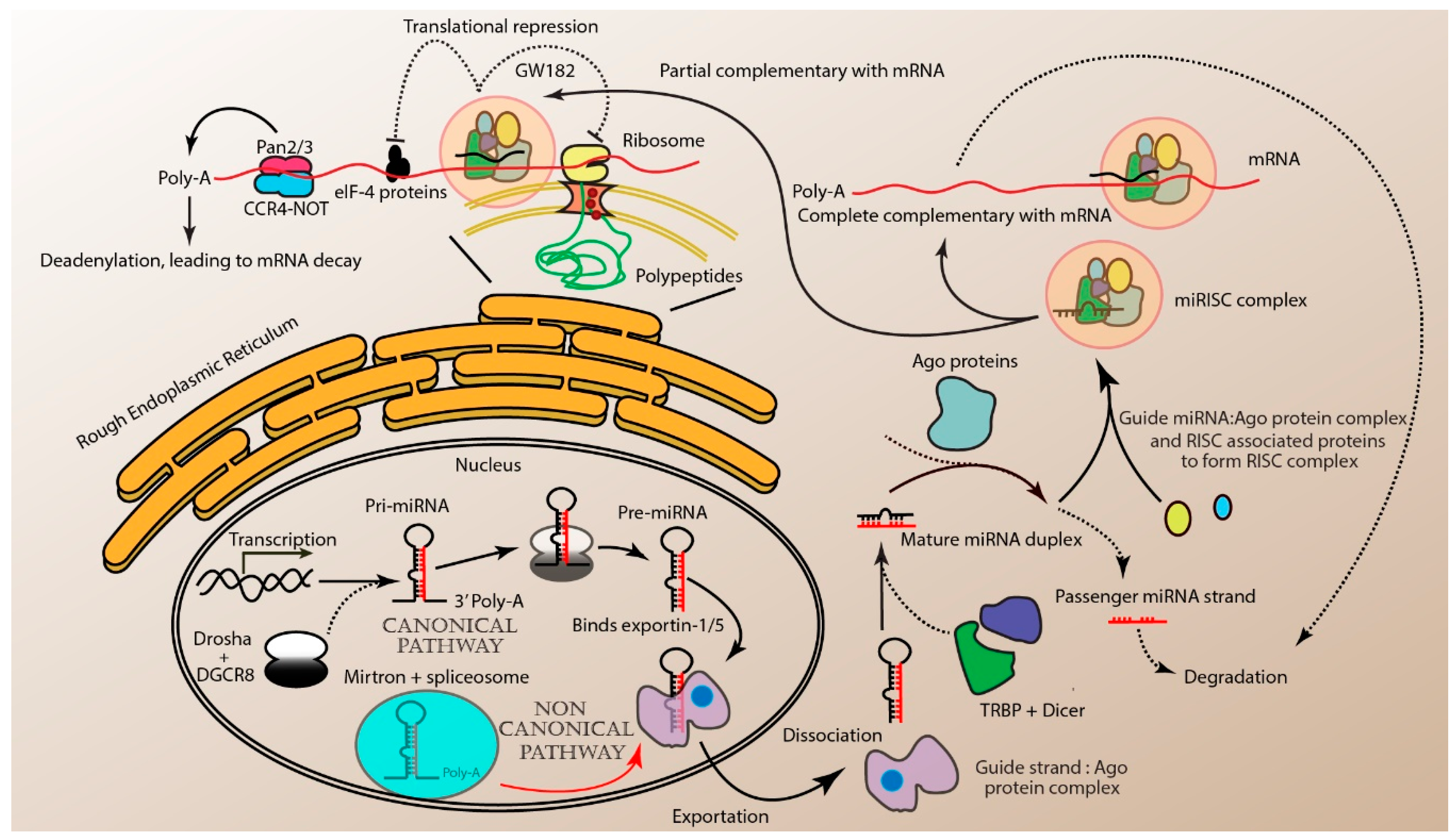
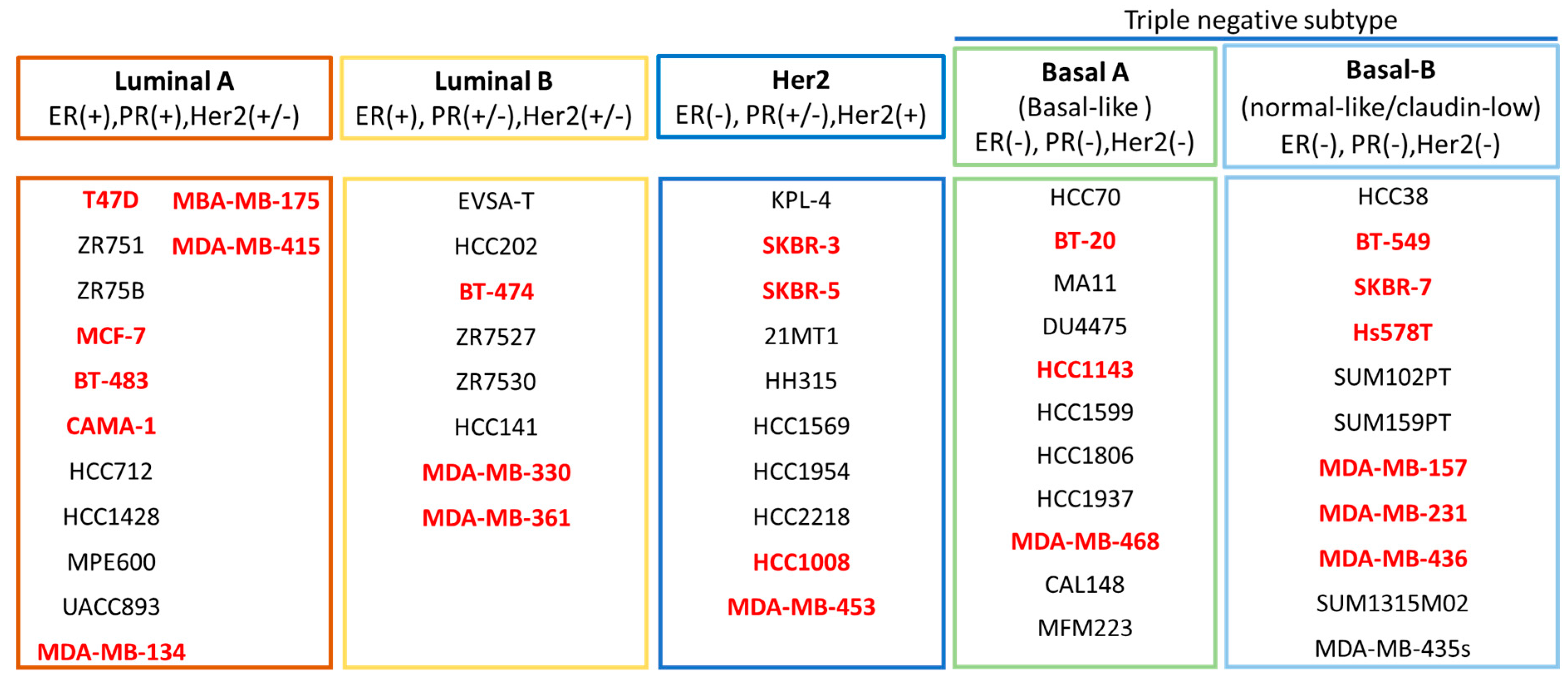
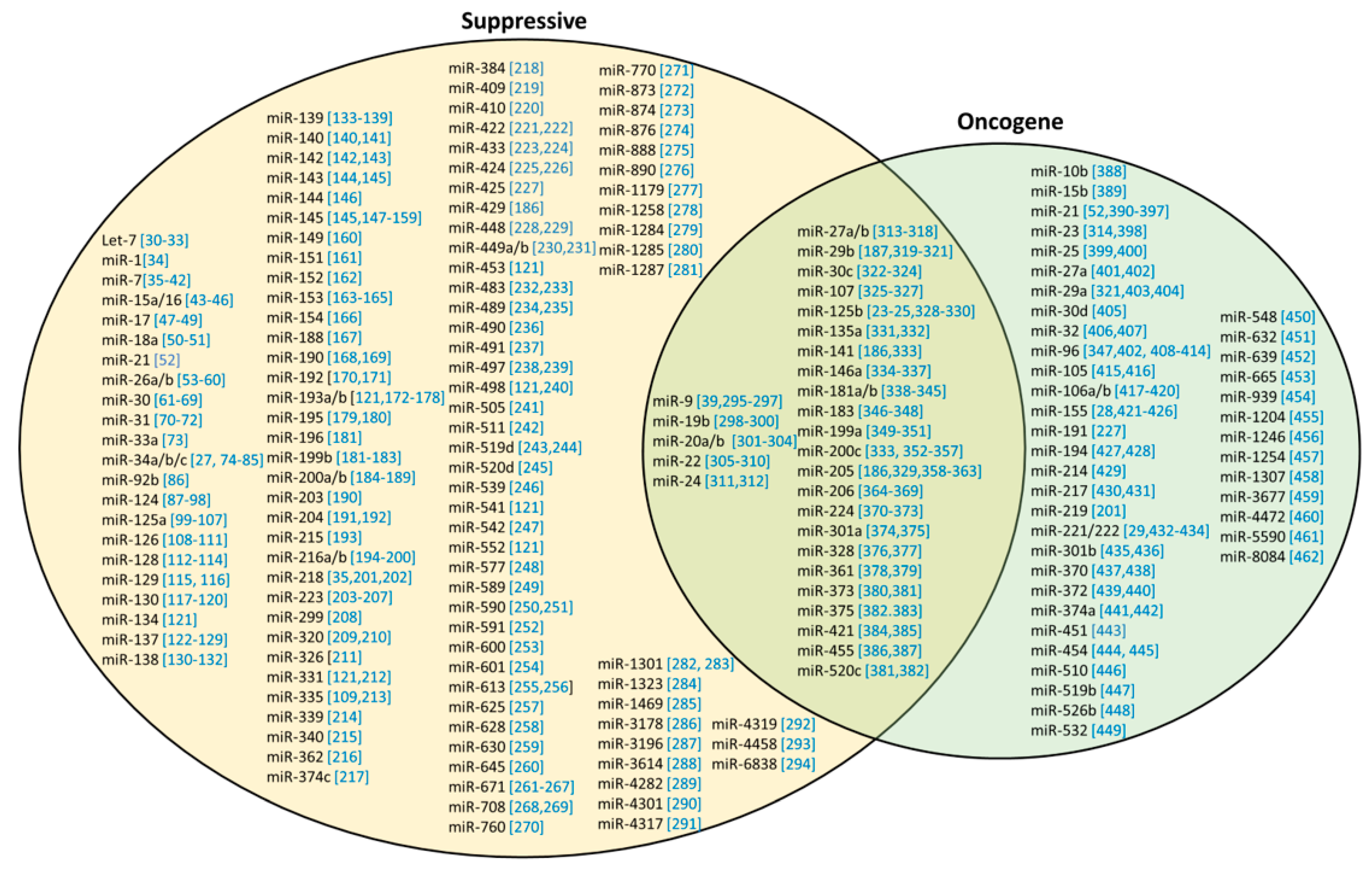
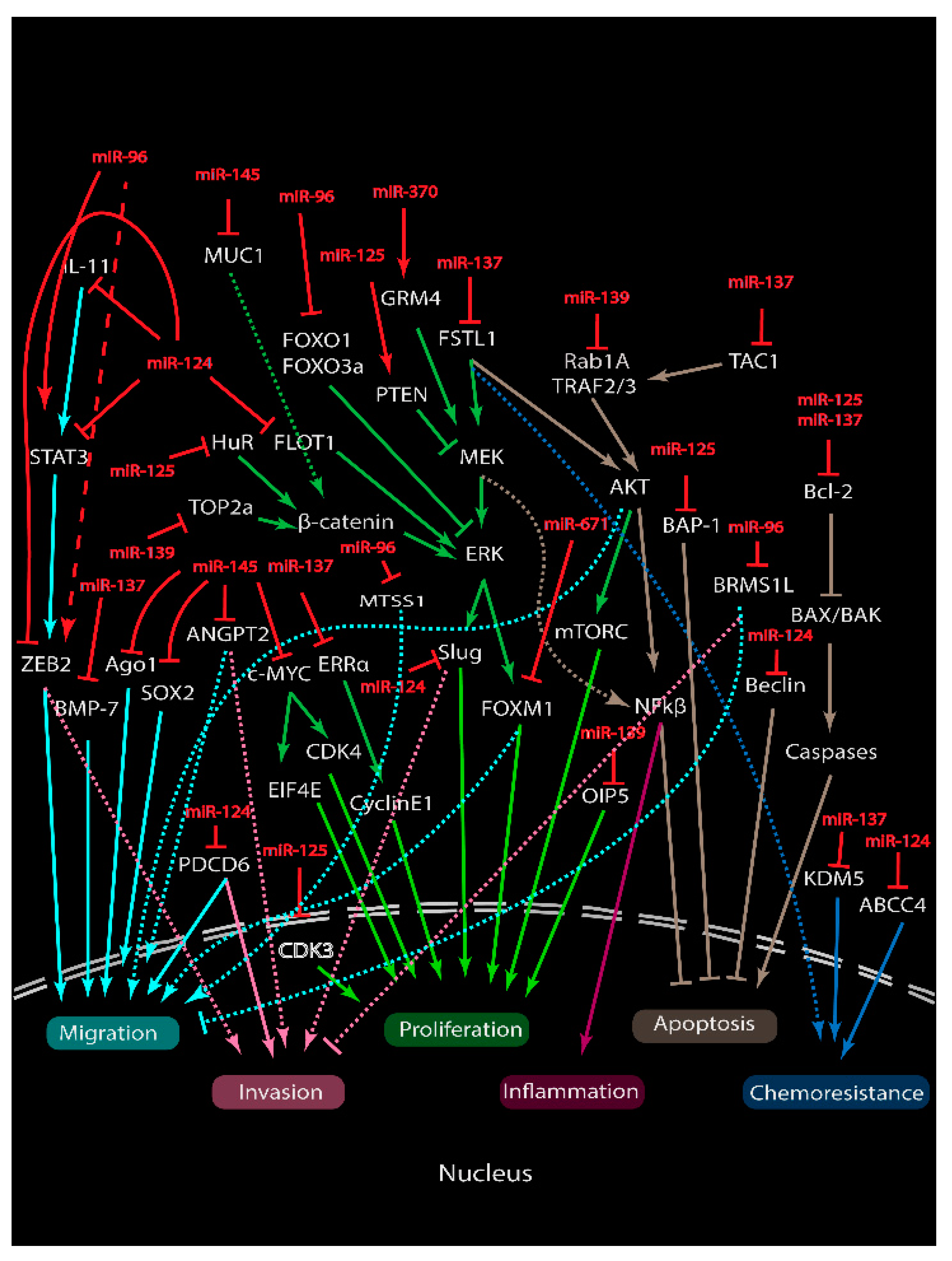
Table 1.
Binding partners of miRNAs in regulating oncogenesis.
| Function | miRNA | Target | Effects | Ref. |
|---|---|---|---|---|
| Tumor suppressive (inhibition) | miR-26a | RNF6 | Proliferation | [53] |
| CHD1, GREB1 and KPNA2 | [54] | |||
| Metadherin | Proliferation and metastasis | [55] | ||
| MCL-1 | [56] | |||
| CCNE2 | Sensitize to trastuzumab in Her2+ subtype | [57] | ||
| ERBB2 | Sensitize to tamoxifen in ER+ subtype | [58] | ||
| miR-193a/b | DDAH1 | Angiogenesis | [172] | |
| PTP1B | Proliferation and survival | [173] | ||
| MORC4 | [174] | |||
| WT1 | Proliferation and metastasis | [175] | ||
| RAB22A | [176] | |||
| MCL-1 | Sensitize to doxorubicin | [177] | ||
| uPA | Proliferation, cell invasion and metastasis | [178] | ||
| miR-216a/b | TLR4 | Suppresses stemness and the release of soluble factors associated with cancer-associated fibroblast activation | [194] | |
| PKCα | Survival and migration | [195] | ||
| PAK2 | Proliferation and metastasis | [196] | ||
| SDCBP | [197] | |||
| P2X7R | Proliferation and survival | [198] | ||
| HDAC8 | Proliferation and colony formation | [199] | ||
| miR-223 | Caprin-1 | Proliferation and invasion | [203] | |
| STIM1 | [204] | |||
| HAX-1 | Survival and sensitize to doxorubicin and cisplatin | [205] | ||
| STAT5A | Survival and sensitize to paclitaxel | [206] | ||
| EGF | Proliferation and sensitize radiation-treated breast cancer cells to lapatinib | [207] | ||
| Oncogenic (promotion) | miR-1246 | CCNG2 | Proliferation, migration, chemoresistance to docetaxel, epirubicin and gemcitabine | [456] |
Abbreviations: Her2: human epidermal growth factor receptor 2; ER: estrogen receptor.
Table 2.
Different types of miRNA-based therapies.
| Treatment | Modes | Methods | miRNA | Types of Studies In Vivo/In Vitro | Ref |
|---|---|---|---|---|---|
| miRNA inhibition therapy | AMOs | 2-OMe | miR-451 | In vitro (MCF-7, SKBR3) In vivo (BALB/c) | [443] |
| miR-21 | In vitro (MCF-7 and HeLa) | [465] | |||
| Gene editing | Crispr-cas9 | miR-23b, miR-27b | In vitro (MCF7) In vivo (nude) | [314] | |
| miRNA replacement therapy | Virus | Lentivirus | miR-126, miR-130a | In vitro (MDA-MB-231) In vivo (PyMT, C57Bl/6) | [466] |
| Adeno-associated virus | miR-1d | In vitro (HEK293T and HeLa) in vivo (PyMT) | [467] | ||
| Retroviral | miR-21 | In vitro (SKBR3, MCF7, Jurkat, HEK-293T) In vivo (nude) | [468] | ||
| Nanoparticle | Gold (PLL) | mir-708 | In vitro (MDA-MB-231, HEK-293, 4T1) In vivo (CB-17 SCID and CB17.Cg-PrkdcscidHrhr/IcrCrl) | [469] | |
| Lipid | miR-203 | In vitro (MDA-MB-231 and Hs578t cells) | [470] | ||
| miR-203 | In vitro (MDA-MB 231) In vivo (BALB/c) | [471] | |||
| Polymer | Creatine | mir-34a | In vitro (4T1.2 and MDA-MB-231) In vivo (BALB/c) | [472] |
Abbreviations: 2-OME: 2’-O-methylation; AMOs: anti-miR oligonucleotides; PPL: positively charged poly-L-lysine.
© 2020 by the authors. Licensee MDPI, Basel, Switzerland. This article is an open access article distributed under the terms and conditions of the Creative Commons Attribution (CC BY) license (http://creativecommons.org/licenses/by/4.0/).
Share and Cite
MDPI and ACS Style
Wong, J.S.; Cheah, Y.K. Potential miRNAs for miRNA-Based Therapeutics in Breast Cancer. Non-Coding RNA 2020, 6, 29. https://0-doi-org.brum.beds.ac.uk/10.3390/ncrna6030029
AMA Style
Wong JS, Cheah YK. Potential miRNAs for miRNA-Based Therapeutics in Breast Cancer. Non-Coding RNA. 2020; 6(3):29. https://0-doi-org.brum.beds.ac.uk/10.3390/ncrna6030029
Chicago/Turabian StyleWong, Jun Sheng, and Yoke Kqueen Cheah. 2020. "Potential miRNAs for miRNA-Based Therapeutics in Breast Cancer" Non-Coding RNA 6, no. 3: 29. https://0-doi-org.brum.beds.ac.uk/10.3390/ncrna6030029
Note that from the first issue of 2016, this journal uses article numbers instead of page numbers. See further details here.