Article

Effect of Drying Methods on Phenolic Compounds and Antioxidant Activity of Urtica dioica L. Leaves

1
Grupo Tecnico Nacional de Plantas Medicinales de la Asociacion Cubana de Tecnicos Agricolas Agroforestales (ACTAF), Calle 98, Esquina 7ma, Playa, La Habana 11300, Cuba
2
Department of Agriculture, Food and Environment, University of Pisa, 56124 Pisa, Italy
3
Department of Biological and Environmental Sciences and Technologies, University of Salento, 73100 Lecce, Italy
4
Interdepartmental Research Center Nutrafood “Nutraceuticals and Food for Health”, University of Pisa, Via del Borghetto 80, 56124 Pisa, Italy
*
Author to whom correspondence should be addressed.
Submission received: 13 December 2020 / Revised: 10 January 2021 / Accepted: 14 January 2021 / Published: 19 January 2021
(This article belongs to the Special Issue Nutritional and Antioxidant Value of Horticulturae Products)

Abstract

:
Stinging nettle (Urtica dioica) is a plant well known in traditional medicine for its many beneficial properties, but the lack of standardization regarding the product to offer to consumers limits its diffusion. To this end, drying appears to be a useful technique to offer a low-cost product that can be stored for long time, but the different drying procedures may give rise to end-products of very different quality as nutraceutical and antioxidant compounds. Nettle leaves have been dehydrated employing freeze-drying (FD), oven-drying (OD) or heat pump drying (HPD) and compared with fresh leaves following water extraction to emulate the use by final consumers. Results indicate that the best dehydration technique is HPD, which apparently gives rise to more than a doubling of total phenols and antioxidant activity in the extract compared to the water extract obtained from fresh leaves but a reduction in the level of ascorbic acid of about 39%. In addition, the content of some phenolic compounds is 10 to over a hundred times higher in the extract after HPD than that obtained from fresh samples. This confirms that the dehydration technique should be tuned in relation to the compounds of greatest interest or value.

Graphical Abstract

1. Introduction

Urtica dioica L., commonly named stinging nettle, is an herbaceous perennial plant belonging to the family of Urticaceae and native of Eurasia. Its medicinal properties are well known, with a wide historical background of the use of its stems, leaves and roots [1]. Recent reviews by Kregiel et al. [2] and Grauso et al. [3] revealed the isolation by stinging nettle leaves of some important phenolic compounds such as caffeic acid, hydroxybenzoic acid, vanillic acid, coumaric acid, gentisic acid, protocatechuic acid, gallic acid, syringic acid, quinic acid and many caffeic and quinic acid derivatives, quercetin, catechin, pelargonidin and apigenin in both the glycosidic and non-glycosidic forms, performed by other authors. The stinging nettle leaves also revealed the presence of important compounds belonging to the terpenoid and carotenoid classes such as 3-oxo-a-ionol, 3-hydroxy-damascone, 3,5,5-trimethyl-4-(1-oxo-2-buten-1-yl)-3-cyclohexen-1-one, 4-(4-hydroxy-2,6,6-trimethyl-1-cyclohexen-1-yl)-3-buten-2-one, 4-(3-hydroxy-1-butyn-1-yl)-3,5,5-trimethyl-2-cyclohexen-1-ol and 3-hydroxy-5,6-epoxy-b-ionol as well as lutein, violaxanthin, neoxanthin, lycopene and β-carotene. Thanks to the phytochemical composition of stinging nettle leaves, this species has traditionally been used to counteract cardiovascular diseases, especially hypertension, but also for its anti-microbial, anti-inflammatory, antirheumatic and acute diuretic effects [2].
Understanding of the molecular mechanisms underlying the nutraceutical effects may open a new horizon for new therapeutic strategies [1].
Natural products, both pure compounds and standardized plant extracts, offer unlimited opportunities as drug sources due to the unequaled availability of chemical diversity. The leaf extract of stinging nettle was one of the herbal remedies for which experimental and clinical trials have complemented each other [1].
Owing to the growing demand for higher productivity of stinging nettle and, at the same time, for higher product quality and lower operating costs, as well as reduced environmental burden, drying technologies have drawn extensive concern. Indeed, oven-drying (OD), the most traditional method, is widely used nowadays for post-harvest processing and storage of a large variety of plant products. However, its long drying time and the use of high temperatures usually decrease product quality because of the degradation of nutritional and nutraceutical compounds, color and flavor. The OD process also has high energy consumption, low efficiency and low productivity. Conversely, the freeze-drying (FD) method may ensure the preservation of the most thermolabile compounds in raw materials, yielding a dried product with high quality. Some authors have studied the quality of plant material following these drying methods, reporting a higher efficiency of the FD method as compared with OD in terms of retention of phenolic compounds, antioxidant activity and other beneficial properties [4,5,6]. A new drying technology named heat pump drying (HPD), consisting of a constant airflow passage in a sealed environment, is increasingly being used for plants and food because it is an energy-efficient and economically feasible drying device for the production of high-quality dried foods and biomaterials [7]. In an example of this process, the temperature was kept at or below 35 °C. In the first passage of air in the dryer, the air had a low moisture and a constant temperature; then, the same air was cooled, and at this step, the moisture was represented by the sum of the moisture of the air and of the leaves. Subsequently, the same air was heated again and sent back to the main chamber to meet new U. dioica leaves to dry [8]. Some authors observed that HPD was more effective in the preservation of phenolic compounds, volatile compounds, ascorbic acid and antioxidant activity in plant materials or fruits than OD and FD [9,10,11]. However, little information about HPD is available in the literature.
The duration and the temperature of the drying process are the most important factors affecting the chemical, nutraceutical and organoleptic quality of dehydrated plant products, and the selection of the optimal drying method depends on quality requirements, the characteristics of the raw material and the market price of the final product [6].
Therefore, an evaluation of the main classes of nutraceutical compounds at the time of storage could help consumers and producers to understand the potential of this species to be marketed in the most appropriate way. Given all of the above, the objective of this study was to evaluate the effects of three different drying methods on the content of phenolic compounds and ascorbic acid and the antioxidant activity of fresh and dried leaves of U. dioica collected in the wild. Leaves were dried with different methods: freeze-drying (FD), oven-drying (OD) and heat pump drying (HPD).

2. Materials and Methods

2.1. Material Preparation
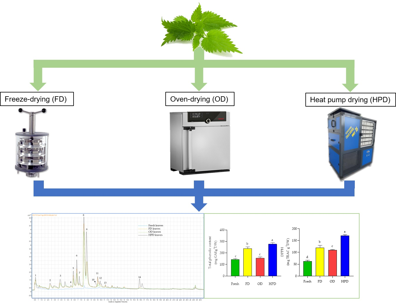
Samples from the species U. dioica L. were collected as wild during July 2020 in the “Il Corniolo” farmhouse, located in Castiglione di Garfagnana, Lucca Province, north of Tuscany, Italy (approximately 400 m. a. s. l). Plants with a similar height and leaf number were randomly sampled with a cut approximately 5 cm above the soil. Samples were placed in damp plastic bags to prevent tissue dehydration and transported to the laboratory within 1–2 h. Once in the laboratory, healthy leaves with uniform color, dimensions and texture were detached from the plants and their petioles were removed. Five pooled samples were prepared with the leaves collected from 20 individual plants; afterwards, sub-samples were analyzed fresh or after dehydration with FD, OD or HPD. Sub-samples of fresh plant material were frozen in liquid nitrogen and stored at −80 °C while dried sub-samples were stored at room temperature until laboratory analyses, which were performed within one month from the harvest.

2.2. Leaf Drying

Leaves were freeze-dried (FD) using a standard unheated chamber of dimensions 215 mm Ø × 300 mm height (Telstar LyoQuest-55, Milan, Italy) at a vacuum pressure of 100 Pa and a final condenser temperature of −55 °C until the plant material reached a constant weight (2 days), determining, in this way, the dry weight (DW) of FD leaves. Oven-drying (OD) was performed at 105 °C using a laboratory electric thermostatic oven (Memmert GmbH + Co. KG Universal Oven UN30, Schwabach, Germany) until the plant material reached a constant weight (2 days), determining the DW of OD leaves. An apparatus manufactured by North West Technology, Cuneo, Italy (model NWT-35), was used for heat pump drying (HPD) at a temperature below 35 °C until the plant material reached 15% moisture (2 days for U. dioica leaves). The DW of HPD-treated and fresh leaves was determined using the same thermostatic electric oven used for the OD treatment at 60 °C until constant weight.

2.3. Extraction Preparation for Total Phenolic Content and Antioxidant Activity Assays

Leaf samples (1 g) were homogenized in 4 mL of an 80% (v/v) aqueous solution of methanol, sonicated with a sonicator (Digital ultrasonic Cleaner, DU-45, Argo Lab, Modena, Italy) for 30 min and then centrifuged with a laboratory centrifuge (MPW 260R, MWP Med. instruments, Warsaw, Poland) at 10,000× g for 15 min at 4 °C. Supernatant was stored at −80 °C until analysis.

2.4. Total Phenolic Content

Total phenolic content was determined according to Dewanto et al. [12] with minor modifications. Extracted samples (10 μL) were added to a solution of 115 μL deionized water and 125 μL Folin–Ciocalteau reagent. Blank solution was utilized with 10 μL distilled water. Samples were stirred for 6 min and then 1.25 mL 7% (w/v) Na2CO3 was added to stop the reaction. Samples were incubated for 90 min at room temperature and the increase in the absorbance at 760 nm was measured against the blank solution with an Ultrospec 2100 Pro spectrophotometer (GE Healthcare Ltd., Little Chalfont, UK). Gallic acid was used as a standard and the results (even for fresh leaves) were expressed as mg gallic acid equivalents per g DW (mg GAE g−1 DW).

2.5. 2,2-Diphenyl-1-Picrylhydrazyl Hydrate (DPPH) Free Radical Scavenging Assay

The 2,2-diphenyl-1-picrylhydrazyl hydrate (DPPH) free radical scavenging capacity of each sample was determined according to Brand-Williams et al. [13]. Briefly, 2–10 µL of extract was added to a 3.12 × 10⁻⁵ M solution of DPPH in methanol:water 80:20 (v/v) to a final volume of 1 mL. The change in absorbance at 515 nm was measured after 30 min of incubation. The antioxidant activity based on the DPPH free radical scavenging ability of the extract (even for fresh leaves) was expressed as mg of Trolox equivalent antioxidant capacity (TEAC) per g DW.

2.6. Ferric Reducing Antioxidant Power (FRAP) Assay

The ferric reducing ability was measured following the method described by Benzie and Strain [14] with some modifications. The ferric reducing antioxidant power (FRAP) reagent was prepared mixing 10 mM 2,4,6-tris(2-pyridyl)-1,3,5-triazine (TPTZ) in 40 mM HCl solution with an identical volume of 20 mM FeCl3 and with ten volumes of 0.3 M acetate buffer (pH 3.6). The mixture was warmed at 37 °C for five minutes prior to the analysis; then, 1 mL of FRAP reagent was mixed with 25–50 μL of sample extract and with distilled water at 37 °C to a final volume of 2 mL. Then, the mixture was incubated at 37 °C for 30 min and absorbance was read at 593 nm. All results were expressed in mg Trolox equivalents per g DW (mg TEAC g−1 DW).

2.7. Extracion of Phenolic Compounds

The extraction of phenolic compounds was performed according to Zhou et al. [15] at a ratio of 1:20 (w/v). Water extraction was preferred because it has no negative impact on health and the environment. Briefly, the samples were finely ground in a mortar and then extracted with water at 100 °C at reflux for 20 min; they were then centrifuged for 20 min at 5000× g, and the supernatant was filtered at 0.45 μm with a polytetrafluoroethylene filter before HPLC analysis.

2.8. Phenolic Compounds’ Characterization

Phenolic characterization and quantification on leaf extracts were carried out using an Agilent 1200 High Performance Liquid Chromatography (HPLC) System (Agilent Technologies, Palo Alto, CA, USA) equipped with a standard autosampler and an Agilent Zorbax Extend-C18 analytical column (5 × 2.1 cm, 1.8 µm), as reported by Negro et al. [16]. The HPLC system was coupled to an Agilent diode-array detector (wavelength 280 and 365 nm) and an Agilent 6320 TOF mass spectrometer. Detection was carried out within a mass range of 50−1700 m/z. Accurate mass measurements of each peak from the total ion chromatograms (TICs) were obtained by means of an ISO Pump (Agilent G1310B) using a dual nebulizer electrospray ionization source that introduces a low flow (20 µL min−1) of a calibration solution containing the internal reference masses at m/z 112.9856, 301.9981, 601.9790 and 1033.9881, in negative ion mode. The compounds were quantified using calibration curves of authentic standards (caffeic acid and rutin). Standards were solubilized in methanol:water 80:20 (v/v) and diluted with water:formic acid 99.9:0.1 (v/v) at a final concentration of 0.5–10 µg/mL.

2.9. Total Ascorbic Acid Content

Ascorbic acid content was measured spectrophotometrically using the method described by Kampfenkel et al. [17] with some modifications. Extractions were carried out with the homogenization of 0.3 g fresh material with 1 mL 6% (v/v) trichloroacetic acid followed by centrifuging for 10 min at 10,000× g at 4 °C. Immediately after extraction, the analysis was performed by adding 50 μL supernatant to 50 μL 10 mM dithiothreitol (DTT) and to 100 μL 0.2 M Na-P buffer (pH 7.4). Samples were stirred and incubated for 15 min at 42 °C in a water bath. Then, 50 μL 0.5% (w/v) N-ethylmaleimide (NEM) was added and samples were stirred again. After 1 min of stirring, 250 μL 10% (v/v) trichloroacetic, 200 μL 42% (w/v) orthophosphoric acid, 200 μL 4% (w/v) 2,2′-bipyridine (diluted in 70% (v/v) ethanol 70% (v/v)) and 100 μL 3% (w/v) FeCl3 were added to samples. The increase in absorbance at 525 nm was measured against a blank solution (with 6% (v/v) trichloroacetic acid instead of supernatant) after 40 min of incubation at 42 °C in a water bath. All results were expressed as mg ascorbic acid per g DW (mg g−1 DW).

2.10. Statistical Analysis

To determine the effect of different drying methods, a one-way ANOVA was performed using GraphPad software (GraphPad, La Jolla, CA, USA). Bartlett’s test was used to verify the normality of the data. Mean values ± standard deviation (SD) of 5 replicates for each treatment were compared using the least significant difference (LSD) test at p = 0.05.

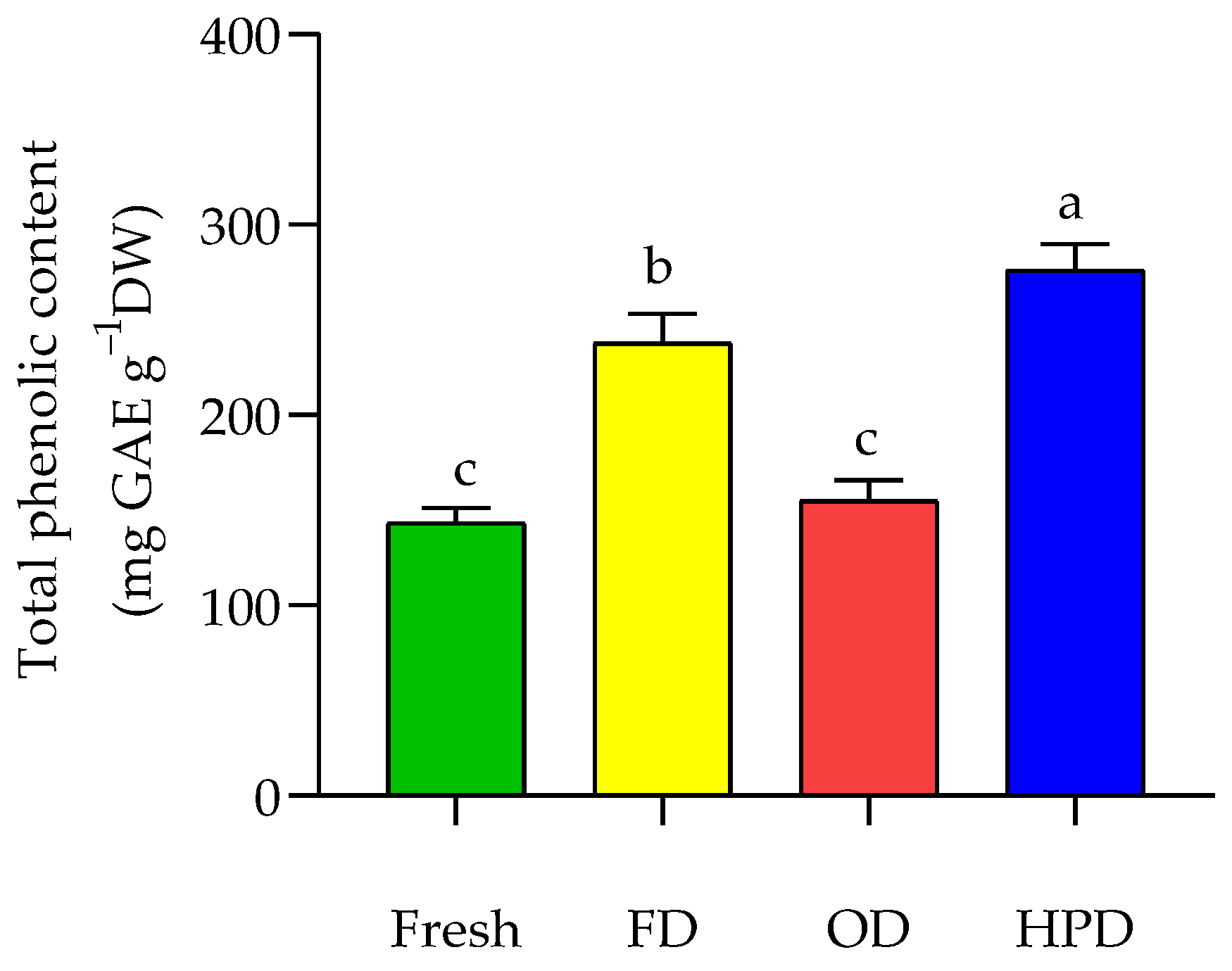
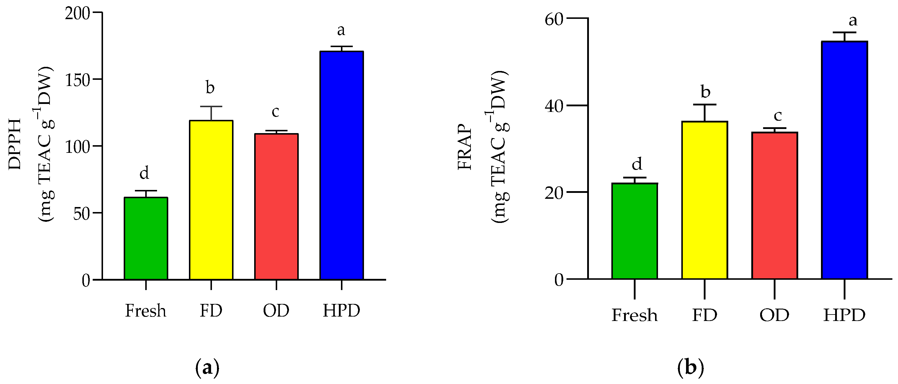
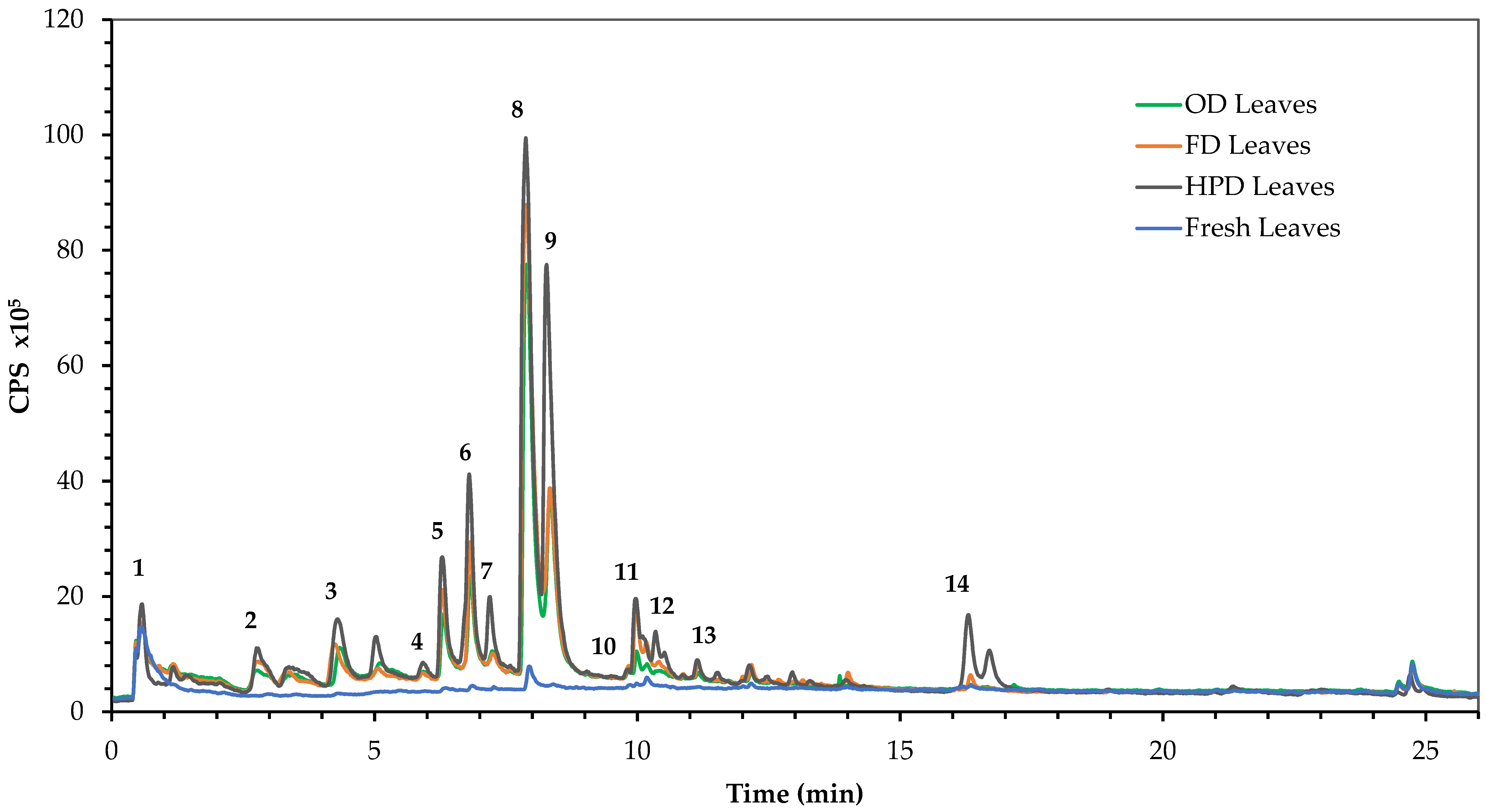
3. Results and Discussion

Temperature and duration of treatment are the determining factors for the selection of the most efficient drying method to retain phenolic compounds and ascorbic acid in plant materials [18,19]. Indeed, specific variations of temperature in the different drying methods may protect against the degradation of these components, leading to the maintenance or the enhancement of the product quality of the analyzed plant material.
In this study with U. dioica leaves, the total phenolic content and antioxidant activity (as analyzed with both DPPH and FRAP assays) were significantly higher in HPD-dried leaves than in fresh leaves and in those dried with FD and OD (Figure 1 and Figure 2). Then, the FD-dried leaves had higher phenolic content and antioxidant activity than the OD-dried leaves, whilst the lowest values of these quantities were found in fresh material (Figure 1 and Figure 2).
The higher total phenolic content of HPD leaves compared to FD or OD leaves could be linked to a more efficient extraction of the insoluble phenolic compounds from the dried or fresh leaves—for instance, phenolic acids or condensed tannins [20,21,22] linked to cell wall polysaccharides or, more specifically, proteins [23,24]. Indeed, the moderate heat treatment (≤35 °C) used by the HPD may have determined the cleavage of the phenolic sugar glycosidic bonds and the formation of phenolic aglycons, which react better with the Folin–Ciocalteu reagent, thus leading to higher values of the total phenolic content [24]. Conversely, the higher (105 °C) temperature in the OD method probably led to the degradation of some phenolic compounds and a more efficient water extraction of phenolic compounds as well as the cleavage of the phenolic sugar glycosidic bonds.
On the other hand, FD resulted to be an intermediate drying method in terms of maintenance of the phenolic compounds or even an enhancement of their extraction from the leaves. In fact, this method involves the formation of small ice crystals inside the cell and their rapid expulsion from the cell during the freezing [6,23]. The rapid expulsion of the ice crystals allows the maintenance of the cell structures and, therefore, the retention of phenolic compounds and vitamins, and the total expulsion of humidity allows a long preservation of plant samples. These characteristics led to the common use of FD as the extraction pre-treatment of plant materials under analysis [25,26]. In the present study, the content of the analyzed phytochemicals was similar in FD leaves and in fresh leaves. The reason for the lower recovery of phenolic compounds from fresh leaves than that from FD leaves remains to be elucidated in further research.
Nevertheless, the phenol content found in fresh U. dioica leaves (Figure 1) is in agreement with previous findings [27,28]. For example, Shonte et al. [28] reported 118.4 mg GAE g−1 DW in fresh stinging nettle leaves and 121.5 and 128.7 mg GAE g−1 DW in FD and OD leaves, respectively. In contrast, lower values (79 mg GAE g−1 DW) were found by Carvalho et al. [29] in wild stinging nettle leaves from the Serra de Estrela region in Portugal and by Hudec et al. [30] in stinging nettle leaves (5.38 mg GAE g−1 DW) of cultivated plants in the area of Nitra, in Slovakia. The differences in the phenolic contents found in this study and by other authors may be due to genetic differences between the plants used and growing conditions.
Similar to the total phenolic content results, the values of antioxidant activity resulted higher in the HPD leaves when compared with fresh, OD and FD samples (Figure 2a,b). The higher antioxidant activity in HPD leaves with respect to the other leaves could be due to different reasons: (i) release of bound antioxidant substances (such as phenolic compounds) by disrupting the cell structure and by obtaining a natural extraction for the analysis [31]; (ii) formation of new antioxidants such as Maillard reaction products (melanoidins) resulting from thermal chemical reaction; (iii) suppression of oxidation by thermal inactivation of oxidative enzymes of antioxidants such as polyphenol oxidase [32]. Indeed, a heat treatment at moderate temperature (50 °C for 5 h) inactivated polyphenol oxidases in extracts from aerial parts of Phyllanthus amarus [32]. However, the HPLC analysis did not reveal the presence of different phenolic compounds in HPD leaves with respect to the other treatments (Figure 3 and Table 1).
The temperature (105 °C) used in OD leaves apparently resulted in too high of an increase in the release of phytochemicals, even though we observed a slightly higher-level small increase in antioxidant activity in comparison to fresh leaves (Figure 2a,b).
Table 1 shows the HPLC analysis of the stinging nettle leaves under investigation, confirming the total phenolic compounds and antioxidant activities data (Figure 1 and Figure 2). The qualitative analysis mainly shows the presence of some caffeic acid isomers, both free and combined with quinic or malic acid, and some flavonoids such as rutin, isoquercetin and isorhamnetin rutinoside [20,21,22].
The most abundant phenolic acid was the caffeoylmalic acid dimer, with values between 19.20 and 35.81 mg g−1 DW for OD and HPD samples, respectively. In addition, the isomers of caffeoylquinic acid were more abundant in the HPD sample, with 20.73 mg g−1 DW, twice the value obtained from the OD sample (10.76 mg g−1 DW), thus confirming that the HPD technique preserves more phenolic compounds. Regarding flavonoids, the most representative one was rutin, whose quantity in the HPD sample reached 2.25 mg g−1 DW. The values obtained were coherent with those reported in the literature. In fact, Grevsen et al. [33] reported amounts of caffeoylmalic acid approximately between 7 and 22 mg g−1 DW and caffeoylquinic acid values between 3 and 18 mg g−1 DW in different accessions of U. dioica. Besides, Orčić et al. [34] reported a content of caffeoylquinic acid variable between 1.2 and 28 mg g−1 DW, while Vajić et al. [35] showed amounts of caffeoylmalic acid up to 1.03 mg g−1 DW in water extract and of 1.64 mg g−1 DW in 50% methanol extract. Orčić et al. [34] indicated rutin content between 0.002 and 4.6 mg g−1 DW, and Grevsen et al. [33] found contents up to 14.5 mg g−1 DW, depending on the harvest time. Virtually all samples showed very high levels after HPD compared to fresh leaves; interestingly, caffeoylmalic acid and its dimer were particularly enriched in HPD samples, up to 42 and 136 times, respectively, while only isorhamnetin rutinoside did not show an increment after dehydration, showing a slight reduction for FD and OD samples and a slight increase for HPD leaves. As discussed previously, this phenomenon deserves further investigation.
While all drying methods increased the total phenolic content and the antioxidant capacity of stinging nettle leaves, the total ascorbic acid content was significantly lower in FD (−36%), OD (−55%) and HPD (−39%) leaves than in fresh leaves (Figure 4). As reported before, FD is usually used as an extraction pre-treatment in plant materials during vitamin and phenolic content analyses. Nevertheless, in the present study, this drying technique resulted in adverse retention of ascorbic acid with a decrease of 36% compared to fresh leaves. However, these findings agree with those reported by Shonte et al. [30]. These authors found a loss of 12% and 22% in FD and OD stinging nettle leaves, respectively, with values of 14.2, 11.8 and 9.9 mg g−1 DW in fresh, FD and OD leaves, respectively. Some studies reported that the loss of total ascorbic acid in dried food products can range from 10% to more than 50% depending on drying temperature [28,36]. This loss is mainly due to chemical degradation involving the oxidation of ascorbic acid to dehydroascorbic acid, followed by hydrolysis to 2,3-diketogulonic acid and subsequent polymerization to form other nutritionally inactive products [37].

4. Conclusions

The presented data clearly indicate that the best dehydration technique is HPD since the resulting aqueous extract exhibited a high content in total phenols and a high antioxidant activity, as well as considerable levels in some phenolic compounds such as caffeoylmalic acid and caffeoylmalic acid dimer. Besides, the OD technique resulted in a high reduction in the ascorbic acid content (55%), and a lower decrease in this antioxidant compound was found when using the FD and HPD techniques compared with the reduction showed using the OD technique. Therefore, all three dehydration techniques appear to be efficient in favoring the extraction of phenols in water but adverse for ascorbic acid. Few studies have reported the quantification of total ascorbic acid in stinging nettle leaves in the literature; thus, this study can implement the knowledge of the quantification of this bioactive compound as well as show the loss of total ascorbic acid after the use of drying methods on plant leaves.

Author Contributions

Conceptualization, L.I. and A.P.; methodology, C.N. and L.M.G.; validation, C.C. and L.M.G.; formal analysis, C.N. and L.M.G.; investigation, C.N. and L.M.G.; resources, L.G. and A.P.; data curation, C.C.; writing—original draft preparation, L.M.G. and C.C.; writing—review and editing, L.D.B., A.P., L.I. and L.G.; supervision, L.G. and A.P.; project administration, L.G. All authors have read and agreed to the published version of the manuscript.

Funding

This research was performed in the framework of the “Erbi Boni” project, funded by FEASR Reg. UE 1305/2013—PSR 2014/2020—Regione Toscana Mis. 19—GAL MontagnAppennino Mis. 16.2.

Institutional Review Board Statement

Not applicable.

Informed Consent Statement

Not applicable.

Data Availability Statement

Data is contained within the article.

Acknowledgments

The authors are grateful to Diletta Piccotino for the useful help in the analysis and to Franca Bernardi, the owner of “Il Corniolo” farmhouse, for her availability and her valuable help during the sampling and the dehydration.

Conflicts of Interest

The authors declare no conflict of interest.

References

  1. Dhouibi, R.; Affes, H.; Salem, M.B.; Hammami, S.; Sahnoun, Z.; Zeghal, K.M.; Ksouda, K. Screening of pharmacological uses of Urtica dioica and others benefits. Prog. Biophys. Mol. Biol. 2020, 150, 67–77. [Google Scholar] [CrossRef]
  2. Kregiel, D.; Pawlikowska, E.; Antolak, H. Urtica spp.: Ordinary plants with extraordinary properties. Molecules 2018, 23, 1664. [Google Scholar] [CrossRef] [PubMed] [Green Version]
  3. Grauso, L.; de Falco, B.; Lanzotti, V.; Motti, R. Stinging nettle, Urtica dioica L.: Botanical, phytochemical and pharmacological overview. Phytochem. Rev. 2020, 19, 1341–1377. [Google Scholar] [CrossRef]
  4. Coklar, H.; Akbulut, M.; Kilinc, S.; Yildirim, A.; Alhassan, I. Effect of freeze, oven and microwave pretreated oven drying on color, browning index, phenolic compounds and antioxidant activity of hawthorn (Crataegus orientalis) fruit. Not. Bot. Horti Agrobot. Cluj-Napoca 2018, 46, 449–456. [Google Scholar] [CrossRef] [Green Version]
  5. An, K.; Zhao, D.; Wang, Z.; Wu, J.; Xu, Y.; Xiao, G. Comparison of different drying methods on Chinese ginger (Zingiber officinale Roscoe): Changes in volatiles, chemical profile, antioxidant properties, and microstructure. Food Chem. 2016, 197, 1292–1300. [Google Scholar] [CrossRef] [PubMed]
  6. Meng, Q.; Fan, H.; Li, Y.; Zhang, L. Effect of drying methods on physico-chemical properties and antioxidant activity of Dendrobium officinale. J. Food Meas. 2018, 12, 1–10. [Google Scholar] [CrossRef]
  7. Perera, C.O.; Shafiur, R.M. Heat pump dehumidifier drying of food. Trends Food Sci. Technol. 1997, 8, 75–79. [Google Scholar] [CrossRef]
  8. Ong, S.P.; Law, C.L. Drying Kinetics and Antioxidant Phytochemicals Retention of Salak Fruit under Different Drying and Pretreatment Conditions. Dry. Technol. 2011, 29, 429–441. [Google Scholar] [CrossRef]
  9. Phoungchandang, S.; Wichayawadee, S.; Borwonsak, L. Kaffir lime leaf (Citrus hystric DC.) drying using tray and heat pump dehumidified drying. Dry. Technol. 2008, 26, 1602–1609. [Google Scholar] [CrossRef]
  10. Hawlader, M.N.A.; Perera, C.O.; Tian, M.; Yeo, K.L. Drying of guava and papaya: Impact of different drying methods. Dry. Technol. 2006, 24, 77–87. [Google Scholar] [CrossRef]
  11. Niruthchara, S.; Srzednicki, G.; Craske, J. Effects of drying treatments on the composition of volatile compounds in dried nectarines. Dry. Technol. 2007, 25, 877–881. [Google Scholar]
  12. Dewanto, V.; Adom, K.K.; Liu, R.H. Thermal processing enhances the nutritional value of tomatoes by increasing total antioxidant activity. J. Agric. Food Chem. 2002, 50, 3010–3014. [Google Scholar] [CrossRef] [PubMed]
  13. Brand Williams, W.; Cuvelier, M.E.; Berset, C. Use of a free radical method to evaluate antioxidant activity. Lebensm. Wiss. Technol. 1995, 28, 25–30. [Google Scholar] [CrossRef]
  14. Benzie, I.F.; Strain, J.J. The ferric reducing ability of plasma (FRAP) as a measure of “antioxidant power”: The FRAP assay. Anal. Biochem. 1996, 239, 70–76. [Google Scholar] [CrossRef] [PubMed] [Green Version]
  15. Zhou, X.; Chan, S.W.; Tseng, H.L.; Deng, Y.; Hoi, P.M.; Choi, P.S.; Or, P.M.; Yang, J.M.; Lam, F.F.; Lee, S.M.; et al. Danshensu is the major marker for the antioxidant and vasorelaxation effects of Danshen (Salvia miltiorrhiza) water-extracts produced by different heat water-extractions. Phytomedicine 2012, 19, 1263–1269. [Google Scholar] [CrossRef]
  16. Negro, C.; Sabella, E.; Nicolì, F.; Pierro, R.; Materazzi, A.; Panattoni, A.; Aprile, A.; Nutricati, E.; Vergine, M.; Miceli, A.; et al. Biochemical Changes in Leaves of Vitis vinifera cv. Sangiovese Infected by Bois Noir Phytoplasma. Pathogens 2020, 9, 269. [Google Scholar] [CrossRef] [Green Version]
  17. Kampfenkel, K.; Van Montagu, M.; Inzé, D. Extraction and determination of ascorbate and dehydroascorbate from plant tissue. Anal. Biochem. 1995, 225, 165–167. [Google Scholar] [CrossRef]
  18. Blank, D.E.; Bellaver, M.; Fraga, S.; Lopes, T.J.; de Moura, N.F. Drying kinetics and bioactive compounds of Bunchosia glandulifera. J. Food Process Eng. 2018, 41, e12676. [Google Scholar] [CrossRef]
  19. López-Vidaña, E.C.; Pilatowsky Figueroa, I.; Cortés, F.B.; Rojano, B.A.; Ocaña, A.N. Effect of temperature on antioxidant capacity during drying process of mortiño (Vaccinium meridionale Swartz). Int. J. Food Prop. 2017, 20, 294–305. [Google Scholar] [CrossRef] [Green Version]
  20. Farag, M.A.; Weigend, M.; Luebert, F.; Brokamp, G.; Wessjohann, L.A. Phytochemical, phylogenetic, and anti-inflammatory evaluation of 43 Urtica accessions (stinging nettle) based on UPLC–Q-TOF-MS metabolomic profiles. Phytochemistry 2013, 96, 170–183. [Google Scholar] [CrossRef]
  21. Komes, D.; Belščak-Cvitanović, A.; Horžić, D.; Rusak, G.; Likić, S.; Berendika, M. Phenolic composition and antioxidant properties of some traditionally used medicinal plants affected by the extraction time and hydrolysis. Phytochem. Anal. 2011, 22, 172–180. [Google Scholar] [CrossRef] [PubMed]
  22. Pinelli, P.; Ieri, F.; Vignolini, P.; Bacci, L.; Baronti, S.; Romani, A. Extraction and HPLC analysis of phenolic compounds in leaves, stalks, and textile fibers of Urtica dioica L. J. Agric. Food Chem. 2008, 56, 9127–9132. [Google Scholar] [CrossRef] [PubMed]
  23. Giada, M.D.L.R. Food phenolic compounds: Main classes, sources and their antioxidant power. Oxidative stress and chronic degenerative diseases—A role for antioxidants. InTech 2013, 87–112. [Google Scholar] [CrossRef] [Green Version]
  24. Singleton, V.L.; Orthofer, R.; Lamuela-Raventós, R.M. Analysis of total phenols and other oxidation substrates and antioxidants by means of Folin-Ciocalteu reagent. Meth. Enzymol. 1999, 299, 152–178. [Google Scholar]
  25. Barros, L.; Dueñas, M.; Carvalho, A.M.; Ferreira, I.C.; Santos-Buelga, C. Characterization of phenolic compounds in flowers of wild medicinal plants from Northeastern Portugal. Food Chem. Toxicol. 2012, 50, 1576–1582. [Google Scholar] [CrossRef] [Green Version]
  26. Martins, N.; Barros, L.; Santos-Buelga, C.; Ferreira, I.C. Antioxidant potential of two Apiaceae plant extracts: A comparative study focused on the phenolic composition. Ind. Crops Prod. 2016, 79, 188–194. [Google Scholar] [CrossRef] [Green Version]
  27. Zeković, Z.; Cvetanović, A.; Švarc-Gajić, J.; Gorjanović, S.; Sužnjević, D.; Mašković, P.; Savic, S.; Radojkovic, M.; Đurović, S. Chemical and biological screening of stinging nettle leaves extracts obtained by modern extraction techniques. Ind. Crops Prod. 2017, 108, 423–430. [Google Scholar] [CrossRef]
  28. Shonte, T.T.; Duodu, K.G.; de Kock, H.L. Effect of drying methods on chemical composition and antioxidant activity of underutilized stinging nettle leaves. Heliyon 2020, 6, e03938. [Google Scholar] [CrossRef]
  29. Carvalho, A.R.; Costa, G.; Figueirinha, A.; Liberal, J.; Prior, J.A.; Lopes, M.C.; Cruz, M.T.; Batista, M.T. Urtica spp.: Phenolic composition, safety, antioxidant and anti-inflammatory activities. Food Res. Intern. 2017, 99, 485–494. [Google Scholar] [CrossRef]
  30. Hudec, J.; Burdová, M.; Kobida, L.U.; Komora, L.; Macho, V.; Kogan, G.; Turianica, I.; Kochanová, R.; Ložek, O.; Habán, M.; et al. Antioxidant capacity changes and phenolic profile of Echinacea purpurea, nettle (Urtica dioica L.), and dandelion (Taraxacum officinale) after application of polyamine and phenolic biosynthesis regulators. J. Agric. Food Chem. 2007, 55, 5689–5696. [Google Scholar] [CrossRef]
  31. Bai, Y.P.; Zhou, H.M.; Zhu, K.R.; Li, Q. Effect of thermal treatment on the physicochemical, ultrastructural and nutritional characteristics of whole grain highland barley. Food Chem. 2020, 128657. [Google Scholar] [CrossRef]
  32. Lim, Y.Y.; Murtijaya, J. Antioxidant properties of Phyllanthus amarus extracts as affected by different drying methods. LWT-Food Sci. Technol. 2007, 40, 1664–1669. [Google Scholar] [CrossRef]
  33. Grevsen, K.; Frette, X.; Christensen, L.P. Concentration and composition of flavonol glycosides and phenolic acids in areal parts of stinging nettle (Urtica dioica L.) are affect by high nitrogen fertilization and harvest time. Eur. J. Hortic. Sci. 2008, 73, 20–27. [Google Scholar]
  34. Orčić, D.; Francišković, M.; Bekvalac, K.; Svirčev, E.; Beara, I.; Lesjak, M.; Mimica-Dukić, N. Quantitative determination of plant phenolics in Urtica dioica extracts by high-performance liquid chromatography coupled with tandem mass spectrometric detection. Food Chem. 2014, 143, 48–53. [Google Scholar] [CrossRef]
  35. Vajić, U.; Grujić-Milanović, J.; Živković, J.; Šavikin, K.; Gođevac, D.; Miloradović, Z.; Bugarski, B.; Mihailović-Stanojević, N. Optimization of extraction of stinging nettle leaf phenolic compounds using response surface methodology. Ind. Crops Prod. 2015, 74, 912–917. [Google Scholar]
  36. Klava, D.; Kampuse, D.; Tomsone, L.; Kince, T.; Ozola, L. Effect of drying technologies on bioactive compounds maintenance in pumpkin by-products. Agron. Res. 2018, 16, 1728–1741. [Google Scholar]
  37. Parsons, H.T.; Fry, S.C. Oxidation of dehydroascorbic acid and 2, 3-diketogulonate under plant apoplastic conditions. Phytochemistry 2012, 75, 41–49. [Google Scholar] [CrossRef]
Figure 1. Total phenolic content of fresh or dried leaves of U. dioica plants collected in the wild. Leaves were dried with different methods: freeze-drying (FD), oven-drying (OD) and heat pump drying (HPD). Means keyed with a different letter are significantly different for p = 0.05 following least significant difference (LSD) test.
Horticulturae 07 00010 g001
Figure 2. Antioxidant activity of fresh or dried leaves of U. dioica plants collected in the wild. Leaves were dried with different methods: freeze-drying (FD), oven-drying (OD) and heat pump drying (HPD). Antioxidant activity was determined with two different assays: 2,2-diphenyl-1-picrylhydrazyl hydrate (DPPH) (a) and ferric reducing antioxidant power (FRAP) (b). Means keyed with a different letter are significantly different for p = 0.05 following LSD test.
Horticulturae 07 00010 g002
Figure 3. Phenolic profile of fresh or dried leaves of U. dioica plants collected in the wild. Leaves were dried with different methods: freeze-drying (FD), oven-drying (OD) and heat pump drying (HPD). The blue line represents the chromatogram of fresh leaves, the orange line the chromatograph of freeze-dried leaves, the green line the chromatogram of the oven-dried leaves and the black line the chromatograph of the heat pump-dried leaves. Numbers refer to the identified phenolic compounds reported in Table 1.
Horticulturae 07 00010 g003
Figure 4. Total ascorbic acid content of U. dioica collected fresh or subjected to three different drying methods. FD: Freeze-drying; OD: Oven-drying; HPD: Heat pump drying. Means keyed with a different letter are significantly different for p = 0.05 following LSD test.
Horticulturae 07 00010 g004
Table 1. High performance liquid chromatography/diode array detector/mass spectrometry-time-of-flight (HPLC/DAD/MS-TOF) tentative identification and quantification of the phenolic compounds extracted from fresh or dried leaves of U. dioica plants collected in the wild. Leaves were dried with different methods: freeze-drying (FD), oven-drying (OD) and heat pump drying (HPD).
PeaksRT (min)Molecular Ion [M-H] (m/z)m/z Exp.m/z Calc.Error (ppm)Tentative IdentificationQuantification (mg/g DW)References
FreshFDODHPD
10.580C7H11O6191.0574191.0561−6.72Quinic acid *0.08 ± 0.05 c0.19 ± 0.05 b0.18 ± 0.04 b0.25 ± 0.03 a[16]
22.534C13H11O9311.0424311.0409−4.86Unknownndndndnd-
33.925C16H17O9353.0916353.0878−10.67Caffeoylquinic acid 10.80 ± 0.05 d3.88 ± 0.04 b3.02 ± 0.03 c7.60 ± 0.02 a[33,34]
45.636C9H7O4179.0364179.0350−8.01Caffeic acid *0.15 ± 0.01 b0.16 ± 0.03 b0.16 ± 0.03 b0.40 ± 0.02 a[33,34]
56.177C16H17O9353.0907353.0878−8.27Caffeolquinic acid 20.10 ± 0.01 d0.88 ± 0.04 b0.60 ± 0.03 c1.92 ± 0.02 a[33,34]
66.754C16H17O9353.0885353.08781.98Caffeoylquinic acid 30.75 ± 0.02 d5.32 ± 0.03 b4.32 ± 0.03 c9.60 ± 0.03 a[33,34]
77.255C16H17O9353.0914353.0878−10.13Caffeoylquinic acid 40.10 ± 0.01 d0.68 ± 0.02 c1.04 ± 0.03 b1.61 ± 0.03 a[33,34]
87.795C26H23O16591.1059591.0992−11.44Caffeoylmalic acid dimer0.85 ± 0.02 d24.72 ± 0.04 b19.20 ± 0.02 c35.81 ± 0.03 a[22,33,35]
98.303C13H11O8295.0470295.04506.77Caffeoylmalic acid0.10 ± 0.01 d3.02 ± 0.02 c8.02 ± 0.04 b13.68 ± 0.05 a[22,33,35]
109.639C10H9O4193.0526193.0506−10.11Ferulic acid *0.02 ± 0.01 c0.08 ± 0.01 a0.05 ± 0.02 b0.10 ± 0.03 a[34]
119.910C27H19O12609.1499609.1461−6.27Rutin *0.30 ± 0.02 d1.40 ± 0.03 b0.44 ± 0.02 c2.25 ± 0.04 a[22,33,34]
1210.020C21H31O16463.0912463.0882−6.58Isoquercetin *0.21 ± 0.03 d0.24 ± 0.03 b0.12 ± 0.04 c0.84 ± 0.02 a[33]
1311.184C28H31O16623.1639623.1618−3.45Isorhamnetin rutinoside0.05 ± 0.01 b0.04 ± 0.02 c0.02 ± 0.01 d0.06 ± 0.01 a[22]
1416.379C14H17O4249.1130249.11321.05Unknownndndndnd-
m/z exp.: m/z experimental; m/z calc.: m/z calculated; Error: difference between the observed mass and the theoretical mass of the compound (ppm); nd: not detected; different letters indicate significant differences for p = 0.05 following least significant difference (LSD) test. * Confirmed by the authentic chemical standard.
Publisher’s Note: MDPI stays neutral with regard to jurisdictional claims in published maps and institutional affiliations.

