Article

Physiological Response to Short-Term Heat Stress in the Leaves of Traditional and Modern Plum (Prunus domestica L.) Cultivars

Agricultural Institute Osijek, Južno Predgrađe 17, 31000 Osijek, Croatia
*
Author to whom correspondence should be addressed.
These authors contributed equally to this work.
Submission received: 14 December 2021 / Revised: 10 January 2022 / Accepted: 12 January 2022 / Published: 13 January 2022

Abstract

:
The aim of this study was to evaluate physiological responses to short-term heat stress in the leaves of traditional (Bistrica) and modern (Toptaste) plum cultivars. In this study, detached plum leaves were incubated at 25 °C (control) and 40 °C (stress). After 1 h of exposure to heat (40 °C), chlorophyll a fluorescence transients were measured, and several biochemical parameters were analyzed. Elevated temperature caused heat stress in both plum cultivars, seen as a decrease in water content (WT), but in the leaves of the cultivar Bistrica, an accumulation of proline and phenols, as well as an accumulation of photosynthetic pigments, suggest the activation of a significant response to unfavorable conditions. Conversely, in the leaves of Toptaste, a significant accumulation of malondialdehyde (MDA) and an activation of guaiacol peroxidase (GPOD), all together with a decreased soluble proteins content, indicate an inadequate response to maintaining homeostasis in the leaf metabolism. The impact of an elevated temperature on photosynthesis was significant in both plum cultivars as reflected in the decrease in performance indexes (PIABS and PItotal) and the maximum quantum yield of PSII (Fv/Fm), with significantly pronounced changes found in Toptaste. Unlike the traditional plum cultivar, Bistrica, in the modern cultivar, Toptaste, short-term heat stress increased the minimal fluorescence (F0) and absorption (ABS/RC), as well as Chl b in total chlorophylls. Additionally, the inactivation of RCs (RC/ABS) suggests that excitation energy was not trapped efficiently in the electron chain transport, which resulted in stronger dissipation (DI0/RC) and the formation of ROSs. Considering all presented results, it can be presumed that the traditional cultivar Bistrica has better tolerance to heat stress than the modern cultivar Toptaste. The cultivar, Bistrica, can be used as a basis in further plum breeding programs, as a source of tolerance for high temperature stress.

1. Introduction

Global climate changes strongly influence plant production worldwide. During the vegetation season, plum orchards are exposed to high temperatures, affecting fruit quality and yield. In the future, cultivars with a better tolerance for elevated temperature will be the only choice in plum production worldwide because plum production is limited to field plantations. Studies on the physiological responses to high temperature and other stressors between traditional, i.e., autochthonous and modern fruit cultivars are rare. Only a few studies have showed that traditional fruit cultivars have better physiological responses, i.e., tolerance to drought than modern ones [1,2].
As the main source of assimilates translocated in fruit, leaves are critical tissue in unfavorable environmental conditions. Photosynthesis is considered to be one of the most heat sensitive physiological processes affecting assimilate production. In the photosynthetic apparatus, photosystem II (PSII) is the critical point of oxidative damage not only by heat but also by various stress factors [3]. Besides a reduced activity of PSII, heat stress also disturbs photosynthetic pigments and impairs the regeneration capacity of Rubisco [4]. All of these events cause overall reactive oxygen species (ROSs) production in a disorganized photosynthetic apparatus in stress conditions, inducing alterations in other metabolic processes seen as the onset of an antioxidative system response in order to re-establish the cellular redox balance and homeostasis [5].
Homeostasis or, more precisely, balance in the biosynthesis and compartmentalization of metabolites is impaired by high temperatures mainly through water relations [6,7,8] and direct influence on osmolytes production (proline, glycine betaine, soluble sugars, etc.) as the primary metabolites [9,10,11]. Osmolytes directly influence proteins, both free and bound in membrane structures, together with lipids [7,12,13,14]. Furthermore, the secondary metabolites, phenolics, play an important role in the heat stress response as soluble phenolics [15] and substrates in the enzymatic activity of peroxidases and other enzymes involved in an antioxidative response [16].
In recent years, numerous papers have studied the effectiveness of photosynthesis in heat stress by measuring chlorophyll fluorescence. OJIP transient is a non-invasive and fast method that at the same time provides detailed information about the functioning and efficiency of the photosynthetic apparatus [17,18,19,20]. The most affected and, therefore, the most informative JIP parameters in a heat stress response are the performance index per absorption basis (PIABS) and the performance index for energy conservation from exciton to the reduction of PSI end acceptors (PItotal) in most of the studied species. They are complex parameters and provide information on the overall energy flow through the photosystems starting with the absorption and trapping of excitation energy altogether with its conversion to electron transport or the dissipation of excess energy [21]. The calculation of several other JIP test parameters give a detailed insight into the stability and functionality of the photosynthetic apparatus [17].
Because of favorable environmental conditions, Croatia has a long tradition of plum growing. The most commonly grown plum cultivar is Bistrica. In recent years, interest in the production of traditional fruit species, including this plum cultivar, has increased and although it is a traditional variety, it is still one of Croatia’s most represented plum cultivars in extensive production [22]. Bistrica gives the best yields in the continental part of the country, with an average summer temperature of about 20 °C [23]. Since the average global temperature has increased in the last decades, it is very important to investigate the impact of high temperatures on the cultivar Bistrica in order to assess its adaptation to unfavorable conditions.
Generally, tolerance mechanisms to abiotic stresses of plum trees are not sufficiently investigated, especially regarding the traditional cultivars. The traditional cultivar, Bistrica, as well as the modern cultivar, Toptaste, have outstanding nutritional value, and consequently are very interesting materials for breeding [24,25]. Therefore, the main objectives of this study were (1) to elucidate the physiological response of traditional and modern plum cultivars to short-term heat stress; and (2) to obtain information about the genetic potential for heat stress tolerance of traditional plum cultivars for the future production and breeding of new, heat-tolerant plum cultivars.

2. Materials and Methods

2.1. Plant Material and Experimental Design

The plum cultivars, Bistrica and Toptaste, were used for the experiments. Plum trees (Prunus domestica L.) were grown in an experimental orchard of the Agricultural Institute Osijek, Croatia (45°31′45.07″ N; 18°45′39.33″ E). The plum cultivars were grafted on Wavit® Prudom rootstock at a planting distance of 1.5 m × 4 m. The experimental plantation was raised in 2020 and fruit trees were grown according to standard agrotechnical measures used in orchards. The soil type was eutric cambisol. The orchard was irrigated with a drip irrigation system. Three plum trees per cultivar were used for the experimental material and five leaves from each tree were pooled into one experimental sample. Fully developed leaves with stalk were harvested and the petioles placed in water. For adjustment, to eliminate external influences, they were kept for 24 h in the dark in an air chamber (ClimaPlus 9000) at 25 °C and 60% humidity with petioles immersed in water). The elevated temperature stress was provoked in the air chamber at 40 °C, 60% humidity and 200 µmolphotons/m2 s of light for the duration of 1 h [17]. The control samples were kept at 25 °C, 60% humidity and 200 µmolphotons m−2 s−1 of light for the duration of 1 h. After 1 h, the chlorophyll a fluorescence was measured, and leaves were immediately flash-frozen in liquid nitrogen and stored at −80 °C until analyzed. Before the analyses, the samples were powdered in liquid nitrogen by mill A11 (IKA, Staufen, Germany), and homogenated samples were used in all other biochemical analyses.

2.2. Chlorophyll a Fluorescence (ChlF)
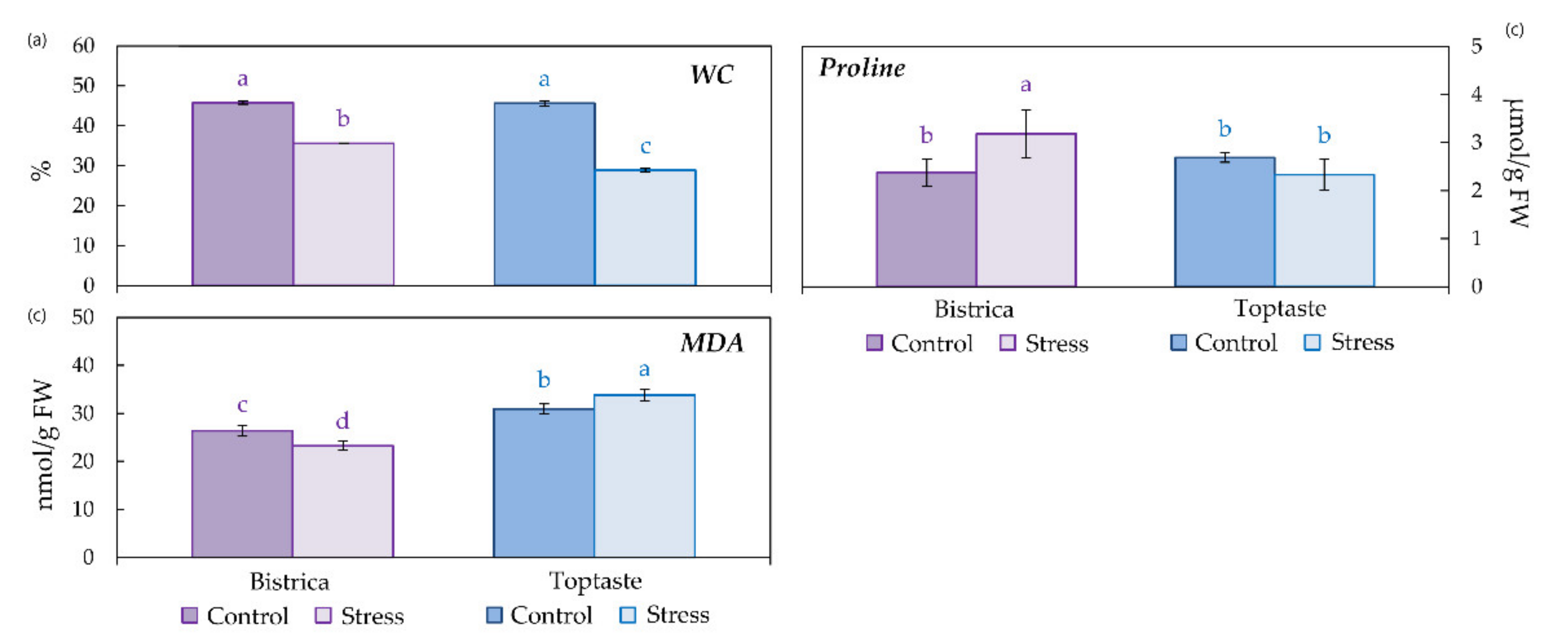
The measurements of chlorophyll a fluorescence were made on 15 leaves per treatment. The polyphasic chlorophyll a fluorescence was measured with a Plant Efficiency Analyzer (Handy PEA, Hansatech Instruments Ltd., King’s Lynn, UK). During the stress treatment, a special leaf clip was placed on each leaf to adapt a small part of the leaf to the dark to ensure that all reaction centers were open. The chlorophyll a fluorescence transient was induced by saturating with red light for 1 s (wavelength in peak at 650 nm, 3200 µmolphoton/m2 s). The fluorescence signals were collected from 10 µs up to 1 s with data acquisition every 10 µs for the first 300 µs, then continued every 100 µs up to 3 ms, and later every 1 ms, with 118 points within 1 s in total. The ChlF transient data were used to calculate the JIP-test parameters (Table 1) according to Strasser et al. [26,27].

2.3. Water Content

A fraction of the homogenized sample was weighted to obtain the fresh weight (FW). Thereafter, the weighted fraction was dried in the oven at 75 °C for 72 h and weighed again to determine the dry weight (DW) [28]. The water content in the leaves was calculated based on the equation: WC (%) = (FW − DW)/FW × 100.

2.4. Proline and Secondary Metabolites

Extraction of the free proline and phenolics from the plum leaves was performed by an ultrasound-assisted solid-liquid extraction for 60 min at 25 °C in a thermostat-controlled ultrasound bath (Sonorex RK 510 H, Bandelin Electronic, Berlin, Germany). In detail, 0.1 g of fresh powdered tissue was homogenated using 1 mL of aqueous ethanol (80% v/v). After sonification, the sample was centrifuged at 14,000× g at 4 °C for 10 min. The supernatant was used for the determination of the proline and phenols content.

2.4.1. Determination of Proline Content

Determination of the free proline content was performed as described by Woodrow et al. [29]. Duplicate extract aliquots of 50 µL, as well as proline standards (in the range of 0.2 to 1 mM) in 80% ethanol (v/v), were dispensed into reaction tubes. In each tube, 100 µL of a reaction mixture (prepared with 1% ninhydrin (w/v), 60% acetic acid (v/v) and 20% ethanol (v/v)) was added. The tube content was mixed and kept in a heating block at 95 °C for 20 min. After cooling at room temperature, the tubes were spun down quickly (1 min, 500 g), and 100 µL of the supernatant was transferred to a polypropylene microplate. The absorbance at 520 nm was measured by an Epoch microplate spectrophotometer (Bio-Tek, Bad Friedrichshall, Germany). The proline content was calculated according to the obtained standard curve and expressed as µmol/g FW.

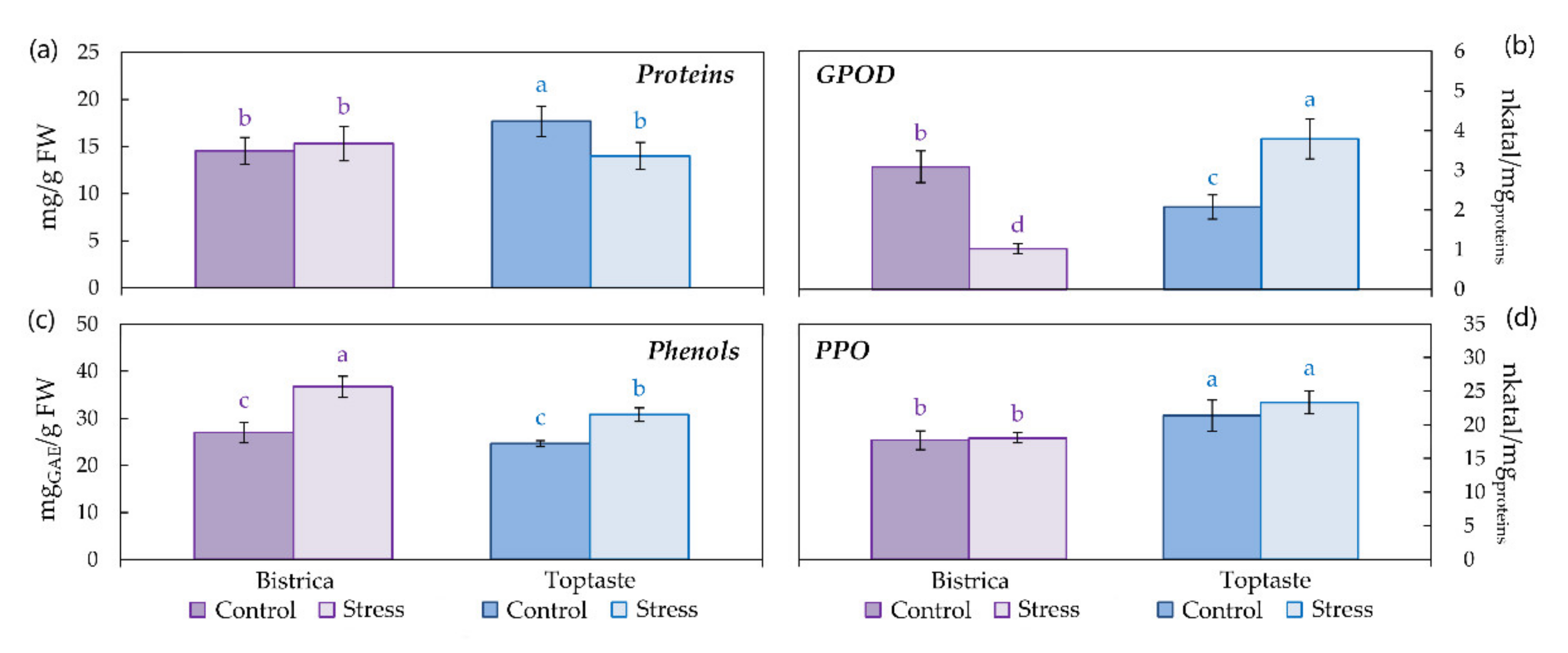
2.4.2. Total Phenols Content

The total phenolic content of the plum leaves was determined by the modified Folin–Ciocalteu method [30]. In brief, 5 µL of the extract was mixed with 795 µL of dH2O and 50 µL of Folin–Ciocalteu reagent (1:1; v/v diluted with water). After 5 min, 150 µL of a sodium carbonate solution (20%; w/v diluted with water) was added. The homogenized reaction mixture was placed for 30 min in a dark place at room temperature, after which absorbance readings at 765 nm were taken in the Epoch microplate spectrophotometer. The total phenolic content was expressed as mg of gallic acid equivalents (GAE) per g FW based on a gallic acid calibration curve (0.05–3 mg/mL).

2.5. Lipid Peroxidation

Extracts for determination of the lipid peroxidation were prepared from fresh powdered plum leaf tissue by 1 mL of 0.1% (w/v) trichloroacetic acid (TCA) per 0.10 g of tissue powder. After a 15 min extraction in an ice bath, homogenates were centrifuged for 15 min at 14,000× g on 4 °C and supernatants were used for the lipid peroxidation assay. Lipid peroxidation was estimated as a production of the malondialdehyde content (MDA) by the thiobarbituric acid (TBA) method [31]. For the determination of MDA in extracts, 0.5 mL of supernatant was mixed with 1 mL of 0.5% TBA in 20% trichloroacetic acid (TCA) and incubated for 30 min at 95 °C, cooled immediately in an ice bath and centrifuged at 14,000× g for 15 min at 4 °C. The supernatant was collected to measure the absorbance at 532 nm followed by subtracting the absorbance at 600 nm. The absorbance of samples was measured in a Specord 200 spectrophotometer (Analytic Jena, Jena, Germany) against the blank (0.5% TBA in 20% TCA). The amount of accumulated MDA was estimated using an extinction coefficient of 155 mM/cm3, and the concentration was expressed as nmol per gram FW.

2.6. Proteins and Enzyme Activities

Crude protein extracts were prepared from fresh powdered plum leaves. Briefly, about 0.2 g of powdered leaf tissue was extracted with 1 mL of a 100 mM potassium phosphate buffer (pH 7.0) with 5 mM ascorbic acid, 0.1 mM EDTA and 2% (w/v) polyvinylpolypyrrolidone (PVP). After 15 min of ice bath extraction and centrifugation for 15 min (14,000× g, 4 °C), the supernatants were taken for an enzymes and soluble protein assay. For all enzyme assays, five extractions per sample were completed and each one was measured in duplicate at a minimum.

2.6.1. Soluble Protein Concentration

Determination of the soluble protein content was completed according to the Bradford method [32], using bovine serum albumin (BSA) as a standard. The reaction mixture contained 5 µL of crude protein extract, 45 µL of dH2O and 1 mL of Coomassie Brilliant Blue reagent. The homogenized reaction mixture was placed for 7 min at room temperature, after which an absorbance reading at 595 nm was determined in the Epoch microplate spectrophotometer. The soluble protein content was expressed as mg per g FW based on the BSA calibration curve (0.01–0.4 mg/mL) and used to calculate and express the enzyme activities in the same crude extract.

2.6.2. Guaiacol Peroxidase Activity

Guaiacol peroxidase (GPOD) activity was measured according to Siegel and Galston [33] based on GPOD scavenging activity by using guaiacol as a hydrogen donor. The enzymatic reaction was initiated by adding 5 µL of crude protein extract into 995 µL of the reaction mixture (5 mM guaiacol and 2.5 mM hydrogen peroxide in a 0.2 M phosphate buffer, pH 5.8). GPOD activity was assessed by the increase in the absorbance of tetra-guaiacol at 470 nm over 1 min and was expressed as nkatal per mg proteins (nkatal/mgproteins).

2.6.3. Polyphenol Oxidase Activity

Polyphenol oxidase (PPO) activity was examined as a rate of the oxidation of pyrogallol to o–quinones at 40 °C [34], seen as an increase in absorbance at 430 nm over two minutes. The enzymatic reaction was initiated by adding 5 µL of a crude protein extract to the reaction mixture, consisting of 895 µL of a 100 mM potassium phosphate buffer (pH 7.0) and 0.1 mL of 0.1 M pyrogallol. PPO activity was expressed as nkatal per mg proteins (nkatal/mgproteins).

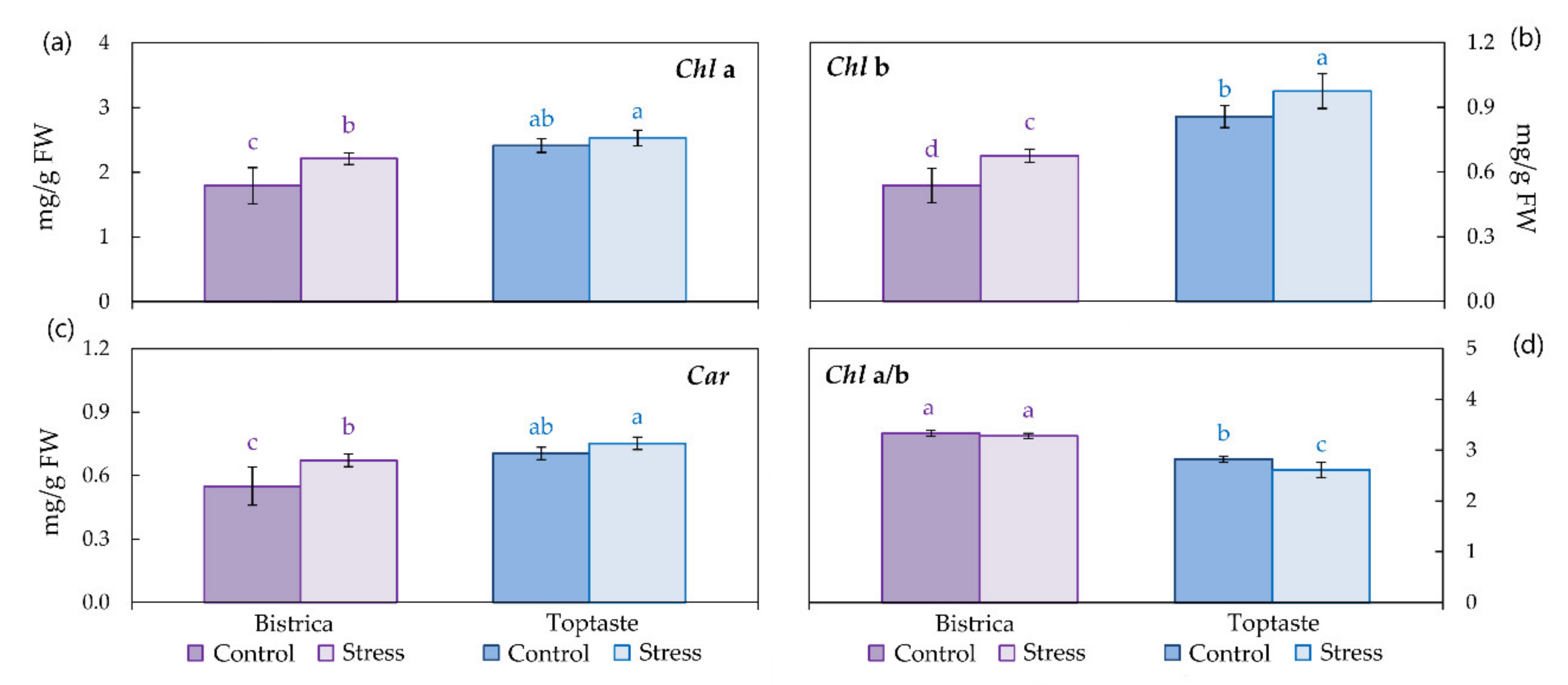
2.7. Photosynthetic Pigments

Photosynthetic pigments in the plum leaves were extracted from fresh powdered leaf tissue with absolute acetone. Concentrations of chlorophyll a (Chl a), chlorophyll b (Chl b), carotenoids (Car) and Chl a/b ratio were calculated from the absorbances at 470 nm, 647 nm, and 663 nm measured in the Specord 200 spectrophotometer. According to Lichtenthaler [35], the equations were used for the calculations and the photosynthetic pigments concentrations were expressed as mg per g FW.

2.8. Data Analyses

Statistical differences between the control and stressed leaves of two plum cultivars were compared by analysis of variance with a post-hoc Fisher’s least significant difference test (LSD). The differences were considered statistically significant when p < 0.05. Data were means ± SD of fifteen biological replicates for the JIP parameters. The same 15 leaves were collected into the composite sample for the biochemical parameters, and five (n = 5) replicates were measured per treatment.

3. Results

3.1. Determination of Stress Occurrence and Severity

Elevated temperature directly influenced leaf water status, therefore the water content (WC) and proline accumulation were evaluated to identify the stress occurrence and severity (Figure 1a,b). The WC significantly decreased in leaves exposed to an elevated temperature in both cultivars compared to control leaves, but less in the cultivar Bistrica (22.33%), and more in the cultivar Toptaste (36.75%). Proline accumulation was significantly enhanced in leaves exposed to the elevated temperature only in the cultivar Bistrica (34.01% compared to control), while in the cultivar Toptaste, it remained at the same significance level as the control. The lipid peroxidation, measured as MDA content as an indicator of stress severity, was significantly decreased in the leaves of the cultivar Bistrica exposed to the elevated temperature (11.76%), while in the stressed leaves of the cultivar Toptaste, the MDA content was significantly increased compared to the control for 9.21% (Figure 1c).

3.2. Proteins, Phenols and Enzymatic Activity

The concentration of total soluble proteins in the leaves of plum cultivars (Figure 2a) was affected by the elevated temperature only in the cultivar Toptaste, which responded to stress by decreasing the protein content by 20.80% compared to the control. Despite no significant change in the soluble proteins in the plum leaves of the cultivar Bistrica, the GPOD activity was significantly decreased by the elevated temperature (66.72%) (Figure 2b). Conversely, in the leaves of the cultivar Toptaste, the elevated temperature increased GPOD activity by 82.44% compared to the control. Plum leaf phenols responded to an elevated temperature similarly in both cultivars with an increase in phenol concentration compared to the control leaves (Figure 2c). This increase was more pronounced in the leaves of the cultivar Bistrica (36.01%) than in the leaves of the cultivar Toptaste (24.97%). The PPO activity in the plum leaves was not influenced by elevated temperature in either cultivar (Figure 2d).

3.3. Photosynthetic Pigments

The response of photosynthetic pigments to elevated temperature in the plum leaves is shown in Figure 3. The Chl a significantly increased in the stressed leaves of the cultivar Bistrica by 23.47% compared to the control leaves, while the leaves of Toptaste remained at the same significance level (Figure 3a). A similar trend in the leaves of the plum cultivar Bistrica exposed to elevated temperature was also found for the Chl b, with an increase of 25.57% compared to the control leaves (Figure 3b). Unlike the changes in Chl a in the cultivar Toptaste under heat stress, the increase in Chl b was 13.77% and significant as compared to the control. Plum leaf carotenoids responded to the elevated temperature with an increase in the Car concentration only in the cultivar Bistrica (22.30%) compared to the control leaves (Figure 3c). A significant difference in Chl a/b in the stressed plum leaves compared to control leaves was found only in the cultivar Toptaste, where Chl a/b decreased compared to the control by 7.54% (Figure 3d).

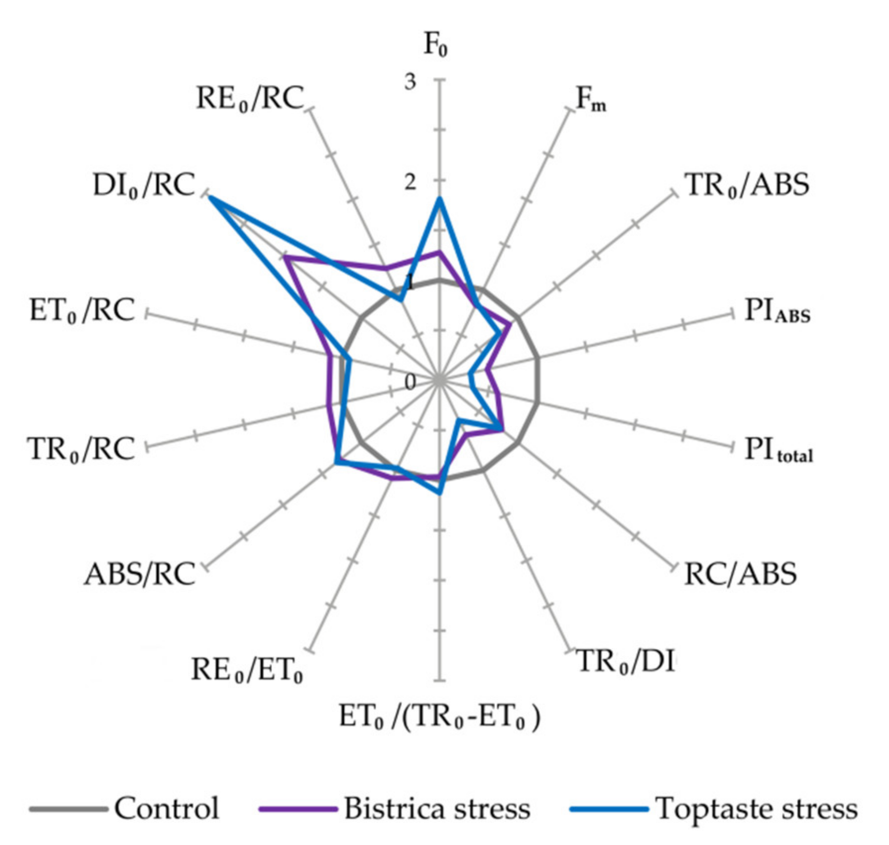
3.4. Photosynthetic Efficiency

The impact of elevated temperature on the photosynthetic process in plum leaves is shown in Figure 4 and Table 2. The minimal (F0) and maximal (Fm) fluorescence are raw data obtained with the measurement of chlorophyll a fluorescence, but they represent significant indicators of stress in plants. In plum leaves exposed to elevated temperature, the F0 significantly increased in the leaves of the cultivar Toptaste (80.91%), while the increase of F0 in the leaves of Bistrica was not significant (27.38%). Moreover, a decrease in Fm was found in the leaves of both cultivars but was more pronounced in the leaves of the cultivar Bistrica (16.66%) than in the leaves of Toptaste (14.73%) compared to their control leaves.
Chlorophyll a fluorescence parameters which describe overall photosynthetic efficiency, namely, PIABS, PItotal, and maximum quantum yield of PS II (Fv/Fm), reacted to the elevated temperature by a decrease in their values (Figure 4). PIABS reduction was more pronounced in the leaves of the cultivar Toptaste (68.20%) than in the leaves of Bistrica (51.32%) compared to their control leaves. The same trend was noted for PItotal as well; however, this parameter was decreased by 66.12% in the leaves of the cultivar Toptaste and 40.92% in Bistrica compared to controls. The maximum quantum yield of PS II followed the same trend as for PIABS and PItotal, i.e., diminution was found in the leaves of both cultivars, but was more pronounced in Toptaste (23.97%) than in Bistrica (11.12%) compared to control treatments.
Within the components of the performance indexes, significant differences between the stress and control leaves were found only for the parameters named QA reducing RCs per PSII antenna Chl, i.e., the density of reaction centers on chlorophyll basis (RC/ABS) and the ratio of trapping and dissipation fluxes (TR0/DI0). The RC/ABS decreased in both cultivars similarly (21.44 and 23.30% in Bistrica and Toptaste, respectively), while diminution of TR0/DI0 was more pronounced in the stressed leaves of Toptaste (55.67%) than in Bistrica (39.64%), compared to the controls. The efficiency of the conversion of excitation energy to electron transport (ET0/(TR0 − ET0)) and the efficiency with which an electron can move from the reduced intersystem electron acceptors to the PS I end electron acceptors (RE0/ET0), remained the same in stressed leaves compared with the control, but the RE0/ET0 was lower in both (control and stressed) leaves of the cultivar Toptaste compared to the leaves of Bistrica.
Energy flow through the photosynthetic system expressed per active reaction centers (RCs) in the plum leaves of control and stress treatments is shown in Figure 4 and Table 2. Absorption per active RCs (ABS/RC) and dissipation per active RCs (DI0/RC) increased in the plum leaves subjected to elevated temperatures compared to the control leaves. The increase of ABS/RC was more pronounced in Toptaste (31.18%) than in Bistrica (28.15%), while the increase of DI0/RC was 194.23% in Toptaste and 99.00% in Bistrica compared to the control leaves. Trapping per active RCs (TR0/RC) and electron flux reducing end electron acceptors at the PSI acceptor side per RCs (RE0/RC) remained unchanged in the leaves of the cultivar Toptaste subjected to an elevated temperature in comparison to the control leaves, while in the leaves of the cultivar Bistrica TR0/RC and RE0/RC significantly increased by 13.79% and 23.63%, respectively. The electron transport per active RC was not affected by elevated temperature in the leaves of either cultivar.

4. Discussion

Elevated temperatures, as a major factor of global climate change, negatively alter important plant physiological processes such as photosynthesis, primary and secondary metabolism, as well as hormone and lipid signaling, provoking antioxidative response mechanisms [36]. The early effects of high temperatures on leaves are alterations in membrane fluidity, chemical solubility and enzyme kinetics which directly influence the photosynthetic processes and water relations [6,18,37]. Decreased leaf WC in our study indicated water loss in both cultivars but was much pronounced in the leaves of the cultivar, Toptaste. A similar decrease in water content (4 h after induction of stress) by temperature elevation to 40 °C was found in sugarcane leaves [6]. Gulen and Eris [7] found a significant decrease in relative water content in strawberry leaves exposed for 48 h to several temperature regimes from 30 to 45 °C, as did Hao et al. [8] in Prunus mira Koehne seedlings exposed to 37 and 42 °C over 8 h. These alterations in water relations imply that elevated temperature causes some metabolic disturbances in plant leaves compared to control leaves. In such situations, the plant activates osmotic adjustment through proline accumulation [9,10]. The alterations in the proline accumulation in leaves of the plum cultivars exposed to short-term heat stress were in accordance with the results obtained by Harsh et al. [11]. They exposed seedlings of 37 moth bean genotypes (Vigna aconitifolia) to short-term heat stress and found a significant difference regarding proline content from an 80% decrease to 1000% increase of proline in stress-treated seedlings compared to control ones. Although Harsh et al. [11] had not found a correlation between the proline content and heat tolerance level, this positive correlation was found in sorghum seedlings exposed to 45 °C for 6 h [38] in the way that tolerant genotypes had a higher proline increase than the more susceptible ones.
Heat stress, as well as other abiotic stresses, induces peroxidation of lipids incorporated in cell membranes and protein denaturation making the membranes unstable [5]. The decreased MDA content in the leaves of the cultivar Bistrica exposed to heat stress implies that the membranes retained their integrity, while in the leaves of Toptaste, the integrity and stability of the membranes were disarranged. Similar increases in the MDA content under heat stress were found in lettuce seedlings [12], wheat [13], purslane [14], while in cotton seedlings heat stress did not influence membrane lipids seen as MDA content [39]. MDA content in buffel grass (Cenchrus ciliaris L.) correlated with plant tolerance to heat stress, therefore it can be used as a potential biochemical indicator for rapid, simple and low-cost identification of heat stress tolerant genotypes [40]. According to the use of water, proline and MDA content as stress markers, in our investigation, the cultivar Bistrica appeared to be more tolerant to heat stress than the cultivar Toptaste.
Higher plants exposed to heat stress exhibit a characteristic set of cellular and metabolic responses to survive undesirable conditions in a way that accelerates the transcription and translation of heat shock proteins (HSPs) to the detriment of “normal” protein synthesis [36]. Our results showed a significant decrease in the total soluble proteins content in the leaves of the cultivar Toptaste exposed to heat stress. Gulen and Eris [7] found a significant decrease in the total soluble protein content in strawberry plants exposed to elevated temperatures applied as a gradual and instant temperature increase in separate treatments. They found that a decrease in total soluble proteins was much pronounced in the treatment with an instant temperature rise even at a difference of 5 °C compared to the control. Despite the significant loss in soluble proteins in the leaves of the cultivar Toptaste, activation of enzymatic antioxidants occurred and was seen as an increase in GPOD activity. A similar increase in the peroxidase activity under heat stress was found in strawberry [41], mulberry [42], purslane [14], cotton [39], sorghum [38], etc. On the contrary, GPOD activity in stress treated leaves of the cultivar Bistrica declined compared to the control. Such opposite GPOD activities between the cultivars experiencing heat stress were also found in moth bean seedlings; however, despite this fact, a significant correlation between GPOD activity and heat stress tolerance in moth bean seedlings exposed to short-term heat stress was found [11]. Interestingly, GPOD activity in the heat stressed leaves of the cultivar Bistrica declined compared to the control despite no change in the total soluble proteins content. This can be explained by the previously mentioned assertion that the heat stress response is usually activated by heat shock protein synthesis on account of other protein structures and molecules [36], and the typical enzymatic antioxidant response is not activated. Drought tolerant sorghum genotypes exposed to heat stress activated the synthesis of many novel heat shock proteins in contrast to drought susceptible genotypes according to [38].
Alterations of the total soluble proteins induced by heat stress did not affect PPO activity in the leaves of either plum cultivar. The increased concentration of phenols in the heat stress exposed plum leaves implies activation of the phenolic pathway response to heat stress. Although no differences in the PPO enzyme activity were found in the leaves exposed to heat treatment, significant soluble phenolic compound accumulation may be attributed to acclimation mechanisms rather than the activity of the PPO [43]. Rivero et al. [43] found that tomato plants exposed daily to heat stress (35 °C) over 30 days increased their total phenols content but decreased PPO activity, while watermelon plants reacted opposite to this. It is evident that metabolic adjustments activated as a response to unfavorable conditions (biotic or abiotic) are dynamic and depend on the severity and type of stress, as well as on the plant species and even cultivar [44]. Consequently, the activation of PPO from heat stress in plum leaves is not species specific, unlike in Basella alba [45], tomato and watermelon [43] or red clover [16] where it is. Phenolic accumulation as the response of plants to heat stress was found in wheat [46], tomato [15], Nicotiana langsdorffii Weinmann [44], hard fescue [47], etc. Zhou et al. [15] found that heat-tolerant tomato genotypes increased their total phenolic content in stress conditions compared to heat-sensitive genotypes in which total phenolic content remained the same. Additionally, through an exhaustive metabolomics study of phenylpropanoid metabolism in tomato seedlings, Martinez et al. [48] revealed that an accumulation of flavonols dominates in contrast to other groups of phenolic compounds under heat stress. That type of stress preferentially led to the accumulation of a specific group of phenol compounds, namely, hydroxycinnamic acids, when tomato seedlings were exposed to drought stress.
Heat stress strongly induces alterations in respiration, photosynthesis and the fraction of reactive oxygen species (ROSs), which in this study caused an accumulation of proline in the Bistrica cultivar, as well as a significant amount of MDA, synthesized as a result of lipid membrane degradation in the Toptaste cultivar, which possibly originated from the photosynthetic apparatus [36]. The early effects of heat stress on the photosynthetic process become obvious by changing the photosynthetic pigments content and alterations of the photosynthetic efficiency. A significant increase in both the Chl a and b, as well as carotenoids in the Bistrica, can represent the first response to short-term heat stress as part of the tolerance mechanism. Reaction to heat stress in the leaves of the cultivar Toptaste was different in the way that the Chl b and Car increased, while Chl a remained the same, which caused a decrease in the Chl a/b compared to the control. The obtained results are partially in accordance with the results of Zhou et al. [15] on tomato seedlings where the significant increase in Chl a and b, and Car were proved in heat-tolerant genotypes exposed to heat stress; however, the obtained results are not in accordance with previously published results on grapevine cultivars [49] or purslane [14], where the photosynthetic pigments declined in heat stress conditions. The explanation for this may lie in the fact that in these studies, the plants were exposed to stress for significantly longer periods than in our study, 20 days for grapevine and 7 days for purslane. Furthermore, Gür et al. [39] exposed cotton seedlings to 38 and 45 °C for 2 h and found an increase in total chlorophylls in plants exposed to 38 °C, which may imply that a plant’s first reaction to short-term heat stress is an increase in chlorophyll as part of its tolerance mechanism response.
It is well known that PSII, the oxygen-evolving complex (OEC) particularly, is the most sensitive part of the electron transport chain of photosynthesis when it is exposed to various abiotic stresses [3,50]. One hour after the exposure of plum leaves to heat stress, a decline in overall photosynthetic efficiency was noted, seen as a significant decline in PIABS, PItotal and Fv/Fm in both cultivars, but much more pronounced in the cultivar Toptaste. Martinazzo et al. [51] exposed detached peach leaves to short term heat stress (40 °C for 30 min) and found significant diminution in PIABS and Fv/Fm. A similar diminution of PIABS and Fv/Fm was found in detached tomato leaves exposed to 40 °C for 1 h [17] but with an increase of PItotal. It is important to note that the elevated temperature decreased the Fv/Fm below the limit of 0.75 in both cultivars, suggesting that the PSII function was significantly impaired. Several investigations on different plant species (peach, tomato, and wheat) found that Fv/Fm is a sensitive indicator of heat stress only at 40 °C and below [17,37,51], respectively. This makes Fv/Fm a less sensitive indicator of heat stress than the performance indexes [52] as our results also confirmed, seen as a diminution of Fv/Fm by 23.97% in contrast to the PIABS and PItotal, which decreased 68.20 and 66.12%, respectively, in the plum leaves of cv. Toptaste exposed to heat compared with control leaves.
The significant decline in Fv/Fm and consequently PIABS and PItotal in the leaves of Toptaste rather than in Bistrica may be the result of significantly increased F0 in Toptaste; however, the increase in F0, together with the decreased Fm, is in accordance with results obtained by Mihaljević et al. [53] for apples exposed to elevated temperatures. Furthermore, many authors have found that heat stress increases F0 and decreases Fm, suggesting that the parameter F0 can be an effective fluorescence parameter for screening for heat stress tolerance [19,37,51].
The performance indexes (PIABS and PItotal) are complex parameters since they provide information on overall energy flow through the photosystems via the absorption and trapping of excitation energy together with its conversion to electron transport or the dissipation of excess energy [21]. The greatest effect on the reduction of PIABS and PItotal in plum leaves exposed to heat stress had parameters RC/ABS and TR0/DI0. Their decrease, visible in the heat exposed leaves of both cultivars compared with the controls, was similar for RC/ABS in both cultivars, while the TR0/DI0 decreased more significantly in the Toptaste leaves, which suggests that TR0/DI0 is the parameter that is most responsible for the stronger reduction in performance indexes in Toptaste. The most significant role of TR0/DI0 among the performance index components in the diminution of PIABS was also found in sour cherry leaves exposed to drought stress [54]; however, ET0/(TR0 − ET0) and RE0/ET0 remained the same in heat stress conditions as in control ones., RE0/ET0 was generally lower in the Toptaste leaves, for both the control and heat treated leaves, indicating a cultivar specific characteristic of Toptaste for a lower electron transfer to end electron acceptors at the PSI acceptor side than in the Bistrica, which was also confirmed by the RE0/RC parameter.
Energy fluxes per RC express specific functional parameters, which describe critical events in the photosynthetic process, i.e., absorbance (ABS) of photons by PSII antenna, intermediate trapping flux (TR), electron transport (ET), the reduction of the end-electron acceptors at the PS I electron acceptor side (RE) and the dissipation of excess energy (DI). According to our results, all fluxes per RCs except for ET0/RC increased in the plum leaves of cv. Bistrica when exposed to elevated temperatures. Undisturbed ET0/RC suggests the activation and synchronization of other processes in order to maintain optimal energy flow through the photosynthetic apparatus, which is in accordance with a stabile ET0/(TR0 − ET0), indicating that heat stress does not impact the electron transport in plum leaves. Although Martinazzo et al. [51] found an increase in ET0/RC in peach leaves exposed to 40 °C for 30 min, our results corroborate with Zushi et al. [17], who did not find a significant impact of heat stress on ET0/RC in tomato leaves exposed to 40 °C for 1 h despite a decreased PIABS in both investigations. Moreover, in the stressed leaves of the cv. Toptaste, a significant effect of an increased ABS/RC and particularly DI0/RC (194.23% compared with the control) without an impact on TR0/RC, ET0/RC and RE0/RC, suggests a high absorption but probably in non-active reaction centers, so that the excitation energy was not trapped in the electron chain transport but rather dissipated as heat.

5. Conclusions

The presented results demonstrated significant differences between the plum cultivars in response to short-term heat stress. Elevated temperatures caused heat stress in both plum cultivars, which was indicated as a decrease in water content. In the leaves of the cultivar Bistrica, accumulation of proline and phenols, as well as an accumulation of photosynthetic pigments, suggest activation of a strong response to unfavorable conditions. Contrary, in the leaves of Toptaste, water content decreased significantly more than in Bistrica, and together with a significant accumulation of MDA, activation of GPOD and decreased soluble proteins content, indicates an inadequate response to maintaining homeostasis in the leaf metabolism. Moreover, although photosynthesis was disturbed in both genotypes, seen as a decrease of the JIP parameters PIABS, PItotal and Fv/Fm, significantly pronounced changes were found in the cultivar Toptaste. In the modern plum cultivar Toptaste, elevated temperatures increased the minimal fluorescence and absorption (ABS/RC), but because of the increase in Chl b in total chlorophylls and/or inactivation of the RCs (RC/ABS), the excitation energy was not trapped efficiently in the electron chain transport, which resulted in stronger dissipation (DI0/RC) and formation of ROSs. After considering all the results, the traditional plum cultivar Bistrica appears to have a better tolerance to heat stress than the modern cultivar Toptaste.

Author Contributions

Conceptualization, I.M.; methodology, I.M. and M.V.V.; software, V.T.; validation, D.H.; formal analysis, M.V.V., I.M. and V.T.; resources, D.H. and D.V.; writing—original draft preparation, M.V.V.; writing—review and editing, M.V.V. and I.M.; visualization, D.H.; supervision D.H., Z.Z. and D.V.; project administration, Z.Z. and D.V. All authors have read and agreed to the published version of the manuscript.

Funding

This research was conducted as a part of the measure M10.2. Support to conservation of genetic resources in agriculture—preservation, sustainable use, and development of genetic resources in agriculture—was provided by the Rural Development Programme of the Republic of Croatia for the Period 2014–2020, which is funded by The European Agricultural Fund for Rural Development, PRR-10.02.01.00.0-03, ID: 1330299.

Institutional Review Board Statement

Not applicable.

Informed Consent Statement

Not applicable.

Data Availability Statement

Not applicable.

Conflicts of Interest

The authors declare no conflict of interest.

References

  1. Mihaljević, I.; Viljevac Vuletić, M.; Šimić, D.; Tomaš, V.; Horvat, D.; Josipović, M.; Zdunić, Z.; Dugalić, K.; Vuković, D. Comparative study of drought stress effects on traditional and modern apple cultivars. Plants 2021, 10, 561. [Google Scholar] [CrossRef]
  2. Mihaljević, I.; Viljevac Vuletić, M.; Tomaš, V.; Horvat, D.; Zdunić, Z.; Vuković, D. PSII photochemistry responses to drought stress in autochthonous and modern sweet cherry cultivars. Photosynthetica 2021, 59, 517–528. [Google Scholar] [CrossRef]
  3. Allakhverdiev, S.I.; Kreslavski, V.D.; Klimov, V.V.; Los, D.A.; Carpentier, R.; Mohanty, P. Heat stress: An overview of molecular responses in photosynthesis. Photosynth. Res. 2008, 98, 541–550. [Google Scholar] [CrossRef]
  4. Fahad, S.; Bajwa, A.A.; Nazir, U.; Anjum, S.A.; Farooq, A.; Zohaib, A.; Sadia, S.; Nasim, W.; Adkins, S.; Saud, S.; et al. Crop production under drought and heat Stress: Plant responses and management options. Front. Plant Sci. 2017, 8, 1147. [Google Scholar] [CrossRef] [Green Version]
  5. Hasanuzzaman, M.; Nahar, K.; Alam, M.M.; Roychowdhury, R.; Fujita, M. Physiological, biochemical, and molecular mechanisms of heat stress tolerance in plants. Int. J. Mol. Sci. 2013, 14, 9643–9684. [Google Scholar] [CrossRef]
  6. Wahid, A.; Close, T.J. Expression of dehydrins under heat stress and their relationship with water relations of sugarcane leaves. Biol. Plant. 2007, 51, 104–109. [Google Scholar] [CrossRef]
  7. Gulen, H.; Eris, A. Some physiological changes in strawberry (Fragaria × ananassa ‘Camarosa’) plants under heat stress. J. Hortic. Sci. Biotech. 2003, 78, 894–898. [Google Scholar] [CrossRef]
  8. Hao, H.-P.; Jiang, C.-D.; Zhang, S.-R.; Tang, Y.-D.; Shi, L. Enhanced thermal tolerance of photosystem II by elevating root zone temperature in Prunus mira Koehne seedlings. Plant Soil 2012, 353, 367–378. [Google Scholar] [CrossRef]
  9. Iqbal, N.; Fatma, M.; Khan, N.A.; Umar, S. Regulatory role of proline in heat stress tolerance: Modulation by salicylic acid. In Plant Signaling Molecules; Khan, M.I.R., Reddy, P.S., Ferrante, A., Khan, N.A., Eds.; Elsevier: Amsterdam, The Netherlands, 2019; pp. 437–448. [Google Scholar] [CrossRef]
  10. Kaur, G.; Asthir, B. Proline: A key player in plant abiotic stress tolerance. Biol. Plant. 2015, 59, 609–619. [Google Scholar] [CrossRef]
  11. Harsh, A.; Sharma, Y.K.; Joshi, U.; Rampuria, S.; Singh, G.; Kumar, S.; Sharma, R. Effect of short-term heat stress on total sugars, proline and some antioxidant enzymes in moth bean (Vigna aconitifolia). Ann. Agric. Sci. 2016, 61, 57–64. [Google Scholar] [CrossRef] [Green Version]
  12. Han, Y.; Fan, S.; Zhang, Q.; Wang, Y. Effect of heat stress on the MDA, proline and soluble sugar content in leaf lettuce seedlings. Agric. Sci. 2013, 4, 112–115. [Google Scholar] [CrossRef] [Green Version]
  13. Sairam, R.K.; Srivastava, G.C.; Saxena, D.C. Increased antioxidant activity under elevated temperatures: A mechanism of heat stress tolerance in wheat genotypes. Biol. Plant. 2000, 43, 245–251. [Google Scholar] [CrossRef]
  14. Jin, R.; Wang, Y.; Liu, R.; Gou, J.; Chan, Z. Physiological and metabolic changes of purslane (Portulaca oleracea L.) in response to drought, heat, and combined stresses. Front. Plant Sci. 2016, 6, 1123. [Google Scholar] [CrossRef] [Green Version]
  15. Zhou, R.; Yu, X.; Kjær, K.H.; Rosenqvist, E.; Ottosen, C.-O. Screening and validation of tomato genotypes under heat stress using Fv/Fm to reveal the physiological mechanism of heat tolerance. Exp. Environ. Bot. 2015, 118, 1–11. [Google Scholar] [CrossRef]
  16. Boeckx, T.; Webster, R.; Winters, A.L.; Webb, K.J.; Gay, A.; Kingston-Smith, A.H. Polyphenol oxidase-mediated protection against oxidative stress is not associated with enhanced photosynthetic efficiency. Ann. Bot. 2015, 116, 529–540. [Google Scholar] [CrossRef] [Green Version]
  17. Zushi, K.; Kajiwara, S.; Matsuzoe, N. Chlorophyll a fluorescence OJIP transient as a tool to characterize and evaluate response to heat and chilling stress in tomato leaf and fruit. Sci. Hortic. 2012, 148, 39–46. [Google Scholar] [CrossRef]
  18. Yan, K.; Chen, P.; Shao, H.; Shao, C.; Zhao, S.; Brestič, M. Dissection of photosynthetic electron transport process in sweet sorghum under heat stress. PLoS ONE 2013, 8, e62100. [Google Scholar] [CrossRef] [Green Version]
  19. Feng, B.; Liu, P.; Li, G.; Dong, S.T.; Wang, F.H.; Kong, L.A.; Zhang, W. Effect of heat stress on the photosynthetic characteristics in flag leaves at the grain-filling stage of different heat-resistant winter wheat varieties. J. Agron. Crop Sci. 2014, 200, 134–155. [Google Scholar] [CrossRef]
  20. Haworth, M.; Marino, G.; Brunetti, C.; Killi, D.; de Carlo, A.; Centritto, M. The impact of heat stress and water deficit on the photosynthetic and stomatal physiology of olive (Olea europaea L.)–A case study of the 2017 heat wave. Plants 2018, 7, 76. [Google Scholar] [CrossRef] [Green Version]
  21. Force, L.; Critchley, C.; van Rensen, J.S. New fluorescence parameters for monitoring photosynthesis in plants. Photosynth. Res. 2003, 78, 17–23. [Google Scholar] [CrossRef]
  22. Biško, A.; Jelačić, T.; Miloloža, D.; Savić, Z.; Brus, K. The plum industry in the Republic of Croatia. Acta Hortic. 2019, 1260, 215–220. [Google Scholar] [CrossRef]
  23. Ivković, F. Šljiva Bistrica—Nacionalni ponos hrvatskih voćara. Glas. Zaštite Bilja 2010, 5, 32–33. [Google Scholar]
  24. Družić, J.; Voća, S.; Čmelik, Z.; Dobričević, N.; Duralija, B.; Skendrović Babojelić, M. Fruit quality of plum cultivars Elena and Bistrica. Agric. Conspec. Sci. 2007, 72, 307–310. [Google Scholar]
  25. Molnár, A.M.; Ladányi, M.; Kovácz, S. Evaluation of the production traits and fruit quality of German plum cultivars. Acta Univ. Agric. Silvic. Mendelianae Brun. 2016, 64, 109–114. [Google Scholar] [CrossRef] [Green Version]
  26. Strasser, R.J.; Srivastava, A.; Tsimilli-Michael, M. The fluorescence transient as a tool to characterize and screen photosynthetic samples. In Probing Photosynthesis: Mechanism, Regulation and Adaptation; Yunus, M., Pathre, U., Mohanty, P., Eds.; Taylor and Francis: London, UK, 2000; pp. 445–483. [Google Scholar]
  27. Strasser, R.J.; Tsimilli-Michael, M.; Srivastava, A. Analysis of the chlorophyll a fluorescence transient. In Chlorophyll a Fluorescence: A Signature of Photosynthesis. Advances in Photosynthesis and Respiration; Papageorgiou, G.C., Govindjee, Eds.; Springer: Dordrecht, The Netherlands, 2004; pp. 321–362. [Google Scholar]
  28. Kumar, D.; Al Hassan, M.; Naranjo, M.A.; Agrawal, V.; Boscaiu, M.; Vicente, O. Effects of salinity and drought on growth, ionic relations, compatible solutes and activation of antioxidant systems in oleander Nerium oleander (L.). PLoS ONE 2017, 12, e0185017. [Google Scholar] [CrossRef]
  29. Woodrow, P.; Ciarmiello, L.F.; Annunziata, M.G.; Pacifico, S.; Iannuzzi, F.; Mirto, A.; D’Amelia, L.; Dell’Aversana, E.; Piccolella, S.; Fuggi, A.; et al. Durum wheat seedling responses to simultaneous high light and salinity involve a fine reconfiguration of amino acids and carbohydrate metabolism. Physiol. Plant. 2017, 159, 290–312. [Google Scholar] [CrossRef]
  30. Singleton, V.L.; Rossi, J.A. Colorimetry of total phenolics with phosphomolybdic–phosphotungstic acid reagents. Am. J. Enol. Vitic. 1965, 16, 144–158. [Google Scholar]
  31. Verma, S.; Dubey, R.S. Leads toxicity induces lipid peroxidation and alters the activities of antioxidant enzymes in growing rice plants. Plant Sci. 2003, 164, 645–655. [Google Scholar] [CrossRef]
  32. Bradford, M.M. A rapid and sensitive method for quantification of microgram quantities of protein utilizing the principle of protein-dye binding. Anal. Biochem. 1976, 72, 248–254. [Google Scholar] [CrossRef]
  33. Siegel, B.Z.; Galston, W. The isoperoxidases of Pisum sativum. Plant Physiol. 1967, 42, 221–226. [Google Scholar] [CrossRef] [Green Version]
  34. Raymond, J.; Rakariyatham, N.; Azanza, J. Purification and some properties of polyphenoloxidase from sunflower seeds. Phytochemistry 1993, 34, 927–931. [Google Scholar] [CrossRef]
  35. Lichtenthaler, H.K. Chlorophylls and carotenoids: Pigments of photosynthetic biomembranes. Meth. Enzymol. 1987, 148, 350–382. [Google Scholar] [CrossRef]
  36. Bita, C.E.; Gerats, T. Plant tolerance to high temperature in a changing environment: Scientific fundamentals and production of heat stress tolerant crops. Front. Plant Sci. 2013, 4, 273. [Google Scholar] [CrossRef] [PubMed] [Green Version]
  37. Mathur, S.; Jajoo, A.; Mehta, P.; Bharti, S. Analysis of elevated temperature-induced inhibition of photosystem II using chlorophyll a fluorescence induction kinetics in wheat leaves (Triticum aestivum). Plant Biol. 2011, 13, 1–6. [Google Scholar] [CrossRef] [PubMed]
  38. Gosavi, G.U.; Jadhav, A.S.; Kale, A.A.; Gadakh, S.R.; Pawar, B.D.; Chimote, V.P. Effect of heat stress on proline, chlorophyll content, heat shock proteins and antioxidant enzyme activity in sorghum (Sorghum bicolor) at seedlings stage. Indian J. Biotechnol. 2014, 13, 356–363. [Google Scholar]
  39. Gür, A.; Demirel, U.; Özden, M.; Kahraman, A.; Çopur, O. Diurnal gradual heat stress affects antioxidant enzymes, proline accumulation and some physiological components in cotton (Gossypium hirsutum L.). Afr. J. Biotechnol. 2010, 9, 1008–1015. [Google Scholar]
  40. Tommasino, E.; Griffa, S.; Grunberg, K.; Ribotta, A.; López Colomba, E.; Carloni, E.; Quiroga, M.; Luna, C.M. Malondialdehyde content as a potential biochemical indicator of tolerant Cenchrus ciliaris L. genotypes under heat stress treatment. Grass Forage Sci. 2012, 67, 456–459. [Google Scholar] [CrossRef]
  41. Gulen, H.; Eris, A. Effect of heat stress on peroxidase activity and total protein content in strawberry plants. Plant Sci. 2004, 166, 739–744. [Google Scholar] [CrossRef]
  42. Chaitanya, K.V.; Sundar, D.; Masilamani, S.; Reddy, A.R. Variation in heat stress-induced antioxidant enzyme activities among three mulberry cultivars. Plant Growth Regul. 2002, 36, 175–180. [Google Scholar] [CrossRef]
  43. Rivero, R.M.; Ruiz, J.M.; García, P.C.; López-Lefebre, L.R.; Sánchez, E.; Romero, L. Resistance to cold and heat stress: Accumulation of phenolic compounds in tomato and watermelon plants. Plant Sci. 2001, 160, 315–321. [Google Scholar] [CrossRef]
  44. Ancillotti, C.; Bogani, P.; Biricolti, S.; Calistri, E.; Checchini, L.; Ciofi, L.; Gonnelli, C.; Del Bubba, M. Changes in polyphenol and sugar concentrations in wild type and genetically modified Nicotiana langsdorffii Weinmann in response to water and heat stress. Plant Physiol. Biochem. 2015, 97, 52–61. [Google Scholar] [CrossRef] [PubMed]
  45. Siddika, M.R.; Rakib, M.A.; Abu Zubair, M.; Islam, M.M.; Haque, M.S.; Al-Khayri, J.M. Regulatory mechanism of enhancing polyphenol oxidase activity in leaf of Basella alba induced by high temperature stress. Emir. J. Food Agric. 2015, 27, 82–93. [Google Scholar] [CrossRef] [Green Version]
  46. Shahid, M.; Saleem, M.F.; Anjum, S.A.; Shahid, M.; Afzal, I. Effect of terminal heat stress on proline, secondary metabolites and yield components of wheat (Triticum aestivum L.) genotypes. Philipp. Agric. Sci. 2017, 100, 278–286. [Google Scholar]
  47. Wang, J.; Yuan, B.; Huang, B. Differential heat-induced changes in phenolic acids associated with genotypic variations in heat tolerance for hard fescue. Crop Sci. 2019, 59, 667–674. [Google Scholar] [CrossRef]
  48. Martinez, V.; Mestre, T.C.; Rubio, F.; Girones-Vilaplana, A.; Moreno, D.A.; Mittler, R.; Rivero, R.M. Accumulation of flavonols over hydroxycinnamic acids favors oxidative damage protection under abiotic stress. Front. Plant Sci. 2016, 7, 838. [Google Scholar] [CrossRef] [PubMed]
  49. Tzortzakis, N.; Chrysargyris, A.; Aziz, A. Adaptive response of a native Mediterranean grapevine cultivar upon short-term exposure to drought and heat stress in the context of climate change. Agronomy 2020, 10, 249. [Google Scholar] [CrossRef] [Green Version]
  50. Wahid, A.; Gelani, S.; Ashraf, M.; Foolad, M.R. Heat tolerance in plants: An overview. Environ. Exp. Bot. 2007, 61, 199–223. [Google Scholar] [CrossRef]
  51. Martinazzo, E.G.; Ramm, A.; Bacarin, M.A. The chlorophyll a fluorescence as an indicatior of the temperature stress in the leaves of Prunus persica. Braz. J. Plant Physiol. 2012, 24, 237–246. [Google Scholar] [CrossRef] [Green Version]
  52. Markulj Kulundžić, A.; Viljevac Vuletić, M.; Matoša Kočar, M.; Mijić, A.; Varga, I.; Sudarić, A.; Cesar, V.; Lepeduš, H. The combination of increased temperatures and high irradiation causes changes in photosynthetic efficiency. Plants 2021, 10, 2076. [Google Scholar] [CrossRef]
  53. Mihaljević, I.; Lepeduš, H.; Šimić, D.; Viljevac Vuletić, M.; Tomaš, V.; Vuković, D.; Dugalić, K.; Teklić, T.; Skendrović Babojelić, M.; Zdunić, Z. Photochemical efficiency of photosystem II in two apple cultivars affected by elevated temperature and excess light in vivo. S. Afr. J. Bot. 2020, 130, 316–326. [Google Scholar] [CrossRef]
  54. Viljevac, M.; Dugalić, K.; Mihaljević, I.; Šimić, D.; Sudar, R.; Jurković, Z.; Lepeduš, H. Chlorophyll content, photosynthetic efficiency and genetic markers in two sour cherry (Prunus cerasus L.) genotypes under drought stress. Acta Bot. Croat. 2013, 72, 221–235. [Google Scholar] [CrossRef] [Green Version]
Figure 1. Water content (a), proline (b), and MDA content (c) in control and stressed leaves of plum cultivars Bistrica and Toptaste. Values are mean ± SD. Different letters indicate significant differences among the treatments and cultivars at p < 0.05 according to the LSD test.
Horticulturae 08 00072 g001
Figure 2. Soluble proteins (a), guaiacol peroxidase activity (b), phenols (c), and polyphenol oxidase activity (d) in control and stressed leaves of the plum cultivars, Bistrica and Toptaste. Values are mean ± SD. Different letters indicate significant differences among the treatments and cultivars at p < 0.05 according to the LSD test.
Horticulturae 08 00072 g002
Figure 3. Photosynthetic pigments, i.e., Chl a (a), Chl b (b), Car (c), and Chl a/b (d) in control and stressed leaves of the plum cultivars, Bistrica and Toptaste. Values are mean ± SD. Different letters indicate significant differences among the treatments and cultivars at p < 0.05 according to the LSD test.
Horticulturae 08 00072 g003
Figure 4. Fluorescence data and JIP test parameters in control and heat-stressed leaves of the plum cultivars Bistrica and Toptaste. The values in spider (n= 15) are plotted relative to their respective controls (set as reference grey circle = 1). Raw data and statistical significances are presented in Table 1.
Horticulturae 08 00072 g004
Table 1. Definitions of JIP-test parameters according to Strasser et al. [26,27]. All fluorescence parameters are in relative units.
F0—Minimal fluorescence intensity (20 µs)
Fm—Maximal fluorescence intensity
TR0/ABS, i.e., Fv/Fm—Maximum quantum yield of PSII
PIABS—Performance index per absorption basis
PItotal—Performance index for energy conservation from exciton to the reduction of PSI end acceptors
RC/ABS—Density of reaction centers on chlorophyll basis
TR0/DI0—Flux ratio trapping per dissipation
ET0/(TR0–ET0)—Efficiency of the conversion of excitation energy to electron transport
RE0/ET0—Efficiency with which an electron from the intersystem electron carriers moves to reduce end electron acceptors at the PSI acceptor side
RC/CS0—Density of reaction centres (QA−reducing PSII reaction) at t = 0
ABS/RC—Absorption per active RC
TR0/RC—Trapping per active RC
ET0/RC—Electron transport per active RC
DI0/RC—Dissipation per active RC
RE0/RC—Electron flux reducing end electron acceptors at PSI acceptor side per RC
All fluorescence parameters are in relative units, RC—reaction center
Table 2. Fluorescence data and JIP test parameters (n = 15) in control and heat-stressed leaves of the plum cultivars Bistrica and Toptaste. Values are mean ± SD. Different letters indicate significant differences among the treatments and cultivars at p < 0.05 according to the LSD test.
BistricaToptaste
ParameterControlStressControlStress
F0450.17 ± 18.22 b573.42 ± 95.85 b474.5 ± 15.93 b858.42 ± 347.18 a
Fm2672.8 ± 117.9 a2227.5 ± 238.4 c2797.3 ± 126.2 a2385.2 ± 212.6 b
TR0/ABS i.e., Fv/Fm0.83 ± 0.01 a0.74 ± 0.06 b0.83 ± 0.01 a0.63 ± 0.18 c
PIABS3.13 ± 0.7 a1.53 ± 0.82 b2.81 ± 0.38 a0.89 ± 0.5 c
PItotal2.57 ± 0.62 a1.52 ± 0.83 b1.79 ± 0.31 a0.61 ± 0.44 c
RC/ABS0.53 ± 0.04 a0.42 ± 0.05 c0.48 ± 0.04 b0.37 ± 0.04 d
TR0/DI04.95 ± 0.34 a2.99 ± 0.80 b4.9 ± 0.21 a2.17 ± 1.10 c
ET0/(TR0 − ET0)1.18 ± 0.18 a1.14 ± 0.31 a1.19 ± 0.10 a1.34 ± 0.85 a
RE0/ET00.45 ± 0.02 a0.49 ± 0.07 a0.39 ± 0.03 b0.38 ± 0.10 b
ABS/RC1.88 ± 0.14 d2.41 ± 0.28 b2.09 ± 0.18 c2.75 ± 0.32 a
TR0/RC1.57 ± 0.12 b1.78 ± 0.24 a1.74 ± 0.15 ab1.7 ± 0.42 ab
ET0/RC0.84 ± 0.08 a0.94 ± 0.22 a0.94 ± 0.07 a0.87 ± 0.14 a
DI0/RC0.32 ± 0.03 c0.63 ± 0.16 b0.36 ± 0.03 c1.05 ± 0.59 a
RE0/RC0.38 ± 0.04 b0.47 ± 0.16 a0.37 ± 0.04 b0.33 ± 0.10 b
Publisher’s Note: MDPI stays neutral with regard to jurisdictional claims in published maps and institutional affiliations.

