Article

Seed Maturity and Its In Vitro Initiation of Chilean Endemic Geophyte Alstroemeriapelegrina L.

1
Laboratorio de Propagación, Escuela de Agronomía, Facultad de Ciencias Agronómicas y de los Alimentos, Pontificia Universidad Católica de Valparaíso, San Francisco S/N, La Palma, Quillota 2260000, Chile
2
Laboratorio de Semillas e Histología Vegetal, Escuela de Agronomía, Facultad de Ciencias Agronómicas y de los Alimentos, Pontificia Universidad Católica de Valparaíso, San Francisco S/N, La Palma, Quillota 2260000, Chile
3
Escuela de Agronomía, Facultad de Ciencias Agronómicas y de los Alimentos, Pontificia Universidad Católica de Valparaíso, San Francisco S/N, La Palma, Quillota 2260000, Chile
*
Author to whom correspondence should be addressed.
Submission received: 21 April 2022 / Revised: 12 May 2022 / Accepted: 16 May 2022 / Published: 21 May 2022
(This article belongs to the Collection Seed Dormancy and Germination of Horticultural Plants)

Abstract

:
Alstroemeria pelegrina (A. pelegrina), a Chilean endemic, is considered vulnerable as its natural habitat is currently threatened. The decline in the reproductive capacity of the species due to anthropogenic impacts and climate change has made it imperative to address the problem by developing large-scale propagation methods. The objective of this study was to establish protocols for breaking the dormancy and in vitro germination of A. pelegrina seeds to speed up the germination and seedling production processes. The research began with morphological observations of the reproductive process, followed by in vitro sowing. The results showed that the seeds reached full maturity in 51 days, and physiological maturity in 41 days, at which point the seeds could be harvested for in vitro germination. The mechanical scarification pretreatment improves the in vitro germination rate to 96% and the germination time to 7 days, showing that the species is characterized by physical seed dormancy. On the other hand, if the seed coat incisions are deeper than 0.5 mm, 30% of the potential plants are lost due to embryo damage. The study provides scientific evidence for the feasibility of large-scale in vitro propagation of the species and establishes an efficient method of seedling production.

1. Introduction

The Chilean native flora comprises more than 4500 taxa and is highly endemic (about 50%) [1], which is why Chile is considered a biogeographic island. This endemism has been concentrated in several botanical families, notably Asteraceae, Calceolariaceae, Alstroemeriaceae, Tropaeolaceae, and Loasaceae. The diversity of endemic species in Chile is due to evolutionary and historical processes that make the country a center of floristic diversity [2].
The genus Alstroemeria consists of about 90 species endemic to South America [3,4]. Found mainly in Chile, Brazil, Argentina, Peru, Bolivia, Paraguay, and Venezuela, it is one of the most diversified genera in Chile, with 58 listed taxa (37 species, 11 subspecies, and 10 varieties), of which almost 82% are endemic to the Chilean Mediterranean climate zone [5]. The species of the Alstroemeria genus have become relevant as ornamental plants in countries such as Holland, England, Japan, and the United States, reaching commercial value due to their cultivation programs and propagation of species [6,7].
A. pelegrina L. (Mariposa de Los Molles), the type-species of the genus, is found between the Coquimbo Region (31°45′ S) and Quintay, south of the Valparaíso Region (33°12′ S), at altitudes of up to 120 m a.s.l. (above sea level) on coastal cliffs, rocks, and mountain slopes [8,9]. Currently this species is considered in a vulnerable state by the Chilean Ministry of the Environment due to indiscriminate extraction and desertification, which has caused a decrease in its natural habitat as a result of real estate development The species is not found in any of the areas protected by the State [8,10].
It is a herbaceous perennial plant 20–60 cm tall. It has a glabrous stem, usually with leaves in the middle or upper third [11]. The leaves, which twist around the stem, are alternate and vivid green in color. It exhibits inflorescence with 1 to 3 uniflorous racemes [5]. The flowers are large, up to 8 cm in diameter, grouped in three umbels, pink to purplish, with a broad purple center, and two upper petals that have yellow streaks with dark stripes [12]. The fruit is a globose, yellow-brown capsule when ripe, 1.8 cm long, with six longitudinal ribs. The seeds are spherical, globose in shape, and 2 to 3 mm in diameter; the seminal testa is dry and hard, yellowish-brown, with dry, densely warty seminal integuments and few cell layers [5,11,13]. The plant disappears from the soil surface in late summer and re-emerges from rhizomes in late fall to flower in spring [12]. Several authors have shown that the fruit exhibits explosive loculicidal dehiscence upon drying, separating from the base with three valves and releasing the seeds and remnants of the fruit [6,9,11].
Various authors have noted that seed multiplication in this genus is characterized by combined dormancy affecting germination due to several limitations, including high genetic variability, long germination time and embryonic dormancy (immature embryos), as well as genotypic and physical constraints (physiological inhibitors in the seed coat) [14,15,16].
In addition, commercial production of Alstroemeria has relied on interspecific hybridization followed by in vitro embryogenesis, mainly due to problems associated with the seed set. The technique focuses on optimizing in vitro embryogenesis conditions by adjusting the culture medium according to the embryo’s developmental stage [16,17,18,19,20,21].
When natural reproductive mechanisms are not robust enough to ensure the survival of a species, rapid vegetative propagation techniques such as micropropagation can come to the rescue [22]. The technique is particularly useful in vulnerable or rare species because the use of the propagation medium helps to achieve higher multiplication rates compared to conventional propagation methods. On the other hand, this method minimizes the collection of explants from wild populations needed to initiate propagation. It is performed in a sterile, pathogen-free environment, requires a small amount of plant material for initial establishment, eliminates the limitations of seasonality, and boosts plant repopulation rates [23,24].
Most studies on in vitro propagation of A. pelegrina are based on wild-collected rhizome explants. However, these are not optimal for vulnerable species because the collection process may involve unintended habitat disturbance. At the same time, methods using seed-grown explants have been overlooked, even though they are equally useful for the genetic improvement of species and commercial breeding [25]. The benefits of in vitro seed cultivation include higher germination percentages, a reduced time, and a uniform germination of species with dormancy problems [26]. In addition, seedlings for micropropagation are available quickly, contributing to the conservation of the natural genetic diversity of vulnerable species. The seedlings can be used to enhance existing populations of vulnerable species or to establish new cultivation sites for conservation or research purposes. Ex situ breeding experiments at such sites will, therefore, not affect the genetic diversity of plant populations in their natural habitats or reduce their evolutionary potential [26,27].
In this study, we propose a method of in vitro germination of A. pelegrina seeds for the subsequent in vitro establishment and cultivation of the species.
The objective of this work is to establish a protocol for breaking dormancy and in vitro germination of A. pelegrina seeds to produce seedlings in a rapid and efficient manner.

2. Materials and Methods

2.1. Morphological Characterization

The study began with the morphological characterization of A. pelegrina obtained from the collection of 15 plants of the Propagation Laboratory of the Pontificia Universidad Católica de Valparaíso, Chile. Plants were received in the vegetative growth stage and grown outdoors in 8-L containers in the city of Quillota (32°54′ S, 71°16′ W) in a temperate Mediterranean climate with semi-arid humidity [28].
Ten seedlings were randomly selected and a total of 25 shoots were marked and observed during their reproductive stages over two years. Characterization studies were conducted over two seasons, from January to March 2021 and from December 2021 to February 2022. The minimum and maximum temperatures recorded during these periods were 11.2 and 26.6 °C, and 10.6 and 27.8 °C, respectively. The Quillota area recorded minimum and maximum temperature of 7.9 °C and 23.4 °C in 2021.
The morphological characterization program of A. pelegrina was based on weekly phenological monitoring, detailing the stages observed throughout the growing season, from flower bud initiation to fruit maturation. Phenological stages were classified according to the following milestones: (i) initiation of flower bud development, (ii) flower bud, (iii) flower bud opening, (iv) anthesis, (v) fruit development stage 1, (vi) fruit development stage 2, (vii) fruit development stage 3, (viii) fruit development stage 4, and (ix) fruit development stage 5.
Photographs were taken as the phenological stages progressed through each season, and the number of days in each stage was recorded to determine the duration of the species’ reproductive growth period in the study area. At the same time, the condition of the seeds at each fruiting stage was evaluated. A meter foot was used to determine the length and width of the samples.

2.2. Morphological Characterization of Fruits and Seeds

2.2.1. Characterization of Fruits

In the summer of 2021–2022, fruits were collected at the five phenological development stages described above and characterized weekly. The Munsell Plant Tissue Color Book 2012 [29] was used to characterize fruit color.

2.2.2. Characterization of Seeds

Fruits were collected randomly from plants at all phenological stages described. Seeds were manually removed from each capsule and characterized according to their developmental stage. One hundred seeds were randomly selected from the fruits collected at each phenological stage of development. Fresh weight was determined using an analytical balance (410AM-FR), followed by drying for 1 h at 130 °C to constant dry weight [30]. The 2012 Munsell Plant Tissue Color Book Table [29] was used for seed color characterization.
The study was conducted according to a completely randomized design. Statistical methods, including one-way analysis of variance (ANOVA) and analysis of variance components, followed by Tukey’s mean comparison test (p < 0.05), were performed using Minitab statistical software (Minitab Inc., State College, PA, USA).

2.3. Seed Experiment

2.3.1. Establishment In Vitro

Based on the reproductive morphological characteristics (2.1), fruits were randomly picked from developmental stages 3 (FR3), 4 (FR4), and 5 (FR5) in the summer of 2021–2022, and seeds were removed from the pods at each stage. Murashige and Skoog (MS) medium [31] with mineral salts (macro- and micronutrients) diluted to 50% concentration was used as the base medium for the study. The medium was gelled with agar 6.5 g L1 and pH adjusted to 5.7 ± 0.1.
Pre-germination scarification treatment to break dormancy included: Cut Level 1 (CL1): control (intact, whole seed); Cut Level 2 (CL2): transverse cut of the seed at the apex (opposite side of the hilum) to a depth of 0.1 to 0.5 mm; Cut Level 3 (CL3): transverse cut at the apex (opposite side of the hilum) to a depth of 0.5 to 1 mm with embryo exposure.
Following testa scarification, seeds were disinfected by immersion in 1% sodium hypochlorite (active ingredient) supplemented with antioxidants ascorbic acid and citric acid (500 mg L−1 each), under constant agitation for 20 min. The plant material was then transferred to the laminar flow hood for in vitro establishment in test tubes with 8 cc of culture medium each. Finally, the tubes were sealed and randomly transferred to a growth chamber for 30 days with a temperature of 25 ± 1 °C, and placed in direct artificial white light (700–900 nm), with a photoperiod of 16:8 h (light/dark cycle). Daily evaluations of the number of germinated seeds were carried out to monitor the progress of the trial.
The evaluation parameter was the seed germination percentage based on the number of seedlings at the time of the unfolding of the first true leaf. The experiment was conducted under a completely randomized design, with a 3 × 3 factorial arrangement, including 3 stages of fruit development (FR3, FR4, and FR5) and 3 levels of cuts (CL1, CL2, and CL3). Fifty randomly seeds were used per treatment with 3 repetitions, with a total of 1350 samples. Percentage values were transformed by natural logarithm. Statistical methods used were two-way analysis of variance (ANOVA) and analysis of variance components, followed by Tukey’s mean comparison test (p < 0.05), and they were performed using Minitab statistical software (Minitab Inc., State College, PA, USA).

2.3.2. Histological Study of Seeds

The histological study was performed on a sample of 10 randomly selected seeds of each stage of maturity. The seeds were fixed in FAA (Formalin-Acetic acid-Alcohol) for 72 h, dehydrated in a series of ethanol (95 to 100%) and xylol (100%) solutions, and embedded in paraffin. Transverse sections of 15 um thickness were made using the HM 325 Thermo ScientificTM rotary microtome with steel disposable knife, and deparaffinization and hydration were performed using xylol and ethanol. Tissue staining was performed with alcoholic safranin and fast green [32,33,34]. Photographs were taken with an optical microscope with a magnification of 10× in the eyepiece and 10× in the objective, for a total magnification of 100×.

3. Results

3.1. Characterization of Phenological Stages

The visual morphological characterization of the reproductive growth of A. pelegrina started at the stage of flower bud formation (Figure 1A) and ended at the stage of mature fruit (Figure 1I). The duration of each stage was based on phenological characterization, as shown in Table 1.

3.2. Morphological Characterization of Fruits and Seeds

3.2.1. Fruits Characterization

The visual characterization of the reproductive stages of the A. pelegrina fruit development (Figure 2) began six days after anthesis (Figure 2 (FR1)) and ended 51 days after anthesis (Figure 2 (FR5)), as shown in Figure 2 and Table 2.

3.2.2. Seed Characterization

Seed characterization was performed at each of the five stages of fruit development, as described above. Thus, five stages of seed development were characterized, as shown in Figure 3 and Table 3.
The seed growth curve developed on the basis of the seeds’ fresh weights at each maturity stage (Figure 4) helped to identify three distinct growth phases: an initial 20-day slow growth phase from day 6 to day 26 after anthesis, with a fresh weight of 14.97 ± 3.9 mg (p≤ 0.05); a second phase of exponential growth between days 26 and 41 after anthesis, with a weight of 37.48 ± 6.1 mg (p ≥ 0.05); and a third phase of decelerated growth between days 41 to 51 after anthesis, which encompasses seed development stages 3 to 5 with average weights of 66.37 ± 7.0 mg (p ≤ 0.05), 58.61 ± 6.8 mg (p ≤ 0.05), and 37.44 ± 6.1 mg (p ≥ 0.05), respectively. Shortly after the end of the third phase, seeds begin to disseminate by fruit dehiscence upon drying.
The seed dry weight curve (Figure 5) shows growth peaking at the FR3 stage, with an average seed dry weight of 31.70 ± 2.8 mg (p ≤ 0.05). This can be considered the physiological seed maturity stage at 41 days after anthesis. However, the seeds in stages FR3, FR4, and FR5 are not homogeneously mature (p ≤ 0.05) (Figure 5), with stage FR3 representing physiological maturity (p ≤ 0.05), stage FR4 representing overall maturity with an average dry weight of 27.83 ± 2.5 mg (p ≤ 0.05), and stage FR5 representing the fruit senescence stage at 51 days after anthesis (p ≤ 0.05), with an average dry weight of 24.38 ± 2.4 mg. Subsequently, between days 5 and 7 after this stage, the capsule undergoes explosive loculicidal dehiscence.
Seed dry weight progression (Figure 5) also showed a double sigmoid curve. Initially, a slow growth period was observed until day 26 after anthesis with a dry weight of 5.38 ± 2.1 mg. Subsequently, an accelerated weight increase was observed between days 26 and 41 after anthesis, with an average weight of 14.18 ± 3.2 mg.
The final average seed dry weight before growth began to decline was 24.38 ± 2.4 mg, corresponding to full seed maturity. Between 3 and 5 days later, the capsule underwent explosive loculicidal dehiscence.

3.3. Seed Testing

3.3.1. In Vitro Establishment

Figure 6 shows the percentage of germinated seeds 30 days after in vitro establishment. The significant differences (p < 0.05) revealed by the test indicate that successful germination greatly depends on seed pretreatment by mechanical scarification. In contrast, the interaction between the fruit maturity stage (FR3, FR4, FR5) and the scarification incision depth (CL1, CL2, CL3) was not significant. Scarification also had no effect on the onset of the fruit maturity stage (p > 0.05).
The results showed that seed germination in this species greatly benefits from apical scarification, with 96% germination achieved by incisions of 0.1–0.5 mm into the seed coat.
It should be noted that A. pelegrina seeds from treatments CL2 and CL3 reached maximum cumulative germination 7 days after in vitro sowing. In contrast, seed germination from the CL1 treatment started later and reached only 14% after 30 days, when the observations were terminated (Figure 7). Thus, scarification significantly improved both the percentage of germinated seeds and the germination time, whereas the germination percentage in the CL1 treatment remained low even after 30 days.

3.3.2. Histological Analysis

Histological analysis of seed samples harvested at the growth stages described above shows that the embryo is located close to the testa or pericarp (Figure 8). At stages FR3, FR4, and FR5, the embryo is located approximately 0.6 mm, 0.5 mm, and 0.4 mm from the apical part of the testa, respectively. A comparison of these observations with the results presented in Figure 5 shows that there is a correlation between the germination rate of A. pelegrina seeds and the degree of disruption of the mechanical seed barrier, i.e., the testa. Thus, the apical incision depth CL2 (0.1–0.5 mm) turns out to be optimal and provides 96% germination as the embryo remains whole. On the other hand, the seeds with the apical incision depth CL3 (0.5–1.0 mm) had only 71% germination. This is due to the fact that apical incisions of 0.5–1 mm depth damage the embryos, resulting in the loss of about 30% of potential seedlings.

4. Discussion

A. pelegrina is a perennial herbaceous plant. Similar to other species in the genus, it has a senescent aerial part that disappears from the soil surface in late summer, only to re-emerge from rhizomes in late fall and flower again in spring [12]. In the study area, the complete reproductive period was 71 days, with individual flowers lasting up to 9 days. The long-lasting flowers were noted by [35], who recorded an average flowering period of 8 to 10 days. In its natural habitat, A. pelegrina flowers in October and November and starts to fructify in October [5], which is consistent with the observations made in this study. This study showed that fruits and seeds reach physiological maturity at 41 days after anthesis and showed an exponential grow between 6 to 41 days after anthesis. This pattern of fruit development is consistent with most fruits that exhibit a double sigmoid growth curve, which is divided into 3 phases: cell division, cell expansion, and ripening [36]. The seeds’ dry matter growth rate was on the increase from the fruit set at the FR1 stage (6 days after anthesis) until the FR3 stage (41 days after anthesis). Thereafter, it began to slow down until the FR5 stage (51 days after anthesis), when the seed dry weight stabilized as the seeds reached maturity.
The high growth rate evidenced by dry matter accumulation confirms the sink strength of the fruits and seeds and emphasizes the plant’s progression through its developmental stages [37,38]. Several authors have pointed out that the fruit, once dry, undergoes explosive loculicidal dehiscence, separating from the base with 3 valves and releasing the seeds and fruit debris to a considerable distance [6,9,11]. In the present study, this situation occurred shortly after 51 days following anthesis.
Our analysis of germination percentages of A. pelegrina seeds found no significant correlations (p > 0.05) between seed maturity at developmental stages FR3, FR4, and FR5 and cutting levels CL1, CL2, and CL3. The embryos were found to be physiologically mature from the FR3 stage onwards and, therefore, could be collected for germination at that time. These results are comparable with the histological analysis by [13], who observed that Alstroemeria and Bomarea embryos are short, linear, closed at the endosperm near the micropyle, and have a sturdy endosperm with abundant thick-walled cells. This can be seen in Figure 8 of this study, where histological cross sections of the seeds at stages FR3, FR4, and FR5 differ solely in terms of the distance from the embryo to the apical part of the testa, with a difference of only 0.1 mm between the stages (embryo position and apical part).
Furthermore, in the present study, the mechanical scarification of A. pelegrina seeds in the apical portion with a transverse incision depth of 0.1 to 0.5 mm resulted in an in vitro germination rate of 96% after 7 days. In the species Leontochir ovallei, A. exerens, A. philippii, A. pulchra, A. werdermannii, and A. magnifica ssp. Magnifica of the same family, lower germination percentages have been reported after mechanical scarification, with values between 6.7% and 68% [39,40]. This may be due to the physical barrier represented by the testa. The present study showed that breaking the hard pericarp by seed scarification guaranteed a higher percentage of in vitro germination compared to the control (CL1) (14% germination). This is because incisions increase the permeability of the structures surrounding the embryo, enabling the embryo to more easily pass through the endosperm and thus break the physical dormancy. Incisions through the hard testa also allow water to enter the interior of the seed, which advances the seed’s response to environmental factors [41].
Mechanical seed scarification by sanding is another method used on seeds with physical dormancy. Several studies have demonstrated the effectiveness of this method on Astragalus gines-lopezii, Astragalus cicer, Parkia biglobosa, Malvastrum coromandelianum ssp, and Phillyrea angustifolia, as well as legume and vigna species. Germination percentages improved from 20% (controls) to between 73% and 100%, respectively [42,43,44,45,46,47,48]. This method eliminates the chalazal zone by creating spaces or cracks (rupture of the palisade tissues) to increase the permeability of the seed testa. These results are consistent with the ones observed in A. pelegrina seeds, where the embryo is encased in a hard endosperm with robust cell walls. Breaking this barrier by seed scarification increases in vitro germination 8-fold after 7 days, whereas only 14% of untreated seeds germinate in vitro after 30 days (CL1). In other words, successful germination depends on breaking the physical seed dormancy that is typical of the species. It is important to mention that germination percentages among species of the Alstroemeriaceae family differ significantly [39,40]. In this study it was not necessary to use stratification treatments, but they may be necessary with other species of the Alstroemeriaceae family [49,50].
In the Adansonia digitata species, the highest germination percentage of 61.7% was achieved by scarifying the seeds with a saw at the hilum end. However, the intervention caused damage to the cotyledon, the first embryonic tissue encountered after breaking through the seed coat [51]. This result compares with the CL3 seed incision in the present study, where 30% of the seeds were lost due to embryo damage. Since cuts to a depth of 0.5–1 mm cause direct embryo injury, it is important to determine the average embryo position within the seed capsule for the Alstroemeria species, and to adapt the mechanical scarification procedure accordingly to reduce seedling loss.
In vitro seed germination is a technique that has been used to germinate and propagate plants with a fragile conservation status. One of its major advantages is the ability to rapidly produce multiple seedlings. This makes it an effective alternative to germplasm conservation, another method to combat the extinction of plant species [52,53,54,55,56,57]. This technique of germination has been successfully used with a variety of species and has shown to be superior to ex vitro germination on substrate or filter paper. The superior result is attributed to lower contamination levels and a faster growth rate compared to the same species grown from seeds in soil, since the medium contains the nutrients necessary for optimal plant growth and development [53,56,58].
In addition, in vitro culture helps maintain genetic variability, reducing genetic bottlenecks and increasing the adaptability of populations, which maintains diversity, reduces the risk of species extinction, and, thus, ensures the success of ex situ conservation efforts [26,27]. Micropropagation has been used successfully for a variety of species, from ornamentals to forest trees, as it produces homogeneous seedlings under sterile conditions. Moreover, this technique can be used throughout the year, regardless of the season or climate [58]. The results of this study provide the basis for the in vitro propagation of the A. pelegrina for conservation purposes. Further research is needed to develop methods for the mass propagation of plants obtained by micropropagation through in vitro germination. These efforts should focus on multiplication, rooting, and acclimation of such plants. On the other hand, in propagation by seeds, studies related to the hormones that limit germination and the possible inhibitors present in the testa are needed.

5. Conclusions

The study showed that physiological seed maturity in A. pelegrina plants is achieved 41 days after anthesis. This information is useful for the timely collection of seeds for subsequent germination.
A. pelegrina seeds are characterized by physical dormancy, which can be overcome by making a transverse incision 0.1 to 0.5 mm depth in the apical portion of the seed coat. The incision boosts the germination rate and indirectly reduces the in vitro germination time, so that a 96% germination percentage can be achieved with this pre-sowing technique.
The in vitro germination protocol established in this study will underpin the development of an in vitro mass propagation method that will use the resulting seedlings. The method is expected to contribute to the conservation, protection, and repopulation of A. pelegrina in degraded ecosystems.

Author Contributions

Conceptualization, F.G. and M.C.; Methodology, F.G. and M.C.; Formal Analysis, F.G.; Research, F.G. and M.C.; Resources, M.C. and P.P.; Writing—Original Draft Preparation, F.G.; M.C.; P.P.; A.V. and R.C.; Drafting: revising and editing, F.G.; M.C.; P.P.; A.V. and R.C.; Acquisition of funds, M.C. All authors have read and agreed to the published version of the manuscript.

Funding

This research has been funded by the Propagation Laboratory of the School of Agronomy, Pontificia Universidad Católica de Valparaíso, Chile.

Institutional Review Board Statement

Not applicable.

Informed Consent Statement

Not applicable.

Data Availability Statement

Not applicable.

Acknowledgments

The authors would like to thank the agronomist Samuel Valdebenito of the Seed and Plant Histology Laboratory for his teaching of the methodologies used in plant tissue histology. We also thank the Propagation Laboratory for the unconditional support given to this research. Finally, we would like to thank the Pontificia Universidad Católica de Valparaíso for their support: “Beneficiario Beca Postgrado PUCV 2021”.

Conflicts of Interest

The authors declare that they have no conflict of interest.

References

  1. Rodríguez, R.; Marticorena, C.; Alarcón, D.; Baeza, C.; Cavieres, L.; Finot, V.; Fuentes, N.; Kiessling, A.; Mihoc, M.; Pauchard, A.; et al. Catálogo de las plantas vasculares de Chile. Gayana Bot. 2018, 75, 1–430. [Google Scholar] [CrossRef] [Green Version]
  2. Muñoz-Schick, M.; Moreira-Muñoz, A. Consideraciones taxonómicas y de distribución geográfica de especies chilenas del género Tropaeolum L. (Tropaeolaceae): T. reicheanum Buchenau ex Reiche; T. looseri Sparre; T. leptophyllum G. Don y T. myriophyllum (Poepp. & Endl.) Sparre. Gayana Bot. 2013, 70, 294–306. [Google Scholar]
  3. Aagesen, L.; Sanso, M. The phylogeny of Alstroemeriaceae; based on Morphology; rps16 Intron; and rbcL sequence data. Syst. Bot. 2003, 28, 47–69. [Google Scholar]
  4. Chacón, J.; Sousa, A.; Baeza, C.M.; Renner, S.S. Ribosomal DNA distribution and a genus-wide phylogeny reveal patterns of chromosomal evolution in Alstroemeria (Alstroemeriaceae). Am.J. Bot. 2012, 99, 1501–1512. [Google Scholar] [CrossRef] [PubMed] [Green Version]
  5. Muñoz, M.; Moreira, A. Alstroemerias de Chile: Diversidad; Distribución y Conservación; Taller La Era: Santiago, Chile, 2003; pp. 16–25. [Google Scholar]
  6. Sanso, A.M. El Género Alstroemeria (Alstroemeriaceae) En Argentina. Darwiniana 1996, 34, 349–382. [Google Scholar]
  7. Buitendijk, J.; Pinsonneaux, N.; Van Donk, A.; Ramanna, M.; Van Lammeren, A. Embryo rescue by half-ovule culture for the production of interspecific hybrids in Alstroemeria. Sci. Hortic. 1995, 64, 65–75. [Google Scholar] [CrossRef]
  8. Ministerio del Medio Ambiente (MMA). Inventario Nacional de especies de Chile. Available online: http://especies.mma.gob.cl/ (accessed on 15 March 2018).
  9. Finot, V.; Baeza, C.; Muñoz-Schick, M.; Ruiz, E.; Espejo, J.; Alarcón, D.; Carrasco, P.; Novoa, P.; Eyzaguirre, M. Guía de Campo Alstroemerias Chilenas, 1st ed.; Corporación Chilena de la Madera: Concepción, Chile, 2018; pp. 176–179. [Google Scholar]
  10. Ravenna, P. New or noteworthy species of Alstroemeria. Phytologia 1988, 64, 281–288. [Google Scholar] [CrossRef]
  11. Bayer, E. Die Gattung Alstroemeria in Chile. Mitt. Bot. Staatssamml. Münch. 1987, 24, 241–362. [Google Scholar]
  12. Riedemann, P.; Aldunate, G. Flora Nativa de Valor Ornamental; Identificación y Propagación: Chile; Zona Centro, 2nd ed.; Ediciones Jardín Botánico Chagual: Santiago, Chile, 2016; pp. 310–311. [Google Scholar]
  13. Sanso, A.; Xifreda, C. Generic delimitation between Alstroemeria and Bomarea (Alstroemeriaceae). Ann. Bot. 2001, 88, 1057–1069. [Google Scholar] [CrossRef] [Green Version]
  14. King, J.; Bridgen, M. Environmental and Genotypic Regulation of Alstroemeria Seed Germination. HortScience 1990, 25, 1607–1609. [Google Scholar] [CrossRef] [Green Version]
  15. Hartmann, H.; Kester, D. Propagación de Plantas: Principios y Prácticas, 2nd ed.; Compañía Editorial Continental: México, Mexico, 1999; pp. 75–204. [Google Scholar]
  16. Latencia de Yemas y Semillas. Available online: http://www.euita.upv.es/varios/biologia/Temas/tema_16.htm#Latenciadeyemas (accessed on 5 March 2021).
  17. Lu, C.; Bridgen, M.P. Effects of genotype; culture medium and embryo developmental stage on the in vitro responses from ovule cultures of interspecific hybrids of Alstroemeria. Plant Sci. 1996, 116, 205–212. [Google Scholar] [CrossRef]
  18. Burchi, G.; Mercuri, A.; Bianchini, C.; Bregliano, R.; Schiva, T. New interspecific hybrids of alstroemeria obtained through in vitro embryo-rescue. Acta Hortic. 2000, 508, 233–236. [Google Scholar] [CrossRef]
  19. Kamstra, S.A.; Ramanna, M.S.; de Jeu, M.J.; Kuipers, A.G.J.; Jacobsen, E. Homoeologous chromosome pairing in the distant hybrid Alstroemeria aurea × A. inodora and the genome composition of its backcross derivatives determined by fluorescence in situ hybridization with species-specific probes. Heredity 1999, 82, 69–78. [Google Scholar] [CrossRef] [PubMed] [Green Version]
  20. De Benedetti, L.; Burchi, G.; Mercuri, A.; Pecchioni, N.; Faccioli, P.; Schiva, T. Random amplified polymorphic DNA (RAPD) analysis for the verification of hybridity in interspecific crosses of Alstroemeria. Plant Breed. 2000, 119, 443–445. [Google Scholar] [CrossRef]
  21. Bridgen, M.; Kollman, E.; Chunsheng, L. Interspecific hybridization of alstroemeria for the development of new; ornamental plants. Acta Hortic. 2009, 836, 73–78. [Google Scholar] [CrossRef]
  22. Grigoriadau, K.; Krigas, N.; Sarropoulou, V.; Papanastasi, K.; Tsoktouridis, G.; Maloupa, E. In vitro propagation of medicinal and aromatic plants: The case of selected Greek species with conservation priority. In Vitro Cell. Dev. Biol. Plant. 2019, 55, 635–646. [Google Scholar] [CrossRef]
  23. Hartmann, H.; Kester, D.; Davies, T.; Geneve, R. Principios y Prácticas de Propagación de Plantas, 7th ed.; Prentice Hall: Upper Saddle River, NJ, USA, 2002; pp. 367–374. [Google Scholar]
  24. Maloupa, E.; Grigoriadou, K.; Papanastassi, K.; Krigas, N. Conservation; Propagation; Development and Utilization of Xe-rophytic Species of the Native Greek Flora towards Commercial Floriculture. Acta Hortic. 2008, 766, 205–214. [Google Scholar] [CrossRef]
  25. Bridgen, M.; Olate, E.; Schiappacasse, F. Flowering Geophytes from Chile. Acta Hortic. 2002, 570, 75–78. [Google Scholar] [CrossRef]
  26. Issac, M.; Kuriakose, P.; Leung, S.; Costa, A.B.; Johnson, S.; Bucalo, K.; Stober, J.M.; Determann, R.O.; Rogers, W.L.; Cruse-Sanders, J.M.; et al. Seed Cryopreservation; Germination; and Micropropagation of Eastern Turkeybeard; Xerophyllum asphodeloides (L.) Nutt.: A Threatened Species from the Southeastern United States. Plants 2021, 10, 1462. [Google Scholar] [CrossRef]
  27. Pullman, G.S.; Bai, K.; Hane, M.; Ruland, D.; Cruse-Sanders, J.M.; Boyd, R.S.; Johnson, S. Seed cryopreservation and mi-cropropagation of the federally threatened species; Price’s potato-bean (Apios priceana B.L. Robins.). In Vitro Cell. Dev. Biol. Plant 2019, 55, 558–568. [Google Scholar] [CrossRef]
  28. Santibáñez, F. Atlas Agroclimático de Chile. Estado Actual y Tendencias del Clima. Tomo III: Regiones de Valparaíso, Metropolitana, O’Higgins y Maule; Fundación para la Innovación Agraria: Santiago, Chile, 2017; p. 37. [Google Scholar]
  29. Munsell Color. Munsell Color Chart for Plant Tissues with Genuine Munsell Color Chips; Munsell Color Company: Grand Rapids, MI, USA, 2012; p. 9. [Google Scholar]
  30. ISTA. International Rules for Seed Testing. Determination of Moisture Content; Rules ISTA: Bassersdorf, Switzerland, 2016; Volume 9, p. 9-1. [Google Scholar]
  31. Murashige, T.; Skoog, F. A revised medium for rapid growth and bioassays with tobacco tissue cultures. Physiol. Plant. 1962, 15, 473–497. [Google Scholar] [CrossRef]
  32. Johansen, D.A. Plant Microtechnique; McGraw-Hill Book Company: New York, NY, USA, 1940. [Google Scholar]
  33. D’Ambrogio de Argüeso, A. Manual de Técnicas en Histología Vegetal; Hemisferio Sur: Buenos Aires, Argentina, 1986. [Google Scholar]
  34. Singh, D.; Mathur, S.B. Histopathology of Seed-Borne Infections, 3rd ed.; CRC Press: Boca Raton, FL, USA, 2004; p. 296. [Google Scholar]
  35. Wagstaff, C.; Chanasut, U.; Harren, F.; Laarhoven, L.; Thomas, B.; Rogers, H.; Stead, A. Ethylene and flower longevity in Alstroemeria: Relationship between tepal senescence; abscission and ethylene biosynthesis. J. Exp. Bot. 2005, 56, 1007–1016. [Google Scholar] [CrossRef] [PubMed] [Green Version]
  36. Gillaspy, G.; Ben-David, H.; Gruissem, W. Fruits: A developmental perspective. Plant Cell 1993, 5, 1439–1451. [Google Scholar] [CrossRef] [PubMed] [Green Version]
  37. Hunt, R. Growth analysis; individual plants. In Encyclopaedia of Applied Plant Sciences; Thomas, B., Murphy, D.J., Murray, B.G., Eds.; Academic Press: London, UK, 2003; pp. 579–588. [Google Scholar]
  38. Araújo, J.; Ramalho, M.; Bruzi, A.; Abreu, N. Dry matter accumulation in genotypes of bean submitted to different levels of nitrogen. Ciênc. Agrotec. 2009, 33, 985–992. [Google Scholar] [CrossRef] [Green Version]
  39. Machuca, A.P. Efecto de la Estratificación de Embriones y Escarificación de Semillas Sobre el Crecimiento de Alstroemeria spp. in vitro. deTìtulo, M., Ed.; Universidad de Chile: Santiago, Chile, 2006. [Google Scholar]
  40. Vargas, M.; Jofré, E.; Navarrete, C.; Bravo, J.; Jamett, F.; Inostroza-Blancheteau, C.; Ibáñez, C. Sexual and asexual reproductive aspects of Leontochir ovallei; a rare and endangered geophyte of the Atacama Desert. Rev. Chil. Hist. Nat. 2018, 91, 5. [Google Scholar] [CrossRef] [Green Version]
  41. Geneve, R.L.; Baskin, C.C.; Baskin, J.M.; Gehan Jayasuriya, K.; Gama-Arachchige, N.S. Functional morpho-anatomy of water-gap complexes in physically dormant seed. Seed Sci. Res. 2018, 28, 186–191. [Google Scholar] [CrossRef]
  42. Martínez-Fernández, V.; Martínez-García, F.; Pérez-García, F. Census; reproductive biology; and germination of Astragalus gines-lopezii (Fabaceae); a narrow and endangered endemic species of SW Spain. Turk. J. Bot. 2014, 38, 686–695. [Google Scholar] [CrossRef] [Green Version]
  43. Statwick, J.M. Germination pretreatments to break hard-seed dormancy in Astragalus cicer L. (Fabaceae). Peer J. 2016, 4, e2621. [Google Scholar] [CrossRef] [Green Version]
  44. Mira, S.; Arnal, A.; Pérez-García, F. Seed germination of Phillyrea angustifolia L.; a species of difficult propagation. For. Syst. 2016, 26, e002. [Google Scholar] [CrossRef] [Green Version]
  45. Galíndez, G.; Lopez-Spahr, D.; Gomez, C.A.; Pastrana-Ignes, V.; Diaz, R.; Bertuzzi, T.; Ortega-Baes, P. Morpho-anatomical structure and physical dormancy breaking of Malvastrum coromandelianum ssp. Coromandelianum (Malvaceae) seeds. B. Soc. Argent. Bot. 2019, 54, 509–517. [Google Scholar] [CrossRef]
  46. Singh, N.; Gore, P.G.; Aravind, J. Breaking seed coat impermeability to aid conservation and utilization of wild Vigna species. Genet. Resour. Crop Evol. 2020, 67, 523–529. [Google Scholar] [CrossRef]
  47. Jaya Rathi, J.; Sasirekha, R.; Ranjith Kumar, R. Effect of physical and chemical treatments on breaking the seed dormancy of Caesalpinia bonduc (L.) Roxb. Plant Sci. Today 2021, 8, 572–577. [Google Scholar] [CrossRef]
  48. Illescas-Gallegos, E.; Rodríguez-Trejo, D.A.; Villanueva-Morales, A.; Borja-de La Rosa, M.A.; Ordóñez-Candelaria, V.R.; Ortega-Aragón, L.A. Factors influencing physical dormancy and its elimination in two legumes genus. Rev. Chapingo Ser. Cie. 2021, 27, 413–429. [Google Scholar]
  49. Schiappacasse, F.; Peñailillo, P.; Yáñez, P.; Bridgen, M. Propagation Studies on Chilean Geophytes. Acta Hortic. 2005, 673, 121–126. [Google Scholar] [CrossRef]
  50. Aros, D.; Rivas, C.; Toledo, M.; Céspedes, C.; Cortés, C.; Santander, M.; Vásquez, M.; Álvarez, N.; Donoso, A.; Prat, M. Chilean native species from the Andes Mountains: An attractive source of germplasm for ornamental plant breeding. Acta Hortic. 2017, 1171, 375–380. [Google Scholar] [CrossRef]
  51. Jansen, L.; Gebauer, J.; Wichern, F. Effect of different mechanical seed scarification methods on germination and emergence dynamics of baobab (Adansonia digitata L.). J. Agric. Rural Dev. Trop. 2021, 122, 183–192. [Google Scholar] [CrossRef]
  52. Filho, A.R.; Vesco, L.D.; Nodari, R.; Lischka, R.; Müller, C.; Guerra, M. Tissue culture for the conservation and mass propa-gation of Vriesea reitzii Leme and Costa; a bromeliad threatened of extinction from the Brazilian Atlantic Forest. Biodivers. Conserv. 2005, 14, 1799–1808. [Google Scholar] [CrossRef]
  53. Sarasan, V.; Cripps, R.; Ramsay, M.M.; Atherton, C.; McMichen, M.; Prendergast, G.; Rowntree, J.K. Conservation In vitro of threatened plants—Progress in the past decade. In Vitro Cell. Dev. Biol. Plant 2006, 42, 206–214. [Google Scholar] [CrossRef]
  54. Tyagi, R.K.; Agrawal, A.; Mahalakshmi, C.; Hussain, Z.; Tyagi, H. Low-cost media for in vitro conservation of turmeric (Curcuma longa L.) and genetic stability assessment using RAPD markers. In Vitro Cell. Dev. Biol. Plant 2007, 43, 51–58. [Google Scholar] [CrossRef]
  55. Engelmann, F. Use of biotechnologies for the conservation of plant biodiversity. In Vitro Cell. Dev. Biol. Plant 2010, 47, 5–16. [Google Scholar] [CrossRef]
  56. Reed, B.M.; Sarasan, V.; Kane, M.; Bunn, E.; Pence, V.C. Biodiversity conservation and conservation biotechnology tools. In Vitro Cell. Dev. Biol. Plant 2011, 47, 1–4. [Google Scholar] [CrossRef]
  57. Pence, V.C.; Ballesteros, D.; Walters, C.; Reed, B.M.; Philpott, M.; Dixon, K.W.; Pritchard, H.W.; Culley, T.M.; Vanhove, A.C. Cryobiotechnologies: Tools for expanding long-term ex situ conservation to all plant species. Biol. Conserv. 2020, 250, 108736. [Google Scholar] [CrossRef]
  58. Hussain, A.; Qarshi, I.A.; Nazir, H.; Ullah, I. Plant tissue culture: Current status and opportunities. In Recent Advances in Plant In Vitro Culture; Leva, A., Rinaldi, L., Eds.; IntechOpen: London, UK, 2012. [Google Scholar]
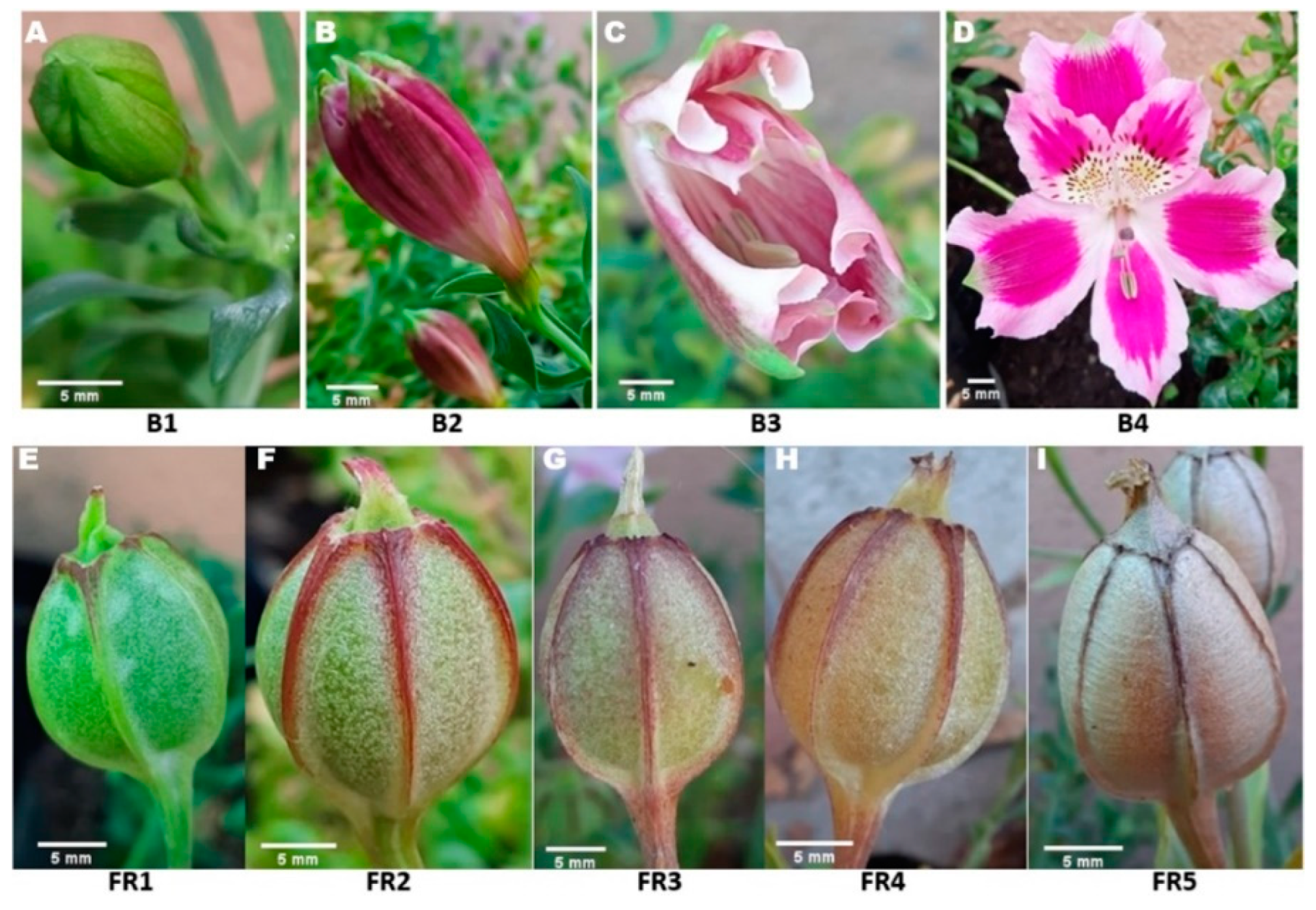
Figure 1. Morphological characterization of the reproductive stages of A. pelegrina. (A) = B1: Initiation of flower bud development; (B) = B2: Flower bud; (C) = B3: Flower bud opening; (D) = B4: Anthesis; (E) = FR1: Fruit stage 1; (F) = FR2: Fruit stage 2; (G) = FR3: Fruit stage 3; (H) = FR4: Fruit stage 4; (I) = FR5: Fruit stage 5. Scale bar: 5 mm.
Horticulturae 08 00464 g001
Figure 2. Morphological characterization of the distinct maturity stages of A. pelegrina fruit. FR1: Fruit stage 1; FR2: Fruit stage 2; FR3: Fruit stage 3; FR4: Fruit stage 4; FR5: Fruit stage 5. Scale bar: 5 mm.
Horticulturae 08 00464 g002
Figure 3. Morphological characterization of distinct seed maturity stages of A. pelegrina. S1: Seed stage 1; S2: Seed stage 2; S3: Seed stage 3; S4: Seed stage 4; S5: Seed stage 5. Scale bar: 1 mm.
Horticulturae 08 00464 g003
Figure 4. Evaluation of fresh weight of A. pelegrina seeds at different maturity stages. S1: Seed stage 1; S2: Seed stage 2; S3: Seed stage 3; S4: Seed stage 4; S5: Seed stage 5. * Values with different letters in the graph indicate statistically significant differences, Tukey (p ≤ 0.05). The bars represent the standard error. Scale bar: 1 mm.
Horticulturae 08 00464 g004
Figure 5. Dry weight of A. pelegrina seeds at different maturity stages. FR1: Fruit stage 1; FR2: Fruit stage 2; FR3: Fruit stage 3; FR4: Fruit stage 4; FR5: Fruit stage 5. * Values with different letters in the graph indicate statistically significant differences, Tukey (p ≤ 0.05). The bars represent the standard error.
Horticulturae 08 00464 g005
Figure 6. In vitro germination percentage of A. pelegrina seeds with different incision depths under the scarification treatment. CL1: control (intact, whole seed); CL2: transverse cut of the seed at the apex (opposite side of the hilum) to a depth of 0.1 to 0.5 mm; CL3: transverse cut at the apex (opposite side of the hilum) to a depth of 0.5 to 1 mm with embryo exposure. * Values with different letters in the graph indicate statistically significant differences, Tukey (p ≤ 0.05). The bars represent the standard error.
Horticulturae 08 00464 g006
Figure 7. Effect of seed scarification on cumulative germination of A. pelegrina grown in vitro. CL1: control (intact, whole seed); CL2: transverse cut of the seed at the apex (opposite side of the hilum) to a depth of 0.1 to 0.5 mm; CL3: transverse cut at the apex (opposite side of the hilum) to a depth of 0.5 to 1 mm with embryo exposure. The bars represent the standard error.
Horticulturae 08 00464 g007
Figure 8. Histological cross section of A. pelegrina seeds at different maturity stages of fruits. FR3: Fruit stage 3; FR4: Fruit stage 4; FR5: Fruit stage 5; Em: embryo; En: endosperm. Red bars represent the distance between the position of the embryo and the apical zone of the seed. Scale bar: 1000 um.
Horticulturae 08 00464 g008
Table 1. Phenological evaluation chart of A. pelegrina reproductive stages.
Reproductive Stage DurationCharacteristics (Mean ± Standard Deviation)
B1: Initiation of flower bud development11 daysLength of flower bud 10.92 ± 1.24 mm; width 7.67 ± 1.30 mm
B2: Flower bud2 daysLength of flower bud 27.76 ± 2.13 mm; width 16.48 ± 1.32 mm
B3: Flower bud opening3 daysLength of flower bud 45.92 ± 1.85 mm; width 20.89 ± 1.28 mm
B4: Flowering-Anthesis9 daysLength 97.23 ± 4.85 mm, upper outer tepal to lower outer tepal; width 45.36 ± 2.18 mm
FR1: Fruit stage 16 daysFruit length 17.65± 2.21 mm; width 14.65 ± 1.12 mm
FR2: Fruit stage 220 daysFruit length 21.01 ± 1.68 mm; width 16.24 ± 1.03 mm
FR3: Fruit stage 315 daysFruit length 23.88 ± 1.36 mm; width 16.86 ± 1.45 mm
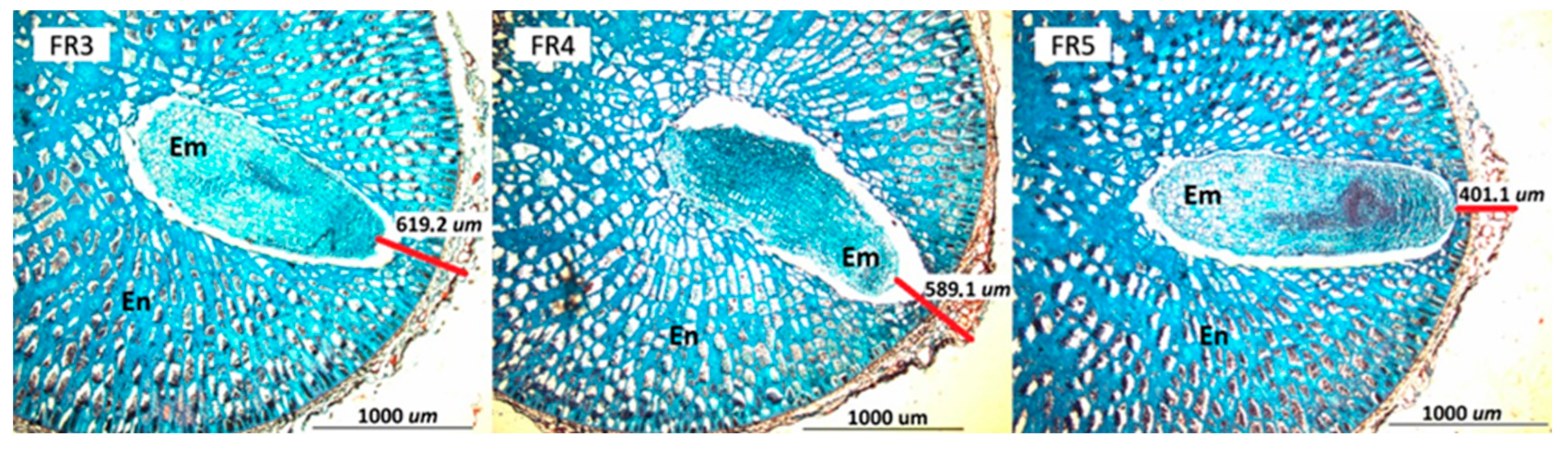
FR4: Fruit stage 45 daysFruit length 20.25 ± 1.96 mm; width 16.62 ± 1.33 mm
FR5: Fruit stage 55 daysFruit length 19.28 mm ± 1.78 mm; width 16.13 ± 0.73 mm
Table 2. Phenological stages of A. pelegrina fruit development and their respective coloration patterns.
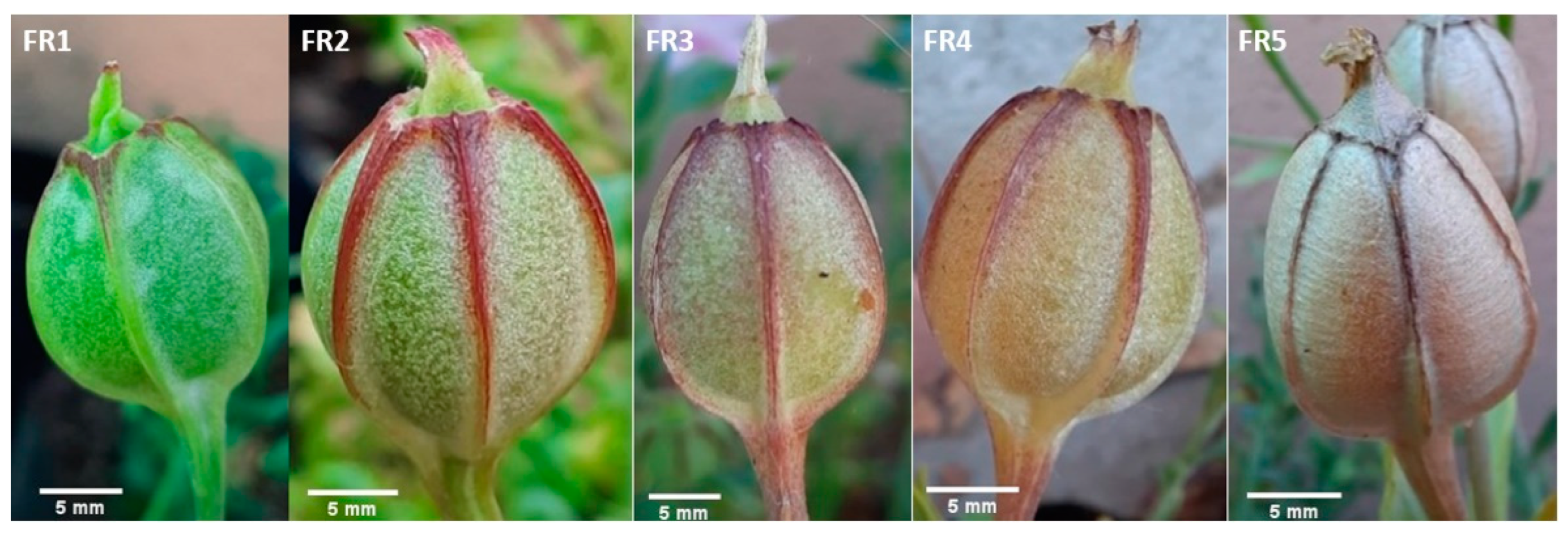
Phenological Stages of A. pelegrina Fruit DevelopmentFruit Color
Fruit stage 1 (FR1)Green (5GY 7/10) with white spots
Fruit stage 2 (FR2)Green (5GY 7/8) with white spots and 5 burgundy veins (2.5R 5/10)
Fruit stage 3 (FR3)Yellow-green (2.5GY 8/8) with 5 burgundy veins (5R 4/10)
Fruit stage 4 (FR4)Yellow (5Y 8/8) with 5 burgundy veins (10R 4/8) extending to the middle of the fruit
Fruit stage 5 (FR5)Brown (2.5YR 7/6) with 5 brown veins (7.5YR 5/2)
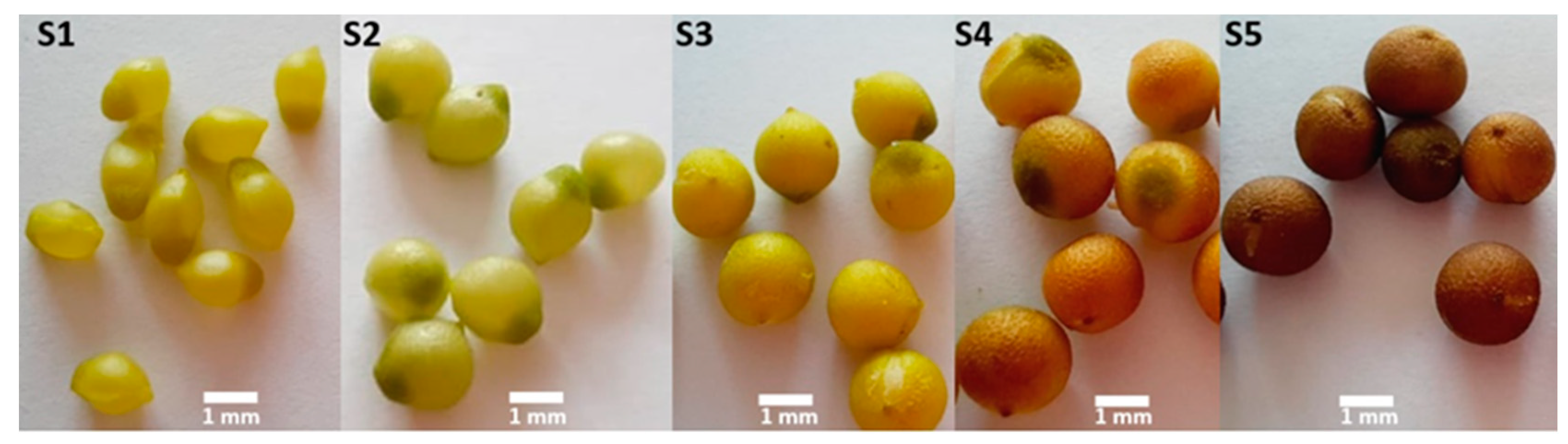
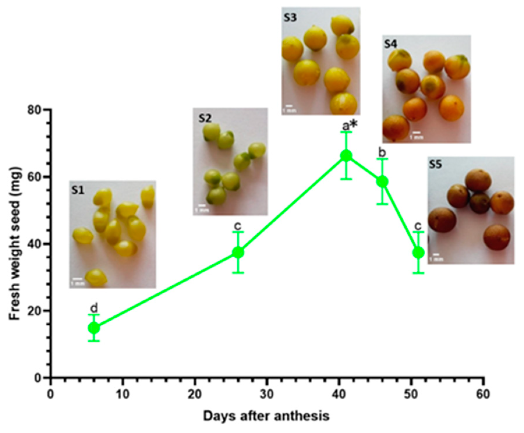
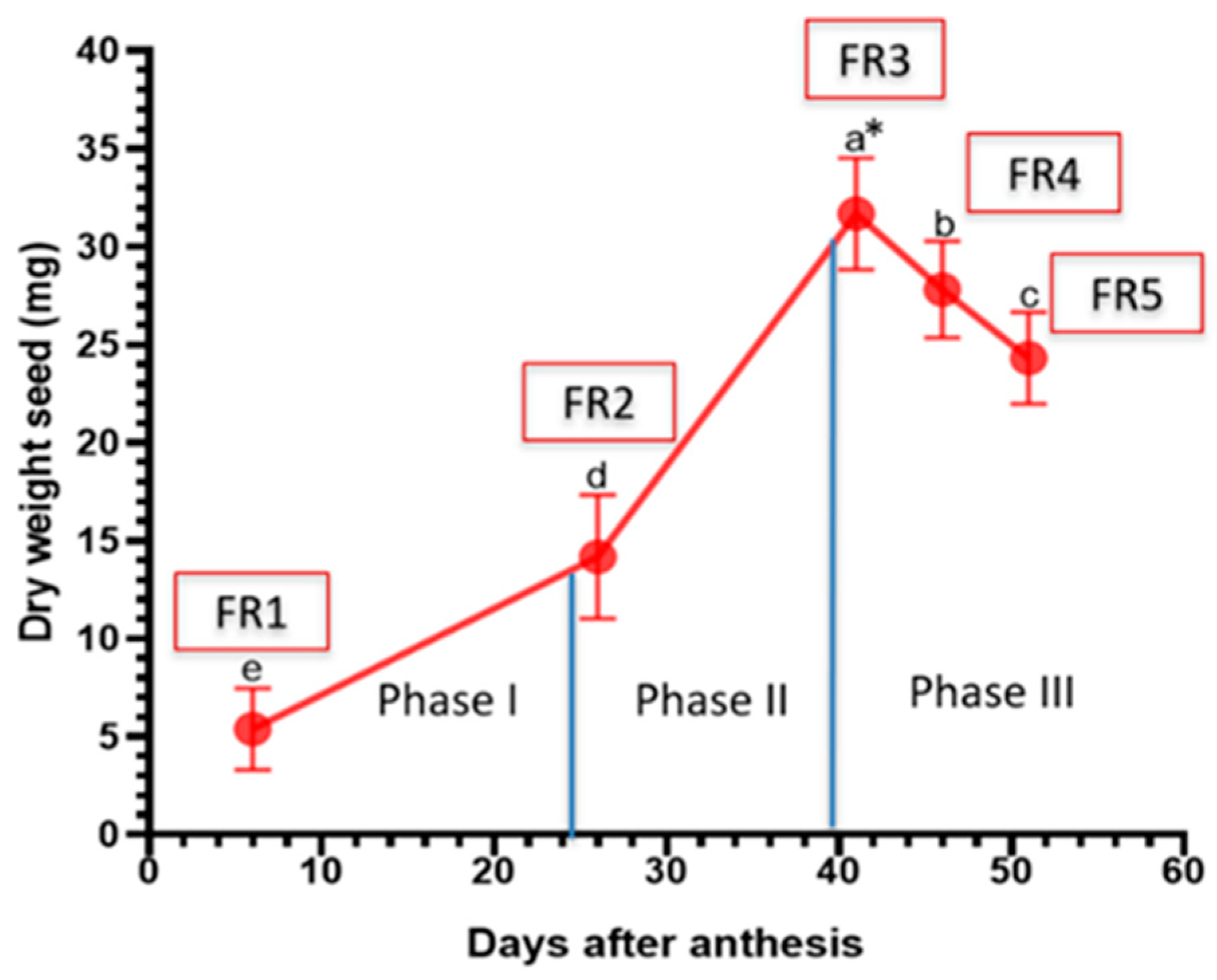
Table 3. Characteristics of A. pelegrina seeds by developmental stage. Mean ± standard deviation.
Developmental StageSeed ColorMean Fresh WeightMean Dry Weight
Seed stage 1 (S1)Lime-green seed color (2.5GY 8/12)14.97 ± 3.9 mg5.38 ± 2.1 mg
Seed stage 2 (S2)Green seed color (2.5GY 7/10)37.48 ± 6.1 mg14.18 ± 3.2 mg
Seed stage 3 (S3)Green seed color (2.5GY 8/10)66.37 ± 7.0 mg31.70 ± 2.8 mg
Seed stage 4 (S4)Brown seed color (2.5Y 6/8)58.61 ± 6.8 mg27.83 ± 2.5 mg
Seed stage 5 (S5)Dark brown seed color (7.5YR 4/4)37.44 ± 6.1 mg24.38 ± 2.4 mg
Publisher’s Note: MDPI stays neutral with regard to jurisdictional claims in published maps and institutional affiliations.

