Improved Acquisition and Reconstruction for Wavelength-Resolved Neutron Tomography †

 , ,
, , 
Abstract
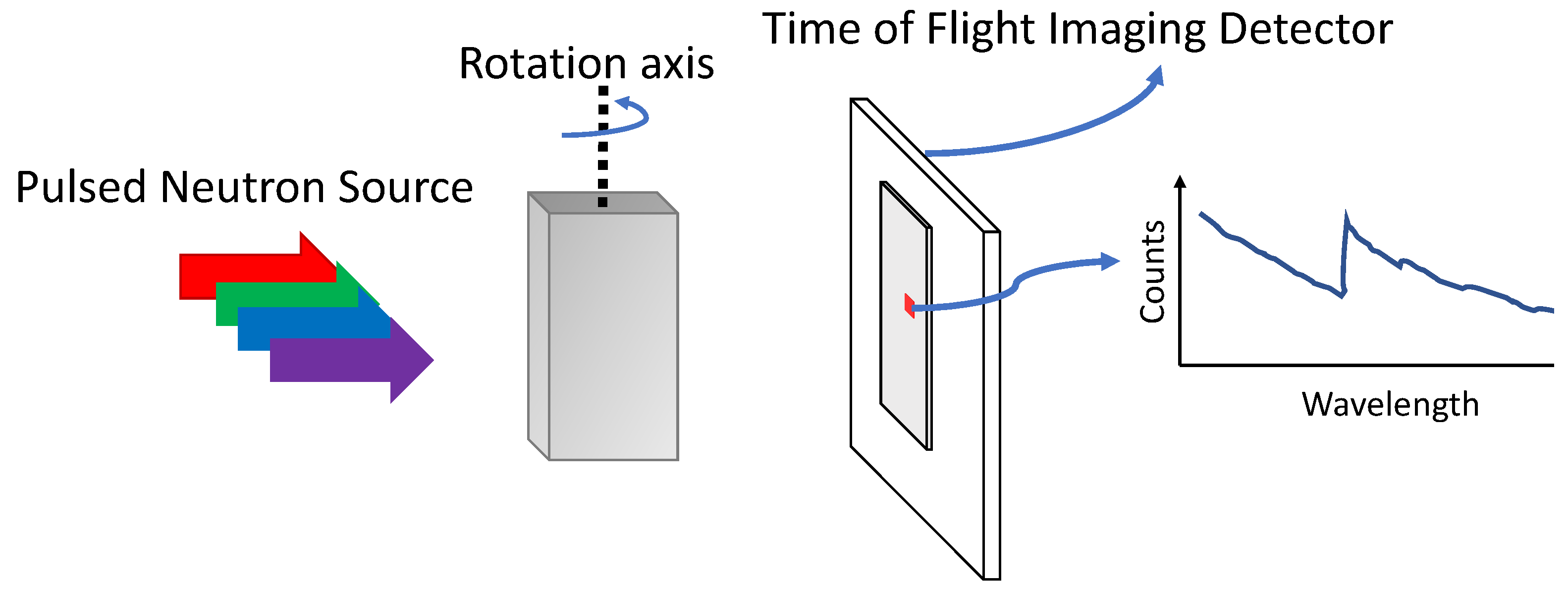
:1. Introduction
2. Interlaced Scanning and Model-Based Image Reconstruction
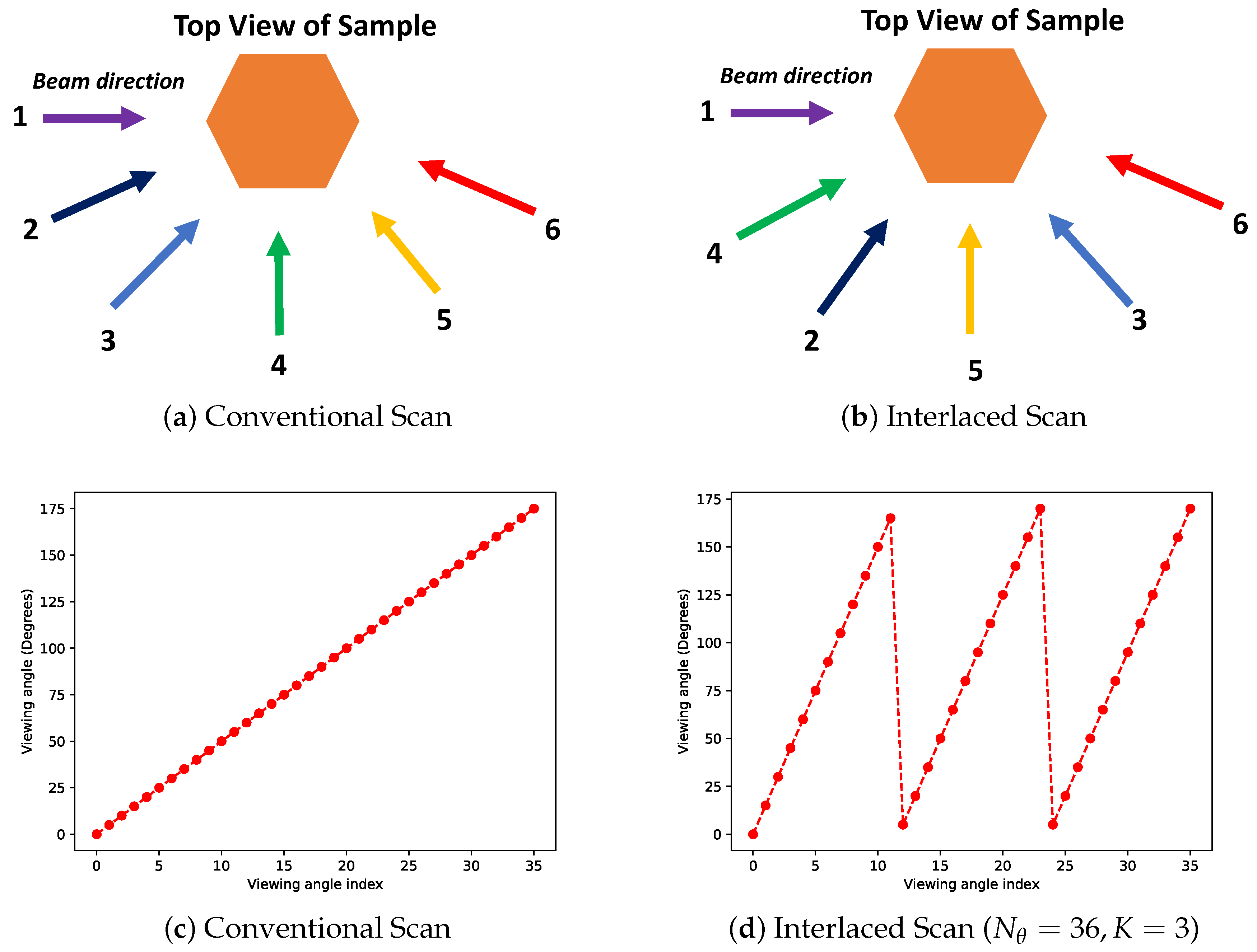
2.1. Interlaced Scanning
2.2. Model-Based Image Reconstruction for Streaming Hyper-Spectral Data
| Algorithm 1: The proposed framework for wavelength-resolved neutron tomography systems. Prior to the scan we decide a fixed number of views to measure and the number of half-rotations over which this will occur K. After each measurement the object can be reconstructed using the MBIR algorithm. The reconstruction from the previous stage is used as a initial condition for the next stage in order to accelerate the convergence i.e., time to reconstruct for the MBIR method. | ||
| 1: function Reconstruct(,K,) | ||
| 2: %Inputs: Number of views , Number of half-rotations K, Regularization parameters | ||
| 3: %Outputs: Wavelength-resolved reconstruction | ||
| 4: Compute according to (1) | ▹ Interlaced scanning angles | |
| 5: Initialize | ||
| 6: Measure open-beam data | ▹ Normalization data for transmission tomography | |
| 7: | ||
| 8: while do | ||
| 9: MEASURE() | ||
| 10: NORMALIZE(,) | ▹ Can include further corrections | |
| 11: MBIR (; ,) | ▹ Equation (8) | |
| 12: | ||
| 13: end while | ||
| 14: Return | ||
| 15: end function | ||
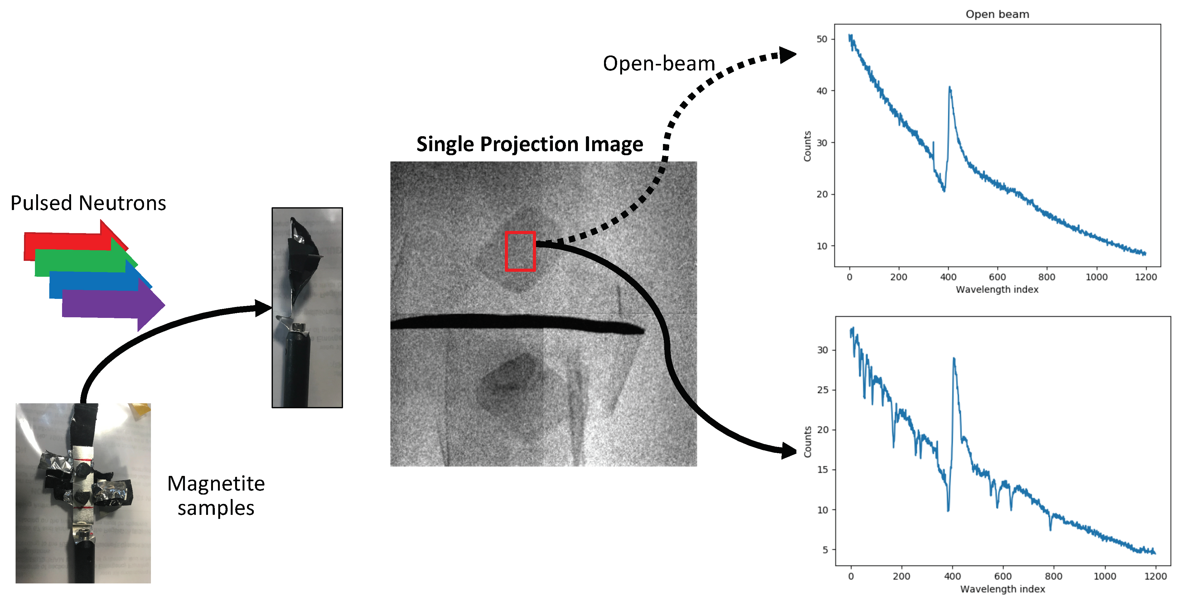
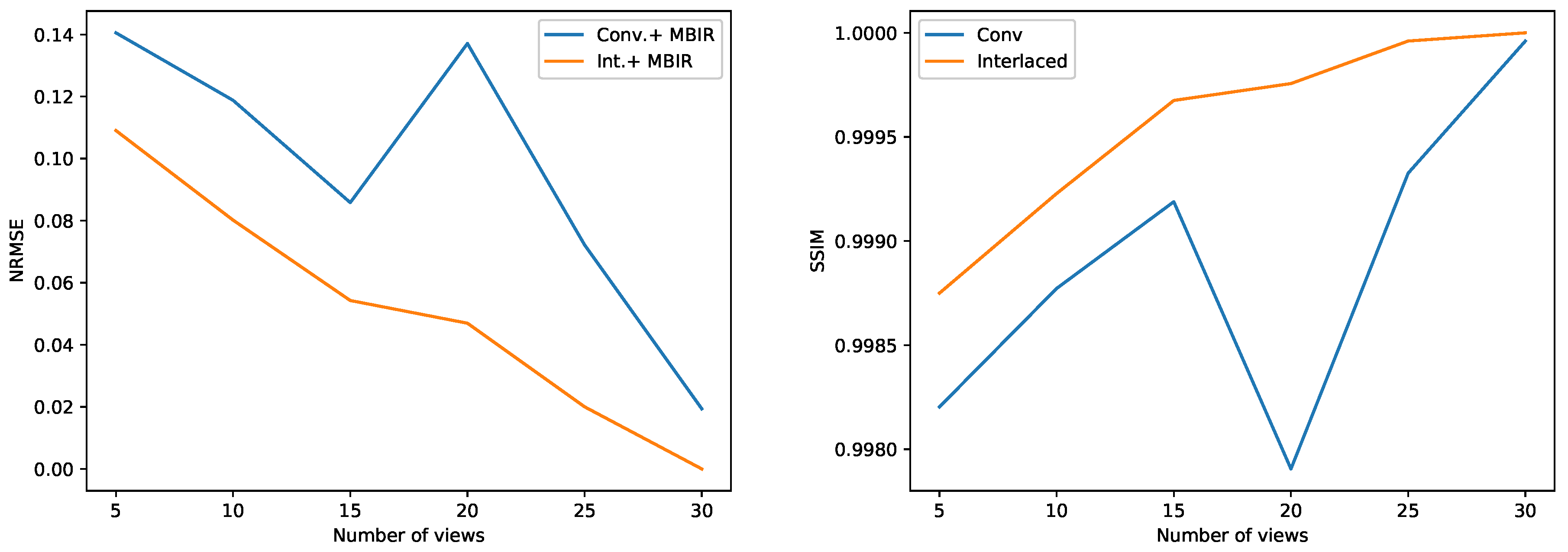
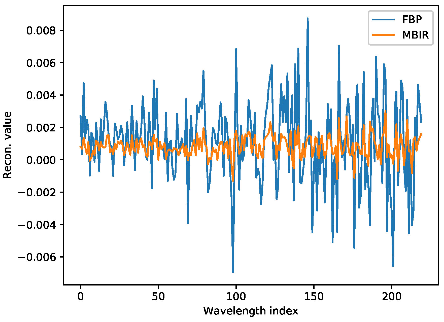
3. Results
4. Conclusions
Author Contributions
Funding
Institutional Review Board Statement
Informed Consent Statement
Data Availability Statement
Acknowledgments
Conflicts of Interest
References
- Strobl, M. Future prospects of imaging at spallation neutron sources. Nucl. Instrum. Methods Phys. Res. Sect. A Accel. Spectrometers Detect. Assoc. Equip. 2009, 604, 646–652. [Google Scholar] [CrossRef]
- Shinohara, T.; Kai, T.; Oikawa, K.; Segawa, M.; Harada, M.; Nakatani, T.; Ooi, M.; Aizawa, K.; Sato, H.; Kamiyama, T.; et al. Final design of the energy-resolved neutron imaging system RADEN at J-PARC. J. Phys. Conf. Ser. 2016, 746, 012007. [Google Scholar] [CrossRef]
- Kockelmann, W.; Minniti, T.; Pooley, D.E.; Burca, G.; Ramadhan, R.; Akeroyd, F.A.; Howells, G.D.; Moreton-Smith, C.; Keymer, D.P.; Kelleher, J.; et al. Time-of-Flight Neutron Imaging on IMAT at ISIS: A New User Facility for Materials Science. J. Imaging 2018, 4, 47. [Google Scholar] [CrossRef] [Green Version]
- Nelson, R.O.; Vogel, S.C.; Hunter, J.F.; Watkins, E.B.; Losko, A.S.; Tremsin, A.S.; Borges, N.P.; Cutler, T.E.; Dickman, L.T.; Espy, M.A.; et al. Neutron Imaging at LANSCE—From Cold to Ultrafast. J. Imaging 2018, 4, 45. [Google Scholar] [CrossRef] [Green Version]
- Tremsin, A.S.; Vallerga, J.V.; McPhate, J.B.; Siegmund, O.H.; Raffanti, R. High Resolution Photon Counting With MCP-Timepix Quad Parallel Readout Operating at >1 KHz Frame Rates. IEEE Trans. Nucl. Sci. 2013, 60, 578–585. [Google Scholar] [CrossRef]
- Myhre, K.G.; Zhang, Y.; Bilheux, H.Z.; Johnson, J.A.; Bilheux, J.C.; Miskowiec, A.J.; Hunt, R.D. Nondestructive Tomographic Mapping of Uranium and Gadolinium Using Energy-Resolved Neutron Imaging; Technical Report; Oak Ridge National Lab. (ORNL): Oak Ridge, TN, USA, 2018. [Google Scholar]
- Woracek, R.; Penumadu, D.; Kardjilov, N.; Hilger, A.; Boin, M.; Banhart, J.; Manke, I. Neutron Bragg-edge tomography for phase mapping. Phys. Procedia 2015, 69, 227–236. [Google Scholar] [CrossRef] [Green Version]
- Song, G.; Lin, J.Y.; Bilheux, J.C.; Xie, Q.; Santodonato, L.J.; Molaison, J.J.; Skorpenske, H.D.; M Dos Santos, A.; Tulk, C.A.; An, K.; et al. Characterization of Crystallographic Structures Using Bragg-Edge Neutron Imaging at the Spallation Neutron Source. J. Imaging 2017, 3, 65. [Google Scholar] [CrossRef] [Green Version]
- Cereser, A.; Strobl, M.; Hall, S.A.; Steuwer, A.; Kiyanagi, R.; Tremsin, A.S.; Knudsen, E.B.; Shinohara, T.; Willendrup, P.K.; Fanta, A.B.S.; et al. Time-of-flight three dimensional neutron diffraction in transmission mode for mapping crystal grain structures. Sci. Rep. 2017, 7, 9561. [Google Scholar] [CrossRef] [PubMed] [Green Version]
- Vogel, S.C.; Bourke, M.A.; Craft, A.E.; Harp, J.M.; Kelsey, C.T.; Lin, J.; Long, A.M.; Losko, A.S.; Hosemann, P.; McClellan, K.J.; et al. Advanced Postirradiation Characterization of Nuclear Fuels Using Pulsed Neutrons. JOM 2020, 72, 187–196. [Google Scholar] [CrossRef]
- Mohan, K.; Venkatakrishnan, S.; Gibbs, J.; Gulsoy, E.; Xiao, X.; De Graef, M.; Voorhees, P.; Bouman, C. TIMBIR: A Method for Time-Space Reconstruction from Interlaced Views. Comput. Imaging IEEE Trans. 2015, 1, 96–111. [Google Scholar] [CrossRef]
- Kohler, T. A projection access scheme for iterative reconstruction based on the golden section. In Proceedings of the IEEE Symposium Conference Record Nuclear Science 2004, Rome, Italy, 16–22 October 2004; Volume 6, pp. 3961–3965. [Google Scholar]
- Kaestner, A.P.; Munch, B.; Trtik, P. Spatiotemporal computed tomography of dynamic processes. Opt. Eng. 2011, 50, 123201. [Google Scholar]
- Kaestner, A.; Lehmann, E.; Hovind, J.; Radebe, M.; De Beer, F.; Sim, C. Verifying neutron tomography performance using test objects. Phys. Procedia 2013, 43, 128–137. [Google Scholar] [CrossRef] [Green Version]
- Kaestner, A.P.; Trtik, P.; Zarebanadkouki, M.; Kazantsev, D.; Snehota, M.; Dobson, K.J.; Lehmann, E.H. Recent developments in neutron imaging with applications for porous media research. Solid Earth 2016, 7, 1281–1292. [Google Scholar] [CrossRef] [Green Version]
- Abir, M.; Islam, F.; Wachs, D.; Lee, H.K. Sparse-view neutron CT reconstruction of irradiated fuel assembly using total variation minimization with Poisson statistics. J. Radioanal. Nucl. Chem. 2016, 307, 1967–1979. [Google Scholar] [CrossRef]
- Kazantsev, D.; Bleichrodt, F.; van Leeuwen, T.; Kaestner, A.; Withers, P.J.; Batenburg, K.J.; Lee, P.D. A novel tomographic reconstruction method based on the robust Student’s t function for suppressing data outliers. IEEE Trans. Comput. Imaging 2017, 3, 682–693. [Google Scholar] [CrossRef] [Green Version]
- Venkatakrishnan, S.; Cakmak, E.; Billheux, H.; Bingham, P.; Archibald, R.K. Model-based iterative reconstruction for neutron laminography. In Proceedings of the 2017 51st Asilomar Conference on Signals, Systems, and Computers, Pacific Grove, CA, USA, 29 October–1 November 2017; pp. 1864–1869. [Google Scholar]
- Barnard, R.C.; Bilheux, H.; Toops, T.; Nafziger, E.; Finney, C.; Splitter, D.; Archibald, R. Total variation-based neutron computed tomography. Rev. Sci. Instruments 2018, 89, 053704. [Google Scholar] [CrossRef] [PubMed] [Green Version]
- Cramblitt, R.M.; Allebach, J.P. Analysis of time-sequential sampling with a spatially hexagonal lattice. JOSA 1983, 73, 1510–1517. [Google Scholar] [CrossRef]
- Bouman, C.A. Model Based Image Processing. 2013. Available online: https://engineering.purdue.edu/~bouman/publications/pdf/MBIP-book.pdf (accessed on 30 October 2020).
- Saghi, Z.; Holland, D.J.; Leary, R.; Falqui, A.; Bertoni, G.; Sederman, A.J.; Gladden, L.F.; Midgley, P.A. Three-Dimensional Morphology of Iron Oxide Nanoparticles with Reactive Concave Surfaces. A Compressed Sensing-Electron Tomography (CS-ET) Approach. Nano Lett. 2011, 11, 4666–4673. [Google Scholar] [CrossRef]
- Goris, B.; den Broek, W.V.; Batenburg, K.; Mezerji, H.H.; Bals, S. Electron tomography based on a total variation minimization reconstruction technique. Ultramicroscopy 2012, 113, 120–130. [Google Scholar] [CrossRef]
- Venkatakrishnan, S.; Drummy, L.; Jackson, M.; De Graef, M.; Simmons, J.; Bouman, C. Model based iterative reconstruction for Bright-Field Electron Tomography. IEEE Trans. Comput. Imaging 2015, 1, 1–15. [Google Scholar] [CrossRef]
- Gürsoy, D.; Biçer, T.; Lanzirotti, A.; Newville, M.G.; De Carlo, F. Hyperspectral image reconstruction for x-ray fluorescence tomography. Opt. Express 2015, 23, 9014–9023. [Google Scholar] [CrossRef] [PubMed]
- Mohan, K.A.; Venkatakrishnan, S.V.; Drummy, L.F.; Simmons, J.; Parkinson, D.Y.; Bouman, C.A. Model-Based Iterative Reconstruction for Synchrotron X-ray Tomography. In Proceedings of the 2014 IEEE International Conference on Acoustics, Speech and Signal Processing (ICASSP), Florence, Italy, 4–9 May 2014. [Google Scholar]
- Sauer, K.; Bouman, C. Bayesian Estimation of Transmission Tomograms Using Segmentation Based Optimization. IEEE Trans. Nucl. Sci. 1992, 39, 1144–1152. [Google Scholar] [CrossRef]
- Thibault, J.B.; Sauer, K.; Bouman, C.; Hsieh, J. A three-dimensional statistical approach to improved image quality for multislice helical CT. Med. Phys. 2007, 34, 4526–4544. [Google Scholar] [CrossRef] [PubMed]
- Golbabaee, M.; Vandergheynst, P. Hyperspectral image compressed sensing via low-rank and joint-sparse matrix recovery. In Proceedings of the 2012 IEEE International Conference on Acoustics, Speech and Signal Processing (ICASSP), Kyoto, Japan, 25–30 March 2012; pp. 2741–2744. [Google Scholar]
- Zhuang, L.; Bioucas-Dias, J.M. Fast hyperspectral image denoising and inpainting based on low-rank and sparse representations. IEEE J. Sel. Top. Appl. Earth Obs. Remote Sens. 2018, 11, 730–742. [Google Scholar] [CrossRef]
- Monier, E.; Oberlin, T.; Brun, N.; Tencé, M.; de Frutos, M.; Dobigeon, N. Reconstruction of Partially Sampled Multiband Images—Application to STEM-EELS Imaging. IEEE Trans. Comput. Imaging 2018, 4, 585–598. [Google Scholar] [CrossRef] [Green Version]
- Kim, D.; Fessler, J.A. An optimized first-order method for image restoration. In Proceedings of the 2015 IEEE International Conference on Image Processing (ICIP), Quebec City, QC, Canada, 27–30 September 2015; pp. 3675–3679. [Google Scholar] [CrossRef]
- Palenstijn, W.; Batenburg, K.; Sijbers, J. Performance improvements for iterative electron tomography reconstruction using graphics processing units (GPUs). J. Struct. Biol. 2011, 176, 250–253. [Google Scholar] [CrossRef] [Green Version]
- van Aarle, W.; Palenstijn, W.J.; Beenhouwer, J.D.; Altantzis, T.; Bals, S.; Batenburg, K.J.; Sijbers, J. The ASTRA Toolbox: A platform for advanced algorithm development in electron tomography. Ultramicroscopy 2015, 157, 35–47. [Google Scholar] [CrossRef] [Green Version]
- Wang, X.; Sridhar, V.; Ronaghi, Z.; Thomas, R.; Deslippe, J.; Parkinson, D.; Buzzard, G.T.; Midkiff, S.P.; Bouman, C.A.; Warfield, S.K. Consensus equilibrium framework for super-resolution and extreme-scale CT reconstruction. In Proceedings of the International Conference for High Performance Computing, Networking, Storage and Analysis, Denver, CO, USA, 9–19 November 2019; pp. 1–23. [Google Scholar]
- Marchesini, S.; Trivedi, A.; Enfedaque, P.; Perciano, T.; Parkinson, D. Sparse Matrix-Based HPC Tomography. arXiv 2020, arXiv:2003.12677. [Google Scholar]
- Venkatakrishnan, S.V. pyMBIR; Technical Report; Oak Ridge National Lab. (ORNL): Oak Ridge, TN, USA, 2019. [Google Scholar]
- Gürsoy, D.; De Carlo, F.; Xiao, X.; Jacobsen, C. TomoPy: A framework for the analysis of synchrotron tomographic data. J. Synchrotron Radiat. 2014, 21, 1188–1193. [Google Scholar] [CrossRef] [Green Version]
- Wang, Z.; Bovik, A.C.; Sheikh, H.R.; Simoncelli, E.P. Image quality assessment: From error visibility to structural similarity. IEEE Trans. Image Process. 2004, 13, 600–612. [Google Scholar] [CrossRef] [Green Version]
- Batenburg, K.J.; Palenstijn, W.J.; Balázs, P.; Sijbers, J. Dynamic angle selection in binary tomography. Comput. Vis. Image Underst. 2013, 117, 306–318. [Google Scholar] [CrossRef] [Green Version]


Publisher’s Note: MDPI stays neutral with regard to jurisdictional claims in published maps and institutional affiliations. |
© 2021 by the authors. Licensee MDPI, Basel, Switzerland. This article is an open access article distributed under the terms and conditions of the Creative Commons Attribution (CC BY) license (http://creativecommons.org/licenses/by/4.0/).
Share and Cite
Venkatakrishnan, S.; Zhang, Y.; Dessieux, L.; Hoffmann, C.; Bingham, P.; Bilheux, H. Improved Acquisition and Reconstruction for Wavelength-Resolved Neutron Tomography. J. Imaging 2021, 7, 10. https://0-doi-org.brum.beds.ac.uk/10.3390/jimaging7010010
Venkatakrishnan S, Zhang Y, Dessieux L, Hoffmann C, Bingham P, Bilheux H. Improved Acquisition and Reconstruction for Wavelength-Resolved Neutron Tomography. Journal of Imaging. 2021; 7(1):10. https://0-doi-org.brum.beds.ac.uk/10.3390/jimaging7010010
Chicago/Turabian StyleVenkatakrishnan, Singanallur, Yuxuan Zhang, Luc Dessieux, Christina Hoffmann, Philip Bingham, and Hassina Bilheux. 2021. "Improved Acquisition and Reconstruction for Wavelength-Resolved Neutron Tomography" Journal of Imaging 7, no. 1: 10. https://0-doi-org.brum.beds.ac.uk/10.3390/jimaging7010010