Next Issue
Volume 8, August
Previous Issue
Volume 8, April


Tomography, Volume 8, Issue 3 (June 2022) – 40 articles

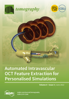
Cover Story (view full-size image): Coronary optical coherence tomography (OCT) is a light-based, intravascular imaging modality that can visualise plaque components in high resolution. This resolution is critical when identifying vulnerable plaque features, such as thin cap fibroatheroma, which dramatically increase the risk of future major adverse cardiac events (MACE). However, extracting these features is often carried out manually, a tedious and time-consuming process that limits its clinical utility. Computational techniques are emerging to automate this process, enabling near-real time image analyses and subsequent three-dimensional simulation to assist in identifying patients most at risk of future MACE. Here, we discuss recent advances in these techniques and their ability to identify the coronary lumen, artery layers, plaque components, and subtypes and stents. View this paper
  • Issues are regarded as officially published after their release is announced to the table of contents alert mailing list.
  • You may sign up for e-mail alerts to receive table of contents of newly released issues.
  • PDF is the official format for papers published in both, html and pdf forms. To view the papers in pdf format, click on the "PDF Full-text" link, and use the free Adobe Reader to open them.
Order results
Result details
Section
Select all
Export citation of selected articles as:
11 pages, 1892 KiB  
Article
Investigation of the Frequency of Coronary Artery Anomalies in MDCT Coronary Angiography and Comparison of Atherosclerotic Involvement between Anomaly Types
by Tuna Şahin and Mehtap Ilgar
Tomography 2022, 8(3), 1631-1641; https://0-doi-org.brum.beds.ac.uk/10.3390/tomography8030135 - 20 Jun 2022
Cited by 1 | Viewed by 1645
Abstract
Coronary artery anomalies (CAAs) are rare anatomical variations characterized by abnormal origin, course, or termination of the coronary arteries. This study aims to identify incidental CAAs in patients who underwent multidetector computed tomography coronary angiography (MDCTCA) to determine their incidence and to evaluate [...] Read more.
Coronary artery anomalies (CAAs) are rare anatomical variations characterized by abnormal origin, course, or termination of the coronary arteries. This study aims to identify incidental CAAs in patients who underwent multidetector computed tomography coronary angiography (MDCTCA) to determine their incidence and to evaluate whether there is a difference between CAA types in terms of coronary atherosclerotic involvement. For this purpose, patients who underwent MDCTCA between December 2018 and January 2022 were retrospectively assessed. Of the 5200 MDCTCAs analyzed, CAAs were detected in 136 patients (2.61%). Of these 136 patients, 37 (27.2%) patients had an origin anomaly, 97 (71.3%) had a course anomaly, and 2 (1.5%) had a termination anomaly. There was no statistically significant difference between CAA types in terms of atherosclerotic involvement (p = 0.220). However, atherosclerotic involvement was high in vessels with anomalies when normal vessels with and without anomalies were compared (p = 0.005). Accurate detection of CAAs is vital for endovascular treatment or surgical intervention. MDCTCA is helpful both in the diagnosis of CAA and in the early detection and development of prevention strategies for coronary atherosclerosis. Full article
(This article belongs to the Section Cardiovascular Imaging)
Show Figures

Figure 1

13 pages, 3123 KiB  
Article
Novel COVID-19 Diagnosis Delivery App Using Computed Tomography Images Analyzed with Saliency-Preprocessing and Deep Learning
by Santiago Tello-Mijares and Fomuy Woo
Tomography 2022, 8(3), 1618-1630; https://0-doi-org.brum.beds.ac.uk/10.3390/tomography8030134 - 20 Jun 2022
Cited by 1 | Viewed by 1476
Abstract
This app project was aimed to remotely deliver diagnoses and disease-progression information to COVID-19 patients to help minimize risk during this and future pandemics. Data collected from chest computed tomography (CT) scans of COVID-19-infected patients were shared through the app. In this article, [...] Read more.
This app project was aimed to remotely deliver diagnoses and disease-progression information to COVID-19 patients to help minimize risk during this and future pandemics. Data collected from chest computed tomography (CT) scans of COVID-19-infected patients were shared through the app. In this article, we focused on image preprocessing techniques to identify and highlight areas with ground glass opacity (GGO) and pulmonary infiltrates (PIs) in CT image sequences of COVID-19 cases. Convolutional neural networks (CNNs) were used to classify the disease progression of pneumonia. Each GGO and PI pattern was highlighted with saliency map fusion, and the resulting map was used to train and test a CNN classification scheme with three classes. In addition to patients, this information was shared between the respiratory triage/radiologist and the COVID-19 multidisciplinary teams with the application so that the severity of the disease could be understood through CT and medical diagnosis. The three-class, disease-level COVID-19 classification results exhibited a macro-precision of more than 94.89% in a two-fold cross-validation. Both the segmentation and classification results were comparable to those made by a medical specialist. Full article
(This article belongs to the Section Artificial Intelligence in Medical Imaging)
Show Figures

Figure 1

10 pages, 1567 KiB  
Article
Follow-Up Assessment of Intracranial Aneurysms Treated with Endovascular Coiling: Comparison of Compressed Sensing and Parallel Imaging Time-of-Flight Magnetic Resonance Angiography
by Gianfranco Vornetti, Fiorina Bartiromo, Francesco Toni, Massimo Dall’Olio, Mario Cirillo, Peter Speier, Ciro Princiotta, Michaela Schmidt, Caterina Tonon, Domenico Zacà, Raffaele Lodi and Luigi Cirillo
Tomography 2022, 8(3), 1608-1617; https://0-doi-org.brum.beds.ac.uk/10.3390/tomography8030133 - 18 Jun 2022
Cited by 1 | Viewed by 1448
Abstract
The aim of our study was to compare compressed sensing (CS) time-of-flight (TOF) magnetic resonance angiography (MRA) with parallel imaging (PI) TOF MRA in the evaluation of patients with intracranial aneurysms treated with coil embolization or stent-assisted coiling. We enrolled 22 patients who [...] Read more.
The aim of our study was to compare compressed sensing (CS) time-of-flight (TOF) magnetic resonance angiography (MRA) with parallel imaging (PI) TOF MRA in the evaluation of patients with intracranial aneurysms treated with coil embolization or stent-assisted coiling. We enrolled 22 patients who underwent follow-up imaging after intracranial aneurysm coil embolization. All patients underwent both PI TOF and CS TOF MRA during the same examination. Image evaluation aimed to compare the performance of CS to PI TOF MRA in determining the degree of aneurysm occlusion, as well as the depiction of parent vessel and vessels adjacent to the aneurysm dome. The reference standard for the evaluation of aneurysm occlusion was PI TOF MRA. The inter-modality agreement between CS and PI TOF MRA in the evaluation of aneurysm occlusion was almost perfect (κ  =  0.98, p  <  0.001) and the overall inter-rater agreement was substantial (κ  =  0.70, p  <  0.001). The visualization of aneurysm parent vessel in CS TOF images compared with PI TOF images was evaluated to be better in 11.4%, equal in 86.4%, and worse in 2.3%. CS TOF MRA, with almost 70% scan time reduction with respect to PI TOF MRA, yields comparable results for assessing the occlusion status of coiled intracranial aneurysms. Short scan times increase patient comfort, reduce the risk of motion artifacts, and increase patient throughput, with a resulting reduction in costs. CS TOF MRA may therefore be a potential replacement for PI TOF MRA as a first-line follow-up examination in patients with intracranial aneurysms treated with coil embolization. Full article
(This article belongs to the Section Brain Imaging)
Show Figures

Figure 1

13 pages, 2023 KiB  
Article
Reclassification of Heart Failure with Preserved Ejection Fraction Following Cardiac Sympathetic Nervous System Activation: A New Cutoff Value of 58%
by Toshihiko Goto, Takafumi Nakayama, Junki Yamamoto, Kento Mori, Yasuhiro Shintani, Shohei Kikuchi, Hiroshi Fujita, Hidekatsu Fukuta and Yoshihiro Seo
Tomography 2022, 8(3), 1595-1607; https://0-doi-org.brum.beds.ac.uk/10.3390/tomography8030132 - 18 Jun 2022
Viewed by 1755
Abstract
Heart failure (HF) with preserved left ventricular ejection fraction (LVEF) is a heterogeneous syndrome. An LVEF of 50% is widely used to categorize patients with HF; however, this is controversial. Previously, we have reported that patients with an LVEF of ≥ 58% have [...] Read more.
Heart failure (HF) with preserved left ventricular ejection fraction (LVEF) is a heterogeneous syndrome. An LVEF of 50% is widely used to categorize patients with HF; however, this is controversial. Previously, we have reported that patients with an LVEF of ≥ 58% have good prognoses. Further, cardiac sympathetic nervous system (SNS) activation is a feature of HF. In this retrospective, observational study, the cardiac SNS activity of HF patients (n = 63, age: 78.4 ± 9.6 years; male 49.2%) with LVEF ≥ 58% (n = 15) and LVEF < 58% (n = 48) were compared using 123I-metaiodobenzylguanidine scintigraphy. During the follow-up period (median, 3.0 years), 18 all-cause deaths occurred. The delayed heart/mediastinum (H/M) ratio was significantly higher in the LVEF ≥ 58% group than in the LVEF < 58% group (2.1 ± 0.3 vs. 1.7 ± 0.4, p = 0.004), and all-cause mortality was significantly lower in patients in the former than those in the latter group (log-rank, p = 0.04). However, when these patients were divided into LVEF ≥ 50% (n = 22) and LVEF < 50% (n = 41) groups, no significant differences were found in the delayed H/M ratio, and the all-cause mortality did not differ between the groups (log-rank, p = 0.09). In conclusion, an LVEF of 58% is suitable for reclassifying patients with HF according to cardiac SNS activity. Full article
(This article belongs to the Section Cardiovascular Imaging)
Show Figures

Figure 1

9 pages, 247 KiB  
Article
Clinical Routine and Necessary Advances in Soft Tissue Tumor Imaging Based on the ESSR Guideline: Initial Findings
by Alexander Korthaus, Sebastian Weiss, Alexej Barg, Johannes Salamon, Carsten Schlickewei, Karl-Heinz Frosch and Matthias Priemel
Tomography 2022, 8(3), 1586-1594; https://0-doi-org.brum.beds.ac.uk/10.3390/tomography8030131 - 17 Jun 2022
Viewed by 1346
Abstract
Soft tissue sarcomas are malignant diseases with a complex classification and various histological subtypes, mostly clinically inconspicuous appearance, and a rare occurrence. To ensure safe patient care, the European Society of Musculoskeletal Radiology (ESSR) issued a guideline for diagnostic imaging of soft tissue [...] Read more.
Soft tissue sarcomas are malignant diseases with a complex classification and various histological subtypes, mostly clinically inconspicuous appearance, and a rare occurrence. To ensure safe patient care, the European Society of Musculoskeletal Radiology (ESSR) issued a guideline for diagnostic imaging of soft tissue tumors in adults in 2015. In this study, we investigated whether implementation of these guidelines resulted in improved MRI protocol and report quality in patients with soft tissue sarcomas in our cancer center. All cases of histologically confirmed soft tissue sarcomas that were treated at our study center from 2006 to 2018 were evaluated retrospectively. The radiological reports were examined for their compliance with the recommendations of the ESSR. Patients were divided into two groups, before and after the introduction of the 2015 ESSR guidelines. In total, 103 cases of histologically confirmed sarcomas were studied. The distribution of, age, gender, number of subjects, performing radiology, and MRI indication on both groups did not show any significant differences. Only using the required MRI sequences showed a significant improvement after the introduction of the guidelines (p = 0.048). All other criteria, especially the requirements for the report of findings, showed no improvement. The guidelines of the European Society for Musculoskeletal Radiology are not regularly followed, and their establishment did not consistently improve MRI quality in our study group. This poses a risk for incorrect or delayed diagnosis and, ultimately, therapy of soft tissue tumors. However, this study is the first of its kind and involves a limited collective. A European-wide multicenter study would be appreciated to confirm these results. Full article
8 pages, 646 KiB  
Article
Quantitative Chest CT Analysis to Measure Short-Term Sequelae of COVID-19 Pneumonia: A Monocentric Prospective Study
by Ezio Lanza, Angela Ammirabile, Maddalena Casana, Daria Pocaterra, Federica Maria Pilar Tordato, Benedetta Varisco, Costanza Lisi, Gaia Messana, Luca Balzarini and Paola Morelli
Tomography 2022, 8(3), 1578-1585; https://0-doi-org.brum.beds.ac.uk/10.3390/tomography8030130 - 17 Jun 2022
Viewed by 1686
Abstract
(1) Background: Quantitative CT analysis (QCT) has demonstrated promising results in the prognosis prediction of patients affected by COVID-19. We implemented QCT not only at diagnosis but also at short-term follow-up, pairing it with a clinical examination in search of a correlation between [...] Read more.
(1) Background: Quantitative CT analysis (QCT) has demonstrated promising results in the prognosis prediction of patients affected by COVID-19. We implemented QCT not only at diagnosis but also at short-term follow-up, pairing it with a clinical examination in search of a correlation between residual respiratory symptoms and abnormal QCT results. (2) Methods: In this prospective monocentric trial performed during the “first wave” of the Italian pandemic, i.e., from March to May 2020, we aimed to test the relationship between %deltaCL (variation of %CL-compromised lung volume) and variations of symptoms-dyspnea, cough and chest pain-at follow-up clinical assessment after hospitalization. (3) Results: 282 patients (95 females, 34%) with a median age of 60 years (IQR, 51–69) were included. We reported a correlation between changing lung abnormalities measured by QCT, and residual symptoms at short-term follow up after COVID-19 pneumonia. Independently from age, a low percentage of surviving patients (1–4%) may present residual respiratory symptoms at approximately two months after discharge. QCT was able to quantify the extent of residual lung damage underlying such symptoms, as the reduction of both %PAL (poorly aerated lung) and %CL volumes was correlated to their disappearance. (4) Conclusions QCT may be used as an objective metric for the measurement of COVID-19 sequelae. Full article
(This article belongs to the Special Issue The Challenge of Advanced Medical Imaging Data Analysis in COVID-19)
Show Figures

Figure 1

8 pages, 1776 KiB  
Case Report
Imaging Neurodegenerative Metabolism in Amyotrophic Lateral Sclerosis with Hyperpolarized [1-13C]pyruvate MRI
by Nikolaj Bøgh, Christoffer Laustsen, Esben S. S. Hansen, Hatice Tankisi, Lotte B. Bertelsen and Jakob U. Blicher
Tomography 2022, 8(3), 1570-1577; https://0-doi-org.brum.beds.ac.uk/10.3390/tomography8030129 - 14 Jun 2022
Cited by 4 | Viewed by 1889
Abstract
The cause of amyotrophic lateral sclerosis (ALS) is still unknown, and consequently, early diagnosis of the disease can be difficult and effective treatment is lacking. The pathology of ALS seems to involve specific disturbances in carbohydrate metabolism, which may be diagnostic and therapeutic [...] Read more.
The cause of amyotrophic lateral sclerosis (ALS) is still unknown, and consequently, early diagnosis of the disease can be difficult and effective treatment is lacking. The pathology of ALS seems to involve specific disturbances in carbohydrate metabolism, which may be diagnostic and therapeutic targets. Magnetic resonance imaging (MRI) with hyperpolarized [1-13C]pyruvate is emerging as a technology for the evaluation of pathway-specific changes in the brain’s metabolism. By imaging pyruvate and the lactate and bicarbonate it is metabolized into, the technology is sensitive to the metabolic changes of inflammation and mitochondrial dysfunction. In this study, we performed hyperpolarized MRI of a patient with newly diagnosed ALS. We found a lateralized difference in [1-13C]pyruvate-to-[1-13C]lactate exchange with no changes in exchange from [1-13C]pyruvate to 13C-bicarbonate. The 40% increase in [1-13C]pyruvate-to-[1-13C]lactate exchange corresponded with the patient’s symptoms and presentation with upper-motor neuron affection and cortical hyperexcitability. The data presented here demonstrate the feasibility of performing hyperpolarized MRI in ALS. They indicate potential in pathway-specific imaging of dysfunctional carbohydrate metabolism in ALS, an enigmatic neurodegenerative disease. Full article
(This article belongs to the Section Brain Imaging)
Show Figures

Figure 1

18 pages, 5066 KiB  
Article
The Influence of Data-Driven Compressed Sensing Reconstruction on Quantitative Pharmacokinetic Analysis in Breast DCE MRI
by Ping Ni Wang, Julia V. Velikina, Leah C. Henze Bancroft, Alexey A. Samsonov, Frederick Kelcz, Roberta M. Strigel and James H. Holmes
Tomography 2022, 8(3), 1552-1569; https://0-doi-org.brum.beds.ac.uk/10.3390/tomography8030128 - 14 Jun 2022
Cited by 2 | Viewed by 1459
Abstract
Radial acquisition with MOCCO reconstruction has been previously proposed for high spatial and temporal resolution breast DCE imaging. In this work, we characterize MOCCO across a wide range of temporal contrast enhancement in a digital reference object (DRO). Time-resolved radial data was simulated [...] Read more.
Radial acquisition with MOCCO reconstruction has been previously proposed for high spatial and temporal resolution breast DCE imaging. In this work, we characterize MOCCO across a wide range of temporal contrast enhancement in a digital reference object (DRO). Time-resolved radial data was simulated using a DRO with lesions in different PK parameters. The under sampled data were reconstructed at 5 s temporal resolution using the data-driven low-rank temporal model for MOCCO, compressed sensing with temporal total variation (CS-TV) and more conventional low-rank reconstruction (PCB). Our results demonstrated that MOCCO was able to recover curves with Ktrans values ranging from 0.01 to 0.8 min−1 and fixed Ve = 0.3, where the fitted results are within a 10% bias error range. MOCCO reconstruction showed less impact on the selection of different temporal models than conventional low-rank reconstruction and the greater error was observed with PCB. CS-TV showed overall underestimation in both Ktrans and Ve. For the Monte-Carlo simulations, MOCCO was found to provide the most accurate reconstruction results for curves with intermediate lesion kinetics in the presence of noise. Initial in vivo experiences are reported in one patient volunteer. Overall, MOCCO was able to provide reconstructed time-series data that resulted in a more accurate measurement of PK parameters than PCB and CS-TV. Full article
(This article belongs to the Section Cancer Imaging)
Show Figures

Figure 1

8 pages, 1602 KiB  
Article
Magnetic Susceptibility Source Separation Solely from Gradient Echo Data: Histological Validation
by Alexey V. Dimov, Kelly M. Gillen, Thanh D. Nguyen, Jerry Kang, Ria Sharma, David Pitt, Susan A. Gauthier and Yi Wang
Tomography 2022, 8(3), 1544-1551; https://0-doi-org.brum.beds.ac.uk/10.3390/tomography8030127 - 14 Jun 2022
Cited by 6 | Viewed by 2167
Abstract
Quantitative susceptibility mapping (QSM) facilitates mapping of the bulk magnetic susceptibility of tissue from the phase of complex gradient echo (GRE) MRI data. QSM phase processing combined with an R2* model of magnitude of multiecho gradient echo data ( [...] Read more.
Quantitative susceptibility mapping (QSM) facilitates mapping of the bulk magnetic susceptibility of tissue from the phase of complex gradient echo (GRE) MRI data. QSM phase processing combined with an R2* model of magnitude of multiecho gradient echo data (R2*QSM) allows separation of dia- and para-magnetic components (e.g., myelin and iron) that contribute constructively to R2* value but destructively to the QSM value of a voxel. This R2*QSM technique is validated against quantitative histology—optical density of myelin basic protein and Perls’ iron histological stains of rim and core of 10 ex vivo multiple sclerosis lesions, as well as neighboring normal appearing white matter. We found that R2*QSM source maps are in good qualitative agreement with histology, e.g., showing increased iron concentration at the edge of the rim+ lesions and myelin loss in the lesions’ core. Furthermore, our results indicate statistically significant correlation between paramagnetic and diamagnetic tissue components estimated with R2*QSM and optical densities of Perls’ and MPB stains. These findings provide direct support for the use of R2*QSM magnetic source separation based solely on GRE complex data to characterize MS lesion composition. Full article
(This article belongs to the Section Brain Imaging)
Show Figures

Figure 1

10 pages, 2058 KiB  
Article
Alert Germ Infections: Chest X-ray and CT Findings in Hospitalized Patients Affected by Multidrug-Resistant Acinetobacter baumannii Pneumonia
by Raffaella Capasso, Antonio Pinto, Nicola Serra, Umberto Atripaldi, Adele Corcione, Giorgio Bocchini, Salvatore Guarino, Roberta Lieto, Gaetano Rea, Giacomo Sica and Tullio Valente
Tomography 2022, 8(3), 1534-1543; https://0-doi-org.brum.beds.ac.uk/10.3390/tomography8030126 - 14 Jun 2022
Viewed by 1662
Abstract
Acinetobacter baumannii (Ab) is an opportunistic Gram-negative pathogen intrinsically resistant to many antimicrobials. The aim of this retrospective study was to describe the imaging features on chest X-ray (CXR) and computed tomography (CT) scans in hospitalized patients with multidrug-resistant (MDR) Ab pneumonia. CXR [...] Read more.
Acinetobacter baumannii (Ab) is an opportunistic Gram-negative pathogen intrinsically resistant to many antimicrobials. The aim of this retrospective study was to describe the imaging features on chest X-ray (CXR) and computed tomography (CT) scans in hospitalized patients with multidrug-resistant (MDR) Ab pneumonia. CXR and CT findings were graded on a three-point scale: 1 represents normal attenuation, 2 represents ground-glass attenuation, and 3 represents consolidation. For each lung zone, with a total of six lung zones in each patient, the extent of disease was graded using a five-point scale: 0, no involvement; 1, involving 25% of the zone; 2, 25–50%; 3, 50–75%; and 4, involving >75% of the zone. Points from all zones were added for a final total cumulative score ranging from 0 to 72. Among 94 patients who tested positive for MDR Ab and underwent CXR (males 52.9%, females 47.1%; mean age 64.2 years; range 1–90 years), 68 patients underwent both CXR and chest CT examinations. The percentage of patients with a positive CT score was significantly higher than that obtained on CXR (67.65% > 35.94%, p-value = 0.00258). CT score (21.88 ± 15.77) was significantly (p-value = 0.0014) higher than CXR score (15.06 ± 18.29). CXR and CT revealed prevalent bilateral abnormal findings mainly located in the inferior and middle zones of the lungs. They primarily consisted of peripheral ground-glass opacities and consolidations which predominated on CXR and CT, respectively. Full article
Show Figures

Figure 1

12 pages, 3088 KiB  
Article
Visual Evaluation of Ultrafast MRI in the Assessment of Residual Breast Cancer after Neoadjuvant Systemic Therapy: A Preliminary Study Association with Subtype
by Maya Honda, Masako Kataoka, Mami Iima, Rie Ota, Akane Ohashi, Ayami Ohno Kishimoto, Kanae Kawai Miyake, Marcel Dominik Nickel, Yosuke Yamada, Masakazu Toi and Yuji Nakamoto
Tomography 2022, 8(3), 1522-1533; https://0-doi-org.brum.beds.ac.uk/10.3390/tomography8030125 - 10 Jun 2022
Cited by 4 | Viewed by 3712
Abstract
The purpose of this study was to investigate the diagnostic performance of ultrafast DCE (UF-DCE) MRI after the completion of neoadjuvant systemic therapy (NST) in breast cancer. In this study, MR examinations of 55 post-NST breast cancers were retrospectively analyzed. Residual tumor sizes [...] Read more.
The purpose of this study was to investigate the diagnostic performance of ultrafast DCE (UF-DCE) MRI after the completion of neoadjuvant systemic therapy (NST) in breast cancer. In this study, MR examinations of 55 post-NST breast cancers were retrospectively analyzed. Residual tumor sizes were measured in the 20th phase of UF-DCE MRI, early and delayed phases of conventional DCE MRI, and high spatial-resolution CE MRI (UF, early, delayed, and HR, respectively). The diagnostic performance for the detection of residual invasive cancer was calculated by ROC analysis. The size difference between MRI and pathological findings was analyzed using the Wilcoxon signed-rank test with the Bonferroni correction. The overall AUC was highest for UF (0.86 and 0.88 for readers 1 and 2, respectively). The difference in imaging and pathological sizes for UF (5.7 ± 8.2 mm) was significantly smaller than those for early, delayed, and HR (p < 0.01). For luminal subtype breast cancer, the size difference was significantly smaller for UF and early than for delayed (p < 0.01). UF-DCE MRI demonstrated higher AUC and specificity for the more accurate detection of residual cancer and the visualization of tumor extent than conventional DCE MRI. Full article
(This article belongs to the Section Cancer Imaging)
Show Figures

Figure 1

13 pages, 300 KiB  
Article
Dynamic Contrast Enhanced Study in Multiparametric Examination of the Prostate—Can We Make Better Use of It?
by Silva Guljaš, Mirta Benšić, Zdravka Krivdić Dupan, Oliver Pavlović, Vinko Krajina, Deni Pavoković, Petra Šmit Takač, Matija Hranić and Tamer Salha
Tomography 2022, 8(3), 1509-1521; https://0-doi-org.brum.beds.ac.uk/10.3390/tomography8030124 - 09 Jun 2022
Cited by 1 | Viewed by 2012
Abstract
We sought to investigate whether quantitative parameters from a dynamic contrast-enhanced study can be used to differentiate cancer from normal tissue and to determine a cut-off value of specific parameters that can predict malignancy more accurately, compared to the obturator internus muscle as [...] Read more.
We sought to investigate whether quantitative parameters from a dynamic contrast-enhanced study can be used to differentiate cancer from normal tissue and to determine a cut-off value of specific parameters that can predict malignancy more accurately, compared to the obturator internus muscle as a reference tissue. This retrospective study included 56 patients with biopsy proven prostate cancer (PCa) after multiparametric magnetic resonance imaging (mpMRI), with a total of 70 lesions; 39 were located in the peripheral zone, and 31 in the transition zone. The quantitative parameters for all patients were calculated in the detected lesion, morphologically normal prostate tissue and the obturator internus muscle. Increase in the Ktrans value was determined in lesion-to-muscle ratio by 3.974368, which is a cut-off value to differentiate between prostate cancer and normal prostate tissue, with specificity of 72.86% and sensitivity of 91.43%. We introduced a model to detect prostate cancer that combines Ktrans lesion-to-muscle ratio value and iAUC lesion-to-muscle ratio value, which is of higher accuracy compared to individual variables. Based on this model, we identified the optimal cut-off value with 100% sensitivity and 64.28% specificity. The use of quantitative DCE pharmacokinetic parameters compared to the obturator internus muscle as reference tissue leads to higher diagnostic accuracy for prostate cancer detection. Full article
(This article belongs to the Special Issue New Advances in Medical Imaging and Applied Radiology in Cancers)
6 pages, 573 KiB  
Case Report
Atypical Cognitive Impairment and Recovery in Two Colorectal Cancer Patients
by Hui Su Lee, Hwan Ho Jo, Ko Woon Kim, Byoung-Soo Shin and Hyun Goo Kang
Tomography 2022, 8(3), 1503-1508; https://0-doi-org.brum.beds.ac.uk/10.3390/tomography8030123 - 09 Jun 2022
Viewed by 1568
Abstract
Cognitive impairment in cancer patients can be caused by various factors; in approximately 30% of cancer patients, the symptoms appear before starting treatment. Paraneoplastic limbic encephalitis (PLE) is a rare disease associated with an autoimmune response, and is characterized by memory loss, depression, [...] Read more.
Cognitive impairment in cancer patients can be caused by various factors; in approximately 30% of cancer patients, the symptoms appear before starting treatment. Paraneoplastic limbic encephalitis (PLE) is a rare disease associated with an autoimmune response, and is characterized by memory loss, depression, and personality changes; it is one of the potential causes of cognitive dysfunction in cancer patients. Two patients were previously diagnosed with mild cognitive impairment and maintained clinical stability; after suffering a rapid change in personality and sudden cognitive decline, colorectal cancer was diagnosed within a few months. The patients did not meet the diagnostic criteria for PLE in several tests. The symptoms improved after the underlying cancer was treated, and the patients returned to their previous stable state. Sudden cognitive impairment may appear as an early cancer symptom, and PLE is considered an atypical cause for these symptoms. However, in patients with unexplained PLE-like symptoms who do not meet the diagnostic criteria for PLE, probable etiologies to be considered are the gut–brain connection, CD8+ T-cell-mediated limbic encephalitis, and somatic mutations in dementia-related genes. Currently, few studies have investigated these symptoms, and further research will offer significant therapeutic strategies for cognitive impairment in cancer patients. Full article
Show Figures

Figure 1

10 pages, 6191 KiB  
Article
What Findings on Chest CTs Can Delay Diagnosis of Pleuropulmonary Paragonimiasis?
by Kai Ke Li, Gong Yong Jin and Keun Sang Kwon
Tomography 2022, 8(3), 1493-1502; https://0-doi-org.brum.beds.ac.uk/10.3390/tomography8030122 - 09 Jun 2022
Cited by 1 | Viewed by 1679
Abstract
Purpose: The purpose of this study was to investigate which findings were delayed in diagnosis with respect to chest CT findings of paragonimiasis. Methods: This retrospective, informed questionnaire study was conducted to evaluate chest CT scans of 103 patients (58 men and 45 [...] Read more.
Purpose: The purpose of this study was to investigate which findings were delayed in diagnosis with respect to chest CT findings of paragonimiasis. Methods: This retrospective, informed questionnaire study was conducted to evaluate chest CT scans of 103 patients (58 men and 45 women; mean age 46.1 ± 14.6 years). The patients were diagnosed with paragonimiasis from 2003 to 2008 in four tertiary hospitals. Statistical analysis was performed using the chi-square test to identify differences between an initially correct diagnosis and an incorrect one of paragonimiasis on chest CT scans, for which we evaluated such variables as the location of lesion, type of parenchymal lesions, and worm migration track. Results: Nodular opacities on chest CT scans were the most common findings (53/94, 56.4%). The sign of worm migration tracks was only present in 18.1% of cases (17/94). Although statistically insignificant, the form of consolidation (18/25, 72%) and mass (6/8, 75%) on CT was common in correct diagnostics, and the form of the worm migration track (12/17, 70.6%) was high in correct diagnostics. Conclusion: A delayed diagnosis of paragonimiasis may often be made in patients with non-nodular, parenchymal lesions who are negative for worm migration track on chest CT scans. Full article
Show Figures

Figure 1

8 pages, 1422 KiB  
Article
Changes of Brain Structures and Psychological Characteristics in Predatory, Affective Violent and Nonviolent Offenders
by Ming-Chung Chou, Tien-Cheng Cheng, Pinchen Yang, Rueih-Chin Lin and Ming-Ting Wu
Tomography 2022, 8(3), 1485-1492; https://0-doi-org.brum.beds.ac.uk/10.3390/tomography8030121 - 08 Jun 2022
Viewed by 1757
Abstract
Purpose: Violent subjects were demonstrated to exhibit abnormal brain structures; however, the brain changes may be different between criminals committing affective (VA), predatory violence (VP), and non-violence (NV). Therefore, the purpose of this study was to compare the differences in brain structures and [...] Read more.
Purpose: Violent subjects were demonstrated to exhibit abnormal brain structures; however, the brain changes may be different between criminals committing affective (VA), predatory violence (VP), and non-violence (NV). Therefore, the purpose of this study was to compare the differences in brain structures and psychological characteristics between VA, VP, and NV offenders. Methods: Twenty male criminal subjects (7 VP; 6 VA; and 7 NV) offenders; and twenty age-matched male healthy non-criminals were enrolled in this study. All subjects received psychological assessments as well as magnetic resonance imaging scans of the brain. Analysis of variance (ANOVA) was performed to understand the differences among four groups with Bonferroni correction. The voxel-based morphometry and voxel-wise diffusion tensor imaging analyses were performed to compare the gray matter (GM) volume and white matter (WM) integrity between the groups. In significant regions, a Spearman correlation analysis was performed to understand the relationship between the brain changes and psychological scores. Results: The ANOVA analysis showed that AUDIT scores were significantly different among four groups, but no significant group difference was noted after Bonferroni correction. The imaging comparisons further demonstrated that the VP and NV offenders exhibited significant alterations of WM and GM tissues in the rectus and superior temporal gyrus, respectively. In addition, the VP offenders exhibited greater GM volumes than VA offenders in the right middle frontal gyrus, and NV offenders had greater GM volumes than VP offenders in the bilateral thalamus. Conclusion: We concluded that the VA, VP, and NV groups exhibited different degrees of alterations in GM and WM tissues in regions involved in emotion and cognition. Full article
(This article belongs to the Section Brain Imaging)
Show Figures

Figure 1

8 pages, 330 KiB  
Article
Effect of Androgen Deprivation Therapy on the Results of PET/CT with 18F-Fluciclovine in Patients with Metastatic Prostate Cancer
by Tore Bach-Gansmo, Katrine Korsan and Trond Velde Bogsrud
Tomography 2022, 8(3), 1477-1484; https://0-doi-org.brum.beds.ac.uk/10.3390/tomography8030120 - 03 Jun 2022
Cited by 1 | Viewed by 2959
Abstract
Background: 18F-fluciclovine is a positron emission tomography (PET) radiotracer approved for the detection of prostate cancer recurrence. No effect of androgen deprivation therapy (ADT) on its performance has been established. Purpose: To study the impact of concurrent ADT on disease detection with 18F-fluciclovine [...] Read more.
Background: 18F-fluciclovine is a positron emission tomography (PET) radiotracer approved for the detection of prostate cancer recurrence. No effect of androgen deprivation therapy (ADT) on its performance has been established. Purpose: To study the impact of concurrent ADT on disease detection with 18F-fluciclovine PET in patients with prostate cancer. Materials and Methods: Data from patients with prostate cancer who had been receiving ADT for ≥3 months at the time of undergoing an 18F-fluciclovine PET/CT at our institution were retrospectively reviewed. Seventy-three scans from 71 patients were included. The scans indicated rising prostate-specific antigen (n = 58), staging advanced disease (n = 4) or therapeutic monitoring (n = 9). Patients’ medical records provided baseline clinical data and post-scan outcomes (median follow-up 40 months). Results: Malignant lesions with increased uptake of 18F-fluciclovine were detected in 60/73 (82%) scans; 33 (45%) had lesions in the prostate/bed and 46 (63%) in extraprostatic sites. Patients received ADT for a median of 2 years (range 3 months to >10 years) pre-scan. The time on ADT did not influence detection; the detection rates were 89% for patients who had received ADT for <1 year, 63% for a treatment period of 1–<2 years, 83% for 2–4 years, 78% for >4–10 years, and 67% for a treatment period of >10 years. Conclusion: 18F-fluciclovine detected recurrent or metastatic lesions in 82% of patients with prostate cancer receiving ADT. The rates achieved in the present study are consistent with widely reported data for 18F-fluciclovine PET/CT, suggesting that withdrawal of ADT before scanning is not necessary. Full article
(This article belongs to the Special Issue Advances in the Radiography of Prostate Cancer)
Show Figures

Figure 1

11 pages, 5482 KiB  
Article
Image Quality and Radiation Dose of Contrast-Enhanced Chest-CT Acquired on a Clinical Photon-Counting Detector CT vs. Second-Generation Dual-Source CT in an Oncologic Cohort: Preliminary Results
by Florian Hagen, Lukas Walder, Jan Fritz, Ralf Gutjahr, Bernhard Schmidt, Sebastian Faby, Fabian Bamberg, Stefan Schoenberg, Konstantin Nikolaou and Marius Horger
Tomography 2022, 8(3), 1466-1476; https://0-doi-org.brum.beds.ac.uk/10.3390/tomography8030119 - 03 Jun 2022
Cited by 15 | Viewed by 2794
Abstract
Our aim was to compare the image quality and patient dose of contrast-enhanced oncologic chest-CT of a first-generation photon-counting detector (PCD-CT) and a second-generation dual-source dual-energy CT (DSCT). For this reason, one hundred consecutive oncologic patients (63 male, 65 ± 11 years, BMI: [...] Read more.
Our aim was to compare the image quality and patient dose of contrast-enhanced oncologic chest-CT of a first-generation photon-counting detector (PCD-CT) and a second-generation dual-source dual-energy CT (DSCT). For this reason, one hundred consecutive oncologic patients (63 male, 65 ± 11 years, BMI: 16–42 kg/m2) were prospectively enrolled and evaluated. Clinically indicated contrast-enhanced chest-CT were obtained with PCD-CT and compared to previously obtained chest-DSCT in the same individuals. The median time interval between the scans was three months. The same contrast media protocol was used for both scans. PCD-CT was performed in QuantumPlus mode (obtaining full spectral information) at 120 kVp. DSCT was performed using 100 kV for Tube A and 140 kV for Tube B. “T3D” PCD-CT images were evaluated, which emulate conventional 120 keV polychromatic images. For DSCT, the convolution algorithm was set at I31f with class 1 iterative reconstruction, whereas comparable Br40 kernel and iterative reconstruction strengths (Q1 and Q3) were applied for PCD-CT. Two radiologists assessed image quality using a five-point Likert scale and performed measurements of vessels and lung parenchyma for signal-to-noise ratio (SNR), contrast-to-noise ratio (CNR), and in the case of pulmonary metastases tumor-to-lung parenchyma contrast ratio. PCD-CT CNRvessel was significantly higher than DSCT CNRvessel (all, p < 0.05). Readers rated image contrast of mediastinum, vessels, and lung parenchyma significantly higher in PCD-CT than DSCT images (p < 0.001). Q3 PCD-CT CNRlung_parenchyma was significantly higher than DSCT CNRlung_parenchyma and Q1 PCD-CT CNRlung_parenchyma (p < 0.01). The tumor-to-lung parenchyma contrast ratio was significantly higher on PCD-CT than DSCT images (0.08 ± 0.04 vs. 0.03 ± 0.02, p < 0.001). CTDI, DLP, SSDE mean values for PCD-CT and DSCT were 4.17 ± 1.29 mGy vs. 7.21 ± 0.49 mGy, 151.01 ± 48.56 mGy * cm vs. 288.64 ± 31.17 mGy * cm and 4.23 ± 0.97 vs. 7.48 ± 1.09, respectively. PCD-CT enables oncologic chest-CT with a significantly reduced dose while maintaining image quality similar to a second-generation DSCT for comparable protocol settings. Full article
Show Figures

Figure 1

3 pages, 212 KiB  
Editorial
Tumor Diagnosis and Treatment: Imaging Assessment
by Filippo Crimì, Federica Vernuccio, Giulio Cabrelle, Chiara Zanon, Alessia Pepe and Emilio Quaia
Tomography 2022, 8(3), 1463-1465; https://0-doi-org.brum.beds.ac.uk/10.3390/tomography8030118 - 30 May 2022
Cited by 1 | Viewed by 1205
Abstract
At present, oncologic imaging is crucial for clinical decision-making [...] Full article
(This article belongs to the Special Issue Tumor Diagnosis and Treatment: Imaging Assessment)
10 pages, 2295 KiB  
Article
Web-Based Application for Biomedical Image Registry, Analysis, and Translation (BiRAT)
by Rahul Pemmaraju, Robert Minahan, Elise Wang, Kornel Schadl, Heike Daldrup-Link and Frezghi Habte
Tomography 2022, 8(3), 1453-1462; https://0-doi-org.brum.beds.ac.uk/10.3390/tomography8030117 - 30 May 2022
Cited by 5 | Viewed by 3959
Abstract
Imaging has become an invaluable tool in preclinical research for its capability to non-invasively detect and monitor disease and assess treatment response. With the increased use of preclinical imaging, large volumes of image data are being generated requiring critical data management tools. Due [...] Read more.
Imaging has become an invaluable tool in preclinical research for its capability to non-invasively detect and monitor disease and assess treatment response. With the increased use of preclinical imaging, large volumes of image data are being generated requiring critical data management tools. Due to proprietary issues and continuous technology development, preclinical images, unlike DICOM-based images, are often stored in an unstructured data file in company-specific proprietary formats. This limits the available DICOM-based image management database to be effectively used for preclinical applications. A centralized image registry and management tool is essential for advances in preclinical imaging research. Specifically, such tools may have a high impact in generating large image datasets for the evolving artificial intelligence applications and performing retrospective analyses of previously acquired images. In this study, a web-based server application is developed to address some of these issues. The application is designed to reflect the actual experimentation workflow maintaining detailed records of both individual images and experimental data relevant to specific studies and/or projects. The application also includes a web-based 3D/4D image viewer to easily and quickly view and evaluate images. This paper briefly describes the initial implementation of the web-based application. Full article
Show Figures

Figure 1

16 pages, 4826 KiB  
Article
Characterization of Cognitive Function in Survivors of Diffuse Gliomas Using Morphometric Correlation Networks
by Chencai Wang, Nicholas S. Cho, Kathleen Van Dyk, Sabah Islam, Catalina Raymond, Justin Choi, Noriko Salamon, Whitney B. Pope, Albert Lai, Timothy F. Cloughesy, Phioanh L. Nghiemphu and Benjamin M. Ellingson
Tomography 2022, 8(3), 1437-1452; https://0-doi-org.brum.beds.ac.uk/10.3390/tomography8030116 - 26 May 2022
Viewed by 1636
Abstract
This pilot study investigates structural alterations and their relationships with cognitive function in survivors of diffuse gliomas. Twenty-four survivors of diffuse gliomas (mean age 44.5 ± 11.5), from whom high-resolution T1-weighted images, neuropsychological tests, and self-report questionnaires were obtained, were analyzed. Patients were [...] Read more.
This pilot study investigates structural alterations and their relationships with cognitive function in survivors of diffuse gliomas. Twenty-four survivors of diffuse gliomas (mean age 44.5 ± 11.5), from whom high-resolution T1-weighted images, neuropsychological tests, and self-report questionnaires were obtained, were analyzed. Patients were grouped by degree of cognitive impairment, and interregional correlations of cortical thickness were computed to generate morphometric correlation networks (MCNs). The results show that the cortical thickness of the right insula (R2 = 0.3025, p = 0.0054) was negatively associated with time since the last treatment, and the cortical thickness of the left superior temporal gyrus (R2 = 0.2839, p = 0.0107) was positively associated with cognitive performance. Multiple cortical regions in the default mode, salience, and language networks were identified as predominant nodes in the MCNs of survivors of diffuse gliomas. Compared to cognitively impaired patients, cognitively non-impaired patients tended to have higher network stability in network nodes removal analysis, especially when the fraction of removed nodes (among 66 nodes in total) exceeded 55%. These findings suggest that structural networks are altered in survivors of diffuse gliomas and that their cortical structures may also be adapting to support cognitive function during survivorship. Full article
(This article belongs to the Section Brain Imaging)
Show Figures

Figure 1

8 pages, 5265 KiB  
Article
Temporal Horn Enlargements Predict Secondary Hydrocephalus Diagnosis Earlier than Evans’ Index
by Paolo Missori, Sergio Paolini, Simone Peschillo, Cristina Mancarella, Anthony Kevin Scafa, Emanuela Rastelli, Stefano Martini, Francesco Fattapposta and Antonio Currà
Tomography 2022, 8(3), 1429-1436; https://0-doi-org.brum.beds.ac.uk/10.3390/tomography8030115 - 25 May 2022
Cited by 4 | Viewed by 3668
Abstract
The aim of this study was to identify early radiological signs of secondary hydrocephalus. We retrieved neuroradiological data from scans performed at various times in patients who underwent surgery for secondary hydrocephalus due to severe traumatic brain injury (TBI), subarachnoid haemorrhage (SAH), or [...] Read more.
The aim of this study was to identify early radiological signs of secondary hydrocephalus. We retrieved neuroradiological data from scans performed at various times in patients who underwent surgery for secondary hydrocephalus due to severe traumatic brain injury (TBI), subarachnoid haemorrhage (SAH), or brain tumour (BT). Baseline measurements, performed on the earliest images acquired after the neurological event (T0), included Evans’ index, the distance between frontal horns, and the widths of both temporal horns. The next neuroimage that showed an increase in at least one of these four parameters—and that lead the surgeon to act—was selected as an indication of ventricular enlargement (T1). Comparisons of T0 and T1 neuroimages showed increases in Evans’ index, in the mean frontal horn distance, and in the mean right and left temporal horn widths. Interestingly, in T1 scans, mean Evans’ index scores > 0.30 were only observed in patients with BT. However, the temporal horn widths increased up to ten-fold in most patients, independent of Evans’ index scores. In conclusion temporal horn enlargements were the earliest, most sensitive findings in predicting ventricular enlargement secondary to TBI, SAH, or BT. To anticipate a secondary hydrocephalus radiological diagnosis, clinicians should measure both Evans’ index and the temporal horn widths, to avoid severe disability and poor outcome related to temporal lobe damage. Full article
(This article belongs to the Section Neuroimaging)
Show Figures

Figure 1

16 pages, 3052 KiB  
Article
Imaging of Transmetallation and Chelation Phenomena Involving Radiological Contrast Agents in Mineral-Rich Fruits
by Subhendra Sarkar, Zoya Vinokur, Bleidis Buitrago, Lin Mousa, Hayley Sanchez, Analia Basilicata, Jodi-Ann Douglas and Seanetta Reddock
Tomography 2022, 8(3), 1413-1428; https://0-doi-org.brum.beds.ac.uk/10.3390/tomography8030114 - 23 May 2022
Cited by 5 | Viewed by 1500
Abstract
Exogenous heavy metals or non-metallic waste products, for example lanthanide or iodinated contrast media for radiological procedures, may interfere with the biochemical pools in patients and in common food sources, creating an excess buildup of exogenous compounds which may reach toxic levels. Although [...] Read more.
Exogenous heavy metals or non-metallic waste products, for example lanthanide or iodinated contrast media for radiological procedures, may interfere with the biochemical pools in patients and in common food sources, creating an excess buildup of exogenous compounds which may reach toxic levels. Although the mechanisms are unknown, our experiments were designed to test if this toxicity can be attributed to “transmetallation” or “chelation” reactions freeing up lanthanides or chelated transition metals in acidic fruits used as phantoms representing the biologically active and mineral-rich carbohydrate matrix. The rapid breakdown of stable contrast agents have been reported at a lower pH. The interaction of such agents with native metals was examined by direct imaging of contrast infused fresh apples and sweet potatoes using low energy X-rays (40–44 kVp) and by magnetic resonance imaging at 1.5 and 3T. The stability of the exogenous agents seemed to depend on endogenous counterions and biometals in these fruits. Proton spin echo MR intensity is sensitive to paramagnetic minerals and low energy X-ray photons are sensitively absorbed by photoelectric effects in all abundant minerals and were compared before and after the infusion of radiologic contrasts. Endogenous iron and manganese are believed to accumulate due to interactions with exogenous iodine and gadolinium in and around the infusion spots. X-ray imaging had lower sensitivity (detection limit approximately 1 part in 104), while MRI sensitivity was two orders of magnitude higher (approximately 1 part in 106), but only for paramagnetic minerals like Mn and Fe in our samples. MRI evidence of such a release of metal ions from the native pool implicates transmetallation and chelation reactions that were triggered by infused contrast agents. Since Fe and Mn play significant roles in the function of metalloenzymes, our results suggest that transmetallation and chelation could be a plausible mechanism for contrast induced toxicity in vivo. Full article
Show Figures

Figure 1

12 pages, 3351 KiB  
Article
Patient Comfort in Modern Computed Tomography: What Really Counts
by Julius Henning Niehoff, Andreas Heuser, Arwed Elias Michael, Simon Lennartz, Jan Borggrefe and Jan Robert Kroeger
Tomography 2022, 8(3), 1401-1412; https://0-doi-org.brum.beds.ac.uk/10.3390/tomography8030113 - 23 May 2022
Cited by 4 | Viewed by 2141
Abstract
Background: The purpose of the present study is to evaluate the patient comfort during CT examinations with a modern CT scanner (Photon-Counting CT (PCCT)) and to compare the perceived patient comfort with a standard CT scanner. Methods: A total of 157 patients participated [...] Read more.
Background: The purpose of the present study is to evaluate the patient comfort during CT examinations with a modern CT scanner (Photon-Counting CT (PCCT)) and to compare the perceived patient comfort with a standard CT scanner. Methods: A total of 157 patients participated in this study and completed a questionnaire on their subjective perceptions after their CT examination. The following aspects of comfort were rated on a 5-point Likert scale: (1) claustrophobia in general and during the examination, (2) the effort to lie down and to get up from the CT table, (3) the speed and comfort of the CT table, (4) the difficulty of holding the breath during the CT scan, and (5) the communication during the examination. Results: Patients rated the modern CT scanner significantly better in terms of speed and comfort of the CT table and in terms of difficulty of holding their breath during the CT scan. The answers regarding the other aspects of comfort did not reveal significant differences. When asked for a comparison, patients who did perceive a difference between both scanners rated the modern CT scanner as more comfortable in all aspects of comfort. Conclusions: The survey did not reveal any major deficits in terms of comfort on the standard CT scanner. However, patients perceived the structural changes positively and appreciated the comfort of a modern CT scanner. Full article
Show Figures

Figure 1

15 pages, 3629 KiB  
Review
Role of CT and MRI in Cardiac Emergencies
by Carlo Liguori, Stefania Tamburrini, Giovanni Ferrandino, Silvio Leboffe, Nicola Rosano and Ines Marano
Tomography 2022, 8(3), 1386-1400; https://0-doi-org.brum.beds.ac.uk/10.3390/tomography8030112 - 23 May 2022
Cited by 4 | Viewed by 3161
Abstract
Current strategies for the evaluation of patients with chest pain have significantly changed thanks to the implemented potentiality of CT and MRI. The possible fatal consequences and high malpractice costs of missed acute coronary syndromes lead to unnecessary hospital admissions every year. CT [...] Read more.
Current strategies for the evaluation of patients with chest pain have significantly changed thanks to the implemented potentiality of CT and MRI. The possible fatal consequences and high malpractice costs of missed acute coronary syndromes lead to unnecessary hospital admissions every year. CT provides consistent diagnostic support, mainly in suspected coronary disease in patients with a low or intermediate pre-test risk. Moreover, it can gain information in the case of cardiac involvement in pulmonary vascular obstructive disease. MRI, on the other hand, has a leading role in the condition of myocardial damage irrespective of the underlying inflammatory or stress related etiology. This article discusses how radiology techniques (CT and MRI) can impact the diagnostic workflow of the most common cardiac and vascular pathologies that are responsible for non-traumatic chest pain admissions to the Emergency Department. Full article
(This article belongs to the Special Issue Imaging in Non-Traumatic Emergencies)
Show Figures

Figure 1

12 pages, 4982 KiB  
Article
Evaluating the Quality of Optimal MRCP Image Using RT-2D-Compressed SENSE(CS)Turbo Spin Echo: Comparing Respiratory Triggering(RT)-2D-SENSE Turbo Spin Echo and Breath Hold-2D-Single-Shot Turbo Spin Echo
by Eun-Hoe Goo and Sung-Soo Kim
Tomography 2022, 8(3), 1374-1385; https://0-doi-org.brum.beds.ac.uk/10.3390/tomography8030111 - 22 May 2022
Viewed by 2020
Abstract
This study aimed to select the pulse sequence providing the optimal MRCP image quality by applying various reduction and denoising level parameters—which could improve image quality and shorten examination time—to BH-2D-SSh TSE, RT- 2D-SENSE TSE, and RT-2D-Compressed SENSE(CS) TSE and then comparing and [...] Read more.
This study aimed to select the pulse sequence providing the optimal MRCP image quality by applying various reduction and denoising level parameters—which could improve image quality and shorten examination time—to BH-2D-SSh TSE, RT- 2D-SENSE TSE, and RT-2D-Compressed SENSE(CS) TSE and then comparing and analyzing the obtained images. This study was carried out using 30 subjects (15 men and 15 women with a mean age of 53 ± 8.76 years) who underwent an MRCP test using 3.0T MRI equipment. These 30 subjects were composed of 20 patients (CHDD: 7; LC: 6; and IPMN: 7) and 10 volunteers without a disease. When the CS technique was used, five reduction values (1.1, 1.2, 1.3, 1.4, and 1.5) were used and four denoising levels (No, Weak, Medium, and Strong) were used. The existing SENSE method was based on a reduction value of 1, and other parameters were set the same. The image data of BH-2D-SSh TSE, RT-2D-SENSE TSE, and RT-CS-2D TSE used for the analysis were acquired in the coronal plane, and the acquired data underwent MIP post-processing for analysis. To compare these techniques, SNR and CNR were measured for six biliary duct images for the purpose of quantitative analysis, and qualitative analysis was performed on the sharpness of the duct, the overall quality of the image, and the motion artifact. The results of the quantitative and standard analyses showed that the RT-2D-CS TSE technique had the highest results for all IPMN, LC, and CHDD diseases (p < 0.05). Moreover, SNR and CNR were the highest when the reduction value was set to 1.3 and the denoising level was set to medium as the CS setting values (p < 0.05). Compared with the conventional RT-2D-SENSE TSE, the test time decreased by 20% and SNR and CNR increased by 14% on average. When conducting RT-2D-CS TSE, we found that it shortened the examination time and improved the image quality compared to the existing RT-2D-SENSE TSE. Unlike previous studies, this study using the RT technique shows that it is a useful MRI Pulse Sequence technique able to replace the BH-2D-SSh TSE and BH-3D-SENSE GRASE techniques, which require the patient to hold their breath during the test. Full article
Show Figures

Figure 1

11 pages, 1897 KiB  
Article
Analysis of Spatial Heterogeneity of Responses in Metastatic Sites in Renal Cell Carcinoma Patients Treated with Nivolumab
by Ankush Jajodia, Varun Goel, Nivedita Patnaik, Sunil Pasricha, Gurudutt Gupta, Ullas Batra and Vineet Talwar
Tomography 2022, 8(3), 1363-1373; https://0-doi-org.brum.beds.ac.uk/10.3390/tomography8030110 - 20 May 2022
Cited by 1 | Viewed by 1707
Abstract
Background: The purpose was to determine whether tumor response to CPI varies by organ and to characterize response patterns in a group of surgically treated metastatic RCC patients treated with Nivolumab. Methods: A retrospective analysis was undertaken between January 2016 and March [...] Read more.
Background: The purpose was to determine whether tumor response to CPI varies by organ and to characterize response patterns in a group of surgically treated metastatic RCC patients treated with Nivolumab. Methods: A retrospective analysis was undertaken between January 2016 and March 2020 on patients receiving Nivolumab for metastatic RCC, following first-line therapy and having at least one baseline and two follow-up scans. A Fisher’s exact test was used to compare categorical variables, and a Kruskal–Wallis test was used to compare continuous variables. Results: Twenty-one out of thirty patients evaluated were eligible, and they were divided into two groups: responders (n = 11) and non-responders (n = 10). According to all iRECIST standards, 18 (85.7 percent) of the 21 patients had PD (10 patients), PR (3 patients), or SD (8 patients). At baseline, 7, 15, 4, 13, 7, and 7 patients, respectively, had detectable hepatic metastasis and lung, brain, lymph node, soft tissue, and other intra-abdominal metastases; these patients were evaluated for organ-specific response. The ORRs for hepatic metastasis and lung, brain, lymph node, soft tissue, adrenals, and other intraperitoneal metastases were correspondingly 10%, 20%, 35%, 0%, and 25%. In total, 13 (61.9%) of them demonstrated varied responses to CPI therapy, with 6 (28.5%) demonstrating intra-organ differential responses. The lymph nodes (35%) had the best objective response (BOR), followed by the adrenals and peritoneum (both 25%), the brain (20%), and the lung (20%). The response rate was highest in adrenal gland lesions (2/4; 50%), followed by lymph nodes (13/19; 68.4 percent) and liver (5/10; 50%), whereas rates were lowest for lesions in the lung (9/25; 36%), intraperitoneal metastases (1/4; 25%), and brain (1/5; 20%). Conclusions: In renal cell carcinoma, checkpoint inhibitors have a variable response at different metastatic sites, with the best response occurring in lymph nodes and the least occurring in soft tissue. Full article
Show Figures

Figure 1

13 pages, 2519 KiB  
Article
Prognostic Accuracy of CTP Summary Maps in Patients with Large Vessel Occlusive Stroke and Poor Revascularization after Mechanical Thrombectomy—Comparison of Three Automated Perfusion Software Applications
by Iris Muehlen, Matthias Borutta, Gabriela Siedler, Tobias Engelhorn, Stefan Hock, Michael Knott, Philip Hoelter, Bastian Volbers, Stefan Schwab and Arnd Doerfler
Tomography 2022, 8(3), 1350-1362; https://0-doi-org.brum.beds.ac.uk/10.3390/tomography8030109 - 17 May 2022
Cited by 6 | Viewed by 2678
Abstract
Background: Innovative automated perfusion software solutions offer support in the management of acute stroke by providing information about the infarct core and penumbra. While the performance of different software solutions has mainly been investigated in patients with successful recanalization, the prognostic accuracy of [...] Read more.
Background: Innovative automated perfusion software solutions offer support in the management of acute stroke by providing information about the infarct core and penumbra. While the performance of different software solutions has mainly been investigated in patients with successful recanalization, the prognostic accuracy of the hypoperfusion maps in cases of futile recanalization has hardly been validated. Methods: In 39 patients with acute ischemic stroke (AIS) due to large vessel occlusion (LVO) in the anterior circulation and poor revascularization (thrombolysis in cerebral infarction (TICI) 0-2a) after mechanical thrombectomy (MT), hypoperfusion analysis was performed using three different automated perfusion software solutions (A: RAPID, B: Brainomix e-CTP, C: Syngo.via). The hypoperfusion volumes (HV) as Tmax > 6 s were compared with the final infarct volumes (FIV) on follow-up CT 36–48 h after futile recanalization. Bland–Altman analysis was applied to display the levels of agreement and to evaluate systematic differences. Based on the median hypoperfusion intensity ratio (HIR, volumetric ratio of tissue with a Tmax > 10 s and Tmax > 6 s) patients were dichotomized into high- and low-HIR groups. Subgroup analysis with favorable (<0.6) and unfavorable (≥0.6) HIR was performed with respect to the FIV. HIR was correlated to clinical baseline and outcome parameters using Pearson’s correlation. Results: Overall, there was good correlation without significant differences between the HVs and the FIVs with package A (r = 0.78, p < 0.001) being slightly superior to B and C. However, levels of agreement were very wide for all software applications in Bland-Altman analysis. In cases of large infarcts exceeding 150 mL the performance of the automated software solutions generally decreased. Subgroup analysis revealed the FIV to be generally underestimated in patients with HIR ≥ 0.6 (p < 0.05). In the subgroup with favorable HIR, however, there was a trend towards an overestimation of the FIV. Nevertheless, packages A and B showed good correlation between the HVs and FIVs without significant differences (p > 0.2), while only package C significantly overestimated the FIV (−54.6 ± 56.0 mL, p = 0.001). The rate of modified Rankin Scale (mRS) 0–3 after 3 months was significantly higher in favorable vs. unfavorable HIR (42.1% vs. 13.3%, p = 0.02). Lower HIR was associated with higher Alberta Stroke Program Early CT Score (ASPECTS) at presentation and on follow-up imaging, lower risk of malignant edema, and better outcome (p < 0.05). Conclusion: Overall, the performance of the automated perfusion software solutions to predict the FIV after futile recanalization is good, with decreasing accuracy in large infarcts exceeding 150 mL. However, depending on the HIR, FIV can be significantly over- and underestimated, with Syngo showing the widest range. Our results indicate that the HIR can serve as valuable parameter for outcome predictions and facilitate the decision whether or not to perform MT in delicate cases. Full article
(This article belongs to the Section Brain Imaging)
Show Figures

Figure 1

43 pages, 3191 KiB  
Review
Automated Coronary Optical Coherence Tomography Feature Extraction with Application to Three-Dimensional Reconstruction
by Harry J. Carpenter, Mergen H. Ghayesh, Anthony C. Zander, Jiawen Li, Giuseppe Di Giovanni and Peter J. Psaltis
Tomography 2022, 8(3), 1307-1349; https://0-doi-org.brum.beds.ac.uk/10.3390/tomography8030108 - 17 May 2022
Cited by 7 | Viewed by 3444
Abstract
Coronary optical coherence tomography (OCT) is an intravascular, near-infrared light-based imaging modality capable of reaching axial resolutions of 10–20 µm. This resolution allows for accurate determination of high-risk plaque features, such as thin cap fibroatheroma; however, visualization of morphological features alone still provides [...] Read more.
Coronary optical coherence tomography (OCT) is an intravascular, near-infrared light-based imaging modality capable of reaching axial resolutions of 10–20 µm. This resolution allows for accurate determination of high-risk plaque features, such as thin cap fibroatheroma; however, visualization of morphological features alone still provides unreliable positive predictive capability for plaque progression or future major adverse cardiovascular events (MACE). Biomechanical simulation could assist in this prediction, but this requires extracting morphological features from intravascular imaging to construct accurate three-dimensional (3D) simulations of patients’ arteries. Extracting these features is a laborious process, often carried out manually by trained experts. To address this challenge, numerous techniques have emerged to automate these processes while simultaneously overcoming difficulties associated with OCT imaging, such as its limited penetration depth. This systematic review summarizes advances in automated segmentation techniques from the past five years (2016–2021) with a focus on their application to the 3D reconstruction of vessels and their subsequent simulation. We discuss four categories based on the feature being processed, namely: coronary lumen; artery layers; plaque characteristics and subtypes; and stents. Areas for future innovation are also discussed as well as their potential for future translation. Full article
(This article belongs to the Section Cardiovascular Imaging)
Show Figures

Figure 1

14 pages, 2734 KiB  
Article
The Importance of Correlation between CBCT Analysis of Bone Density and Primary Stability When Choosing the Design of Dental Implants—Ex Vivo Study
by Mirko Mikic, Zoran Vlahovic, Momir Stevanović, Zoran Arsic and Rasa Mladenovic
Tomography 2022, 8(3), 1293-1306; https://0-doi-org.brum.beds.ac.uk/10.3390/tomography8030107 - 11 May 2022
Cited by 2 | Viewed by 4938
Abstract
This study aims to determine the correlation between the mean value of bone density measured on the CBCT device and the primary stability of dental implants determined by resonant frequency analysis. An experimental study was conducted on a material of animal origin: bovine [...] Read more.
This study aims to determine the correlation between the mean value of bone density measured on the CBCT device and the primary stability of dental implants determined by resonant frequency analysis. An experimental study was conducted on a material of animal origin: bovine femur and pig ribs. Two types of implants were used in this study: self-tapping and non-self-tapping of the same dimensions. Results of the experimental study showed a statistically significant correlation between bone density expressed in HU units and the primary stability of self-tapping and non-self- tapping dental implants expressed in ISQ units in bovine femur bones and self-tapping implants and pig rib bones. There was no statistically significant correlation between non-self-tapping dental implants in pig rib bones. Self-tapping and non-self-tapping implants did not show statistical significance in the primary stability in bones of different qualities. The analysis of bone density from CBCT images in the software of the apparatus expressed in HU units can be used to predict the degree of primary stability of self-tapping and non-self-tapping dental implants in bones of densities D1 and D2, and self-tapping dental implants in bones of the lower quality D4. Full article
Show Figures

Figure 1

16 pages, 2381 KiB  
Article
Chemical Exchange Saturation Transfer for Lactate-Weighted Imaging at 3 T MRI: Comprehensive In Silico, In Vitro, In Situ, and In Vivo Evaluations
by Karl Ludger Radke, Daniel B. Abrar, Miriam Frenken, Lena Marie Wilms, Benedikt Kamp, Matthias Boschheidgen, Patrick Liebig, Alexandra Ljimani, Timm Joachim Filler, Gerald Antoch, Sven Nebelung, Hans-Jörg Wittsack and Anja Müller-Lutz
Tomography 2022, 8(3), 1277-1292; https://0-doi-org.brum.beds.ac.uk/10.3390/tomography8030106 - 07 May 2022
Cited by 6 | Viewed by 1889
Abstract
Based on in silico, in vitro, in situ, and in vivo evaluations, this study aims to establish and optimize the chemical exchange saturation transfer (CEST) imaging of lactate (Lactate-CEST—LATEST). To this end, we optimized LATEST sequences using Bloch–McConnell simulations for optimal detection of [...] Read more.
Based on in silico, in vitro, in situ, and in vivo evaluations, this study aims to establish and optimize the chemical exchange saturation transfer (CEST) imaging of lactate (Lactate-CEST—LATEST). To this end, we optimized LATEST sequences using Bloch–McConnell simulations for optimal detection of lactate with a clinical 3 T MRI scanner. The optimized sequences were used to image variable lactate concentrations in vitro (using phantom measurements), in situ (using nine human cadaveric lower leg specimens), and in vivo (using four healthy volunteers after exertional exercise) that were then statistically analyzed using the non-parametric Friedman test and Kendall Tau-b rank correlation. Within the simulated Bloch–McConnell equations framework, the magnetization transfer ratio asymmetry (MTRasym) value was quantified as 0.4% in the lactate-specific range of 0.5–1 ppm, both in vitro and in situ, and served as the imaging surrogate of the lactate level. In situ, significant differences (p < 0.001) and strong correlations (τ = 0.67) were observed between the MTRasym values and standardized intra-muscular lactate concentrations. In vivo, a temporary increase in the MTRasym values was detected after exertional exercise. In this bench-to-bedside comprehensive feasibility study, different lactate concentrations were detected using an optimized LATEST imaging protocol in vitro, in situ, and in vivo at 3 T, which prospectively paves the way towards non-invasive quantification and monitoring of lactate levels across a broad spectrum of diseases. Full article
(This article belongs to the Special Issue Quantitative Imaging in Oncology)
Show Figures

Figure 1

Previous Issue
Next Issue
Back to TopTop