Article

Montivipera bornmuelleri Venom: Inhibitory Effect on Staphylococcus epidermidis and Escherichia coli F1F0-ATPases and Cytotoxicity on HCT116 Cancer Cell Lines

1
Azm Center for Research in Biotechnology and Its Applications, Laboratory of Applied Biotechnology (LBA3B), EDST, Lebanese University, Tripoli 1300, Lebanon
2
Department of Biology, American University of Beirut, Beirut 1107, Lebanon
3
Faculté de Médecine Secteur Nord, Institut de NeuroPhysiopathologie, Université Aix-Marseille, UMR 7051, Boulevard Pierre Dramard-CS80011, CEDEX 15, 13344 Marseille, France
4
Bioactive Molecules Research Laboratory, Faculty of Sciences, Section II, Lebanese University, B.P. 90656, Jdeidet El-Matn 1202, Lebanon
5
Department of Biology, Faculty of Sciences, Section III, Michel Slayman Tripoli Campus, Lebanese University, Ras Maska 1352, Lebanon
*
Authors to whom correspondence should be addressed.
Equal contribution.
Submission received: 1 June 2021 / Revised: 5 July 2021 / Accepted: 7 July 2021 / Published: 14 July 2021

Abstract

:
In this work, we pursued the biological characterization of the venom of Montivipera bornmuelleri, a viper from the Lebanese mountains. In relation to its antibacterial potential, the inhibitory effect of this venom on the F1F0-ATPase enzymes of Gram-positive Staphylocoocus epidermidis and Gram-negative Escherichia coli bacteria was examined. In order to determine the degree of cytotoxicity of the venom on the HCT116 human colon cancer cell lines, the biological MTT proliferation and cell viability test were implemented. After validation of the enzymatic F1F0-ATPase model by the spectrophotometric method, using quercetin as the reference ligand, results revealed that M. bornmuelleri venom is able to inhibit the activity of the enzyme of these two bacteria with a concentration of the order of 100–150 µg/mL. In addition, a venom concentration of 10 µg/mL was sufficient to kill the totality of HCT116 cell lines cultivated in vitro. These data show that M. bornmuelleri venom is a mixture of diverse molecules presenting activities of interest, and is a potential source to explore in order to discover new drug candidates.

1. Introduction

Montivipera bornmuelleri is a rather small, venomous snake (maximum length of 75 cm) belonging to the viperidae family [1,2]. It is endemic to the Mount Lebanon region, found only at high altitudes (above 1800 m) and listed as endangered by the IUCN due to its limited geographical range [2]. The venom of M. bornmuelleri is a mixture of different compounds, with the major being Phospholipase A2 (PLA2), serine-protease, metalloproteinase III [1] and L-amino acid oxidase (LAAO) [3] and certainly other various molecules unexplored to date. This venom, as well as its components, displays a wide range of biological activities. In fact, crude venom of M. bornmuelleri possesses antibacterial activity against Gram-positive and Gram-negative bacteria [4], plays an anticancer effect against HaCaT cells [5], induces upregulation of pro-inflammatory cytokines [6], has pro- and anti-coagulant activities, indirect hemolytic activity [7] and a relaxant effect on vascular contractility [8]. As for its characterized components, PLA2 displays antibacterial, hemolytic, anti-coagulant and pro-inflammatory activities [9] and the purified LAAO from M. bornmuelleri venom shows also an anti-bacterial activity [3].
Since the discovery of penicillin in 1928 by Alexander Fleming [10], many bacteria have developed resistance to one or more classes of antibiotics, rendering the treatment of bacterial infections increasingly difficult [11]. In fact, 25,000 patients die annually in the EU due to infection by multidrug-resistant (MDR) bacteria, indicating that mortality rates are very high [10]. Therefore, the need for novel antibacterial agents has emerged, especially those based on new bacterial biological processes [11].
Staphylococcus epidermidis, a Gram-positive staphylococcus found on the human skin and mucosal surfaces [12], capable of forming biofilms and causing nosocomial infections [13] and Escherichia coli, a Gram-negative bacillus, a part of the mammalian gut microbiome, but also possessing pathological strains [14], both developed a resistance to antibiotics [13,15] and both possess an F1F0-ATPase [16,17]. This membrane-associated enzyme found in mitochondria, bacteria and thylakoids [18] consists of a membrane-bound F0 sector, a proton channel, and a soluble F1 sector. The soluble portion is responsible for both the hydrolysis and formation of ATP. Both activities are achieved by a rotary mechanism of the F0 and F1 sectors [19]. Its important role in cell life and death makes it an interesting target for new antimicrobial drugs [20].
Cancer has a high incidence and mortality, and both are increasing rapidly worldwide. It is important to note that cancer has a very high and negative impact on public health, as it is considered the second cause of death (21%) among diseases that are not transmissible and not caused by infectious agents. Among the numerous human cancers, colon cancer is a major cause of death (9.2% mortality rate, after lung cancer) and has a very high incidence (10.2%) in both males and females, preceded by lung and female breast cancer [21,22]. Some of the current therapies used to treat this type of cancer include radical surgery, considered to be the most effective method of treatment, and adjuvant chemotherapy (Palliative chemotherapy, anti-EGFR therapy and anti-angiogenic therapy) [23]. However, it is known that treatments such as chemotherapy have significant side effects and influence the quality of life of patients. In fact, many patients express heavy concerns towards the known side effects of chemotherapy, such as loss of hair (50% of patients), constant fatigue (42% of patients) and disturbance of work duties (39% of patients) [24]. Some chemotherapeutic agents may even cause cardiotoxicity and other cardiac side effects [25]. As these types of treatments continue to prove unsatisfactory and even dangerous, it is important to continue screening for new anticancer medication and improved therapies to treat colon cancer. Since then, the search for a natural extract that has a cytotoxic effect on the human colon cancer cell lines will undoubtedly contribute to the discovery of new molecules which could possibly be explored as drug candidates for this type of cancer.
This work aims to assess M. bornmuelleri venom’s ability to inhibit two bacterial F1F0-ATPases, those of S. epidermidis and E. coli, and its cytotoxic effect on the HCT116 colorectal cancer cell lines.

2. Materials and Methods

2.1. Reagents and Chemicals

Extraction of the venom from Montivipera bornmuelleri snake was carried out by Dr. Riyad Sadek (American University of Beirut). The venom sample was stored at −20 °C until use.
Adenosine 5′-triphosphate disodium salt hydrate ATP (99%), ammonium molybdate tetrahydrate (99%), dimethyl sulfoxide DMSO (99%), ethylenediaminetetraacetic acid disodium salt dehydrate EDTA (99%), potassium phosphate monobasic KH2PO4, Trizma base (99%) and quercetin (95%) were purchased from Sigma-Aldrich (Saint Louis, MO, USA). L-Ascorbic acid (99%) was provided by HiMedia (Mumbai, India) and Glycerol was obtained from Loba Chemie (Mumbai, India). Magnesium chloride MgCl2 was supplied by Acros organics (Trenton, NJ, USA). Hydrochloric acid HCl and sulfuric acid H2SO4 (96.3%) were provided by VWR chemicals.

2.2. Bacterial Cultures

Staphylococcus epdiermidis (ATCC® 14990™) and Escherichia coli (ATCC® 25922™) were obtained from the American type culture collection ATCC (Manassas, VA, USA). Both bacterial strains are nonpathogenic and thus classified as biosafety level 1 (BSL-1). Nutrient broth and agar were purchased from CONDA (Madrid, Spain).
S. epidermidis: 50 µL from frozen stock tubes are inoculated on nutrient agar (NA) plates. In order to prepare the pre-cultures, 8–10 colonies of the bacteria from NA culture plates are inoculated in sterile tubes containing 10 mL TSB. Both plates and tubes are incubated for 48 h with partial aeration at standard conditions and 37 °C, and finally stored at 5 °C until use.
E. coli: 2–3 colonies of the bacteria from NA culture plates are inoculated in 5 mL of nutrient medium (TSB) and incubated at 37 °C for 18 h, with slightly open caps. In order to determine the number of bacteria, a UV-VIS spectrophotometer is used to measure the optical density (OD) of the obtained pre-cultures at 620 nm. This value corresponds to the number of bacteria × 109 (CFU/mL).

2.3. Preparation of the Bacterial Sample

In order to study the membrane-bound F1F0-ATPase, it must first be isolated from bacterial cells. The protocol used followed the method by Issa et al. [26] with some modifications. A volume of 500 µL of S. epidermidis bacterial suspension was centrifuged at 10,000× g and 4 °C for 10 min. Supernatant was removed and the pellet is resuspended in 500 µL of Tris-HCL (pH 8.5) then sonicated on ice 6 times for 30 s each, and 10 s of rest between each cycle. The suspension was again centrifuged at 10,000× g and 4 °C for 10 min and the supernatant removed. The obtained pellet contains the F1F0-ATPase enzyme.
Similarly, 500 µL of E. coli bacterial suspension was centrifuged at 10,000× g and 4 °C for 15 min. 400 µL of a Tris-HCL buffer (50 mM; pH 8.5) containing 4% (v/v) of EDTA was used to resuspend the pellet after the supernatant was removed, followed by incubation for 10 min at room temperature. A volume of 100 µL of 4% (v/v) glycerol was added to the bacterial suspension. Next, a freeze-thaw cycle is performed on the suspension, which is then sonicated 3 times on ice for 1 min each. Finally, the suspension was subjected to another centrifugation at 10,000× g and 4 °C for 15 min and the supernatant was removed. The obtained pellet contains the F1F0-ATPase enzyme and was washed twice with Tris-HCL buffer (50 mM; pH 8.5) to remove glycerol and EDTA.

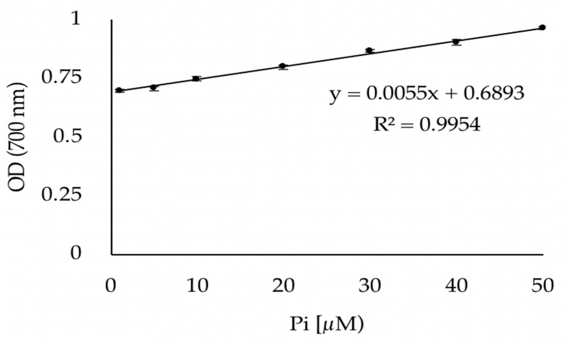
2.4. Phosphate Quantification Protocol

The hydrolysis of ATP by F1F0-ATPase leads to the formation of ADP and inorganic phosphate (Pi). The latter is used to quantify the enzymatic reaction. A linear regression showing absorbance values as a function of standard Pi solution concentrations ranging from 10 to 50 µM is used to deduce the concentration of Pi. Standard Pi solutions are prepared in tris-HCL buffer (50 mM, pH 8.5) and their concentrations are quantified using the method of Lowry et al. [27]. In each tube containing 1000 µL of the different Pi concentrations, and one tube containing 1000 µL of Tris-HCL buffer serving as a negative control, 100 µL of a 1% ammonium molybdate solution is added, and the contents are thoroughly mixed, allowing the formation of a phosphomolybdic acid complex. After adding 100 µL of a 1% ascorbic acid solution to each tube and incubating for 10 min at room temperature, a blue molybdous compound is formed. Both ammonium molybdate and ascorbic acid solutions are prepared in 0.25 N of H2SO4. A spectrophotometer is used to measure the optical density of each tube at 700 nm. The assays for each Pi concentration were carried out in triplicate.

2.5. Study of the Inhibition of the Membrane-Bound S. epidermidis and E. coli ATP Synthases by Quercetin

After obtaining a pellet containing the isolated F1F0-ATPase enzyme as previously described in Section 2.3, inhibitory assays were carried out using quercetin, a natural inhibitor of the enzyme, at different volumes, for both S. epidermidis and E. coli. The pellet was first resuspended in Tris-HCL buffer (50 mM; pH 8.5) and MgCl2 (15 mM) and incubated at 37 °C for 10 min. Then, to each sample a different volume of quercetin solution was added, so that different concentrations were obtained (1–500 µM), as well as 100 µM of ATP to start the reaction. The concentration of ATP used was saturating for the enzyme. Next, each tube was incubated at 37 °C for 40 min, and 1% (v/v) of SDS was added to each sample in order to stop the reaction. The samples were then centrifuged at 10,000× g for 10 min and finally the pellet was removed and the supernatant, containing the Pi released by the enzymatic reaction, was tested as described in Section 2.4 to study the inhibition of the enzyme. Two control tubes were prepared, one lacking the inhibitor quercetin to obtain the maximum activity of the enzyme, and one lacking both the inhibitor and ATP, which was used as a blank. All tests were performed in triplicate.
Inhibitory concentration (IC50) value of quercetin was calculated using GraphPad Prism (GraphPad Software, San Diego, CA, USA). This was achieved by using a curve of the enzymatic activity (%) as a function of log of different concentrations of quercetin (µM).

2.6. Study of the Potential Inhibitory Effect of M. bornmuelleri Venom on the Membrane-Bound S. epidermidis and E. coli F1F0-ATPases

The pellet containing the isolated F1F0-ATPase as described in Section 2.3 is tested with M. bornmuelleri venom as described in Section 2.4 to study its effect on the enzyme. The same protocol was applied on both S. epidermidis and E. coli membrane-bound F1F0-ATPase: To each tube containing the membrane-bound enzyme different concentrations of M. bornmuelleri venom (0–200 µg/mL) are added, prepared in Tris-HCL buffer (50 mM; pH 8.5), MgCl2 (15 mM), Tris-HCL buffer (50 mM; pH 8.5) and lastly ATP (100 µM, a saturating concentration) to start the reaction, and for a final volume of 1 mL for each tube. Samples are then incubated at 37 °C for 40 min, and 1% (v/v) of SDS is added to each sample in order to stop the reaction. Lastly, the tubes are centrifuged at 10,000× g and 4 °C for 10 min. Two blanks, one containing Tris-HCL MgCl2 and ATP and the other without ATP were prepared. A positive control tube, containing the enzyme, Tris-HCL, MgCl2, and ATP was prepared to determine the maximum activity of the enzyme. All assays were carried out in triplicate.
For both bacteria, curves of the enzymatic activity (%) as a function of different concentrations of the venom (µg/mL) were established.

2.7. HCT116 Cell Culture

To the medium used for HCT116 Cell line culture 1% Penicillin Streptomycin (100 U·mL−1) and 10% heat-inactivated fetal bovine serum (FBS) were added in order to amplify it. DMEM acquired at Sigma Aldrich (Gibco Dulbecco’s modified Eagle medium) was used to culture the HCT116 cell line at 37 °C in a humidified atmosphere with 5% CO2 and 95% air.

2.8. Cellular Viability Assay

The MTT test for cellular viability [28] was performed as follows: 96-well plates were used to seed HCT116 cells at a density of 104 cells/well, when they reached 60–80% confluency the cells were treated for 24 h and experiments were carried out with different concentrations of the venom extract. The medium was then discarded and to each well 100 µL of MTT solution was added. Finally, the absorbance was measured by ELISA READER at 570 nm. All tests were carried out in triplicate.

2.9. Statistical Analysis

Data were analyzed using GraphPad Prism (GraphPad Software, San Diego, CA, USA) and represented as mean ± SD of at least 3 independent experiments performed in triplicate for each condition. One-way ANOVA tests were performed. Differences were considered significant at p < 0.05(*), p < 0.01(**), p < 0.001(***) and p < 0.0001(****).

3. Results

3.1. Validation of the Phosphate Dosage Method

The F1F0-ATPase enzyme catalyzes the hydrolysis of ATP, which results in inorganic phosphate (Pi) that can be measured in order to assess enzymatic activity. This measurement must be linear in order to use it for quantitative analysis on assays. Therefore, we used the colorimetric method described by Lowry et al. 1945 [27] with some optimizations elaborated previously by our team [29]. Experiments were performed with solutions of low concentrations of Pi of 10, 20, 30, 40 and 50 µM.
Results represented in Figure 1 show a positive linear correlation between the optic density (OD) and the Pi concentration (R2 > 0.9954). In addition, the relative standard deviation (RSD%) is less than 3%, indicating that the measurements performed are highly repeatable; and it is important to add that all Pi solutions were assayed in triplicate. Thus, we determined that this linear regression is suitable for the analysis of S. epidermidis and E. coli membrane-bound F1F0-ATPases’ activity by way of quantifying the Pi produced during the catalytic reaction.

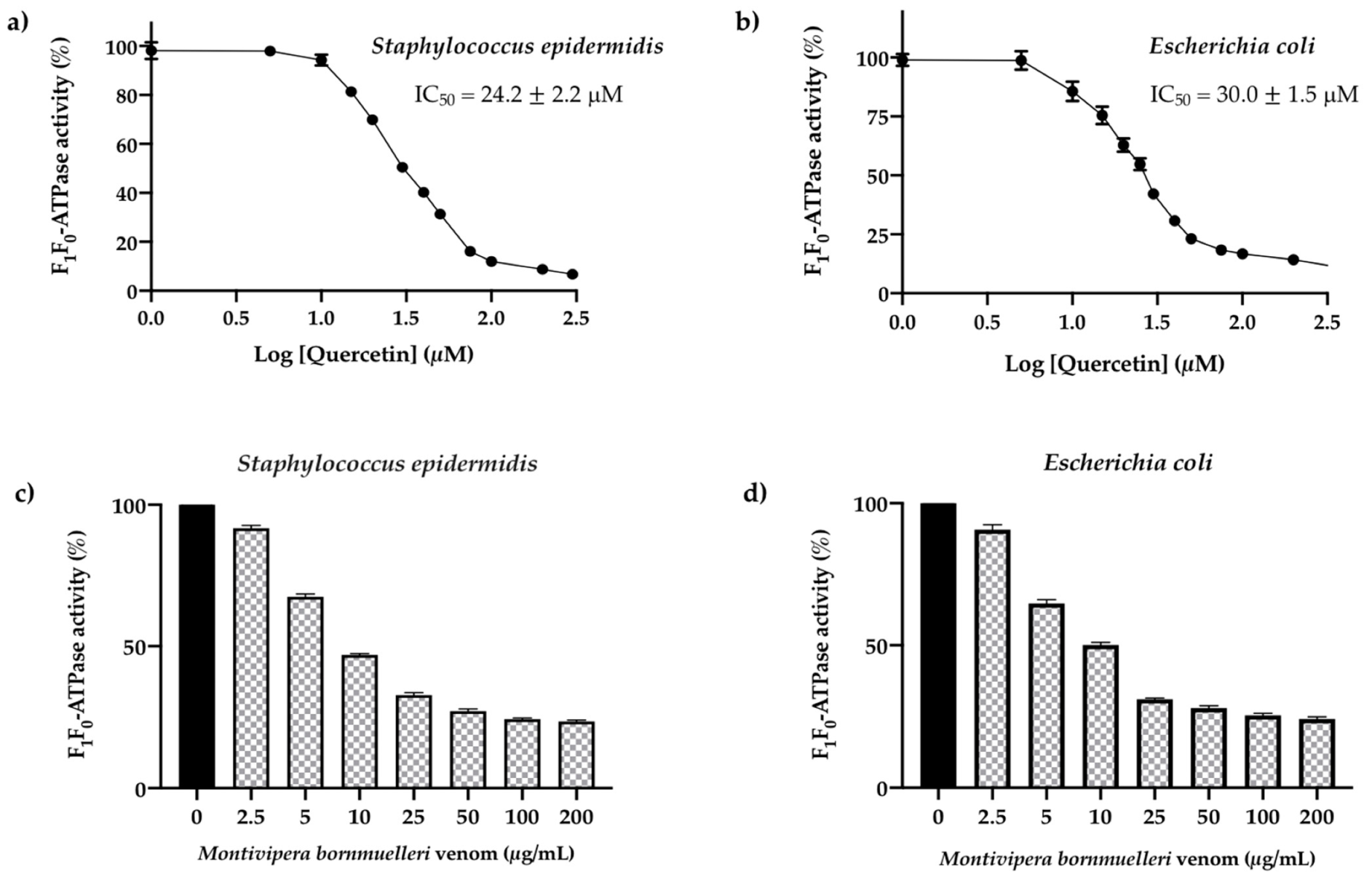
3.2. Screening of the Reference Inhibitor Quercetin on S. epidermidis and E. coli F1F0-ATPase Enzymes

The natural quercetin was chosen as reference inhibitor of the two bacteria F1F0-ATPases in order to validate the experimental model. The dose–response curves representing the enzymatic activity (%) of S. epidermidis and E. coli F1F0-ATPases as a function of log of reference inhibitor concentrations are represented in Figure 2a,b, respectively. The corresponding IC50 values obtained are 24.2 ± 2.2 μM for S. epidermidis and 30.0 ± 1.5 μM for E. coli. These values are in good agreement with those reported in literature [30] which validate the developed method. Good repeatability was obtained on assays with RSD < 5% for both bacteria.

3.3. Effect of the M. bornmuelleri Venom on the Membrane S. epidermidis and E. coli. F1F0-ATPase Enzymes

The M. bornmuelleri venom test on the membrane S. epidermidis and E. coli F1F0-ATPase enzymes shows that this extract is a potentially active inhibitor on those enzymes. In fact, seven concentrations in μg/mL of M. bornmulleri venom were tested (2.5, 5, 10, 25, 50, 100, 200). When the snake venom concentration increases, the enzyme activity for the two strains of bacteria Gram-positive S. epidermidis and Gram-negative E. coli decreases and reaches a plateau with a minimum enzymatic activity of approximately 20% (maximum inhibition of 80%) (Figure 2c,d).

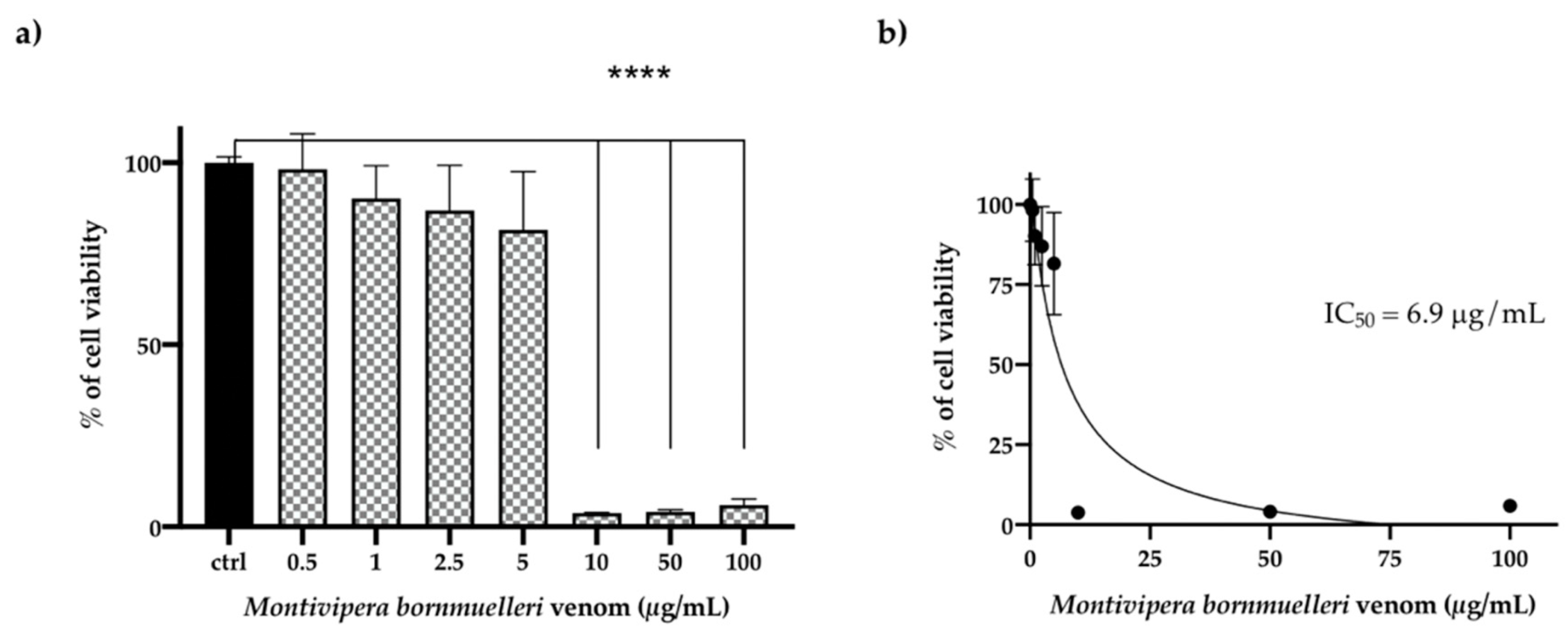
3.4. Effect of the M. bornmulleri Venom on HCT116 Cancer Cell Lines

The MTT (3-(4,5-dimethylthiazol-2-yl)-2,5-diphenyltetrazolium bromide) assay was the method used to elucidate the cytotoxic activity of crude M. bornmuelleri venom on human colon cancer HCT116 cells. These latter were exposed to a range of snake venom concentrations (0.5, 1, 2.5, 5, 10, 50 and 50 µg/mL) for 24 h and the obtained results were represented as the percentage of cell viability as a function of concentration of M. bronumelleri venom (µg/mL). Untreated cells with 100 % viability were used as a control to compare with HCT116 cells.
The data showed that the M. bornmuelleri venom inhibits the total cell viability of HCT116 cells with a low concentration of 10 µg/mL of the M. bornmuelleri venom. In fact, this concentration was able to induce a significant decrease in cell viability (95% of viability) in comparison to the control (Figure 3a). The results reveal the strong cytotoxic effect of the Lebanese snake M. bornmuelleri venom on HCT116 cells with an IC50 of 6.9 µg/mL calculated by Prism software (Figure 3b).

4. Discussion

Montivipera bornmuelleri is among three viper species found in Lebanon that inhabits mountainous regions [2]. Our previous studies aimed to characterize this snake and its venom in order to prove its important therapeutic applications. Indeed, an ecological study found that the snake prefers rocky regions with few thorny cushion plants and its maximum size is around 75 cm [1,2]. Its venom was analyzed and it was shown that it contains mainly three enzymes: PLA2, Metalloprotease III and serine protease, which possess many biological activities. In fact, the venom displayed both pro- and anti-coagulant activities depending on its concentration, as well as indirect hemolytic activity, possibly due to the presence of PLA2 [7]. It was shown by in vivo experiments on mice that it induced an upregulation of pro-inflammatory cytokines (INF-γ, TNF-α, IL-1β, IL-4 and IL-17), while also downregulating the anti-inflammatory cytokine IL-10 [6]. In addition, the M. bornmulleri venom exhibits antibacterial effect against both Gram-positive and Gram-negative bacteria, as well as antifungal activity [4]. Moreover, given the resistance to antibiotics of a multitude of bacteria and—now more than ever—the need to discover new antibacterial agents [31], such as those capable of acting on the F1F0-ATPase enzymes which are essential for the pathogenic microorganisms survival, we were interested in testing the venom effect of M. bornmuelleri on the F1F0-ATPases of bacteria from both Gram-positive (S. epidermidis) and Gram-negative (E. coli) genera. In agreement with our previous results [4], we found here that the venom inhibited the activity of the F1F0-ATPase enzyme presented in the membranes of these two bacteria. This membrane-bound enzyme plays an important role in cell life and death by both synthesizing and hydrolyzing ATP. Since ATP deprivation is linked to cell death [20], M. bornmuelleri venom having the ability to inhibit the activity of this enzyme translates to its ability as an effective antimicrobial agent. Thus, we showed that this crude venom managed to reduce the enzymatic activity to a minimum of 20% at concentrations of 100 to 150 µg/mL. However, this inhibitory effect was only assayed on isolated F1F0-ATPase of both bacteria, and not on total cells. The ability of the venom to inhibit the ATPase of Gram-negative bacteria, which are more resistant to antibiotics than Gram-positive ones due to the presence of an outer membrane that must be passed by drugs to attain their target [31], with the same level of inhibition (maximum of 80%) as Gram-positive bacteria, confers it a broad spectrum of activity and an important property. Indeed, it can be used against both types of bacteria instead of acting against only one, and this expands its potential application. Another potential benefit for this activity is the fact that the transfer of antibiotic resistance genes is possible between Gram-positive and Gram-negative bacteria [32] or between the same types of bacteria, such as S. epidermidis which is able to transfer these genes to S. aureus, and thus spread antibiotic resistance [13], so an antibacterial agent that is not susceptible to resistance by the various bacteria proves to be even more useful. As for compounds responsible for the inhibition of the bacterial enzyme, many aromatic derivatives from various plants have been found to exhibit this type of activity, such as resveratrol, piceatannol and quercetin [30]; however, similar compounds have not been characterized in M. bornmuelleri venom to date. Instead, we suggest that compounds found in the venom and known for their antibacterial effect, such as PLA2 and LAAO [4], are inhibiting the bacterial F1F0-ATPase. Indeed, PLA2, an enzyme purified from Protobothrops mucrosquamatus snake venom is able to control both Gram-positive (Bacillus subtilis) and Gram-negative (Pseudomonas aeruginosa) bacterial strains [33]. Metalloproteinase isolated from Agkistrodon halys, also inhibited Gram-positive (Staphylococcus aureus) and Gram-negative (Proteus vulgaris) bacteria [34]. LAAO, purified from Bothriechis schlegelii venom, also exerts an inhibitory effect against both Gram-positive (S. aureus) and Gram-negative (Acinetobacter baumannii) bacteria [35]. All of these enzymes are found in M. bronmuelleri venom and could be responsible for its antibacterial activity, which is in accordance with these findings in the literature [2].
In terms of cytotoxicity, our previous studies showed that the M. bornmuelleri venom has a specific cytotoxicity against B16 skin melanoma cells and 3-MCA-induced murine fibrosarcoma cell lines, which were more sensitive to the venom when overexpressing ovalbumin. However, it did not manage to reduce tumor size in vivo [36]. It has been shown that the venom possesses a vasorelaxant effect on K+ depolarized smooth muscle and inhibits the contraction of smooth muscle tissue induced by CaCl2, phenylephrine and AngI [8], but did not have a strong cytotoxicity towards human erythrocytes [3]. Here, different concentrations of the crude venom of M. bornmuelleri were tested against human colon cancer cell line HCT116, and it was found that a low concentration of 10 µg/mL was able to totally reduce cancer cell viability (by 95%) compared to untreated control cells and with an IC50 value of 6.9 µg/mL. This is in agreement with the literature, where the snake venom toxin from Vipera lebetina turanica was also tested on HCT116 colon cancer cells and reduced their viability with an IC50 value of 1.14 µg/mL. This cell growth inhibition was achieved by inducing apoptosis in the treated cells through an increase in ROS generation, upregulation of death receptors DR4 and DR5, cleavage of caspases-3, 8 and 9 and activation of the JNK pathway [37]. M. bornmuelleri venom could act through similar pathways and it would be beneficial to elucidate its mechanism of action in future studies. In addition, crude venom from the Malaysian Cryptelytrops purpureomaculatus demonstrated cytotoxic activity in SW480 and SW620 colon cancer cell lines with EC50 values of 29.43 µg/mL and 23.19 µg/mL respectively, which is in good agreement with our results. In this study, LAAO was also isolated from the venom and decreased the viability of the SW480 and SW620 colon cancer cell lines with lower EC50 values of 13.56 µg/mL and 13.17 µg/mL respectively through apoptosis [38]. The purification of a single component from M. bornmuelleri venom in order to test it on HCT116 cells could similarly prove to possess an enhanced cytotoxic effect compared to the crude venom. However, another study in which Malaysian mangrove pit viper Trimeresurus purpureomaculatus venom was assayed on HT-29 colon cancer cell lines, showed that viability was reduced with a low IC50 value of 0.42 µg/mL, suggesting that high cytotoxicity could result from synergistic interactions between various compounds present in the venom instead of only one component. Indeed, the different fractions obtained by HPLC had demonstrated each variable’s cytotoxic activity [39]. A viable concern for the use of snake venom in anticancer therapy is the potential cytotoxicity of its components against normal cells, in other words, its lack of selectivity for cancer cells. Indeed, PLA2 found in the venom is known to act on membrane phospholipids, and LAAO acts by producing H2O2, which is directly correlated with the deterioration of cell membranes. However such effects have been found to be most potent against cancer cells in particular, since these latter possess asymmetric plasma membranes [40]. One of the previous studies carried out on skin cancer cells demonstrated that the crude venom of M. bornmuelleri caused the least damage to HaCaT cells (non-tumorigenic), reducing their viability to 70.63% as opposed to 44% for A5 cells and 21.33% for II4 cells, both tumorigenic, using the same venom concentration of 60 µg/mL, further proving the venom’s selective cytotoxicity against cancer, and not normal, cells [5]. Since colon cancer is a major cause of cancer-related death and its incidence and mortality are increasing quickly [22], and its treatment faces the problem of the resistance of this disease to chemotherapy [41]—which is also unsatisfactory since it has many adverse side effects and lowers the quality of life of patients [24]—finding new treatments for this life-threatening disease is an important step that must be taken to improve public health, and natural sources such as snake venoms, and specifically M. bornmuelleri venom, as we showed in this study, prove to be of special interest in terms of discovering such therapeutic agents.

Author Contributions

Conceptualization, H.N. and Z.F.; methodology, M.K., C.M., M.R. and R.S.; software, C.M. and Z.F.; formal analysis, H.N. and Z.F.; investigation, Z.F., H.N. and J.-M.S.; resources, Z.F.; writing—original draft preparation, M.K.; writing—review and editing, H.N. and Z.F.; supervision, Z.F.; project administration, Z.F.; funding acquisition, Z.F. All authors have read and agreed to the published version of the manuscript.

Funding

This research was funded by Lebanese University.

Institutional Review Board Statement

Not applicable.

Informed Consent Statement

Not applicable.

Data Availability Statement

Not applicable.

Acknowledgments

We gratefully thank Lara Bitar and Rim Wehbe for their significant discussion and help.

Conflicts of Interest

The authors declare no conflict of interest.

References

  1. Rima, M.; Naini, S.M.A.; Karam, M.; Sadek, R.; Sabatier, J.-M.; Fajloun, Z. Vipers of the Middle East: A Rich Source of Bioactive Molecules. Molecules 2018, 23, 2721. [Google Scholar] [CrossRef] [PubMed] [Green Version]
  2. Hraoui-bloquet, S.; Sadek, R.; Hleihel, W.; Fajloun, Z. An ecological study of the Lebanon mountain viper Montivipera bornmuelleri (werner, 1898) with a preliminary biochemical characterization of its venom. Lebanese Sci. J. 2012, 13, 89–101. [Google Scholar]
  3. Rima, M.; Accary, C.; Haddad, K.; Sadek, R.; Hraoui-Bloquet, S.; Desfontis, J.C.; Fajloun, Z. Identification of L-amino acid oxidase (Mb-LAAO) with antibacterial activity in the venom of Montivipera bornmuelleri, a viper from Lebanon. Infect. Disord. Drug Targets 2014, 13, 337–343. [Google Scholar] [CrossRef] [PubMed]
  4. Accary, C.; Hraoui-Bloquet, S.; Hamze, M.; Mallem, Y.; Omar, F.; Sabatier, J.-M.; Desfontis, J.-C.; Fajloun, Z. Protein Content Analysis and Antimicrobial Activity of the Crude Venom of Montivipera bornmuelleri; a Viper from Lebanon. Infect. Disord. Drug Targets 2014, 14, 49–55. [Google Scholar] [CrossRef] [PubMed]
  5. Sawan, S.; Yaacoub, T.; Hraoui-Bloquet, S.; Sadek, R.; Hleihel, W.; Fajloun, Z.; Karam, M. Montivipera bornmuelleri venom selectively exhibits high cytotoxic effects on keratinocytes cancer cell lines. Exp. Toxicol. Pathol. 2017, 69, 173–178. [Google Scholar] [CrossRef]
  6. Yacoub, T.; Rima, M.; Sadek, R.; Hleihel, W.; Fajloun, Z.; Karam, M. Montivipera bornmuelleri venom has immunomodulatory effects mainly up-regulating pro-inflammatory cytokines in the spleens of mice. Toxicol. Rep. 2018, 5, 318–323. [Google Scholar] [CrossRef] [PubMed]
  7. Accary, C.; Rima1, M.; Kouzayha, A.; Hleihel, W.; Sadek, S.; Desfontis, J.-C.; Fajloun, Z.; Hraoui-Bloquet, S. Effect of the Montivipera bornmuelleri snake venom on human blood: Coagulation disorders and hemolytic activities. Open J. Hematol. 2014, 5. [Google Scholar] [CrossRef] [Green Version]
  8. Accary, C.; Hraoui-Bloquet, S.; Sadek, R.; Alameddine, A.; Fajloun, Z.; Desfontis, J.-C.; Mallem, Y. The relaxant effect of the Montivipera bornmuelleri snake venom on vascular contractility. J. Venom Res. 2016, 7, 10–15. [Google Scholar]
  9. Accary, C.; Mantash, A.; Mallem, Y.; Fajloun, Z.; Elkak, A. Separation and Biological Activities of Phospholipase A2 (Mb-PLA2) from the Venom of Montivipera bornmuelleri, a Lebanese Viper. J. Liq. Chromatogr. Relat. Technol. 2015, 38, 833–839. [Google Scholar] [CrossRef]
  10. Aminov, R.I. A Brief History of the Antibiotic Era: Lessons Learned and Challenges for the Future. Front. Microbiol. 2010, 1, 134. [Google Scholar] [CrossRef] [Green Version]
  11. Alanis, A.J. Resistance to Antibiotics: Are We in the Post-Antibiotic Era? Arch. Med. Res. 2005, 36, 697–705. [Google Scholar] [CrossRef]
  12. Namvar, A.E.; Bastarahang, S.; Abbasi, N.; Ghehi, G.S.; Farhadbakhtiarian, S.; Arezi, P.; Hosseini, M.; Baravati, S.Z.; Jokar, Z.; Chermahin, S.G. Clinical characteristics of Staphylococcus epidermidis: A systematic review. GMS Hyg. Infect. Control 2014, 9, Doc23. [Google Scholar] [CrossRef] [PubMed]
  13. Penelitian, K. Strategi Implementasi., Baragina Widyaningrum, Program Pascasarjana. Nat. Rev. Microbiol. 2008, 7, 555–567. [Google Scholar] [CrossRef]
  14. Blount, Z.D. The unexhausted potential of E. coli. eLife 2015, 4, e05826. [Google Scholar] [CrossRef]
  15. Reinthaler, F.; Posch, J.; Feierl, G.; Wüst, G.; Haas, D.; Ruckenbauer, G.; Mascher, F.; Marth, E. Antibiotic resistance of E. coli in sewage and sludge. Water Res. 2003, 37, 1685–1690. [Google Scholar] [CrossRef]
  16. Hermolin, J.; Fillingame, R.H. H+-ATPase activity of Escherichia coli F1F0 is blocked after reaction of dicyclohexylcarbodiimide with a single proteolipid (subunit c) of the F0 complex. J. Biol. Chem. 1989, 264, 3896–3903. [Google Scholar] [CrossRef]
  17. Uribe-Alvarez, C.; Chiquete-Félix, N.; Contreras-Zentella, M.; Guerrero-Castillo, S.; Peña, A.; Uribe-Carvajal, S. Staphylococcus epidermidis: Metabolic adaptation and biofilm formation in response to different oxygen concentrations. Pathog. Dis. 2015, 74, ftv111. [Google Scholar] [CrossRef] [PubMed] [Green Version]
  18. Hong, S.; Pedersen, P.L. ATP Synthase and the Actions of Inhibitors Utilized To Study Its Roles in Human Health, Disease, and Other Scientific Areas. Microbiol. Mol. Biol. Rev. 2008, 72, 590–641. [Google Scholar] [CrossRef] [Green Version]
  19. Devenish, R.J.; Prescott, M.; Rodgers, A.J. The Structure and Function of Mitochondrial F1F0-ATP Synthases. Int. Rev. Cell Mol. Biol. 2008, 267, 1–58. [Google Scholar] [CrossRef]
  20. Nesci, S.; Trombetti, F.; Algieri, C.; Pagliarani, A. A Therapeutic Role for the F1FO-ATP Synthase. SLAS Discov. Adv. Life Sci. R&D 2019, 24, 893–903. [Google Scholar] [CrossRef]
  21. Viegas, S.; Ladeira, C.; Costa-Veiga, A.; Perelman, J.; Gajski, G. Forgotten public health impacts of cancer—An overview. Arch. Ind. Hyg. Toxicol. 2017, 68, 287–297. [Google Scholar] [CrossRef] [Green Version]
  22. Bray, F.; Ferlay, J.; Soerjomataram, I.; Siegel, R.L.; Torre, L.A.; Jemal, A. Global cancer statistics 2018: GLOBOCAN estimates of incidence and mortality worldwide for 36 cancers in 185 countries. CA Cancer J. Clin. 2018, 68, 394–424. [Google Scholar] [CrossRef] [Green Version]
  23. Shen, H.; Yang, J.; Huang, Q.; Jiang, M.-J.; Tan, Y.-N.; Fu, J.-F.; Zhu, L.-Z.; Fang, X.-F.; Yuan, Y. Different treatment strategies and molecular features between right-sided and left-sided colon cancers. World J. Gastroenterol. 2015, 21, 6470–6478. [Google Scholar] [CrossRef]
  24. Lindley, C.; McCune, J.S.; Thomason, T.E.; Lauder, D.; Sauls, A.; Adkins, S.; Msawyer, W.T.S. Perception of Chemotherapy Side Effects Cancer versus Noncancer Patients. Cancer Pract. 1999, 7, 59–65. [Google Scholar] [CrossRef]
  25. Monsuez, J.-J.; Charniot, J.-C.; Vignat, N.; Artigou, J.-Y. Cardiac side-effects of cancer chemotherapy. Int. J. Cardiol. 2010, 144, 3–15. [Google Scholar] [CrossRef]
  26. Issa, D.; Najjar, A.; Greige-Gerges, H.; Nehme, H.; Nehme, H. Screening of Some Essential Oil Constituents as Potential Inhibitors of the ATP Synthase of Escherichia coli. J. Food Sci. 2019, 84, 138–146. [Google Scholar] [CrossRef] [PubMed] [Green Version]
  27. The Determination of Inorganic Phosphate in the Presence of Labile Phosphate Esters–PubMed. Available online: https://pubmed.ncbi.nlm.nih.gov/21018750/ (accessed on 30 May 2021).
  28. Mosmann, T. Rapid colorimetric assay for cellular growth and survival: Application to proliferation and cytotoxicity assays. J. Immunol. Methods 1983, 65, 55–63. [Google Scholar] [CrossRef]
  29. Nehme, H.; Ayde, H.; El Obeid, D.; Sabatier, J.M.; Fajloun, Z. Potential Inhibitory Effect of Apis mellifera’s Venom and of Its Two Main Components—Melittin and PLA2—On Escherichia coli F1F0-ATPase. Antibiotics 2020, 9, 824. [Google Scholar] [CrossRef] [PubMed]
  30. Dadi, P.K.; Ahmad, M.; Ahmad, Z. Inhibition of ATPase activity of Escherichia coli ATP synthase by polyphenols. Int. J. Biol. Macromol. 2009, 45, 72–79. [Google Scholar] [CrossRef]
  31. Breijyeh, Z.; Jubeh, B.; Karaman, R. Resistance of Gram-Negative Bacteria to Current Antibacterial Agents and Approaches to Resolve It. Molecules 2020, 25, 1340. [Google Scholar] [CrossRef] [Green Version]
  32. Courvalin, P. Transfer of Antibiotic Resistance Genes between Gram-Positive and Gram-Negative Bacteriat. Antimicrob. Agents Chemother. 1994, 38, 1447–1451. [Google Scholar] [CrossRef] [Green Version]
  33. Junior, N.G.D.O.; Cardoso, M.H.E.S.; Franco, O.L. Snake venoms: Attractive antimicrobial proteinaceous compounds for therapeutic purposes. Cell. Mol. Life Sci. 2013, 70, 4645–4658. [Google Scholar] [CrossRef] [PubMed]
  34. Samy, R.P.; Gopalakrishnakone, P.; Chow, V.; Ho, B. Viper metalloproteinase (Agkistrodon halys pallas) with antimicrobial activity against multi-drug resistant human pathogens. J. Cell. Physiol. 2008, 216, 54–68. [Google Scholar] [CrossRef]
  35. Alam, I.; Ojha, I.; Alam, A.; Quasimi, H.; Alam, O. Therapeutic potential of snake venoms as antimicrobial agents. Front. Drug Chem. Clin. Res. 2019, 2, 1–9. [Google Scholar] [CrossRef]
  36. Haddoub, C.; Rima, M.; Heurtebise, S.; Lawand, M.; Jundi, D.; Sadek, R.; Amigorena, S.; Fajloun, Z.; Karam, M.C. Cytotoxic effect of Montivipera bornmuelleri’s venom on cancer cell lines: In vitro and in vivo studies. PeerJ 2020, 8, e9909. [Google Scholar] [CrossRef]
  37. Park, M.H.; Jo, M.; Won, D.; Song, H.S.; Han, S.B.; Song, M.J.; Hong, J.T. Snake venom toxin from vipera lebetina turanicainduces apoptosis of colon cancer cells via upregulation of ROS- and JNK-mediated death receptor expression. BMC Cancer 2012, 12, 228. [Google Scholar] [CrossRef] [Green Version]
  38. Abidin, S.A.Z.; Rajadurai, P.; Chowdhury, E.H.; Othman, I.; Naidu, R. Cytotoxic, Anti-Proliferative and Apoptosis Activity of l-Amino Acid Oxidase from Malaysian Cryptelytrops purpureomaculatus (CP-LAAO) Venom on Human Colon Cancer Cells. Molecules 2018, 23, 1388. [Google Scholar] [CrossRef] [PubMed] [Green Version]
  39. Tan, C.H.; Liew, J.L.; Navanesan, S.; Sim, K.S.; Tan, N.H.; Tan, K.Y. Cytotoxic and anticancer properties of the Malaysian mangrove pit viper (Trimeresurus purpureomaculatus) venom and its disintegrin (purpureomaculin). J. Venom. Anim. Toxins Incl. Trop. Dis. 2020, 26, e20200013. [Google Scholar] [CrossRef]
  40. Hiu, J.J.; Yap, M.K.K. Cytotoxicity of snake venom enzymatic toxins: Phospholipase A2 and l-amino acid oxidase. Biochem. Soc. Trans. 2020, 48, 719–731. [Google Scholar] [CrossRef] [Green Version]
  41. Galligan, L.; Longley, D.B.; McEwan, M.; Wilson, T.R.; McLaughlin, K.; Johnston, P.G. Chemotherapy and TRAIL-mediated colon cancer cell death: The roles of p53, TRAIL receptors, and c-FLIP. Mol. Cancer Ther. 2005, 4, 2026–2036. [Google Scholar] [CrossRef] [Green Version]
Figure 1. Linearity of the Pi dosage. Standard Pi solutions of 10 µM to 50 µM prepared in tris-HCl buffer (pH = 8.5; 50 mM). Incubation time: 10 min. λ = 700 nm. Error bars show the standard deviation obtained from experiments carried out in triplicate.
Sci 03 00031 g001
Figure 2. Effect of the M. bornmuelleri venom on the F1F0-ATPase enzymatic activities of S. epidermidis and E. coli bacteria strains. The IC50 of the quercetin—used as an inhibitory reference ligand—on the enzymes is shown. (a) Dose-response curve showing the F1F0-ATPase activity (%) of S. epidermidis as a function of the log of the concentration of quercetin. (b) Dose-response curve showing the F1F0-ATPase activity (%) of E. coli as a function of the log of the concentration of quercetin. (c) Histogram representing the effect of M. bornmuelleri venom (µg/mL) on the activity (%) of membrane-bound S. epidermidis F1F0-ATPase. (d) Histogram representing the effect of M. bornmuelleri venom (µg/mL) on the activity (%) of membrane-bound E. coli F1F0-ATPase. For the four graphs represented here, the error bars show the standard deviation obtained from experiments carried out in triplicate. Data are expressed as mean ± SD (n = 3). Quercetin was used as a positive control. One-way ANOVA test: ns (not significant), when compared with the control. The M. bornmuelleri venom exerts a dose dependent inhibition on ATPase activity.
Sci 03 00031 g002
Figure 3. Cell viability of HCT116 colon cancer cells was measured by MTT assay after treatment with increased concentrations of M. bornmuelleri venom. (a) Results are expressed as mean ± SD of three independent experiments (n = 3). One-way ANOVA test: ns (no significant), **** p < 0.0001 when compared with the control. At 10, 50 and 100 μg/mL, the M. bornmuelleri venom showed a high cytotoxic activity against HCT116 cancers cells. (b) Curves for MTT assay showing IC50 values and the % of cell viability in function of concentrations M. bornmuelleri venom.
Sci 03 00031 g003
Publisher’s Note: MDPI stays neutral with regard to jurisdictional claims in published maps and institutional affiliations.

