Proceeding Paper

Amygdalin as a Plant-Based Bioactive Constituent: A Mini-Review on Intervention with Gut Microbiota, Anticancer Mechanisms, Bioavailability, and Microencapsulation †

1
Department of Food Science and Human Nutrition, College of Agriculture and Veterinary Medicine, Qassim University, Buraydah 51452, Saudi Arabia
2
Food Technology Department, Faculty of Agriculture, Benha University, Moshtohor, 13736 Qaliuobia, Egypt
Presented at the 1st International Electronic Conference on Nutrients—Nutritional and Microbiota Effects on Chronic Disease, 2–15 November 2020; Available online: https://iecn2020.sciforum.net/.
Published: 30 October 2020

Abstract

:
Amygdalin—a plant-based bioactive constituent particularly abundant in bitter almond has been identified as featuring the cyanogenic glycoside chemical organic compound which was originally intended to be a medication for cancer treatment once hydrolyzed to hydrogen cyanide (HCN). Unfortunately, studies have revealed that HCN can similarly affect normal cells, rendering it harming the human body. Both in vivo and in vitro investigations are extremely controversial and make its use unsafe. An updated substantial review on the source, structure, intervention with gut microbiota, anticancer therapy, bioavailability, and microencapsulation of amygdalin was summarized. Amygdalin provided anti-tumor, anti-fibrotic, anti-atherosclerosis, anti-inflammatory, immunomodulatory, analgesic, ameliorating digestive and reproductive systems, and enhancing neurodegeneration as well as myocardial hypertrophy. Studies confirmed the toxicity of amygdalin produced by its HCN after oral ingestion. However, the intravenous route of administration was much less toxic than the oral route and can be avoided with an oral dosage ranging from 0.6 to 1 g daily. The diversity of gut consortium is a key factor in inducing toxicity by amygdalin. Indeed, there is no guaranteed way to point out the microbial consortium for each person and provide a safe oral dosage. Recently, the encapsulating of amygdalin using alginate–chitosaninanoparticles (ACNPs) as transporter was investigated. As an active drug delivery mechanism for regulated and constant release of amygdalin with its enhanced cytotoxic effect on cancerous cells, biocompatible and biodegradable ACNPs can be applied while protecting normal cells and tissues. In conclusion, still unproven and conflicting facts give way to a broad avenue of research for a compound that could potentially be the next stage of cancer therapy.

1. Introduction

Amygdalin is one of the major pharmacological components in almonds and normally available in the seeds of Rosaceae plants [1,2]. It is a widespread substance in the environment, occurring in seeds of many plants, including black cherries and apricots, etc. [3]. It is a cyanogenic glycoside chemical organic compound with a molecular formula of C20H27O11 (Figure 1A) and a molecular mass of 457.42 g mol−1. Amygdalin consists of benzaldehyde, hydrocyanic acid, ianditwo glucose molecules (D-mandelonitrile-β-D-glucoside-6-β-glucoside), also known as laetrile, which is derived mainly from almond, apricots, and apple seeds [4]. It has antioxidant, antibacterial, anti-inflammatory, and immunoregulatory functions [3]. Amygdalin is not a toxic substance but has toxic properties of HCN, produced during enzymatic hydrolysis. Years of studies on the effects of amygdalin have shown its efficient properties, including its supportive role in treating asthma, bronchitis, leprosy, or colorectal cancer [5,6]. It has analgesic effects due to the presence of benzaldehyde in its molecule [7]. However, its anticancer function is still controversial and considered a subject of more research. This capacity may be related to enzymatic hydrolysis, resulting in the release of hydrocyanic acid [8]. However, the use of amygdalin as an anticancer drug has been questioned due to its toxicity to normal cells and limited pharmacokinetic properties. The anticancerous effects are mainly considered to be apoptotic inducing cancer cell death, inhibiting the growth of cancer cells, by degrading carcinogenic compounds [9], blocking cancer cells’ nutrient source, thereby decreasing the incidence of multiple cancers [10]. The current review provides a review on structure, intervention with gut microbiota, anticancer therapy, bioavailability, and microencapsulation of amygdalin as a plant-based bioactive constituent and its updated application in the field of dietetic therapy, hoping that updated data would offer theoretical and practical clues for exploring amygdalin’s functional utility and applications in terms of its antitumor functionality.

2. The Microbiome and Oral Amygdalin Administration: Intervention with Gut Microbiota

In the gastrointestinal tract (GIT), bacteria are extremely diverse and dominant, with about 1014 bacteria and 500 distinct species dominated by the anaerobes. Intestinal microbiota obtains their nutrition through the action of enzymes that produce such as sulfatase, esterase, α-rhamnosidase, β-glucosidase, and β-glucuronidase by hydrolysis of sulfates, amides, glucuronidases, esters, and lactones [11]. Probiotics are organisms such as Lactobacillus and Bifidobacterium which help to modulate bacterial intestinal enzymes and absorb or bind carcinogenic substances. However, when amygdalin was taken orally as a drug is affected by the lumen, gut wall, hepatic, and gut microbiome enzymes of GIT. Owing to minimal intestinal enzymatic activity, the undigested compounds flow down to the colon and specific digestion by gut microbial enzymes takes place. β-glucosidases present in kernels are also responsible for amygdalin hydrolysis [12]. Even the gut microflora determines the level of toxicity and the amount of whole blood cyanide [13]. They have high β-glucosidase activity, mainly the Bacteroides, and release cyanide via amygdalin symbiotic digestion. Different substrates have been shown for the intestinal and microbial β-glucosidases. Zhang et al. found that the gut microbiota of a termite-consuming polysaccharide had mainly members of the genus Bacteriodetes. They were found to be a rich source of β-glucosidase genes. Bacteriodetes have been shown to generate β-glucosidase enzyme also in the human intestine, which because of its work as a cellulose hydrolase plays an important role in hemicellulose or cellulose degradation [14]. Experiments on germ-free and control rats have shown that the gut microbiome induces the host to increase the production of glucose and tri-acyl glycerol [15]. This may explain why amygdalin, due to its glucose content, is strongly hydrolyzed. In obese and non-obese patients, it has been observed that, with a rise in body weight, the ratio of Bacteriodetes to Firmicutes decreases [16,17,18]. High-fiber diets such as those in Europe and rural Africa indicate a healthy population of Bacteriodetes in the gut [18]. This indicates that the interactions between diet, immune system, gut microbiota, and metabolism have significant effects on health and bacterial metabolism. Lactulose is also known to decrease the amount of β-glucosidase that generates Bacteriodetes [16]. Amygdalin is used as conventional and alternative medicine (CAM) therapy because the cyanide obtained in hydrolysis can bind to cytochrome oxidase c and a3, interfere with respirationiand DNA synthesis due to reactive oxygen species formation, obstruct cell nutrition and lead to lysis [19,20]. This is useful for parenteral administration but will hit the gut unchanged upon ingestion. The gut microbiota is anaerobic, producing high levels of lactic acid through pyruvate fermentation [21]. This pH raises β-glucosidase activity because of which amygdalin is hydrolyzed. to cyanide and contributes to toxicity. Blaheta et al. have shown.that amygdalin still has anti-tumor properties in the absence of β-glucosidase, giving rise to the possibility that it is not the cyanide that is responsible for that effect [12]. It is suspected that by inhibiting collagenase and hyaluronidase, which weakens the matrix and contributes to benign tumors, the strength of the intracellular matrix into which cancer cells are embedded is increased [22,23]. Usually, these two CAM therapies are administered together. Huge doses of vitamin C are now notorious for depleting the body’s cysteine reserves. Cysteine plays a role in thiocyanate formation in the rate-limiting stage [21]. Amygdalin can be hydrolyzed by gut bacteria, combined with the physiological effects of vitamin C, resulting in increased exposure to cyanide [21]. The same is proven by studies by Ward et al. [24]. Studies have provided contradictory findings for use of both therapies alone and in combination. Some reveal that this may be due to oral intake rather than intravenous, which contributes to cyanide toxicity rather than cancer rectification [25]. Owing to the lack of β-glucosidase and the presence of Rhodanese, intravenous routes lead to low HCN levels. Big oral doses are hard to detoxify since there is little to no rhodanese in the GIT [26]. It can be difficult to diagnose cyanide poisoning, and the study of arterial blood gas can be used for quick and effective prognosis [27]. Treatment for which a cyanide antidote kit was initially used after diagnosis is needed but it has its own toxicityiissues. Cynokit (hydroxocobalamin) is widely used and there are no clinically relevant adverse effects other than chromaturia and discoloration of the red skin [11,28]. Due to the higher binding affinity, vitamin B12 can chelate cyanide, form cyanocobalamin, and leave the system through the kidneys [29]. Due to this property, it is used as a cyanide antidote [19]. Another alternative therapy may be probiotic ingestion, which is known to decrease Bacteroides levels. However, it is understood that Lactobacillus develops β-glucosidases. Studies on HCN levels in a cancer patient with low Bacteriodetes and strong β-glucosidase generating amygdalin-ingested Lactobacillus need to be conducted further.

3. Anti-Cancer Mechanisms of Amygdalin: A Molecular Approach

Initially, amygdalin was intended to be a safe medication for cancer treatment and was accepted by natural medicine followers because it was believed to be hydrolyzed only in cancer cells releasing toxic HCN and therefore killed. Unfortunately, reviewed studies by Liczbińsk et al. [30] have shown that HCN is also produced in normal cells, so it may not be safe for human beings. However, scientific work on the anti-cancer properties of this compound has still been performed. In vitro studies have shown induction of apoptosis by amygdalin as a result of increased Bax protein and caspase-3 expression and decreased antiapoptotic Bcl-2 protein expression. It has also been shown that amygdalin prevents the adhesion of breast cancer cells, lung cancer cells, and bladder cancer cells by decreasing integrin expression, reducing catenin levels, and inhibiting the Akt-mTOR pathway, which may result in inhibition of cancer cell metastases. It was also discovered that amygdalin increased the expression of p19 protein in renal cancer cells, resulting in inhibition of cell transfer from G1 to S-phase, and thus inhibited cell proliferation. Other studies have shown that amygdalin inhibits the signaling pathways of NF-kb and NLRP3 and thus has an anti-inflammatory effect due to decreased expression of pro-inflammatory cytokines such as pro-IL-1b. In addition, the effect of amygdalin on the TGFb/CTGF pathway, anti-fibrous activity, and follistatin expression resulting in muscle cell growth activation was recorded. This compound may contribute to the treatment of different types of cancer cells. On the other side, amygdalin might be used as an alternative therapy for bone fractures mediated by TGF-beta/Smad signaling [31]. However, its anti-inflammatory and analgesic activities were reviewed [32]. Table 1, lists the key sources relating to the anti-inflammatory and analgesic activities of amygdalin.

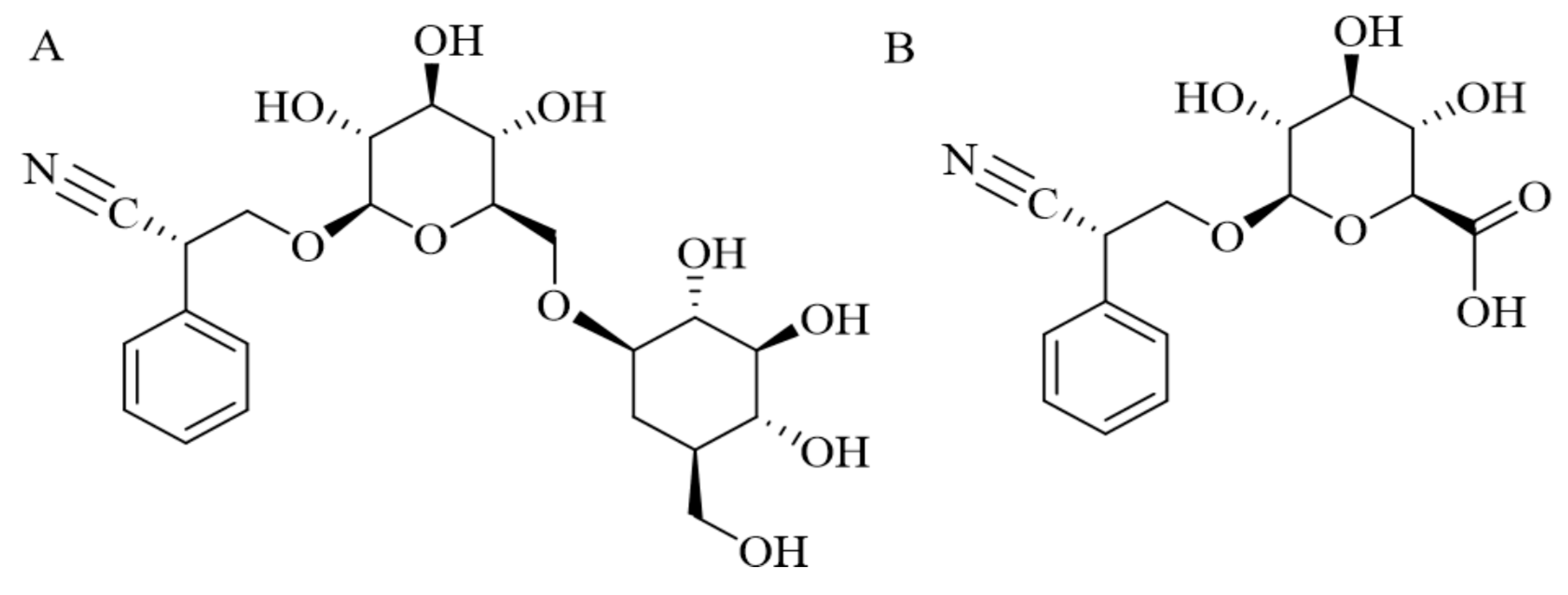
4. Microencapsulation and Bioavailability

The anticancerous activities of amygdalin are mostly measured to be apoptotic persuading cancer cell death, hindering the cancer cell’s growth, by debasing carcinogenic components [9], hindering cancer cells’ nutrients, thus declining multiple cancers incidence [10]. It has been utilized for asthma, bronchitis, emphysema eprosy, colorectal cancer, and vitiligo, and multiple cancers’ treatment [5]. However, using amygdalin as an anticancer drug has been quizzed owing to its toxicity to normal cells and imperfect pharmacokinetic belongings. Amygdalin has hardly been utilized and has not been explored yet with any drug delivery nanocarriers. To cause an upsurge in the therapeutic activities and decrease the side-effects of amygdalin, the encapsulation of amygdalin is needed. Nanocarriers as smart drug delivery systems are being designed, developed, and investigated for many applications including therapeutics [38]. Nanoparticulate drug delivery has been exposed to vastly increase therapeutic activity duration, drug-stability, allowing the drug’s parent or enteral administration, cumulative drug penetrability, thus minimizing or preventing drug metabolism, cellular efflux, as well as squalor [39]. Nanoparticles (1–200 nm) are usually laden with drugs by either in core encapsulation or by surface adsorption. Besides metallic, normal degradable biopolymers, polysaccharides, synthetic polymers, and lipids are being utilized in making nanoparticles. Colloidal polymeric nanoparticles, besides their small size, can deliver constant drug release over time owing to their possible multi-functional properties [40]. Furthermore, biodegradability, nontoxicity, biocompatibility, low cost, and high abundance in nature increase their usage in making drug delivery systems [40,41]. Amygdalin, despite having anticancerous features, has been observed as a contentious option due to the attendance of the cyanide group. The potential of alginate-chitosan nanoparticles (ACNPs) as drug delivery agents for amygdalin encapsulation and its delivery to cancer cells was investigated [37]. Amygdalin-loaded ACNPs were made with anionic and/or cationic outer layer to further explore charge dependence on drug delivery and cytotoxicity. ACNPs-encapsulating amygdalin was monodispersing, colloidally stable by 90% drug encapsulation efficacy, and was wholly ended from natural ingredients. The nanoparticles showed constant drug release for a period of 10 h and significant bump rates in neutral and somewhat acidic settings. The ACNPs positively obeyed to porcine mucin-type I when measured for its muco-adhesion and exposed to drift with an average velocity of 1.68 μm s−1 in uncoated channels, under bio-mimicked flow circumstances. To examine charge dependence on drug delivery and cytotoxicity, amygdalin-loaded ACNPs were ended with both anionic and cationic outer layers and measured. ACNPs demonstrated better yet sustained anticancerous activity on H1299-cells in a dose-dependent manner than free-amygdalin, signifying better cellular approval of the previous. The author demonstrated that stable anionic and cationic surface amygdalin-loaded ACNPs can be synthesized by ionic gelation method with high colloidal stability, as publicized by its zeta potential (−36 and +32) having a size of ~119 nm and high drug encapsulation efficiency [38]. ACNPs presented amplified swelling and sustained drug release shape with pH (3.1, 5.0, 7.4). The amygdalin-loaded ACNPs exhibited effectual passage under 10 dynes cm−2 shear flow stress and good mucoadhesive property in the BioFlux system. Finally, the author proposed ACNPs as an active drug delivery carrier for amygdalin, present sustain release, and better efficacy and delivery. This inspires further in vivo analysis of the prepared amygdalin-loaded ACNPs on cancer tumor models. Recently, Zhou et al. [42] showed that by chemical coupling methods, starch-coated magnetic nanoparticles (MNPs) were successively conjugated with β-glucosidase (β-Glu) and polyethylene glycol (PEG). Consequently, amygdalin-mediated immobilized β-Glu activated prostate cancer cell death. Tumor-targeting studies have shown that PEG modification increased the accumulation of nanoparticles loaded with β-Glu in targeted tumor tissue subjected to an external magnetic field and decreased the accumulation of nanoparticles in the liver and spleen. Protection analyses found that this technique had some effect on the function of the liver and heart but did not cause obvious organ damage. All results suggest that this technique of magnetically directed enzyme/prodrug therapy has the potential to become a promising new method for prostate cancer targeted therapy.

5. Future Perspectives and Conclusions

Amygdalin has customarily been used for cough, asthma, leprosy, bronchitis, nausea, and leukoderma treatment. Earlier in vivo and in vitro studies have authenticated its pharmacological effects of anti-tumor, anti-fibrotic, anti-inflammatory, analgesic, immunomodulatory, anti-atherosclerosis, ameliorating the peptic and reproductive systems, refining neurodegeneration and myocardial hypertrophy, as well as reducing blood glucose. However, the obtainable research has mostly fixated on the pharmacological activity and toxicity of amygdalin and the clarification of the molecular mechanisms of its actions are still inadequate. Studies are extremely controversial, rendering it hazardous for use as a therapeutic agent. Neither systemic data concerning the pharmacokinetics of amygdalin nor estimations of its target-organ toxicity are sufficiently known. Hence, more novel research needs to be directed in the future to assess its possible therapeutic effects, side effects, or toxicity. Moreover, in recent years, cumulative but still only an imperfect number of studies have fixated on its effect on inflammation, digestion, neurodegeneration, reproduction, myocardial hypertrophy, and blood glucose; more studies should be done to obtain a more complete understanding of this component. Current literature points to the fact that amygdalin causes toxicity on oral consumption and not IV administration; however, its mode of action and toxicity-causing dose is still not confirmed and are gut consortium-dependent. Amygdalin has hardly been used and has not been widely investigated yet with any drug delivery nanocarriers, in vivo. Recent experiments using amygdalin-loaded ACNPs and an amygdalin/β-Glu-based MDEPT strategy may have good prospects as promising studies for clinical use in cancer therapies. Therefore, studies investigating its encapsulation and anticancerous efficacy in the encapsulated form to increase the therapeutic effect and reduce the side effects of amygdalin should be focused on further.

Funding

This research received no external funding.

References

  1. Jaswal, V.; Palanivelu, J.; Ramalingam, C. Effects of the gut microbiota on amygdalin and its use as an anti-cancer therapy: substantial review on the key components involved in altering dose efficacy and toxicity. Biochem. Biophys. Rep. 2018, 14, 125–132. [Google Scholar] [CrossRef] [PubMed]
  2. Toomey, V.M.; Nickum, E.A.; Flurer, C.L. Cyanide and amygdalin as indicators of the presence of bitter almonds in imported raw almonds. J. Forensic Sci. 2012, 57, 1313–1317. [Google Scholar] [CrossRef] [PubMed]
  3. Orlikova, B.; Legrand, N.; Panning, J.; Dicato, M.; Diederich, M. Anti-inflammatory and anticancer drugs from nature. In Advances in Nutrition and Cancer; Springer: Berlin/Heidelberg, Germany, 2014; pp. 123–143. [Google Scholar]
  4. Qadir, M.; Fatima, K. Review on pharmacological activity of amygdalin. Arch. Cancer Res. 2017, 5, 160. [Google Scholar] [CrossRef]
  5. Li, X.B.; Liu, C.H.; Zhang, R.; Huang, X.T.; Li, Y.Y.; Han, L.; Xu, M.L.; Mi, S.Q.; Wang, N.S. Determination and pharmacokinetics of amygdalin in rats by LC–MS-MS. J. Chromatogr. Sci. 2014, 52, 476–481. [Google Scholar] [CrossRef]
  6. Song, Z.; Xu, X. Advanced research on anti-tumor effects of amygdalin. J. Cancer Res. Ther. 2014, 10, 3. [Google Scholar] [PubMed]
  7. Chang, H.K.; Shin, M.S.; Yang, H.Y.; Lee, J.W.; Kim, Y.S.; Lee, M.H.; Kim, J.; Kim, K.H.; Kim, C.J. Amygdalin induces apoptosis through regulation of Bax and Bcl-2 expressions in human DU145 and LNCaP prostate cancer cells. Biol. Pharm. Bull. 2006, 29, 1597–1602. [Google Scholar] [CrossRef]
  8. Zhou, C.; Qian, L.; Ma, H.; Yu, X.; Zhang, Y.; Qu, W.; Zhang, X.; Xia, W. Enhancement of amygdalin activated with β-D-glucosidase on HepG2 cells proliferation and apoptosis. Carbohydr. Polym. 2012, 90, 516–523. [Google Scholar] [CrossRef]
  9. Park, H.J.; Yoon, S.H.; Han, L.S.; Zheng, L.T.; Jung, K.H.; Uhm, Y.K.; Lee, J.H.; Jeong, J.S.; Joo, W.S.; Yim, S.V. Amygdalin inhibits genes related to cell cycle in SNU-C4 human colon cancer cells. World J. Gastroenterol. WJG 2005, 11, 5156. [Google Scholar]
  10. Chen, Y.; Ma, J.; Wang, F.; Hu, J.; Cui, A.; Wei, C.; Yang, Q.; Li, F. Amygdalin induces apoptosis in human cervical cancer cell line HeLa cells. Immunopharmacol. Immunotoxicol. 2013, 35, 43–51. [Google Scholar] [CrossRef]
  11. Ushakova, N.; Nekrasov, R.; Pravdin, I.; Sverchkova, N.; Kolomiyets, E.; Pavlov, D. Mechanisms of the effects of probiotics on symbiotic digestion. Biol. Bull. 2015, 42, 394–400. [Google Scholar] [CrossRef]
  12. Blaheta, R.A.; Nelson, K.; Haferkamp, A.; Juengel, E. Amygdalin, quackery or cure? Phytomedicine 2016, 23, 367–376. [Google Scholar] [CrossRef] [PubMed]
  13. Tokpohozin, S.E.; Fischer, S.; Sacher, B.; Becker, T. β-d-Glucosidase as “key enzyme” for sorghum cyanogenic glucoside (dhurrin) removal and beer bioflavouring. Food Chem. Toxicol. 2016, 97, 217–223. [Google Scholar] [CrossRef] [PubMed]
  14. Zhang, M.; Liu, N.; Qian, C.; Wang, Q.; Wang, Q.; Long, Y.; Huang, Y.; Zhou, Z.; Yan, X. Phylogenetic and functional analysis of gut microbiota of a fungus-growing higher termite: Bacteroidetes from higher termites are a rich source of β-glucosidase genes. Microb. Ecol. 2014, 68, 416–425. [Google Scholar] [CrossRef] [PubMed]
  15. Sorbo, B. On the properties of rhodanese: Partial Purification, Inhibitors and Intracellular Distribution. Acta Chem. Scand. 1951, 5, 724–726. [Google Scholar] [CrossRef]
  16. Duda-Chodak, A.; Tarko, T.; Satora, P.; Sroka, P. Interaction of dietary compounds, especially polyphenols, with the intestinal microbiota: A review. Eur. J. Nutr. 2015, 54, 325–341. [Google Scholar] [CrossRef]
  17. Greenberg, D.M. The vitamin fraud in cancer quackery. West. J. Med. 1975, 122, 345. [Google Scholar]
  18. Chong, E.S.L. A potential role of probiotics in colorectal cancer prevention: Review of possible mechanisms of action. World J. Microbiol. Biotechnol. 2014, 30, 351–374. [Google Scholar] [CrossRef]
  19. Kuugbee, E.D.; Shang, X.; Gamallat, Y.; Bamba, D.; Awadasseid, A.; Suliman, M.A.; Zang, S.; Ma, Y.; Chiwala, G.; Xin, Y. Structural change in microbiota by a probiotic cocktail enhances the gut barrier and reduces cancer via TLR2 signaling in a rat model of colon cancer. Dig. Dis. Sci. 2016, 61, 2908–2920. [Google Scholar] [CrossRef]
  20. Bromley, J.; Hughes, B.G.; Leong, D.C.; Buckley, N.A. Life-threatening interaction between complementary medicines: Cyanide toxicity following ingestion of amygdalin and vitamin C. Ann. Pharmacother. 2005, 39, 1566–1569. [Google Scholar] [CrossRef]
  21. Padayatty, S.J.; Sun, A.Y.; Chen, Q.; Espey, M.G.; Drisko, J.; Levine, M. Vitamin C: Intravenous use by complementary and alternative medicine practitioners and adverse effects. PLoS ONE 2010, 5, e11414. [Google Scholar] [CrossRef]
  22. Richards, E. The politics of therapeutic evaluation: The vitamin C and cancer controversy. Soc. Stud. Sci. 1988, 18, 653–701. [Google Scholar] [CrossRef]
  23. Calabrese, E.J. Conjoint use of laetrile and megadoses of ascorbic acid in cancer treatment: Possible side effects. Med. Hypotheses 1979, 5, 995–997. [Google Scholar] [CrossRef]
  24. Ward, E.; Jemal, A.; Cokkinides, V.; Singh, G.K.; Cardinez, C.; Ghafoor, A.; Thun, M. Cancer disparities by race/ethnicity and socioeconomic status. CA Cancer J. Clin. 2004, 54, 78–93. [Google Scholar] [CrossRef]
  25. Holstege, C.P.; Forrester, J.D.; Borek, H.A.; Lawrence, D.T. A case of cyanide poisoning and the use of arterial blood gas analysis to direct therapy. Hosp. Pract. 2010, 38, 69–74. [Google Scholar] [CrossRef] [PubMed]
  26. Kleessen, B.; Sykura, B.; Zunft, H.J.; Blaut, M. Effects of inulin and lactose on fecal microflora, microbial activity, and bowel habit in elderly constipated persons. Am. J. Clin. Nutr. 1997, 65, 1397–1402. [Google Scholar] [CrossRef]
  27. Borron, S.W.; Baud, F.J.; Mégarbane, B.; Bismuth, C. Hydroxocobalamin for severe acute cyanide poisoning by ingestion or inhalation. Am. J. Emerg. Med. 2007, 25, 551–558. [Google Scholar] [CrossRef]
  28. Oyewole, O.; Olayinka, E. Hydroxocobalamin (vit B12a) effectively reduced extent of cyanide poisoning arising from oral amygdalin ingestion in rats. J. Toxicol. Environ. Health Sci. 2009, 1, 008–011. [Google Scholar]
  29. Chan, T.Y. A probable case of amygdalin-induced peripheral neuropathy in a vegetarian with vitamin B12 deficiency. Ther. Drug Monit. 2006, 28, 140–141. [Google Scholar] [CrossRef]
  30. Liczbiński, P.; Bukowska, B. Molecular mechanism of amygdalin action in vitro: Review of the latest research. Immunopharmacol. Immunotoxicol. 2018, 40, 212–218. [Google Scholar] [CrossRef]
  31. Ying, J.; Ge, Q.; Hu, S.; Luo, C.; Lu, F.; Yu, Y.; Xu, T.; Lv, S.; Zhang, L.; Shen, J.; et al. Amygdalin promotes fracture healing through TGF-beta/Smad signaling in mesenchymal stem cells. Stem Cells Int. 2020, 2020, 8811963. [Google Scholar] [CrossRef]
  32. He, X.Y.; Wu, L.J.; Wang, W.X.; Xie, P.J.; Chen, Y.H.; Wang, F. Amygdalin—A pharmacological and toxicological review. J. Ethnopharmacol. 2020, 254, 112717. [Google Scholar] [CrossRef] [PubMed]
  33. Yang, H.Y.; Chang, H.K.; Lee, J.W.; Kim, Y.S.; Kim, H.; Lee, M.H.; Shin, M.S.; Ham, D.H.; Park, H.K.; Lee, H.; et al. Amygdalin suppresses lipopolysaccharide-induced expressions of cyclooxygenase-2 and inducible nitric oxide synthase in mouse BV2 microglial cells. Neurol. Res. 2007, 29, 59–64. [Google Scholar] [CrossRef] [PubMed]
  34. Hwang, H.J.; Lee, H.J.; Kim, C.J.; Shim, I.; Hahm, D.H.J.J.M.B. Inhibitory effect of amygdalin on lipopolysaccharide-inducible TNF-alpha and IL-1beta mRNA expression and carrageenan-induced rat arthritis. J. Microbiol. Biotechnol. 2008, 18, 1641–1647. [Google Scholar] [PubMed]
  35. Luo, D.; Shan, Z.; Jinlian, G.; Liu, Q.; Luo, L. Effects of amygdalin on TNF-α and sICAM-1 of rats with type I collagen-induced arthritis. Chin. J. Inf. Tradit. Chin. Med. 2015, 7, 75–77. [Google Scholar]
  36. Zhang, A.; Pan, W.; Lv, J.; Wu, H. Protective effect of amygdalin on LPS-induced acute lung injury by inhibiting NF-κB and NLRP3 signaling pathways. Inflammation 2017, 40, 745–751. [Google Scholar] [CrossRef]
  37. Zhong, X.Q.; Li, L.; Lu, C.J.; Lu, Y.; Wei, J.A.; Han, L. Anti-inflammation effect of amygdalin on macrophage 264.7 Cells Stimulated by Lipopolysaccharide. Tradit. Chin. Drug Res. Clin. Pharmacol. 2018, 29, 257–263. [Google Scholar]
  38. Sohail, R.; Abbas, S.R. Evaluation of amygdalin-loaded alginate-chitosan nanoparticles as biocompatible drug delivery carriers for anticancerous efficacy. Int. J. Biol. Macromol. 2020, 153, 36–45. [Google Scholar] [CrossRef]
  39. Sarmento, B.; Ribeiro, A.; Veiga, F.; Ferreira, D.; Neufeld, R. Insulin-loaded nanoparticles are prepared by alginate ionotropic pre-gelation followed by chitosan polyelectrolyte complexation. J. Nanosci. Nanotechnol. 2007, 7, 2833–2841. [Google Scholar] [CrossRef]
  40. De Jong, W.H.; Borm, P.J. Drug delivery and nanoparticles: Applications and hazards. Int. J. Nanomed. 2008, 3, 133. [Google Scholar] [CrossRef]
  41. Liu, Z.; Jiao, Y.; Wang, Y.; Zhou, C.; Zhang, Z. Polysaccharides-based nanoparticles as drug delivery systems. Adv. Drug Deliv. Rev. 2008, 60, 1650–1662. [Google Scholar] [CrossRef]
  42. Zhou, J.; Hou, J.; Rao, J.; Zhou, C.; Liu, Y.; Gao, W. Magnetically directed enzyme/prodrug prostate cancer therapy based on beta-Glucosidase/amygdalin. Int. J. Nanomed. 2020, 15, 4639–4657. [Google Scholar] [CrossRef] [PubMed]
Figure 1. (A) Chemical structure of amygdalin, (B) Chemical structure of laetrile.
Proceedings 61 00015 g001
Table 1. Key sources relating to the anti-inflammatory and analgesic activities of amygdalin.
ModelDose/Method/PeriodActivityRefs.
BV2 glial cells 1, 10, 100, 1000 μg mL−1; culture; 24 h COX-2 mRNA, iNOS mRNA ↓
the synthesis of prostaglandin E2 ↓
the production of nitric oxide ↓
[33]
RAW 264.7 cells 1, 10, 100 mmol L−1; culture; 6 h At a concentration of 1 mM, TNF-α and IL-1β mRNA ↓
Amygdalin does not inhibit TNF-α and IL-1β mRNA expressioniiin a dose-dependent manner.
[34]
Arthritis pain model (Carrageenan-induced), SD male rats 0.005, 0.05, and 0.1 mg kg−1; i.m; 8 h At a concentration of 0.005 mg/kg, Fos, TNF-α and
IL-1β ↓; However, no analgesic effect of amygdalin was observed at doses greater than 0.005 mg/kg. ↓
[35]
Pain model (plantar injection of formalin), SD male rats 0.1, 0.5, 1.0, and 10.0 mg kg−1; Plantar injection c-Fos, TNF-α, IL-1β
Laetrile reduces pain in a dose-dependent manner in a dose range of less than 1 mg/kg.
[34]
CIA rat model (type II collagen-induced), Wistar rats 120 mg kg−1; gavage; 28 days TNF-α and sICAM-1 ↓ [35]
BALB/c mice 0.5, 1, and 2 mg kg−1; ip; 7 h NF-κB ↓
Reduced pulmonary edema in a dose-dependent manner.
[36]
RAW 264.7 cells 6.25, 12.5, 25, 50, 100, 200, 400 µmol L−1; culture; 24 h IL-17A, IL-23, CCL2, and CCL5 mRNA ↓ p-p38 ↓
the viability of RAW264.7 cell ↓
[37]
Publisher’s Note: MDPI stays neutral with regard to jurisdictional claims in published maps and institutional affiliations.

