Proceeding Paper

Phenolic Profile and Antioxidant Activity of Ethanolic Extract of Larrea cuneifolia Cav. Leaves †

by
Maria Emilia Lorenzo
1,2,*,
Patricia Elizabeth Gómez
1,2,
Eugenia Sabatino
3,
Adrián Federico Segovia
1,
Lara Carolina Figueroa
1 and
María Verónica Baroni
3,4,5
1
Facultad de Ciencias Agrarias, Universidad Nacional de Catamarca, 4700 Catamarca, Argentina
2
CITCA/CONICET-UNCA, 4700 Catamarca, Argentina
3
CONICET, ICYTAC, 5000 Córdoba, Argentina
4
Facultad de Ciencias Químicas, Universidad Nacional de Córdoba, 5000 Córdoba, Argentina
5
ISIDSA, SECyT, 5000 Córdoba, Argentina
*
Author to whom correspondence should be addressed.
Presented at the 1st International Electronic Conference on Food Science and Functional Foods, 10–25 November 2020; Available online: https://foods_2020.sciforum.net/.
Published: 9 November 2020

Abstract

:
The genus of the Zygophyllaceae family includes evergreen shrub species. Background highlights the antioxidant and anti-tumor activity of Larrea divaricate and nordihydroguayaretic acid (NDGA) due to their potential as a dietary supplement and food preservative, but little is known about Larrea cuneifolia. The aim of this work was to determine the antioxidant characteristics of ethanolic extracts of L. cuneifolia leaves collected in the central valley of Catamarca (Argentina). Total polyphenols content (TP) was determined by Folin-Ciocâlteu and the phenolic profile by HPLC-PDA-QTOF. The antioxidant activity was measured by in vitro (FRAP, TEAC and DPPH) and cellular (HepG2 and Caco2 cells) assays. The phenolic compounds identified were mainly derivatives of NDGA and flavonols derivatives of quercetin, kaempferol, isorhamnetin and gossypetin. TP content and antioxidant activity exceeded the values reported for L. divaricata. With regard to cytotoxicity, an increase in this parameter could be observed with the increase in the concentration of polyphenols in both cell types. Furthermore, in cells exposed to H2O2, a significant decrease in reactive oxygen species (ROS) concentration was observed for HepG2 cells. This effect can be used to study compounds with bioactivity on tumor cells. L. cuneifolia is a species rich in phenolic compounds, with antioxidant properties, and is a potential source of bioactive compounds for the production of functional foods.

1. Introduction

Larrea cuneifolia Cav. is a species that belongs to the Zygophyllaceae family, which includes species of evergreen shrubs distributed throughout the American continent. Larrea species are considered medicinal plants and have been used since ancient indigenous communities mainly as infusions [1]. Most pharmacological studies focus on the antioxidant, antimicrobial and antitumor activity of extracts of L. divaricata Cav. and one of the main chemical components, nordihydroguayaretic acid (ANHG) [2,3,4,5,6]. However, few studies are devoted to the antioxidant properties of L. cuneifolia [7,8,9].
Polyphenols are a group of compounds that have important organoleptic and health properties, generated as a product of the secondary metabolism of plants [10] and are attributed, among other properties, the ability to be antioxidants. Some also have other associated biological activities, such as anti-microbial, anti-inflammatory and antitumor activities, and their value has increased by demonstrating the ability to manipulate gene expression in mammalian cells [11]. As a result, there is a growing interest in characterizing the phenolic compounds present in different plant tissues [12].
The objective of this work was to determine the total polyphenols content (TPC) and the main phenolic composition of L. cuneifolia leaf extracts. Furthermore, the antioxidant activity was evaluated through in vitro chemical and cell culture tests (Caco-2 and HepG2).

2. Materials and Methods

2.1. Samples, Processing and Extraction of Phenolic Compounds

The plant material Larrea cuneifolia Cav was collected from the central valley of Catamarca, Argentina during the fall season (April–May) in two consecutive years (2017–2018). The plant material was identified by Drs. G. Barboza and L. Ariza Espinar. A voucher specimen has been deposited in the herbarium CORD of the National University of Córdoba, Argentina with sample code CORD 00097491. The previously conditioned plant material was grounded, lyophilized and stored in desiccators protected from light until processing. The extraction of phenolic compounds was carried out as described by Perez et al. 2014 [13] with modifications. Briefly, leaves samples were ground with an electric grinder to obtain a fine powder. Afterwards, 0.1 g of sample was extracted using 5 mL of ethanol 50%. These were sonicated for 15 min in an ultrasonic bath (Cleanson, Villa Maipú, Argentina). Then, the extracts were centrifuged (Gelec, Buenos Aires, Argentina) for 10 min at 800× g, and the supernatants were collected. This process was repeated three times and all supernatants were combined, filtered and stored at −80 °C until antioxidant properties measurement and HPLC-MS/MS determinations.

2.2. Polyphenol Content and Phenolic Composition

Total polyphenol content (TPC) was measured by the Folin-Ciocâlteu method according to the methodology described by Singleton et al. 2019 [14]. Samples were determined in duplicate, at 750 nm against a reagent blank. The results were expressed as µg gallic acid/g dry weight (DW).
The phenolic compounds were analyzed by HPLC-PDA-QTOF, using an Agilent 1200 LC Series system (Agilent, Santa Clara, CA, USA), coupled to a diode array UV/Vis detector (PDA) (Agilent Series 1200) in tandem with an electrospray ionization source (ESI), connected to a Micro-QTOF II high-resolution mass spectrometer (Bruker Daltonics, Billerica, MA, USA) (MS and MS/MS) according to Lingua et al. 2016 [15].
Polyphenols present in samples were tentatively identified according to their retention times, UV/Vis spectra, high resolution MS and MS/MS spectra, by comparison with pure compounds, when available, or by comparison with compounds reported in the literature [5,6,16,17,18,19].

2.3. Determination of Antioxidant Capacity by In Vitro Chemical Methods

The in vitro antioxidant activity was measured by three methodologies: free radical elimination activity assay on 1,1-diphenyl-2-picrylhydrazylradical (DPPH) following the methodology described by Brand-Williams et al. 1995 [20], ferric reduction capacity of plasma assay (FRAP) was performed according to Benzie and Strain 1996 [21] and Trolox equivalent antioxidant capacity test method (TEAC) was performed according to Re et al. 1999 [22]. For all assays, results were obtained from a calibration curve made using Trolox. Results were expressed in mmol Trolox equivalents 100 g−1 of dry weight (DW). All samples were analyzed in duplicate, after a 30 min reaction time.

2.4. Determination of Antioxidant Capacity by Cell Culture Assays (Caco-2 and HepG2).

The cytotoxicity induced by the ethanolic extract of L. cuneifolia leaf dissolved in DMSO 0.01% (vehicle) was studied. The cells were cultured for 72 h in complete Dulbecco’s Modified Eagle’s medium (DMEM) with 10% Fetal Bovine Serum and then incubated for 24 h in complete DMEM with the polyphenol extracts (0, 0.1, 0.5 and 1 mg/mL). The cytotoxicity of the vehicle was evaluated in the same way, but replacing the extract with 0.01% DMSO in DMEM. A full 100% viability was determined from wells in which the cells were cultured with the culture medium (DMEM) and the vehicle. Then the antioxidant capacities of the extracts were evaluated by inducing oxidative stress for one hour with a H2O2 solution.
Viability was evaluated by the Trypan Blue method. The cells were dislodged from the medium by enzymatic digestion with trypsin and were resuspended in Phosphate buffer saline (PBS) with Trypan Blue [23]. Intracellular oxidation was determined by fluorescence using the probe 2′7′-dichlorofluorescein diacetate (DCFH-DA) [24].

2.5. Statistical Analysis

The determinations by in vitro chemical methods were analyzed with INFOSTAT [25]. The analysis of variance (ANOVA) was applied to the values obtained in order to evaluate the variation between extracts (p < 0.05) by the method of multiple comparisons Di Rienzo, Guzmán and Casanoves test (DGC). For variables with non-parametric distribution, the data were analyzed using the variance comparison procedure of general and mixed linear models, with a Fisher post-test (p < 0.05).

3. Results and Discussions

3.1. Total Polyphenols Content (TPC) and Phenolic Compounds

3.1.1. TPC by Folin-Ciocâlteu

The TPC of leaf ethanolic extracts of L. cuneifolia from Catamarca was evaluated, obtaining 228.1 ± 33.8 µg gallic acid mg-1 dry leaf sample. This value is in agreement with that reported by Rossi et al. 2008 [26] in the same species from La Rioja, Argentina and was slightly higher than those reported in other investigations for L. divaricate [26,27,28].

3.1.2. Identification of Phenolic Compounds by HPLC-ESI-MS/MS

In leaf extracts of L. cuneifolia a total of 18 compounds were tentatively identified. Table 1 shows the identified phenolic compounds and the spectral parameters used for their identification such as retention times (RT), exact mass and fragmentation pattern.
Two hydroxicinnamic acids, specifically isomers of caffeoyl quinic acids, were tentatively identified (compounds 1, 2, Table 1). These acids have not been previously identified in Larrea species; however, other authors have identified different kinds of cinnamic acids [6]. A total of 12 flavonoid compounds were found, mainly glycosylated and methylated derivatives of quercetin, kaempferol, isorhamnetin and gossypetin (compounds 39 and 1113). Most flavonols tentatively identified in this work have been previously described in Larrea species [16,17]. The naringenin is a flavanone (compound 10) and this was previously described in Larrea sp. [19]. One flavone was tentatively identified in this samples (compound 15) previously described in Larrea tridentata leaves [17]. Finally, three lignans and a flavonolignan (compounds 14, 16, 18 and 17) have been previously identified in other Larrea species [5,18,19].

3.2. Antioxidant Capacity

3.2.1. Antioxidant Activity by In Vitro Chemical Methods

The antioxidant activity of the ethanol extracts of L. cuneifolia determined by the DPPH test showed a mean value and SD of 94.7 ± 11.6 mmol of Trolox 100 g−1 sample. In another investigation [27] lower values were found for L. divaricata leaf infusions by the same method. In FRAP assay, the value obtained was 77.3 ± 9.3 mmol of Trolox 100 g−1 sample, which is similar to the values presented for aqueous extracts of Ligaria cuneifolia (186 mmol of Trolox 100 g−1 sample) according to [29], and exceed the values presented by [27] for L. divaricata. Finally, in TEAC assay, a mean value of 114.4 ± 23.7 was obtained, widely exceeding the values reported for L. divaricata of 70 mmol of Trolox 100 g−1 dry aqueous extract [27].

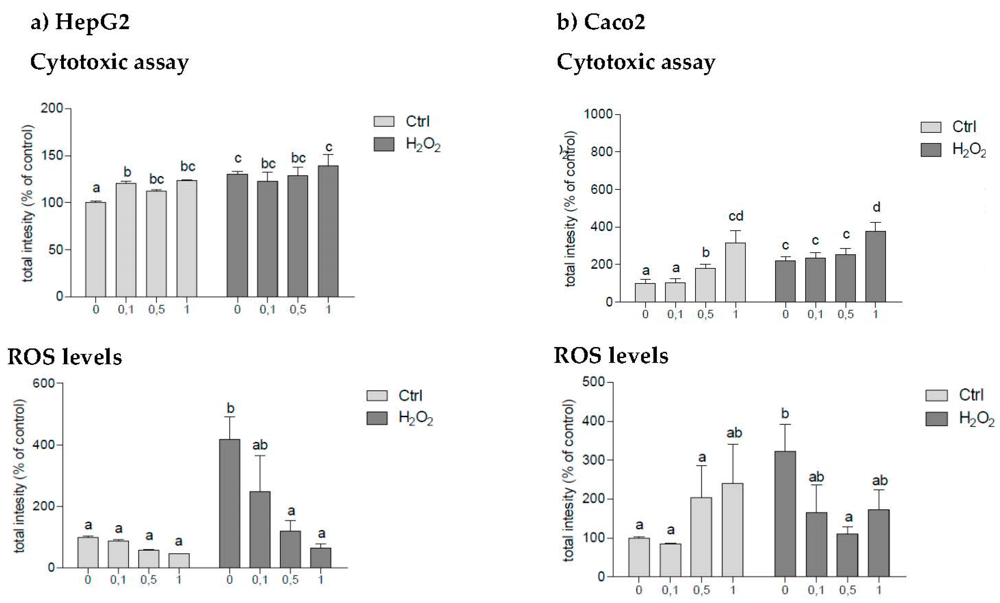
3.2.2. Antioxidant Activity Determined by Cell Culture Assays (Caco-2 and HepG2)

The AC or bioactivity of samples (as chemoprotectors or exogenous antioxidants) in protecting HepG2 and Caco2 cells against oxidative stress induced by H2O2 was studied, measuring the cell viability and reactive oxygen species (ROS) levels. The results are shown in Figure 1a, b. First, we checked the basal effect of the polyphenols extracts on cell viability, evaluating the cytotoxic effects with different poliphenols concentrations.
In HepG2 lines (Figure 1a) a cytotoxicity effect was observed when cells were exposed only to the polyphenol extracts, increasing in a dose-response manner. On the other hand, when cells were exposed only to H2O2, a cytotoxicity effect was also observed derived from oxidative stress stimulus. The supplementation with polyphenols did not recover cells from this oxidative stress. However, a significant decrease in ROS levels was observed with increasing extract concentration in those cells exposed to H2O2, showing an antioxidant effect of L. cuneifolia extracts.
In the Caco-2 cell line (Figure 1b), in both the control cells and in those exposed to H2O2, a cytotoxic effect of polyphenols was observed rising with increasing concentration. On the other hand, as expected, exposure of cells to H2O2 produces a cytotoxic effect increasing cell death. In this case, polyphenols are only capable of reducing the ROS level in a concentration of 0.5 ug/mL.
It should be noted that the increase in cytotoxicity is a positive outcome when the final objective is the search for treatments for tumor cells, such as the HepG2 and Caco 2 lines, probably with the use of higher doses of bioactive compounds in tests.

4. Conclusions

L. cuneifolia Cav. is a species that grows in arid and degraded soils, is rich in phenolic compounds and possesses important antioxidant activity in vitro. The main polyphenolic components are the nordihydroguayaretic acid (NDGA) derivatives and flavonols (flavonols and flavones) in addition to lignans, flavonolignans and cinnamic acids. In the activity on HepG2 cell culture lines, a significant decrease in ROS concentration was observed when cells are exposed to H2O2. On the other hand, a cytotoxic effect is primarily observed on Caco2 cells. This effect can be used to study compounds with bioactivity in the search for new oncological treatments.

Author Contributions

Conceptualization, E.L., P.G. and V.B.; methodology, V.B., P.G., E.S., M.L. and A.Q.; software, V.B., E.S. and E.L.; validation, V.B., P.G. and E.L.; formal analysis, V.B. and P.G.; investigation, E.L. and P.G.; resources, V.B. and P.G.; writing—original draft preparation, E.L. and P.G.; writing—review and editing, V.B. and P.G.; supervision, V.B.; project administration, V.B. and P.G.; funding acquisition, V.B. and P.G. All authors have read and agreed to the published version of the manuscript.

Funding

This work was mainly supported by CONICET [PIP2015-11220150100684]; FonCyT [PICT-2015-2817, 2017-1637, 2017-0123]; SECyT, UNC [33620180100522CB (2018–2021)]; SECyT UNCa [02/L117(2016–2018)]. Emilia Lorenzo has a fellowship from CONICET.

Institutional Review Board Statement

Not applicable.

Informed Consent Statement

Not applicable.

Data Availability Statement

Not applicable.

Conflicts of Interest

The authors declare no conflict of interest.

References

  1. Carabajal, M.P.A.; Perea, M.C.; Isla, M.I.; Zampini, I.C. The use of jarilla native plants in a Diaguita-Calchaquí indigenous community from northwestern Argentina: An ethnobotanical, phytochemical and biological approach. J. Ethnopharmacol. 2020, 247, 112258. [Google Scholar] [CrossRef] [PubMed]
  2. Ríos, J.M.; Mangione, A.M.; Gianello, J.C. Effects of natural phenolic compounds from a desert dominant shrub Larrea divaricata Cav. on toxicity and survival in mice. Rev. Chil. Hist. Nat. 2008, 81, 2. [Google Scholar] [CrossRef]
  3. Davicino, R.; Martino, R.; Anesini, C. Larrea divaricata Cav.: Scientific evidences howingits beneficial effects and its wide potential application. Boletín Latinoam. Caribe Plantas Med. Aromáticas 2011, 10, 92–103. [Google Scholar]
  4. Micucci, P.G.; Alonso, M.D.R.; Turner, S.A.; Davicino, R.C.; Anesini, C.A. Antioxidant and antimicrobial activities of Larreadivaricata Cav. aqueous extract on vitamin C from natural orange juice. Food Nutr. Sci. 2011, 2, 35–46. [Google Scholar]
  5. Vogt, V.; Cifuente, D.; Tonn, C.; Sabini, L.; Rosas, S. Antifungal activity in vitro and in vivo of extracts and lignans isolated from Larrea divaricata Cav. against phytopathogenic fungus. Ind. Crops Prod. 2013, 42, 583–586. [Google Scholar] [CrossRef]
  6. Hapon, M.V.; Boiteux, J.J.; Fernández, M.A.; Lucero, G.; Silva, M.F.; Pizzuolo, P.H. Effect of phenolic compounds present in Argentinian plant extracts on mycelial growth of the plant pathogen Botrytis cinerea Pers. Phyton 2018, 86, 270–277. [Google Scholar]
  7. Amani, S.M.; Isla, M.I.; Vattuone, M.A.; Poch, M.P.; Cudmani, N.G.; Sampietro, A.R. Antimicrobial activities in some Argentine medicinal plants. In II WOCMAP Congress Medicinal and Aromatic Plants, Part 2: Pharmacognosy, Pharmacology, Phytomedicine, Toxicology; International Society for Horticultural Science: Leuven, Belgium, 1997; Volume 501, pp. 115–122. [Google Scholar]
  8. Zampini, I.C.; Cudmani, N.; Isla, M.I. Antimicrobial activity of argentine medicinal plants on antibiotic-resistant bacteria. Acta Bioquím Clín. Lat. Am. 2007, 41, 385–393. [Google Scholar]
  9. Jiménez, C.M.; Sgariglia, M.A. Evaluación de Actividad Antifúngica de Larrea divaricata Cav. y Larrea cuneifolia Cav. Sobre Especies del Género Fusarium; Cuartas Jornadas de Jóvenes Investigadores UTN-Conicet: Tucumán, Argentina, 2010. [Google Scholar]
  10. De-Torres-Sánchez, Antonia. Influencia de los Factores Tecnológicos en la Calidad y en el Contenido en Antioxidantes del Aceite de Oliva Virgen. Ph.D. Thesis, Universidad de Jaén, Jaén, Spain, 2013. Available online: http://hdl.handle.net/10953/523 (accessed on 10 November 2020).
  11. Demmig-Adams, B.; Adams, W.W. Antioxidants in photosynthesis and human nutrition. Science 2002, 298, 2149–2153. [Google Scholar] [CrossRef]
  12. Pinelo, M.; Del Fabbro, P.; Manzocco, L.; Nuñez, M.J.; Nicoli, M.C. Optimization of continuous phenol extraction from Vitisvinifera byproducts. Food Chem. 2005, 92, 109–117. [Google Scholar] [CrossRef]
  13. Pérez, M.J.; Cuello, A.S.; Zampini, I.C.; Ordoñez, R.M.; Alberto, M.R.; Quispe, C.; Schmeda-Hirschmann, G.; Isla, M.I. Polyphenolic compounds and anthocyanin content of Prosopisnigra and Prosopis alba pods flour and their antioxidant and anti-inflammatory capacities. Food Res. Int. 2014, 64, 762–771. [Google Scholar] [CrossRef]
  14. Singleton, V.L.; Orthofer, R.; Lamuela-Raventós, R.M. Analysis of total phenols and other oxidation substrates and antioxidants by means of folin-ciocalteu reagent. Method Enzym. 1999, 299, 152–178. [Google Scholar]
  15. Lingua, M.S.; Fabani, M.P.; Wunderlin, D.A.; Baroni, M.V. From grape to wine: Changes in phenolic composition and its influence on antioxidant activity. Food Chem. 2016, 208, 228–238. [Google Scholar] [CrossRef] [PubMed]
  16. Sakakibara, M.; Difeo, D., Jr.; Nakatani, N.; Timmermann, B.; Mabry, T.J. Flavonoid methyl ethers on the external leaf surface of Larrea tridentata and L. divaricata. Phytochemistry 1976, 15, 727–731. [Google Scholar] [CrossRef]
  17. Abou-Gazar, H.; Bedir, E.; Takamatsu, S.; Ferreira, D.; Khan, I.A. Antioxidant lignans from Larrea tridentata. Phytochemistry 2004, 65, 2499–2505. [Google Scholar] [CrossRef] [PubMed]
  18. Agüero, M.B.; Svetaz, L.; Sánchez, M.; Luna, L.; Lima, B.; López, M.L.; Tapia, A. Argentinean Andean propolis associated with the medicinal plant Larreanitida Cav. (Zygophyllaceae). HPLC–MS and GC–MS characterization and antifungal activity. Food Chem. Toxicol. 2011, 49, 1970–1978. [Google Scholar] [CrossRef]
  19. Martins, S.; Amorim, E.L.; Sobrinho, T.J.P.; Saraiva, A.M.; Pisciottano, M.N.; Aguilar, C.N.; Mussatto, S.I. Antibacterial activity of crude methanolic extract and fractions obtained from Larreatridentata leaves. Ind. Crops Prod. 2013, 41, 306–311. [Google Scholar] [CrossRef]
  20. Brand-Williams, W.; Cuvelier, M.E.; Berset, C. Use of a Free Radical Method to Evaluate Antioxidant Activity. LWT Food Sci. Technol. 1995, 28, 25–30. [Google Scholar] [CrossRef]
  21. Benzie, I.F.F.; Strain, J.J. The ferric reducing ability of plasma (FRAP) as a measure of antioxidant power: The FRAP assay. Anal. Biochem. 1996, 239, 70–76. [Google Scholar] [CrossRef]
  22. Re, R.; Pellegrini, N.; Proteggente, A.; Pannala, A.; Yang, M.; Rice-Evans, C. Antioxidant activity applying an improved ABTS radical cation decolorization assay. Free Radic. Biol. Med. 1999, 26, 1231–1237. [Google Scholar] [CrossRef]
  23. Avelar-Freitas, B.A.; Almeida, V.G.; Pinto MC, X.; Mourão FA, G.; Massensini, A.R.; Martins-Filho, O.A.; Brito-Melo, G.E.A. Trypan blue exclusion assay by flow cytometry. Braz. J. Med. Biol. Res. 2014, 47, 307–315. [Google Scholar] [CrossRef]
  24. Jakubowski, W.; Bartosz, G. 2, 7-dichlorofluorescin oxidation and reactive oxygen species: What does it measure? Cell Biol. Int. 2000, 24, 757–760. [Google Scholar] [CrossRef] [PubMed]
  25. Di Rienzo, J.A.; Casanoves, F.; Balzarini, M.G.; Gonzalez, L.; Tablada, M.; Robledo, Y.C. InfoStat versión 2016. Grupo InfoStat, FCA, Universidad Nacional de Córdoba, Argentina. 2016. Available online: http://www. infostat.com.ar (accessed on 8 November 2020).
  26. Rossi, C.A.; Pereyra, A.M.; González, G.L.; De León, M.; Chagra Dib, P. Composición química, contenido de polifenoles totales y valor nutritivo en especies de ramoneo del Sistema silvopastoril del Chaco árido argentino. Zootec. Trop. 2008, 26, 105–115. [Google Scholar]
  27. Dadé, M.M.; Fioravanti, D.E.; Schinella, G.R.; Tournier, H.A. Total antioxidant capacity and polyphenol content of 21 aqueous extracts obtained from native plants of Traslasierra valley (Argentina). Bollatinoam Caribe 2009, 8, 529–539. [Google Scholar]
  28. Peralta, B.I.N. Larrea divaricata Cav.: Estudio de la Actividad antioxidante frente a estrés oxidativo inducido y Estudio de Los parámetros farmacocinéticos. Ph.D. Thesis, Universidad de Buenos Aires, Buenos Aires, Argentina, 2019. Available online: https://ri.conicet.gov.ar/handle/11336/81114 (accessed on 4 th November 2020).
  29. Borneo, R.; León, A.; Aguirre, A.; Ribotta, P.; Cantero, J. Antioxidant capacity of medicinal plants from de province of Córdoba (Argentina) and their in vitro testing in a model food system. Food Chem. 2009, 112, 664–670. [Google Scholar] [CrossRef]
Figure 1. Cytotoxicity determined by Trypan Blue test and intensity of ROS levels measured by 2′7′-dichlorofluorescein diacetate (DCFH) at different concentrations of the extract of L. cuneifolia leaf (0, 0.1, 0.5 and 1 µg/mL) in HepG2 cell lines (a) and Caco-2 (b) in medium, with and without H2O2.
Proceedings 70 00037 g001
Table 1. Phenolic compounds and derivatives, identified with HPLC-ESI-MS/MS, in ethanolic leaf extract of L. cuneifolia.
RT (min)Tentatively Identified
Compound
Molecular Formula[M-H] (m/z) Theoretical[M-H] (m/z) ExperimentalError ppmMS/MS
111.44-caffeoylquinicacidC16H17O93.530.878353.08410.2191
212.83-caffeoylquinicacidC16H17O93.530.878353.0849.6191
318.9Quercetin rutinosideC27H29O166.091.461609.147−0.9301
419.2Quercetin glucosideC21H19O124.630.882463.089−1.7301
520.0Kaempferolhexoside isomer IIC21H19O114.470.933447.0942.5285
620.2DihydroisorhamnetinC16H13O73.170.667317.0683.9299, 289, 273, 258, 231, 207
721.2Isorhamnetinrhamnosyl glucosideC28H31O166.231.618623.163−2.5315
824.4Dimethyl gossypetinC17H13O83.450.616345.0620.4
924.7TrimethylgossypetinC18H15O83.590.772359.0793.7315, 273
1024.8NaringeninC15H11O52.710.612271.062−1.6177, 151, 227
1125.5Quercetin methylether isomer IC16H11O7315.051315.05616.7300
1227.6KaempferolC15H9O62.850.405285.0411.7
1327.8Quercetin methylether isomer IIC16H11O7315.051315.0535.8300
1428.1meso-(rel7S,8S,7′R,8′R)-3,4,3′,4′-tetrahydroxy7,7′-epoxylignanC19H22O53.291.394329.141−3.3177
1528.9Trihydroxytrimethoxy flavoneC18H15O83.590.772359.0781.7344, 329, 316, 301, 273
1631.0Nordihydroguayaretic acid (NDGA)C18H21O43.011.445301.1466273, 268, 299
1732.0Trimethyl quercetinC18H15O73.430.823343.0869.2328, 313
1834.23-methylnordihydroguayaretic acid (MNDGA)C19H24O43.151.602315.1626.7300
Publisher’s Note: MDPI stays neutral with regard to jurisdictional claims in published maps and institutional affiliations.

