Article

A SiO2/pHEMA-Based Polymer-Infiltrated Ceramic Network Composite for Dental Restorative Materials

1
Division of Biomaterials, Department of Oral Functions, Kyushu Dental University, Fukuoka 803-8580, Japan
2
Division of Oral Reconstruction and Rehabilitation, Department of Oral Functions, Kyushu Dental University, Fukuoka 803-8580, Japan
*
Author to whom correspondence should be addressed.
Submission received: 19 December 2021 / Revised: 2 January 2022 / Accepted: 4 January 2022 / Published: 5 January 2022
(This article belongs to the Special Issue Feature Papers in Journal of Composites Science in 2021)

Abstract

:
SiO2-poly(2-hydroxyethyl methacrylate) (pHEMA)-based composites have been widely used as biomaterials owing to their biocompatibility. However, they have not yet been applied as tooth restorative materials because of their poor mechanical properties. In the present paper, we develop a novel SiO2/pHEMA-based composite with a polymer-infiltrated network (PICN) structure for use in dental restorative materials. A mixture of SiO2 nanoparticles and a poly(vinyl alcohol) binder was sintered at 950 °C to fabricate a porous SiO2 block. A monomer mixture containing 70 wt%-HEMA/30 wt%-ethylene glycol dimethacrylate and a benzoyl peroxide initiator was infiltrated into the porous SiO2 block and heat-polymerized to fabricate the SiO2/pHEMA-based composite with a PICN structure. The composite was characterized according to its mechanical properties, surface free energy, and bonding properties with a dental adhesive. The flexural strength was 112.5 ± 18.7 MPa, the flexural modulus was 13.6 ± 3.4 GPa, and the Vickers hardness was 168.2 ± 16.1, which are similar values to human teeth. The surface free energy of the polar component of the composite was 19.6 ± 2.5 mN/m, suggesting that this composite has an active surface for bonding with the adhesive. The composite bonded well to the adhesive, in the presence of a silane coupling agent. The SiO2/pHEMA-based composite was demonstrated to be a potential candidate for dental restorative materials.

1. Introduction

Biomimetic concepts have been widely adapted for the development of advanced biomaterials that imitate the biological functions, mechanical properties, or tissue morphologies of natural life forms [1]. For example, many studies have focused on developing biomaterials that mimic the hard tissues of enamel and dentin for tooth restoration, wherein a mechanical compatibility with teeth is one of the most important properties of a tooth restorative material. In contemporary dental restorative materials, polymer-infiltrated ceramic network (PICN) composites are the closest materials to human teeth (enamel and dentin) in terms of their mechanical properties, such as their hardness and elasticity [2,3]. A PICN composite consists of dual network structures of a ceramic skeleton and a polymer (resin) that infiltrates into the continuous pores of the ceramic [2,4,5]. This microstructure differs significantly from a conventional dispersed-filler (DF) composite, which consists of ceramic particles dispersed in a polymer matrix. Owing to the unique PICN structure, the mechanical properties of PICN composites, such as their hardness and elastic modulus, are superior to those of DF composites and are relatively close to those of enamel. The excellent mechanical compatibility of the PICN composite allows its application as a restorative material for dental inlays, onlays, and crowns [6,7]. PICN composites have also been studied with the aim of improving their mechanical properties through focusing on their microstructures, especially the ceramic skeleton. As a result, silica (SiO2)-based [8,9,10], aluminosilicate-based [11,12], and zirconia-based [13,14] composites have been developed. Among the previously reported PICN composites, the SiO2-based PICN composites consisting of appropriate polymers, such as SiO2/PMMA [8], SiO2/pTEGDMA [10], and silica/pUDMA [9], are similar to enamel in terms of their hardness, and comparable to dentin in the context of the elastic modulus. Hence, SiO2-based PICN composites are expected to be applied as restorative materials for teeth.
Poly(2-hydroxyethyl methacrylate) (pHEMA) is a hydrophilic polymer that is frequently used in biomaterials [15,16]. SiO2/pHEMA-based composites have also been developed for use in biomaterials owing to their superior biocompatibility compared to pHEMA alone [17,18,19]. In terms of dental applications, pHEMA has been frequently used in dental adhesives, such as resin cements [20,21]. However, SiO2/pHEMA composites have yet to be established for application in tooth restorative materials, such as dental crowns. As previously reported, the DF composites composed of SiO2/pHEMA exhibit water-swelling properties [19], which result in a soft-gel behavior in an aqueous environment, and prevent the use of SiO2/pHEMA-based composites in the mouth, since tooth restorative materials must be rigid. In contrast, PICN structural composites tend to be rigid due to the ceramic skeleton present in their structure. However, SiO2/pHEMA-based composites with PICN structures have yet to be reported.
Thus, the aim of this study is to develop a novel PICN composite composed of SiO2/pHEMA for application as a tooth restorative material. Initially, the composite is prepared via the sol–gel method, followed by sintering and polymer infiltration processes. In the infiltration process, the sintered porous SiO2 monolith was immersed into the appropriate liquid monomer mixture, followed by heat polymerization. The obtained composite was then characterized according to its mechanical properties and bonding properties, to demonstrate the possibility of its application in tooth restorative materials.

2. Materials and Methods

2.1. Materials

The reagents used for the preparation of the SiO2/pHEMA-based composites are listed in Table 1.

2.2. Preparation of the SiO2/pHEMA-Based Composite

The SiO2/pHEMA composite was prepared using a modification of our previously reported procedure, for the preparation of a SiO2/PMMA composite with a PICN structure [8]. More specifically, SiO2 nanoparticles (6 g) with 7 nm of mean diameter were added to purified water (54 g) at pH 3 and dispersed under ultrasonication for 1 h. In addition, a 10 wt% poly(vinyl alcohol) (PVA) solution (11.4 g) was obtained by dissolving PVA powder in purified water. The prepared SiO2 suspension and PVA solution were then mixed, and a 4 wt% aqueous solution of Na2B4O5(OH)4·8H2O (1 mL) was added under magnetic stirring at 30 °C for 24 h to obtain a SiO2–PVA slurry. This slurry was then poured into a Teflon container and dried in an oven at 30 °C for 7 d. Consequently, a solid-state monolithic SiO2-PVA green body was obtained. Thermal debinding of the green body was performed in an electric furnace at 600 °C for 3 h under air, followed by sintering at 950 °C to form a porous silica block. The resulting porous silica block was immersed in a liquid monomer mixture (30 wt% HEMA and 70 wt% ethylene glycol dimethacrylate (EGDMA) and 0.5 wt% benzoyl peroxide (BPO)) for 1 d at 25 °C. Thermal polymerization of the monomer mixture-infiltrated block was performed at 60 °C for 2 d, followed by treatment at 80 °C for 1 d to give the desired SiO2/pHEMA-based composite.

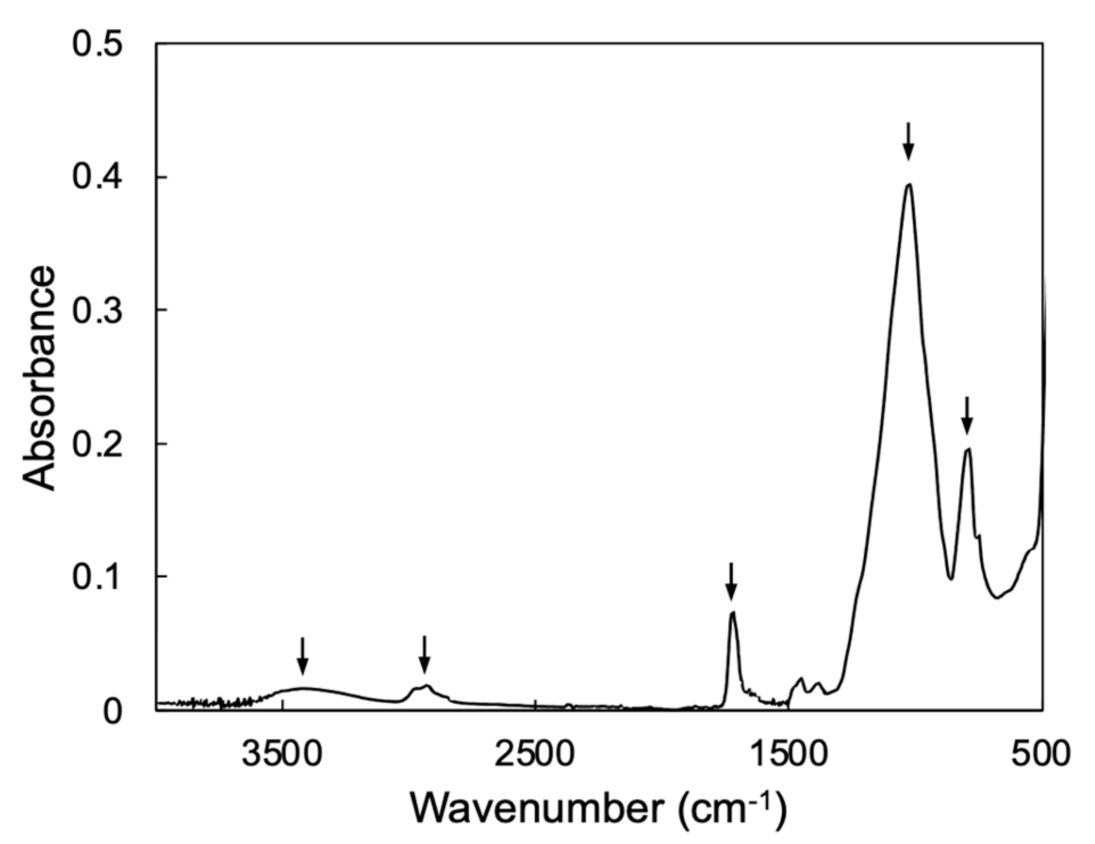
2.3. Structural Characterization

The prepared SiO2/pHEMA-based composite was analyzed by Fourier transform infrared (FT-IR) spectroscopy, using a spectrometer (IRSpirit, Shimadzu Corp., Kyoto, Japan) equipped with an attenuated total reflection (ATR) unit.

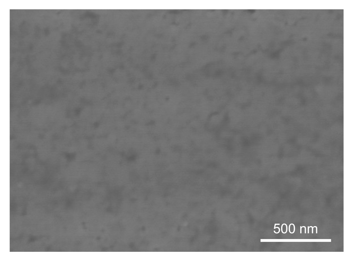
2.4. SEM Observation

The microstructure of the filler was observed using a field emission scanning electron microscopy (FE-SEM; S-4300, Hitachi High-Tech Corp., Tokyo, Japan). Prior to the observation, the sample was coated with platinum via a sputtering.

2.5. Mechanical Properties

The prepared SiO2/pHEMA-based composite was cut and polished using emery papers up to #2000 to fabricate bar-shaped samples (width = 4 mm, length = 14 mm, thickness = 1.2 mm). The flexural strength and modulus of each bar sample was measured using a universal testing machine (AGS-H, Shimadzu Corp., Kyoto, Japan), according to the three-point bending test procedure given in the ISO standard [22]. A support span of 12 mm and a crosshead speed of 1 mm/min were used. After the 3-point bending test, the fractured sample was used for the Vickers hardness test, which was carried out using a hardness tester (HMV-G21ST, Shimadzu Corp., Japan) with a load of 200 g and a dwell time of 15 s. Ten samples were used for each mechanical test (n = 10).

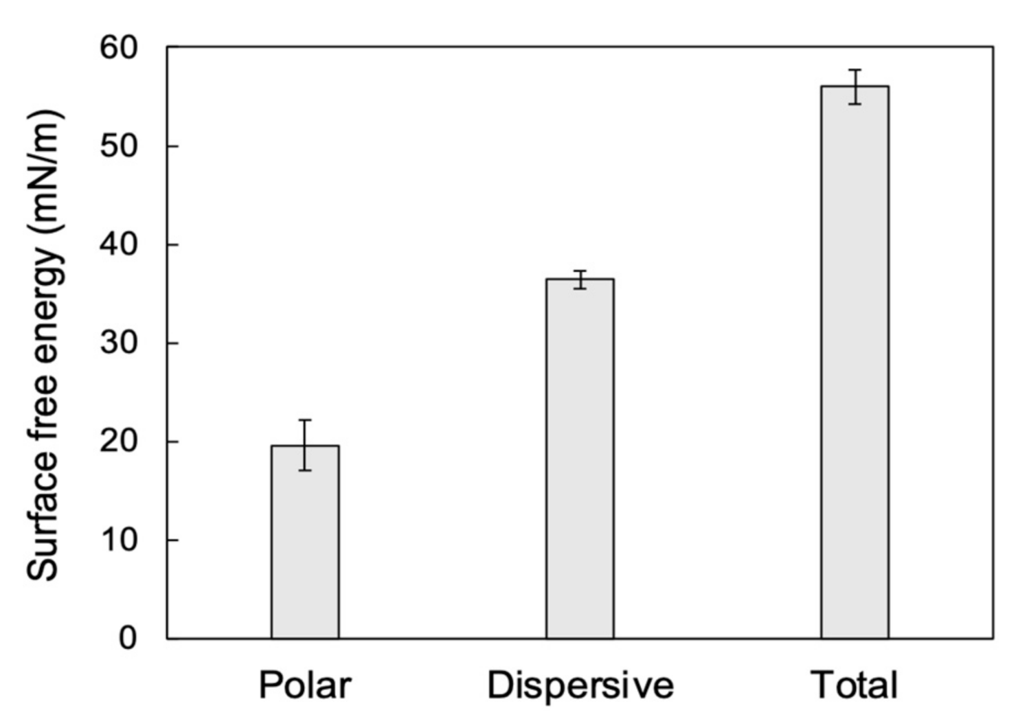
2.6. Surface Characterization

The surface of the prepared SiO2/pHEMA-based composite was characterized according to its surface free energy (SFE) via contact angle measurements, as described in a previous study [23]. More specifically, the contact angles of two test liquids (distilled water and diiodomethane) were measured using a contact angle meter (DMe-211, Kyowa Interface Science Co., Ltd., Saitama, Japan). The surface free energies, dispersive components (γd), polar components (γp), and total surface free energies (γtotal), were calculated from the measured contact angles using the Owens–Wendt theory [24]. Ten samples were used in the test (n = 10).

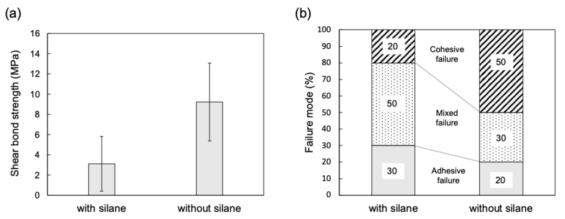
2.7. Shear Bond Strength

The shear bond strength between the prepared SiO2/pHEMA-based composite and the commercial dental adhesive was measured according to a previously reported procedure [23]. The composite was cut into a disk shape (diameter = 10 mm, thickness = 1.5 mm) and polished using emery papers up to #1000. A commercial silane primer (Porcelain primer, SHOFU Inc., Kyoto, Japan) containing a silane coupling agent was applied to the sample surface. Subsequently, resin cement (Resicem, SHOFU Inc., Japan) was loaded onto the sample surface and light-cured using a light irradiator for 5 min. After this time, the cement-cured sample was held under ambient conditions for 60 min and then stored in distilled water at 37 °C for 24 h. The shear bond strength (SBS) tests were conducted using the universal testing machine at a crosshead speed of 1.0 mm/min with a blade-shaped loading device. After SBS testing, the debonded surface was observed using an optical microscope to classify the failure modes as one of three types: adhesive failure at the adhesive–sample interface, cohesive failure within the sample, or mixed adhesive and cohesive failure.

3. Results and Discussion

In the practical application of dental materials, tooth restorative materials, such as dental crowns, are milled from a block-shaped material using a computer-aided design/computer-aided manufacturing (CAD/CAM) milling system. Hence, the composite should ideally possess a block shape. To satisfy this requirement, we carried out a preliminary experiment to fabricate monolithic blocks of the SiO2/pHEMA composites. In the first preliminary experiment, the sintering temperature of the green body was optimized using temperatures of 750, 850, 950, and 1050 °C, wherein a monolithic block was successfully obtained at a sintering temperature of 950 °C. In contrast, a lack of pores was observed for the samples prepared at other sintering temperatures, and, in many cases, crack formation also took place. Subsequently, we optimized the composition of the infiltration monomer by changing the mixture ratio of HEMA to EGDMA. In the case of 100 wt% HEMA without the EGDMA cross-linker, the infiltration monomer was not polymerized under the present heat-polymerization conditions. Indeed, this was achieved only in the presence of EGDMA, wherein the optimal composition to obtain a cured monomer-infiltrated sample was found to be 70 wt%-HEMA + 30 wt%-EGDMA.
The FT-IR spectrum of the SiO2/pHEMA-based composite is shown in Figure 1, wherein characteristic peaks can be observed at ~800 and 1020 cm−1, which were assigned to the Si–O–Si symmetric and asymmetric vibrations of SiO2 [25,26]. In addition, the peaks at ~1720 and 2950 cm−1 were assigned to the C = O and C–H stretching vibrations in the ester compounds of pHEMA and pEGDMA [27,28]. Furthermore, the broad peak at ~3400 cm−1 was assigned to the O–H stretching vibration of the composite. This result clearly indicates that the prepared composite was composed of SiO2 and copoly(HEMA/EGDMA).
The microstructure of the SiO2/pHEMA-based composite is shown in Figure 2. The composite has the PICN structure, composed of a SiO2 skeleton and pHEMA-based resin inside the pores of the skeleton, which forms a nanoscale dual network structure.
The mechanical properties, flexural strength, elastic modulus (flexural modulus), and hardness are the most important properties of a tooth restorative material. Thus, the mechanical properties of the SiO2/pHEMA-based composite were examined and are listed in Table 2. More specifically, it was found that the flexural strength of the composite (112.5 MPa) was relatively close to that of human dentin (109–160 MPa) [29]. In addition, the flexural (elastic) modulus of the composite (13.6 GPa) was closer to that of dentin (ca.10–20 GPa) [30] rather than that of enamel (ca. 40–100 GPa) [30], while the Vickers hardness value of the composite (168.2) was between those of enamel (~300–400) [30] and dentin (~50–100) [30]. These results therefore indicate that the mechanical properties of the composite are similar to those of human teeth. Furthermore, the mechanical properties of the composite meet the criteria for dental restorative materials based on the ISO standard [31]. Hence, the SiO2/pHEMA-based composite has sufficient mechanical properties for application as a tooth restorative material. The mechanical properties of the SiO2/pHEMA-based composite are similar to those of a SiO2/PMMA-based PICN composite, which contained a silica skeleton infiltrated with PMMA resin [8]. Considering the microstructure, these PICN composites have different resin components, but the same ceramic skeleton. Therefore, it is considered that the unique properties of the SiO2-based PICN composite was affected by the microstructure of the ceramic skeleton.
In the clinical treatment of tooth restorations, the dental crown is bonded onto the abutment tooth of the patient using a dental adhesive, and so the dental restorative material should exhibit a sufficient bonding capability to the dental adhesive. Thus, we evaluated the bonding properties of the SiO2/pHEMA-based composite in terms of the SFE and the bond strength. The SFE values of the composite are shown in Figure 3, wherein the dispersive and polar components were 36.4 ± 0.9 and 19.6 ± 2.5 mN/m, respectively. The value for the polar component is higher than that of a conventional dental composite with a DF structure [23], and this unique surface characteristic has a positive effect on the bonding properties of the composite, as can be seen in Figure 4. More specifically, the SBS (Figure 4a) significantly increased when using the silane primer, as did the number of cohesive failures, thereby indicating that the composite bonded strongly to the dental adhesive, and that the silane coupling agent in the primer was suitable for combination with this composite and resin cement. It was assumed that the silane coupling agent reacted with the silica skeleton in the PICN structure. Indeed, similar results can be found in the bond characteristics between a conventional PICN composite and an adhesive [23,32,33].
Overall, these results indicate that we successfully developed a novel SiO2/pHEMA-based composite with a good mechanical compatibility to teeth and a superior bonding capability toward dental adhesive. Although SiO2/pHEMA-based composites have been reported for DF composites [17,19,34,35,36,37,38,39,40,41,42], there have been no reports into the use of PICN composites. Since the present SiO2/pHEMA-based composite has a PICN structure, the mechanical properties of the present composite are superior to those of the conventional DF composites. This advance provides the opportunity to apply the SiO2/pHEMA-based composite on biomaterials for hard tissues, such as teeth and bones. This study is therefore the first to report a SiO2/pHEMA-based composite, and our results demonstrate that the prepared composite has the potential for application in dental restorative materials, such as inlays, onlays, and crowns. However, further studies are required relating to the biocompatibility of this material, such as its cytotoxicity, toward human gingiva. In addition, the PICN composite has unique microstructure and properties, enabling its application in industrial fields, such as environmentally friendly materials [43]. This possibility will be further investigated in future works.

4. Conclusions

A polymer-infiltrated ceramic network composite composed of SiO2 and copoly(HEMA/EGDMA) was successfully prepared. The flexural strength, flexural modulus, and Vickers hardness of the composite were 112.5 ± 18.7 MPa, 13.6 ± 3.4 GPa, and 168.2 ± 16.1, respectively. These mechanical properties of the composite were comparable to those of human teeth and met the criteria for tooth restorative materials. The prepared composite bonded well to the dental adhesive, and it was demonstrated that this composite is a potential candidate material for dental applications.

Author Contributions

Conceptualization, H.I., R.H. and H.S.; Data curation, H.I., Y.K., M.K.S. and H.T.Y.; Formal analysis, H.I., Y.K. and M.K.S.; Funding acquisition, M.K.S. and H.T.Y.; Investigation, H.I., Y.K. and Y.N.; Methodology, Y.N. and C.M.; Project administration, H.S.; Resources, H.I. and C.M.; Software, M.K.S., H.T.Y. and Y.N.; Supervision, H.S. and R.H.; Validation, H.I., Y.K. and R.H.; Visualization, R.H.; Writing— original draft, H.I., Y.K., M.K.S. and H.T.Y.; Writing—review & editing, Y.N., C.M., R.H. and H.S. All authors have read and agreed to the published version of the manuscript.

Funding

This work was supported by JSPS KAKENHI, grant numbers 21K21047 and 21K21093.

Data Availability Statement

The data presented in this study are available on request from the corresponding author.

Conflicts of Interest

The authors declare no conflict of interest.

References

  1. Wilmers, J.; Bargmann, S. Nature’s design solutions in dental enamel: Uniting high strength and extreme damage resistance. Acta Biomater. 2020, 107, 1–24. [Google Scholar] [CrossRef] [PubMed]
  2. Coldea, A.; Swain, M.; Thiel, N. Mechanical properties of polymer-infiltrated-ceramic-network materials. Dent. Mater. 2013, 29, 419–426. [Google Scholar] [CrossRef]
  3. Mainjot, A.; Dupont, N.; Oudkerk, J.; Dewael, T.; Sadoun, M. From Artisanal to CAD-CAM Blocks: State of the Art of Indirect Composites. J. Dent. Res. 2016, 95, 487–495. [Google Scholar] [CrossRef]
  4. He, L.-H.; Swain, M. A novel polymer infiltrated ceramic dental material. Dent. Mater. 2011, 27, 527–534. [Google Scholar] [CrossRef] [PubMed]
  5. Kang, L.; Zhou, Y.; Lan, J.; Yu, Y.; Cai, Q.; Yang, X. Effect of resin composition on performance of polymer-infiltrated feldspar-network composites for dental restoration. Dent. Mater. J. 2020, 39, 900–908. [Google Scholar] [CrossRef] [PubMed]
  6. Goujat, A.; Abouelleil, H.; Colon, P.; Jeannin, C.; Pradelle, N.; Seux, D.; Grosgogeat, B. Mechanical properties and internal fit of 4 CAD-CAM block materials. J. Prosthet. Dent. 2018, 119, 384–389. [Google Scholar] [CrossRef] [PubMed]
  7. Zheng, Z.; He, Y.; Ruan, W.; Ling, Z.; Zheng, C.; Gai, Y.; Yan, W. Biomechanical behavior of endocrown restorations with different CAD-CAM materials: A 3D finite element and in vitro analysis. J. Prosthet. Dent. 2021, 125, 890–899. [Google Scholar] [CrossRef]
  8. Ikeda, H.; Nagamatsu, Y.; Shimizu, H. Preparation of silica–poly(methyl methacrylate) composite with a nanoscale dual-network structure and hardness comparable to human enamel. Dent. Mater. 2019, 35, 893–899. [Google Scholar] [CrossRef]
  9. Kawajiri, Y.; Ikeda, H.; Nagamatsu, Y.; Masaki, C.; Hosokawa, R.; Shimizu, H. PICN Nanocomposite as Dental CAD/CAM Block Comparable to Human Tooth in Terms of Hardness and Flexural Modulus. Materials 2021, 14, 1182. [Google Scholar] [CrossRef] [PubMed]
  10. Sodeyama, M.; Ikeda, H.; Nagamatsu, Y.; Masaki, C.; Hosokawa, R.; Shimizu, H. Printable PICN Composite Mechanically Compatible with Human Teeth. J. Dent. Res. 2021, 100, 1475–1481. [Google Scholar] [CrossRef] [PubMed]
  11. Nguyen, J.; Ruse, N.D.; Phan, A.; Sadoun, M. High-temperature-pressure Polymerized Resin-infiltrated Ceramic Networks. J. Dent. Res. 2014, 93, 62–67. [Google Scholar] [CrossRef] [PubMed] [Green Version]
  12. Cui, B.; Li, J.; Wang, H.; Lin, Y.; Shen, Y.; Li, M.; Deng, X.; Nan, C. Mechanical properties of polymer-infiltrated-ceramic (sodium aluminum silicate) composites for dental restoration. J. Dent. 2017, 62, 91–97. [Google Scholar] [CrossRef]
  13. Li, S.; Zhao, Y.; Zhang, J.-F.; Xie, C.; Zhao, X. Machinability of poly(methyl methacrylate) infiltrated zirconia hybrid composites. Mater. Lett. 2014, 131, 347–349. [Google Scholar] [CrossRef]
  14. Li, J.; Cui, B.-C.; Lin, Y.-H.; Deng, X.-L.; Li, M.; Nan, C.-W. High strength and toughness in chromatic polymer-infiltrated zirconia ceramics. Dent. Mater. 2016, 32, 1555–1563. [Google Scholar] [CrossRef] [PubMed]
  15. Luensmann, D.; Jones, L. Albumin adsorption to contact lens materials: A review. Contact Lens Anterior Eye 2008, 31, 179–187. [Google Scholar] [CrossRef]
  16. Kirschning, A.; Dibbert, N.; Dräger, G. Chemical Functionalization of Polysaccharides-Towards Biocompatible Hydrogels for Biomedical Applications. Chem. Eur. J. 2018, 24, 1231–1240. [Google Scholar] [CrossRef]
  17. Liu, Y.-Y.; Liu, T.-Y.; Chen, S.-Y.; Liu, D.-M. Synthesis and characterization of nanoporous SiO2/pHEMA biocomposites. J. Mater. Sci. Mater. Electron. 2008, 19, 2903–2911. [Google Scholar] [CrossRef] [Green Version]
  18. Luciani, G.; Costantini, A.; Silvestri, B.; Tescione, F.; Branda, F.; Pezzella, A. Synthesis, structure and bioactivity of pHEMA/SiO2 hybrids derived through in situ sol–gel process. J. Sol-Gel Sci. Technol. 2008, 46, 166–175. [Google Scholar] [CrossRef] [Green Version]
  19. Silvestri, B.; Luciani, G.; Costantini, A.; Tescione, F.; Branda, F.; Pezzella, A. In-situ sol-gel synthesis and characterization of bioactive pHEMA/SiO 2 blend hybrids. J. Biomed. Mater. Res. Part B Appl. Biomater. 2009, 89B, 369–378. [Google Scholar] [CrossRef]
  20. Moszner, N.; Hirt, T. New polymer-chemical developments in clinical dental polymer materials: Enamel-dentin adhesives and restorative composites. J. Polym. Sci. Part A Polym. Chem. 2012, 50, 4369–4402. [Google Scholar] [CrossRef]
  21. Sidhu, S.K.; Nicholson, J.W. A Review of Glass-Ionomer Cements for Clinical Dentistry. J. Funct. Biomater. 2016, 7, 16. [Google Scholar] [CrossRef] [PubMed]
  22. ISO 6872:2015; Dentistry–Ceramic Materials; International Organization for Standarization: Geneva, Switzerland, 2015.
  23. Yano, H.T.; Ikeda, H.; Nagamatsu, Y.; Masaki, C.; Hosokawa, R.; Shimizu, H. Correlation between microstructure of CAD/CAM composites and the silanization effect on adhesive bonding. J. Mech. Behav. Biomed. Mater. 2020, 101, 103441. [Google Scholar] [CrossRef] [PubMed]
  24. Owens, D.K.; Wendt, D.T. Estimation of the surface free energy of polymers. J. Appl. Polym. Sci. 1969, 13, 1741–1747. [Google Scholar] [CrossRef]
  25. Uchino, T.; Aboshi, A.; Kohara, S.; Ohishi, Y.; Sakashita, M.; Aoki, K. Microscopic structure of nanometer-sized silica particles. Phys. Rev. B 2004, 69, 155409. [Google Scholar] [CrossRef] [Green Version]
  26. Yamada, T.; Nakajima, M.; Suemoto, T.; Uchino, T. Formation and Photoluminescence Characterization of Transparent Silica Glass Prepared by Solid-Phase Reaction of Nanometer-Sized Silica Particles. J. Phys. Chem. C 2007, 111, 12973–12979. [Google Scholar] [CrossRef]
  27. Abay, I.; Denizli, A.; Bişkin, E.; Salih, B. Removal and pre-concentration of phenolic species onto β-cyclodextrin modified poly(hydroxyethylmethacrylate–ethyleneglycoldimethacrylate) microbeads. Chemosphere 2005, 61, 1263–1272. [Google Scholar] [CrossRef]
  28. Suhag, G.S.; Bhatnagar, A.; Singh, H. Poly(hydroxyethyl methacrylate)-based co-polymeric hydrogels for transdermal delivery of salbutamol sulphate. J. Biomater. Sci. Polym. Ed. 2008, 19, 1189–1200. [Google Scholar] [CrossRef] [PubMed]
  29. Arola, D.D.; Reprogel, R.K. Tubule orientation and the fatigue strength of human dentin. Biomaterials 2006, 27, 2131–2140. [Google Scholar] [CrossRef]
  30. Zhang, Y.-R.; Du, W.; Zhou, X.-D.; Yu, H.-Y. Review of research on the mechanical properties of the human tooth. Int. J. Oral Sci. 2014, 6, 61–69. [Google Scholar] [CrossRef] [Green Version]
  31. ISO 10477:2018; Dentistry—Polymer-Based Crown and Veneering Materials; International Organization for Standarization: Geneva, Switzerland, 2018.
  32. Bello, Y.D.; Di Domenico, M.; Magro, L.D.; Lise, M.W.; Corazza, P.H. Bond strength between composite repair and polymer-infiltrated ceramic-network material: Effect of different surface treatments. J. Esthet. Restor. Dent. 2018, 31, 275–279. [Google Scholar] [CrossRef]
  33. Şişmanoğlu, S.; Gürcan, A.T.; Yıldırım-Bilmez, Z.; Turunç-Oğuzman, R.; Gümüştaş, B. Effect of surface treatments and universal adhesive application on the microshear bond strength of CAD/CAM materials. J. Adv. Prosthodont. 2020, 12, 22–32. [Google Scholar] [CrossRef] [Green Version]
  34. Ji, X.-L.; Jiang, S.-C.; Qiu, X.-P.; Dong, D.-W.; Yu, D.-H.; Jiang, B.-Z. Structure and properties of hybrid poly(2-hydroxyethyl methacry-late)/SiO2monoliths. J. Appl. Polym. Sci. 2003, 88, 3168–3175. [Google Scholar] [CrossRef]
  35. Costa, R.O.R.; Pereira, M.M.; Lameiras, F.S.; Vasconcelos, W.L. Apatite formation on poly(2-hydroxyethyl methacrylate)-silica hybrids prepared by sol-gel process. J. Mater. Sci. Mater. Electron. 2005, 16, 927–932. [Google Scholar] [CrossRef]
  36. Costantini, A.; Luciani, G.; Annunziata, G.; Silvestri, B.; Branda, F. Swelling properties and bioactivity of silica gel/pHEMA nanocomposites. J. Mater. Sci. Mater. Electron. 2006, 17, 319–325. [Google Scholar] [CrossRef]
  37. Costantini, A.; Luciani, G.; Silvestri, B.; Tescione, F.; Branda, F. Bioactive poly(2-hydroxyethylmethacrylate)/silica gel hybrid nanocomposites prepared by sol-gel process. J. Biomed. Mater. Res. Part B Appl. Biomater. 2008, 86B, 98–104. [Google Scholar] [CrossRef] [PubMed]
  38. Huang, S.-L.; Chin, W.-K.; Yang, W. Structural characteristics and properties of silica/poly(2-hydroxyethyl methacrylate) (PHEMA) nanocomposites prepared by mixing colloidal silica or tetraethyloxysilane (TEOS) with PHEMA. Polymer 2005, 46, 1865–1877. [Google Scholar] [CrossRef]
  39. Costa, R.O.; Lameiras, F.S.; Nunes, E.H.; Vasconcelos, D.C.; Vasconcelos, W. Preparation of silica-poly(2-hydroxyethyl methacrylate) hybrids modified with 3-methacryloxypropyltrimethoxysilane. Ceram. Int. 2016, 42, 3465–3472. [Google Scholar] [CrossRef]
  40. Schiraldi, C.; D’Agostino, A.; Oliva, A.; Flamma, F.; De Rosa, A.; Apicella, A.; Aversa, R.; De Rosa, M. Development of hybrid materials based on hydroxyethylmethacrylate as supports for improving cell adhesion and proliferation. Biomaterials 2004, 25, 3645–3653. [Google Scholar] [CrossRef]
  41. Li, S.; Shah, A.; Hsieh, A.J.; Haghighat, R.; Praveen, S.S.; Mukherjee, I.; Wei, E.; Zhang, Z.; Wei, Y. Characterization of poly(2-hydroxyethyl methacrylate-silica) hybrid materials with different silica contents. Polymer 2007, 48, 3982–3989. [Google Scholar] [CrossRef]
  42. Chang, C.-C.; Oyang, T.-Y.; Hwang, F.-H.; Chen, C.-C.; Cheng, L.-P. Preparation of polymer/silica hybrid hard coatings with enhanced hydrophobicity on plastic substrates. J. Non-Cryst. Solids 2012, 358, 72–76. [Google Scholar] [CrossRef]
  43. Soltani, N.; Bahrami, A.; Pech-Canul, M.; González, L. Review on the physicochemical treatments of rice husk for production of advanced materials. Chem. Eng. J. 2015, 264, 899–935. [Google Scholar] [CrossRef]
Figure 1. FT-IR spectrum of the SiO2/pHEMA-based composite. The arrows in the figure show the composite assignments.
Jcs 06 00017 g001
Figure 2. SEM image of the SiO2/pHEMA-based composite.
Jcs 06 00017 g002
Figure 3. Surface free energies of the SiO2/pHEMA-based composite obtained using the Owens–Wendt theory.
Jcs 06 00017 g003
Figure 4. Bonding properties of the SiO2/pHEMA-based composite. (a) Shear bond strengths between the composite and the dental adhesive with/without the silane primer. (b) Sample failure modes after shear bond strength testing.
Jcs 06 00017 g004
Table 1. Reagents used to prepare the SiO2/pHEMA-based composite.
AcronymMaterial TypeRegent Name (Product Name)Purity (%)Product Company
SiO2CeramicSilica nanoparticles (Aerosil® 300)99.8NIPPON AEROSIL CO., LTD., Tokyo, Japan
PVABinderPoly(vinyl alcohol)78–82 *FUJIFILM Wako Pure Chemical Corp., Tokyo, Japan
HEMAMonomer2-hydroxyethyl methacrylate95.0FUJIFILM Wako Pure Chemical Corp., Osaka, Japan
EGDMACross-linkerEthylene glycol dimethacrylate97Tokyo Chemical Industry Co., Ltd., Tokyo, Japan
BPOInitiatorBenzoyl peroxide97.0Alfa Aesar, Haverhill, MA, USA
* Saponification degree.
Table 2. Mechanical properties of the SiO2/pHEMA-based composite given as mean values (with standard deviations).
Flexural Strength, MPaFlexural Modulus, GPaVickers Hardness
112.5 (18.7)13.6 (3.4)168.2 (16.1)
Publisher’s Note: MDPI stays neutral with regard to jurisdictional claims in published maps and institutional affiliations.

