1. Introduction
Maxillofacial materials are used to replace missing facial parts which have been lost through disease, trauma or congenital malformation. Today, polydimethylsiloxane (PDMS) elastomers are the most commonly used materials for the fabrication of facial or somatoprostheses. Although widely used, these materials are far from ideal presenting poor tear resistance and discoloration in clinical service [
1,
2,
3,
4,
5].
Polysiloxanes relatively more prone to bacterial attachment, biofilm formation, nonspecific adhesion of proteins and biomolecules than many other polymers [
6]. When an implant is placed into a living tissue, various proteins such as collagen, fibrinogen and fibronectin are absorbed on its interface with the tissue. This leads to the formation of a compact protein layer, which is susceptible to the attachment of planktonic bacteria. These micro-organisms rapidly multiply, produce other cells and form a sticky mass, which finally becomes a three-dimensional colony of attached cells, called biofilm [
7].
In the last two decades, most material research in this area has focused on improving the physical, mechanical, antimicrobial properties and color stability of prosthesis material, so that it more closely resembles human skin and has longer clinical life [
8,
9]. Previous research indicated promising results with addition of nanoparticles such as TiO
2, ZrO
2, ZnO, SiO
2, CeO
2 and Al
2O
3 to polydimethylsiloxanes elastomers in terms of physical, mechanical and optical properties [
10,
11,
12,
13,
14,
15,
16].
A number of studies investigated the thermomechanical properties and color stability of polydimethylsiloxane prosthetic elastomers by adding nano-sized TiO2 of various concentrations.
Cevik et al. [
17] evaluated the effect of nano-sized TiO
2 addition on the color stability of a commercial RTV maxillofacial silicone elastomer submitted to an accelerated aging. The results of the study revealed that the addition of 10% by volume nano-sized TiO
2 used as reinforcing agent did not cause any color degradation of A-2000 silicone after 168h of artificial aging.
Han et al. [
18] studied the effects of TiO
2 nano-oxide at 1%, 2%, 2.5% by weight concentrations, combined with each of five intrinsic silicone pigments on the color stability of silicone A-2186 prosthetic elastomer before and after artificial aging. 2% and 2.5% nano-TiO
2 was used as opacifier for silicone A-2186 maxillofacial prostheses with mixed pigments, which exhibited the least color changes when subjected to artificial aging at total exposure of 450 kJ/m
2.
In another study, Akash and Guttal added 2% by weight of nano-TiO
2 to Cosmesil M511 intrinsically colored silicone elastomer, and evaluated the color stability subjected to outdoor weathering for 6 months. Findings suggested that incorporation of nano-TiO
2 improved the color stability of Cosmesil M511 silicone elastomer, and also acted as an opacifier [
19].
Eltayyar et al. investigated the effect of nano-TiO
2 at different concentrations (2%, 2.5 and 3% by weight) on the color stability of pigmented silicone MDX4-4210 prosthetic elastomer after aging of sunlight, UV-A for one month. The TiO
2 groups exposed to UV-A and sunlight, especially the subgroup (2%) exhibited the least degree of color change. As the concentration of TiO
2 increased (2.5% and 3%), significant color changes were observed [
20].
Cytotoxicity tests on silicone elastomer filled with nano-TiO
2 particles showed that the thermal aging and stress fatigue had no impact on the cytotoxicity of composite materials tested, though the viability of cells declines through UV ageing [
21].
Several approaches to combat biofilms have been tried out, but it is generally recognized that it is difficult to completely eradicate biofilms. Pessoa et al. [
22] used Atomic Layer Deposition (ALD) to deposit TiO
2 thin films on polyurethane and polydimethylsiloxane substrates, and their effect on Candida albicans growth was studied. They observed that chlorine-doped amorphous TiO
2 films applied on polymeric surfaces may offer fungistatic properties.
Silicone maxillofacial elastomers are colored to match the surrounding facial skin by using usually metal oxides inorganic pigments presenting as suspensions, pastes or dry and applying intrinsic or extrinsic coloration. Color changes of facial structures like ears, nose, chin or cheeks caused by a cold surrounding temperature are individual, and this could be mimicked in facial prostheses. Fabricating such prostheses to have a natural looking mimicking red color changes of the skin in cold weather could be achieved by incorporating thermochromic pigment. Thermochromism is the reversible change of color with temperature of some materials [
23]. Very few attempts reported in the relevant literature investigated the possibility of coloring silicone facial prostheses with thermochromic pigment.
Kantola et al. investigated a thermochromic pigment incorporated in a fair skin color silicone facial elastomer at 0.2 and 0.6 wt% concentrations, and reported that the first gave a slight red color change after freezing, which could be used in silicone prostheses fabrication [
24]. Again, Kantola et al., using the same thermochromic pigment, concentrations and facial elastomer studied the color stability after UV irradiation for 46 days in 6 h cycles, reporting that the silicone containing thermochromic pigment was very prone to visually noticeable color changes, and as such is not recommended to be used in facial prostheses [
25]. The thermochromic pigment investigated was a leuco dye within microcapsules.
Lötzsch and Seeboth [
26] reported that the light stability of leuco dyes is rather poor to both visible and UV radiation. The above findings corroborate those of Lopes et al. [
27], who investigated the use of microencapsulated thermochromic pigments sprayed on polycarbonate plates and aging with UV-A irradiation. After a few degradation cycles, the color of the substrate becomes a yellowish hue, which suggests that chemical degradation of the components of the microcapsule has occurred.
In this study, the effect of titania nanoparticles on thermomechanical properties and antibacterial (E. coli) activity of polysiloxane matrix nanocomposites was examined. TiO2/polysiloxane nanocomposites colored with thermochromic pigments, were exposed in accelerated ageing conditions, and their color change versus time was recorded. Therefore, the response of thermochromic pigments in accelerated ageing conditions of pigmented TiO2/PDMS nanocomposites can give crucial information for the design of biomaterials appropriate for maxillofacial prosthesis applications.
2. Materials and Methods
2.1. Materials
Silanol-terminated PDMS-grade DMS-S31 (Gelest Inc., Morrisville, PA, USA) of 26,000 g/mol molecular weight and 1000 cSt viscosity at 25 °C was the silicon base elastomers used in this work. The vulcanization reaction system was tetrapropoxysilane, (CH3CH2CH2O)4Si, (TPOS) (Aldrich) as crosslinker and dibutyltin dilaurate, (CH3CH2CH2CH2)2Sn[OCO(CH2)10CH3]2, (DBTDL) (Aldrich) as catalyst. The nanofiller used in this work was titanium dioxide AEROXIDE P25 (Degussa AG, Frankfurt) powder, with a weight ratio of anatase to rutile approximately 80/20, specific surface 50 ± 15 m2/g and primary particles diameter 21 nm. Thermochromic pigment (Chromazone Free Flowing Powder Pigment red 15 C; Thermographic Measurements Co Ltd., Honiton, UK) was added in two different concentrations (0.2 and 0.6 wt%) into a silicone elastomer unreinforced and reinforced with 2 wt% TiO2 specimens.
2.2. Preparation of Nanocomposites
The composition of mixtures was: 10 g of silicone elastomer, 1 g crosslinker, 0.1 g accelerator and the appropriate amount of titania filler. These ingredients were placed in a beaker and mechanically stirred for homogenization. To further improve the dispersion of nanoparticles, sonication by the use of an ultrasound probe was performed for 8 min at room temperature. The homogenized mixtures were then poured into molds and left to cure at room temperature for 12 h. By this procedure, sheets of 3.5 mm thickness were obtained.
2.3. Composites Characterization
2.3.1. X-ray Diffraction
XRD of TiO2 and TiO2/PDMS nanocomposites was performed in a BRUKER D8-ADVANCE (twin/twin) diffractometer (40 kV, 40 mA), with a Cu X-ray tube (λ = 1.5418 Å). The diffractograms were scanned in the 2θ range from 2–60°, at a rate of 0.02°/sec.
2.3.2. Differential Scanning Calorimetry (DSC)
DSC tests were performed by using a DSC 1 model Mettler Toledo differential scanning calorimeter. The size of samples, accurately weighed in an analytical balance, was about 10 mg. The samples were encapsulated in aluminum pans, and nitrogen was added at a flow rate of 20 cm3/min, in order to ensure an inert atmosphere and inhibit thermo-oxidative degradation. Cooling of the samples from 30 to −150 °C at a rate of −10 °C/min took place, and then they remained at this temperature for 5 min, which enables to erase previous thermal history. Then, heating of the samples was performed from −150 to 30 °C at 5 °C/min. The thermographs obtained by the above procedures allowed the calculation of the temperatures of glass transition (Tg), crystallization (Tc) and melting (Tm), as well as the heat of fusion (ΔHm) of the tested samples.
2.3.3. Thermogravimetric Analysis (TGA)
Tests of thermogravimetric analysis of the investigated nanocomposites were run in a Mettler Toledo thermogravimetric analyzer (model TGA-DTA). Samples of 10 mg were used, with a heating rate of 10 °C/min and a range of temperature from 25 °C to 700 °C. Nitrogen was also used in order to establish an inert atmosphere.
2.3.4. Tensile Properties
For the determination of tensile properties of the samples, an Instron (model 4466) tensometer was used, equipped with a load cell of maximum capacity of 10 kN, operating at grip separation speed of 100 mm/min. All tests were performed according to ASTM D 412 specification, at the temperature of 25 °C.
2.3.5. Swelling Experiments
The solvent uptake of immersed PDMS nanocomposite samples was also measured at 25 °C. After weighing, the nanocomposite specimens were placed in a beaker containing toluene. The specimens were removed periodically for the solvent, and after a fast wiping with filter paper to minimize evaporation of the absorbed toluene, they reweighed. This procedure lasted a few days, and the final weight of the swollen sample was recorded at the equilibrium state.
Mc Calculation
The average molecular weight between crosslinking points (M
c) was calculated using the classic Flory-Rehner equation, assuming an ideal polymer network without fillers
where ρ is the density of the polymer network, χ is the Flory-Huggins polymer-solvent interaction parameter and V is the molar volume of the swelling solvent.
The polymer volume fraction in the swollen network (u
2) was given by the following equation
where V
dry, V
filler, V
swollen is the volume of dry polymer, filler and swollen sample at equilibrium, respectively.
Mp, Mf, Ms is the weight of polymer, filler and solvent respectively
ρp, ρf, ρs is the density of polymer, filler and solvent respectively.
2.4. Antibacterial Activity
The quantitative antibacterial activity of the samples was determined using a viable cell count method. Samples were cut into cylindrical pieces (diameter 40 mm and thickness 2 mm) and exposed to a UV light environment (0.01 mW/cm2) for 4 h, and then they were placed in individual sterile flasks to be used in the test for microbial inhibition. The samples were sterilized by 75% ethanol for 10 min, and then washed by PBS (Phosphate-buffered saline) for 5 min.
Escherichia coli was cultivated in LB broth at 37 °C in an orbital shaker at 180 rpm, until it reaches the midlog phase. Each 30 mL tube of bacterial cell culture was then centrifuged for 5 min at 4 °C and 7000×
g, decanted, washed with PBS, centrifuged for 5 min and decanted. The cell pellet was placed into 100 mL of LB broth and diluted to 10% of the original broth concentration with 900 mL of sterile distilled water to obtain an inoculum of approx. (1.0–2.5) × 10
7 colony-forming units (CFU)/mL. Then, 100 mL of the inoculum was aseptically added to each of the flasks containing the samples. An inoculum of cell suspension in a flask with PDMS was used as a control. The flasks were placed on an orbital shaker operated at 180 rpm and 37 °C. Aliquots of 0.1 mL of cell suspension were periodically (1, 3, 6 h) withdrawn from the flasks, diluted serially with PBS and plated in duplicate on LB-agar. The plates were incubated for 24 h at 37 °C. The number of colonies on each plate was counted and reported as CFU per milliliter. All experiments were performed in triplicate for each set of data [
28].
The percent reduction of the colonies (%R) was calculated by the following equation, which relates the number of colonies from the neat polymer (PDMS) (CFU
PDMS) with the number of colonies from the TiO
2/PDMS samples (CPU
TiO2/PDMS) [
29].
2.5. Accelerated Aging Process
Specimens were cut from the mold and placed in a weatherometer chamber, type QUV-Weathering Testers (Q-Panel, Lab Products), and tested under accelerated aging conditions, using the appropriate method, in order to assess their ageing resistance.
The aging experiments were performed according to ASTM D 4587-01 and ASTM D 4329-99. The exposure of the specimens consisted of repeated cycles with 8 h, consisting of combined exposure to UV-radiation and damp heating. UV-radiation was produced using a lamp type UVΒ-313. Water in the bottom of the test chamber was heated, in order to fill the chamber with hot vapor and create 100 percent humidity at 50 °C.
The specific set-up of the accelerated aging procedure performed in this work was:
2.6. Color Tests
The determination of color changes (ΔΕ) was made by the use of a microcolor tristimulus colorimeter (Micromatch plus, Sheen Instruments). This device is capable of measuring the color parameters in the CIE LAB color system, based on the designation ASTM D 2244-68. The system is an approximately uniform color space and utilizes three parameters (L, a, b) in order to determine the characteristics of color. More specifically, (L) is related to the light-dark character of a certain color; (
a) reflects the red-green character (a high +a value means an intense red chroma and a high -a means an intense green chroma); whereas (
b) corresponds to the yellow-blue part (a high +b means an intense yellow chroma and a high absolute value of -b means an intense blue chroma). For the calculation of color changes, the following equation was used:
3. Results and Discussion
Polysiloxane belongs to a group of elastomers, being among the first polymeric biomaterials used in maxillofacial prosthetics, such as implants in plastic surgery, catheters and other biomedical applications. The aim of the present study was to investigate the modification of polysiloxanes with titania and/or thermochromic pigment, in order to optimize their behavior in maxillofacial applications.
Basic characteristics of polysiloxane are the high thermal and oxidative stability at high temperatures, as well as the maintenance of its elasticity and flexibility at very low temperatures. However, silicone polymers display poor mechanical properties, and moreover, allow the growth of infections on the interface with adjacent tissues. Furthermore, since in maxillofacial applications it is necessary to include the addition of pigments that give the outer surface of the prosthesis the shade of the skin, color stability during their exposure to environmental conditions, such as humidity and UV radiation, is important. To improve the behavior of these systems, specific thermochromic pigments have been developed, whose color changes with the change of temperature. These systems might be able to satisfy the requirement for the prosthesis to obtain the red hue at a low ambient temperature (e.g., in winter), so that they do not differ in color from adjacent tissues.
In this work, titania/polysiloxane nanocomposites, at concentration of 2, 5 and 10 wt%, were produced by sonicating using an ultrasonic probe. Low molecular weight polysiloxane, with hydroxyl end groups, which follows condensation vulcanization, was used as a polymeric matrix. For these systems, selected functional properties were studied to highlight the vulcanization parameters and understand how they affect those properties, in order to identify the optimal system for the biomedical application in question.
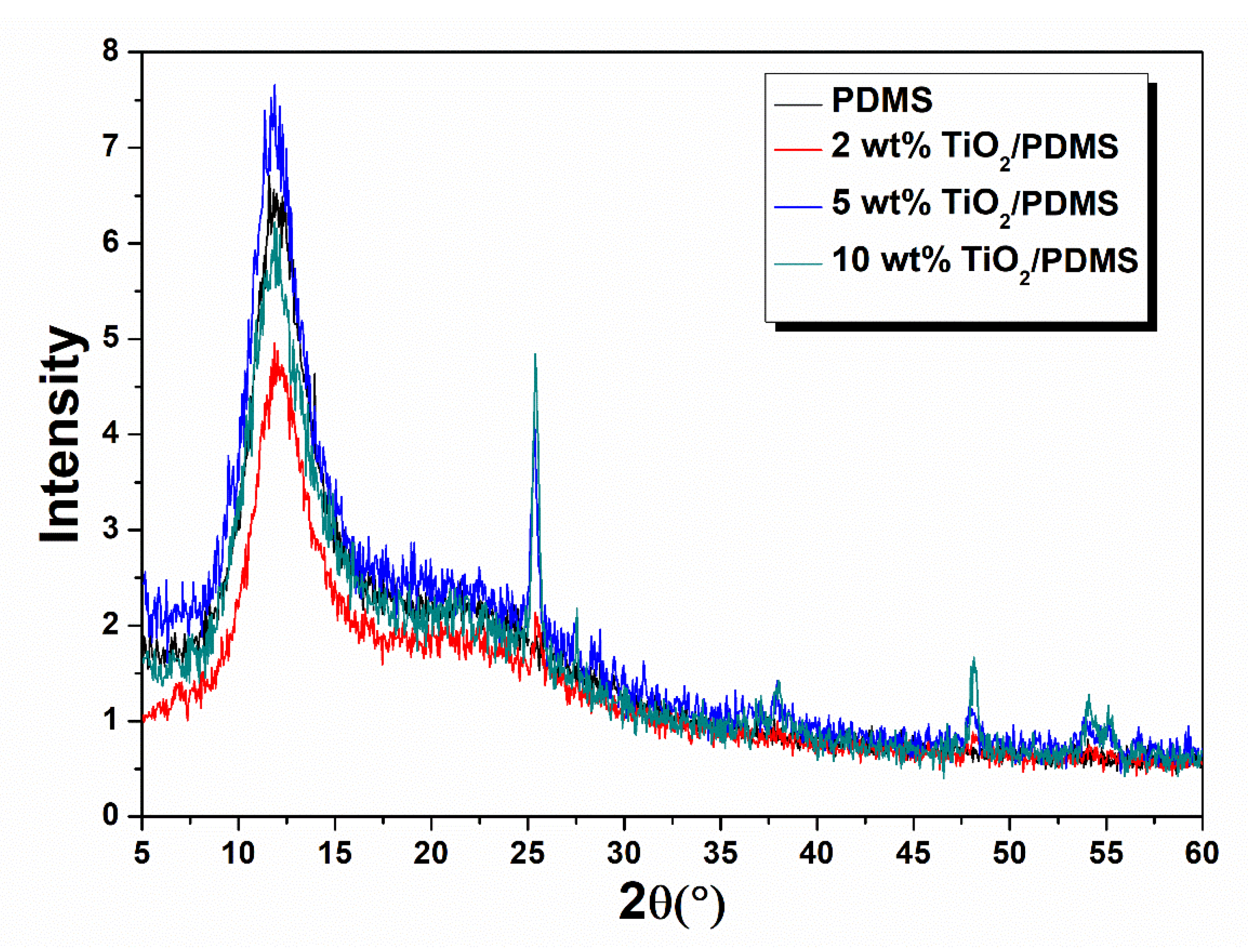
3.1. XRD
The structural investigation of the nanocomposites was carried out via X-ray diffraction (XRD). The XRD patterns of TiO
2/PDMS nanocomposites (
Figure 1) present the characteristic peaks of anatase (2θ: 25.4°, 38°, 48°) and rutile (2θ: 27.5°, 54°) types of TiO
2.
3.2. DSC
Concerning the effect of TiO
2 nanoparticles on the thermal properties, it can be observed by DSC analysis (
Figure 2,
Table 1) that the glass transition temperature (T
g) remained almost unaffected, while the crystallization temperature (T
c) of PDMS matrix of 2 wt% TiO
2/PDMS nanocomposites shifted to lower values, indicating a facilitation of the crystallization process. For higher TiO
2 content, the incorporation of nanoparticles seems to hinder the crystallization process, and T
c shifts to higher values. The melting enthalpy of PDMS presented a slight increase in the case of nanocomposites, in comparison with that of the unfilled elastomer. An increase of T
g with the increase of titania content, accompanied by broadening of the glass transition step, has also been mentioned by Klonos et al. [
30], as a result of their DSC study on the thermal transitions of titania/PDMS nanocomposites. They also claimed that TiO
2 nanoparticles did not act as crystallization nuclei, and crystallization of PDMS does not proceed close to the surface of nanoparticles.
3.3. TGA
By Thermogravimetric Analysis (TGA), it was found that the incorporation of titania into the polysiloxane resulted in a significant enhancement of its thermal stability, especially at loadings of 5 & 10%, where a shift of the thermal degradation at higher temperatures was observed (
Figure 3a,b). This effect was reflected in the results of
Table 2, where an increase of the decomposition temperature at the maximum degradation rate through the entire range of filler concentrations can be seen.
3.4. Tensile and Swelling Properties
Tensile tests of polysiloxane nanocomposites revealed that the incorporation of titania nanoparticles increases tensile strength and elastic modulus of the elastomer (
Table 3). Due to interactions of silica nanoparticles with the PDMS molecules, those particles act as crosslinking points and increase the network density, which led to enhanced tensile properties of the reinforced elastomer.
In order to evaluate the effect of the mesh density of polysiloxane on the properties of titania/polysiloxane nanocomposites, swelling experiments using toluene were carried out at room temperature. The nanocomposites showed lower swelling with the increase of TiO
2, as can be seen in
Figure 4, which is associated with the increase in the mesh density due to the above-mentioned physicochemical interactions and also to the increase in the diffusion path of the solvent resulting from the incorporation of titania nanoparticles. Based on the swelling behavior of the nanocomposites, the average molar mass between crosslinking points was calculated, and the results are presented in
Table 4.
3.5. Antibacterial Activity (E. coli)
A study of the antibacterial properties of the nanocomposite 10 wt% titania/PDMS was carried out, in comparison with pure polysiloxane. The strain used for this study was Escherichia coli.
The results of the antimicrobial properties against
E. coli of the 10% wt TiO
2/PDMS samples irradiated with UVA, for different times of exposure, are shown in
Table 5. TiO
2/PDMS samples containing 10 wt% of TiO
2 were quite effective, killing approximately 68% of
E. coli after 1 hr exposure. The bacterial reduction percentage remained constant after 3 or 6 h exposure of
E. coli cells.
Therefore, after photoactivation of the specimens by exposure to UV radiation, the resulting oxidative species led to a reduction of the number of bacterial colonies after the first hour of study, confirming the bactericidal action of the nanoparticles of titania. Then, the number of bacterial populations showed a small increase, which means that consumption of oxidative radicals takes place. Despite the slight increase of the number of bacterial colonies in titania/PDMS samples, fewer bacterial colonies are observed compared to pure PDMS samples.
The bactericidal effects of TiO
2 on
E. coli have also been demonstrated by other researchers [
29,
31,
32,
33]. The antibacterial effect of UV/TiO
2 photocatalysis is due to the production of reactive oxygen species (ROS: O
2•−, H
2O
2 and HO
•) generated by actual light-activated TiO
2. These highly active species cause cell death by decomposition of the cell wall first, followed by decomposition of the cell membrane [
22,
29]. The antibacterial effect against
E. coli was found to be more intensive at higher contents of TiO
2 in PDMS matrix. In addition, it was reported that silicone rubber matrix demonstrated resistance against UV light and ROS [
33].
3.6. Color Changes and Stability of Thermochromic Pigmented Silicone Elastomers
3.6.1. Temperature Change Effect
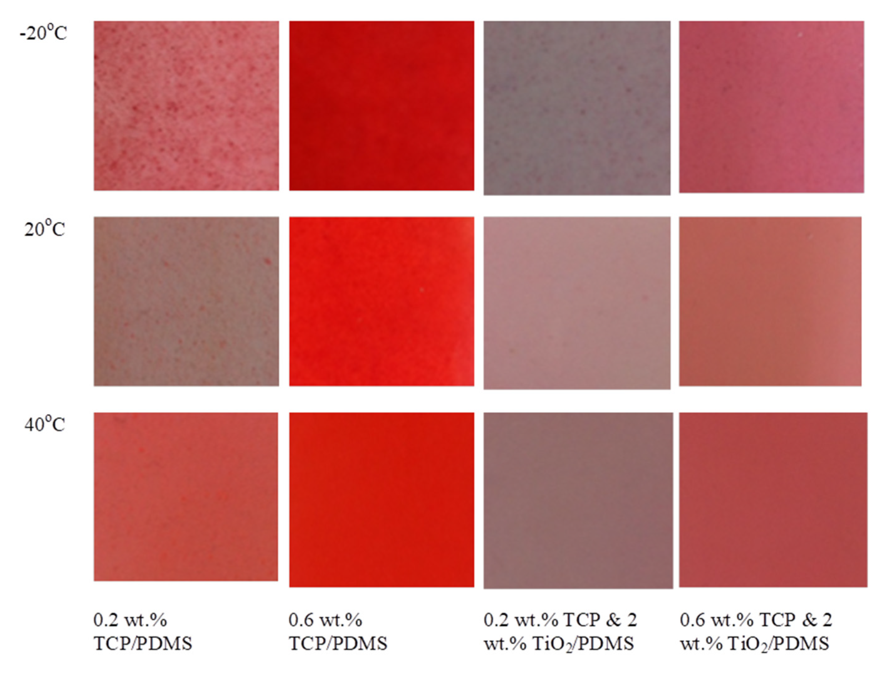
The color change of polysiloxane and its nanocomposite (2 wt% TiO2/PDMS) colored with red thermochromic pigment (at 0.2 and 0.6 wt%) at −20 and 40 °C was studied, taking the ambient temperature as a reference.
From
Figure 5, it is obvious that polysiloxane specimens colored with red thermochromic pigment using two different concentrations (0.2 and 0.6 wt%) showed a color change, as the temperature decreases from 20 to −20 °C. These changes were depicted in all the lab color coordinates, and are presented in
Table 6. Specimens with low pigment content (0.2 wt%) are more sensitive to the decrease of temperature, in comparison with those colored with 0.6 wt% TCP. Titania nanoparticles in polysiloxane acted as opacifiers, and reduced the color change due to the temperature decrease.
From the results presented in
Table 6, it is obvious that the values of coordinates a and L are associated with the total color change of the pigmented polysiloxane specimens.
By increasing the temperature from 20 to 40 °C, all the examined specimens, except that with 0.2 wt% TCP, presented a minor color change, as can be seen in
Table 7. The investigated red thermochromic pigment reversibly changes its color, and depending on the decrease or increase in temperature, the color becomes brighter or lighter, respectively. The addition of titania nanoparticles “fades” the shade of the color of the elastomer. Maximum color changes were observed in samples with the lowest concentration (0.2 wt%) of thermochromic pigment. For all the tested samples, the highest color changes were recorded at −20 °C. In the examined temperature range, the addition of titania limits the color variation of the samples with the lowest pigment content.
3.6.2. Accelerating Aging Effect
The color deterioration of facial elastomer prosthesis has been attributed to certain environmental factors, such as solar irradiation, temperature and water. Ultraviolet radiation is only a small part of the radiation received from the sun, but it has a large impact on the color stability of silicon elastomer prosthesis. Colored with red thermochromic pigment polysiloxane and TiO
2/polysiloxane nanocomposites specimens were imposed to accelerated aging conditions by exposure to repeated cycles of UV radiation and humidity at 50 °C. By visual inspection of specimens (
Figure 6) of pigmented polysiloxane, after the first hours of exposure to UVB radiation, the red color faded and brown spots were observed. In pigmented TiO
2/polysiloxane nanocomposites, discoloration was slower, and brown spots of oxidation products were not observed.
From
Figure 7, it is observed that the specimen with 0.2% TCP showed faster and more significant change in the values of color coordinate Δa*, in comparison with specimens colored with 0.6% TCP, and this effect was restricted with the addition of titania to the elastomer. On the other hand, polysiloxane specimens colored with 0.6% TCP presented no significant color change during the first 325 h of ageing. No color change was observed in the reference polysiloxane specimen contining TCP, as well as in those without TCP and reinforced with 2% TiO
2.
The examined samples presented minor changes in the values of coordinate Δb
*, as can be seen in
Figure 8. As shown in
Figure 9, the coordinate L is affected by the time of exposure in accelerating ageing conditions for samples of pure PDMS colored with TCP, while in the case of colored 2 wt.% TiO
2/PDMS composites, it remains more stable. The addition of TiO
2 in specimens colored with thermochromic pigments enhanced the light character of their color, and this effect is stronger for elastomers colored with 0.6 wt% pigment. From
Figure 10, showing the total color change ΔΕ versus time of ageing, it is observed that the unreinforced PDMS specimen colored with 0.2 wt% TCP presented the higher color change, and this effect was restricted in the case of reinforced with 2 wt% titania nanocomposites. Higher amount of TCP (0.6 wt%) gave more stable color characteristics to PDMS at the investigated aging conditions. From colorimetry results, it was found that samples containing only thermochromic pigment as an additive (0.2 wt% TCP/PDMS and 0.6 wt% TCP/PDMS) showed poor color stability from the first hours of exposure, while discoloration occurred over time, and eventually the specimens became almost transparent, with a pale pink-brown hue. The addition of titania to polysiloxane specimens had a positive effect, and enhanced the stability of the thermochromic pigment, especially at a low concentration (0.2 wt%) in the nanocomposite.
The results of DSC tests with specimens containing 2 wt% TiO
2 after ageing are presented in
Table 8, and reveal an increase in T
g of the elastomer, probably due to the formation of new crosslinks after exposure to UVB radiation, resulting in the restriction of polymers end chains. The decrease in T
m was explained by the deterioration in crystalline structure of the elastomer due to the exposure to ageing conditions.
4. Conclusions
The main conclusions of this work are that the incorporation of TiO2 nanoparticles in PDMS matrix improves the thermal stability of the elastomer composites, by increasing the initial temperature of thermal degradation (Τonset), as well as the temperature at maxinum thermal degradation rate (Tpeak), as shown by the results obtained from TGA experiments. TiO2/PDMS composites present enhanced tensile strength, stiffness and solvent resistance. In addition, titania provides antibacterial properties to the elastomer, leading to a 72% reduction of the bacterial colony (E. coli) after 3 h of exposure. It also improves the color stability of the specimens colored with a red thermochromic pigment and subject to accelerated aging by exposure to UVB radiation and moisture. Therefore, the combination of these two additives can be useful for the optimization of the design of maxillofacial silicone elastomer prostheses. Based on the above results, for maxillofacial application, the recommended formulation could be the use of 0.2 wt% thermochromic pigment, due to the fact that this content proved to be more sensitive in temperature changes, in comparison with specimens colored with a higher pigment content. The amount of TiO2 nanoparticles will be kept at lower levels i.e., (2 wt%), in order to leave intact the hue of the pigment, and to retain the modulus of elasticity at lower levels, since it is required in maxillofacial prostheses applications.


 ,
, 


