Case Report

Bilateral Breast Edema: Case Report and Review of the Literature

by
Andriani D. Vouxinou
1,
Georgios M. Iatrakis
1,*,
Stefanos Zervoudis
1,2,
Anastasia Bothou
1,
Sofia Tsitsiou
2,
Anisa Markja
1,
Zois Margelis
2,
Christos A. Tooulias
3 and
Evangelia Antoniou
1
1
Department of Midwifery Athens, University of West Attica, 12243 Egaleo, Greece
2
Breast Department, Rea Hospital, 17564 Athens, Greece
3
Ophthalmology Department, Attikon Hospital, 12462 Athens, Greece
*
Author to whom correspondence should be addressed.
Submission received: 21 May 2020 / Revised: 14 June 2020 / Accepted: 23 June 2020 / Published: 27 June 2020

Abstract

:
Both benign and malignant conditions related to regional or systemic disorders could be included in the differential diagnosis of bilateral breast edema. Some of them are often unilateral, including stromal infiltration and lymphatic obstruction presented in “peau d’ orange”, which is the usual presentation of breast cancer. However, the term “idiopathic” could be included in the spectrum of diagnoses. Here, we present a woman of 78 years old who came into our breast unit with a bilateral, painless edema of the breasts (appeared one month ago). Clinical examination revealed that both breasts were swollen with widespread erythema and the appearance of an orange peel/“peau d’ orange”. On palpation, the breasts were not sensitive, and no tumor was palpable. However, clinically palpable lymph nodes were found in both axillas. Her temperature was normal. The breast edema could not be explained from her medical history nor the medications taken. Breast ultrasound, Mammography and Magnetic Resonance Imaging were non-conclusive (BI-RADS 0) and bilateral core biopsy was negative for cancer. Anti-inflammatory plus antibiotic therapy was prescribed for 10 days and at the end of treatment, regional redness and edema were disappeared and reduced, respectively. Total recovery was found one month after the initial findings. It can be concluded that bilateral breast edema is correlated to regional or systemic conditions or it is presented as an “idiopathic” disorder of unknown etiology.

1. Introduction

Both benign and malignant conditions could be included in the differential diagnosis of bilateral breast edema, although some of them are very often unilateral [1,2,3]. As an example, in “peau d’orange” [2], stromal infiltration and lymphatic obstruction is almost exclusively a unilateral involvement of both skin and breast parenchyma (which is the usual presentation of breast cancer as a unilateral tumor). Bilateral breast edema can be correlated to rare regional or systemic conditions [4,5] or can occur after breast-conserving surgery with radiotherapy or in the postpartum period.
The purpose of this study is to briefly review possible etiologies of bilateral breast edema apropos of one case presented in our breast unit and included in the thesis of the first author.

2. Case Presentation Section

2.1. Clinical History

Here, we present a 78 years old woman who came in our breast unit with bilateral edema of the breast, which had appeared one month ago. Her past medical history was negative for heart/lungs/kidney/liver/endocrine disease without any therapeutic or preventing medicine apart from an antihypertensive agent (prescribed, for the first time, three months ago, and after normalization of blood pressure it was discontinued without hypertension relapse, 2 months ago). Breast cancer personal and family history were negative. No pharmaceutical or other interventions were reported to be related to the above-mentioned condition.

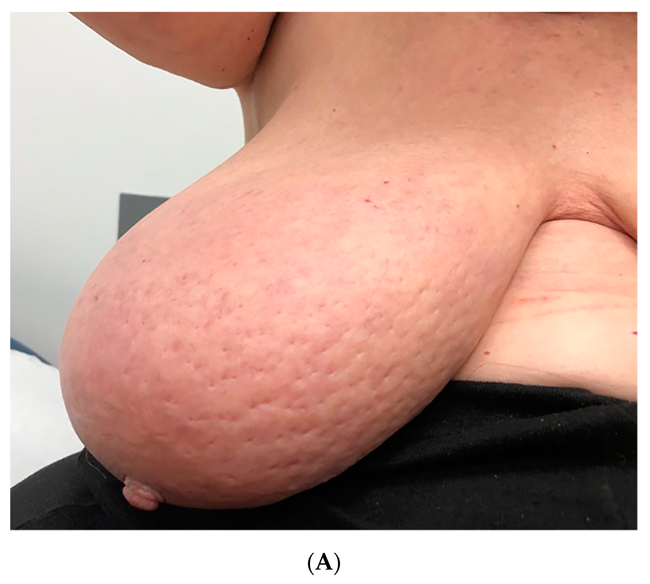
2.2. Symptoms and Duration

The patient complained about discomfort, swollen breasts and breast heaviness but no pain for the last four weeks before her arrival in our unit. Her temperature was normal (36.8 °C). No other accompanying symptoms were reported, apart from sleep difficulties during the last month which were interpreted by the woman as a consequence of her condition.

2.3. Clinical Examination and Initial Assessment
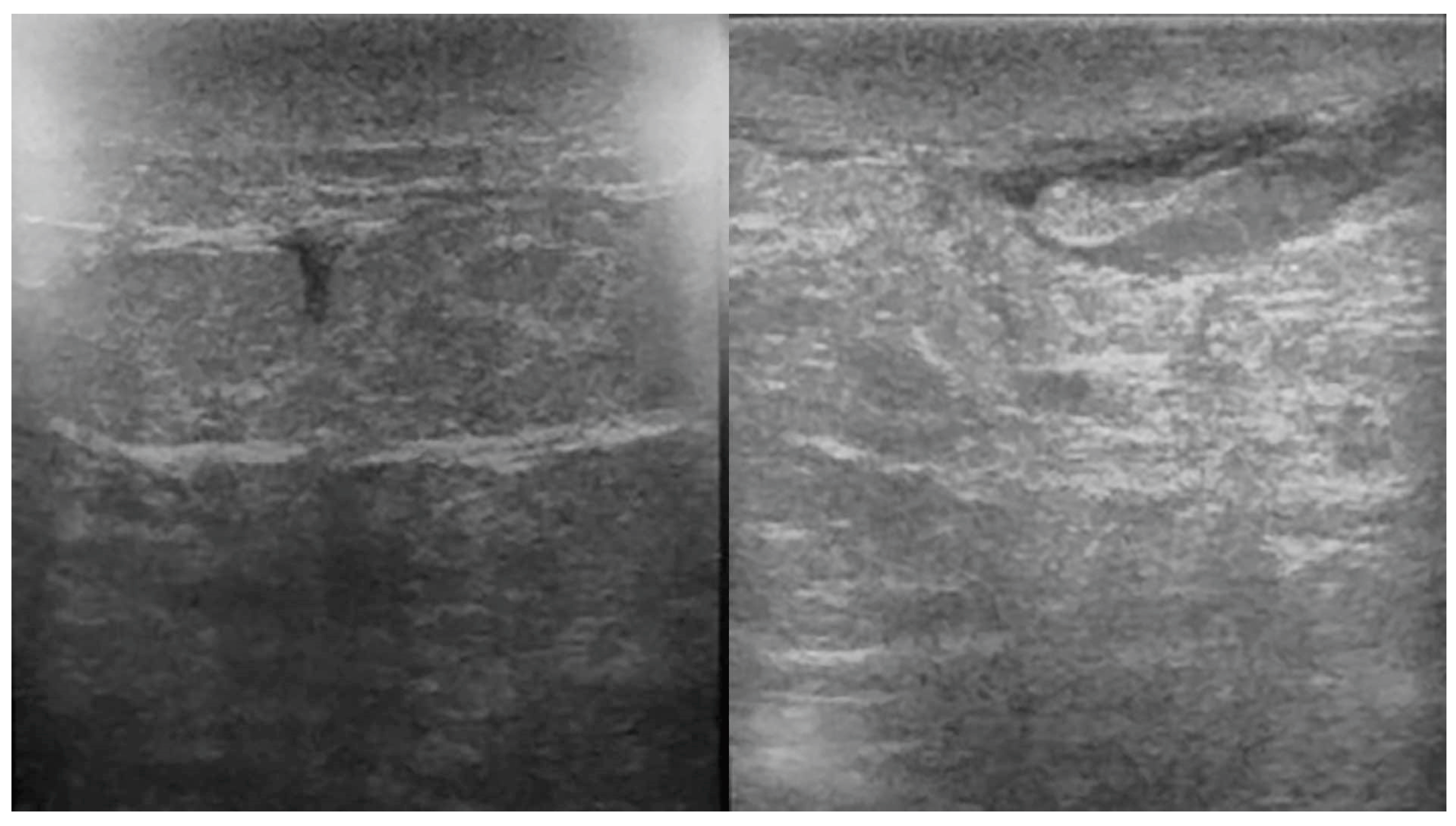
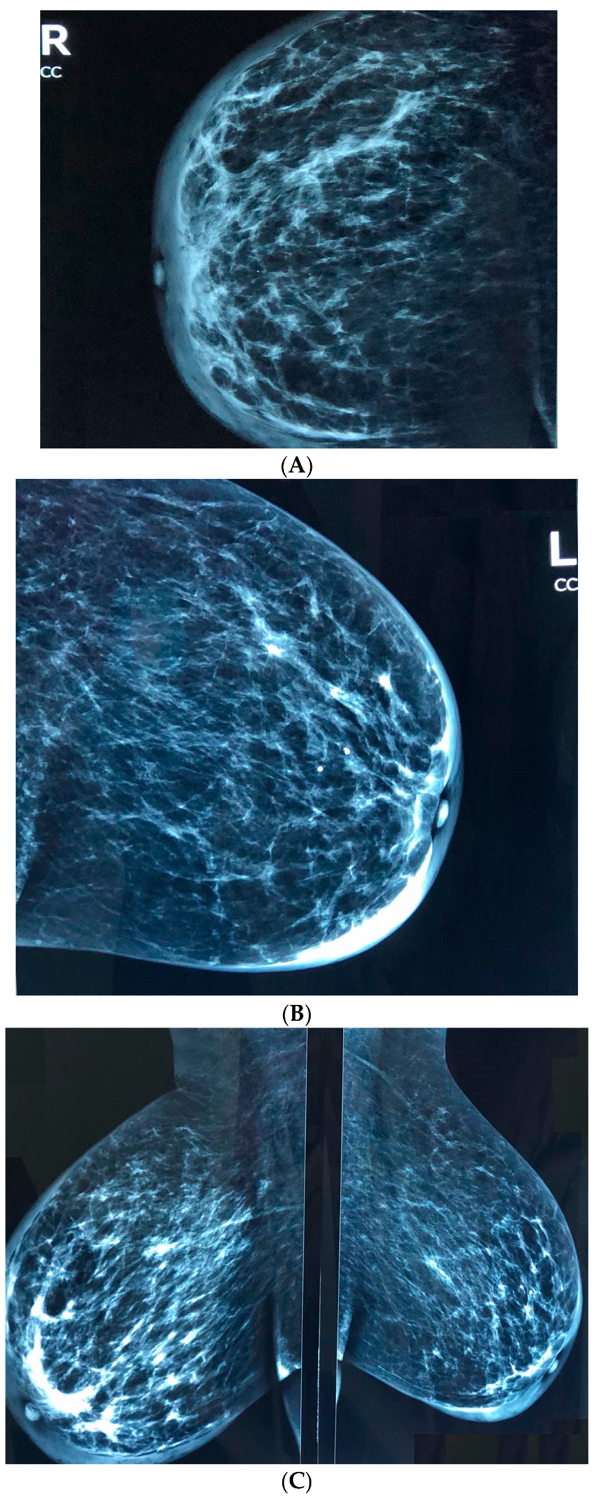
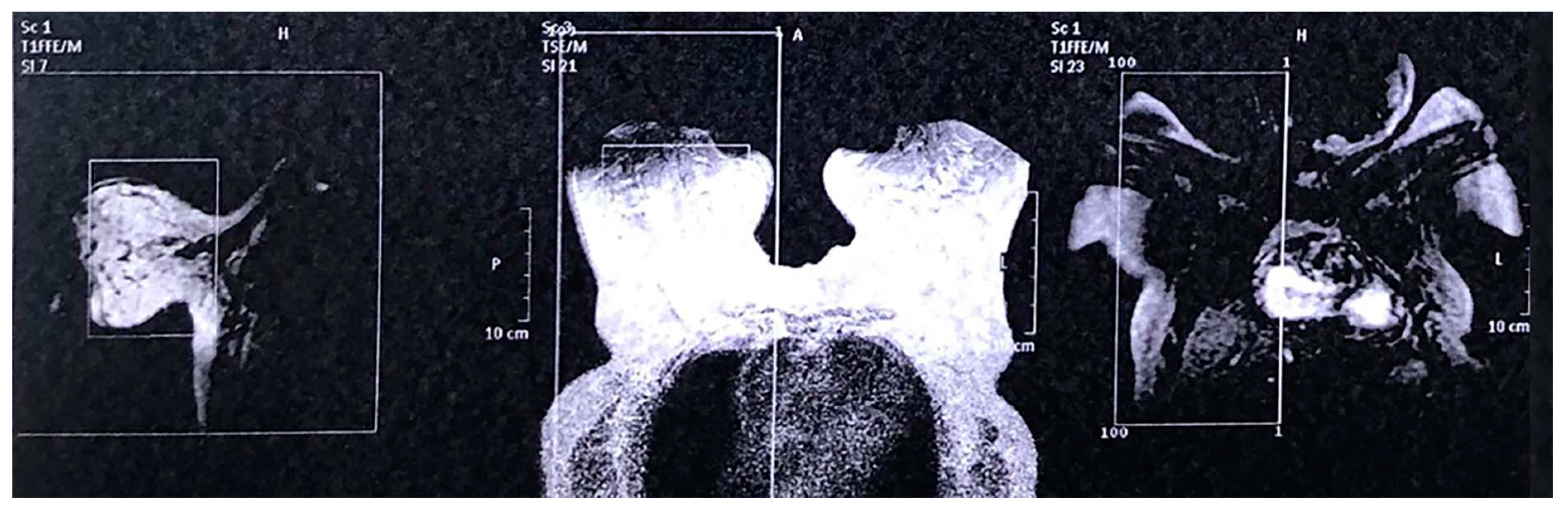
Clinical examination revealed that both breasts were swollen (increased volume of the breasts) with extended redness and the appearance of an orange peel/“peau d’ orange” (Figure 1A,B). On palpation, the breasts were sensitive and no tumor was palpable. However, clinically palpable lymph nodes were found in both axillas. Relevant blood tests including whole blood count, inflammatory markers (including C-reactive protein and erythrocyte sedimentation rate), liver and renal function were all normal. The breast edema could not be explained from her medical history nor the medications taken in the past. Apart from a short discussion about her increased body weight (85 kg with body mass index 31 kg/m2) and her sedentary lifetyle, no other behavioral modifications were deemed necessary. Informed consent was obtained from the patient for the following steps of diagnostic evaluation:

2.4. Tests

Breast ultrasound (Figure 2), Mammography (Figure 3A–C) and Magnetic Resonance Imaging (Figure 4) were non-conclusive (BI-RADS 0). Considering the rare case of a bilateral malignancy and the peau d’ orange appearance, core biopsy was deemed necessary for both breasts, including both actual breast tissue and skin. The day of core biopsy, clinically palpable lymph nodes were significantly shrunken in the left axilla and were not palpable in the right axilla and no further investigation of the axilla region was deemed necessary.

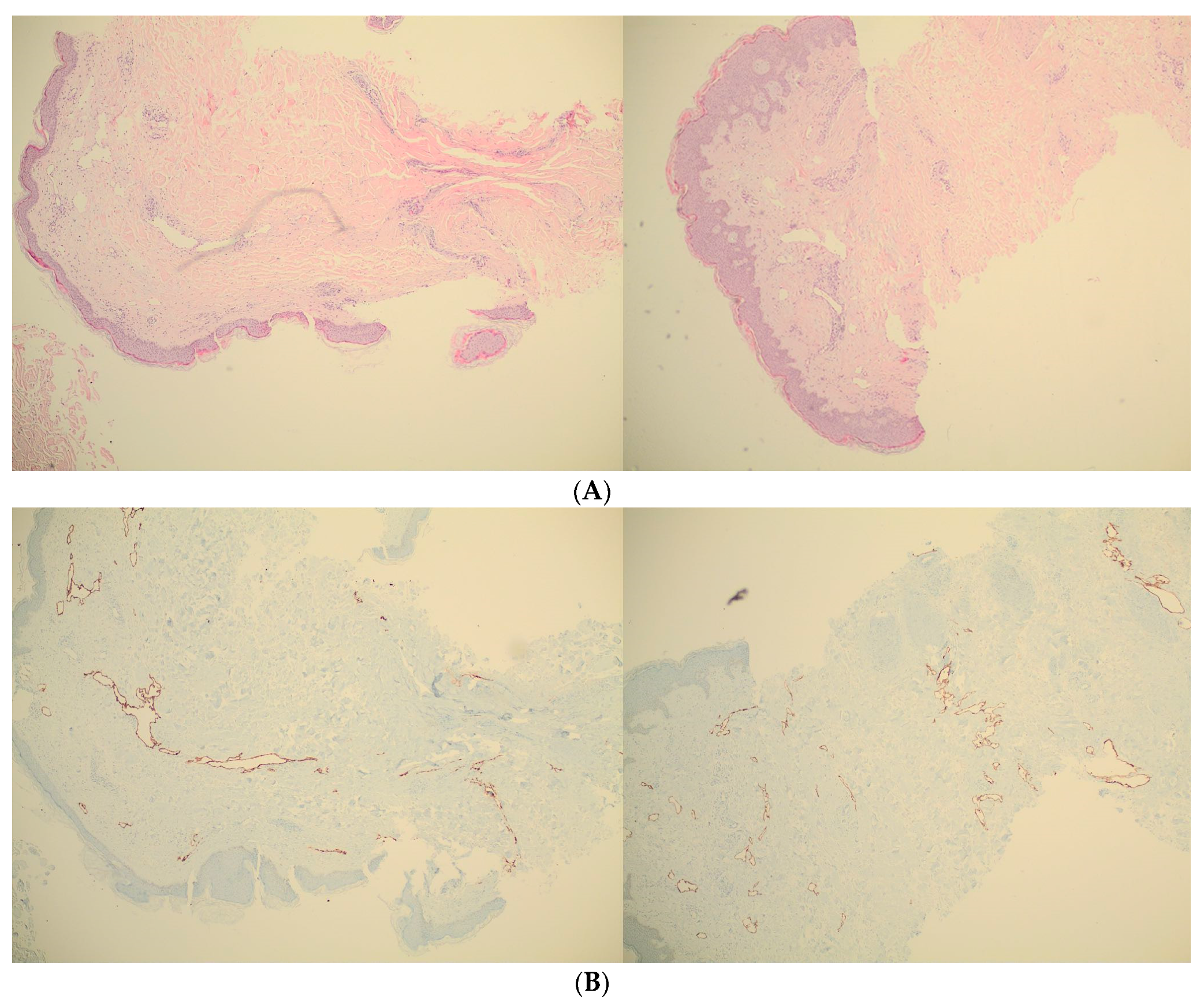
2.5. Pathology Results

Pathology results were negative for cancer and for any specific pathology. The glandular component revealed atrophic glands with fibrosis. Dilated blood vessels and a mild chronic perivascular inflammation (mainly lymphocytes’ infiltration) were reported for the chorion and skin (Figure 5A). The immunochemistry staining with AE1/E3, CD34 and podoplanin was negative (Figure 5B). No malignant cells or precancerous lesions were reported.

3. Medical Therapy and Follow-Up

The patient was informed that no diagnosis was done, and we proposed a blind antibiotherapy (Amoxicillin-Clavulanate 500 mg orally every 12 h) plus anti-inflammatory (Ibuprofen 200 mg orally every 6 h) treatment as a therapeutic test for 7 days. The patient complied with the treatment and no side-effects were reported. After the end of medical therapy, precisely on day 12, regional redness and edema were disappeared and reduced, respectively. Total recovery was found one month after the initial findings (Table 1) and no recurrence was reported one and two months later.

4. Discussion

Bilateral breast edema can be correlated to rare regional or systemic conditions [4,5] or can be occurred after breast-conserving surgery with radiotherapy [6] or in the postpartum period [7,8]. Systemic diseases related to the condition include congestive heart failure, central venous obstruction (superior vena cava syndrome) and dermatosis [9]. In rare cases, thyroid dermopathy (almost always associated with ophthalmopathy) can present as “peau d’ orange” [10] in different areas of the skin in both sexes. Moreover, bilateral inflammatory breast cancer is a very rare entity [11] but it should be ruled out in all cases because of poor prognosis and the necessity of immediate systemic treatment. Although there is no consensus on the definition of breast edema and standardized assessment criteria, common criteria for breast edema found in the literature are an increased volume of the breast, peau d’orange, heaviness of the breast and redness of the skin [12], all included in our case report. Clinical and imaging findings [13] are essential for the diagnosis and the possible avoidance of invasive procedures. However, despite normal imaging findings, only the biopsy could exclude or affirm the diagnosis of inflammatory breast cancer. In our case, clinical, imaging and pathologic findings were negative for a formal diagnosis and bilateral breast edema (including the sign of “peau d’ orange”) was characterized as “idiopathic”. The major limitations of our case report presentation are that no cause–effect relationship was established and that it is not definitely known if the breast edema resolved solely as a consequence of medical therapy. However, the strength of our approach to this case is the meticulous clinical, imaging and pathologic investigation and the close follow-up of the patient that give us the confidence to characterize the case as idiopathic. Considering the age of the patient (“Geriatric case”) and that this is the first case published with a bilateral idiopathic breast edema, thoroughly investigated by the second, the third and the fourth authors, the term “ZerIatric Breast” Disease (ZIBD) was adopted by the writing team of the paper, in parallel with other diseases and/or syndromes of unknown causal relationship. The hypothesis of bilateral breast edema of unknown etiology or “idiopathic” breast edema should be further investigated in a broader spectrum of ages.

5. Conclusions

Bilateral breast edema can be correlated to some regional or systemic conditions, including, rarely, to bilateral inflammatory breast cancer, or it can be presented as a rare temporary disorder of unknown etiology (idiopathic), as in our case. After a negative biopsy, a trial treatment could be proposed along with a close follow-up. The term “idiopathic” could be included in the spectrum of diagnoses.

Author Contributions

A.D.V., G.M.I., S.Z., A.B. and E.A. contributed to the conception and design of the study; acquisition, analysis and interpretation of data; drafting the article and/or making critical revisions related to important intellectual content of the manuscript and approval of the final version. S.T., A.M. and Z.M. contributed to acquisition, analysis and interpretation of data and approved the final version. C.A.T. contributed to interpretation of data and approved the final version of the paper. All authors have read and agree to the published version of the manuscript.

Funding

This research received no external funding.

Acknowledgments

Special thanks to Demetrius Iatrakis (National [Metsovian] Technical University of Athens [Zografou Campus 9, Iroon Polytechniou Street, 15780 Zografou, Greece]) for technical assistance and text formatting.

Conflicts of Interest

The authors declare no competing interests.

References

  1. Panourgias, E. Inflammatory intramammary lymph node—A benign lesion simulating malignancy ON breast MRI. Acta Endocrinol. Buchar. 2009, 5, 259–264. [Google Scholar] [CrossRef]
  2. Schairer, C.; Soliman, A.S.; Omar, S.; Khaled, H.; Eissa, S.; Ayed, F.B.; Khalafallah, S.; Ayoub, W.B.; Kantor, E.D.; Merajver, S.; et al. Assessment of Diagnosis of Inflammatory Breast Cancer Cases at Two Cancer Centers in Egypt and Tunisia. Cancer Med. 2013, 2, 178–184. [Google Scholar] [CrossRef] [PubMed]
  3. Zervoudis, S.; Iatrakis, G.; Tomara, E.; Bothou, A.; Papadopoulos, G.; Tsakiris, G. Main Controversies in Breast Cancer. World J. Clin. Oncol. 2014, 5, 359–373. [Google Scholar] [CrossRef] [PubMed]
  4. Goktan, C.; Pekindil, G.; Orguc, S.; Coskun, T.; Serter, S. Bilateral Breast Edema in Intestinal Lymphangiectasia. Breast J. 2005, 11, 360. [Google Scholar] [CrossRef] [PubMed]
  5. Maeda, M.; Fugimoto, N.; Satoh, T. Bilateral Breast Edema Associated with Nephrotic Syndrome. Eur. J. Dermatol. EJD 2013, 23, 739–740. [Google Scholar] [CrossRef] [PubMed]
  6. Young-Afat, D.A.; Gregorowitsch, M.L.; van den Bongard, D.H.; Burgmans, I.; van der Pol, C.C.; Witkamp, A.J.; Bijlsma, R.M.; Koelemij, R.; Schoenmaeckers, E.J.; Jonasse, Y.; et al. Breast Edema Following Breast-Conserving Surgery and Radiotherapy: Patient-Reported Prevalence, Determinants, and Effect on Health-Related Quality of Life. JNCI Cancer Spectr. 2019, 3, pkz011. [Google Scholar] [CrossRef] [PubMed]
  7. Miller, V.; Riordan, J. Treating Postpartum Breast Edema with Areolar Compression. J. Hum. Lact. Off. J. Int. Lact. Consult. Assoc. 2004, 20, 223–226. [Google Scholar] [CrossRef] [PubMed]
  8. Kujawa-Myles, S.; Noel-Weiss, J.; Dunn, S.; Peterson, W.E.; Cotterman, K.J. Maternal intravenous fluids and postpartum breast changes: A pilot observational study. Int. Breastfeed. J. 2015, 10, 18. [Google Scholar] [CrossRef] [PubMed] [Green Version]
  9. Ramos, R.M.L.; Armán, F.J.A.; García, M.T.R.; Carrero, E.B.; Fariñas, I. Breast Edema. A Pictorial Review with Pathologic Correlation. Available online: https://www.semanticscholar.org/paper/Breast-edema.-A-pictorial-review-with-pathologic-Ramos-Arm%C3%A1n/2447fda13d9ab77d23f43e9741c34197e6ba9c6c (accessed on 17 May 2020).
  10. Dhali, T.K.; Chahar, M. Thyroid Dermopathy-a Diagnostic Clue of Hidden Hyperthyroidism. Dermato-Endocrinology 2014, 6, e981078. [Google Scholar] [CrossRef] [PubMed] [Green Version]
  11. Masannat, Y.A.; Peter, M.; Turton, P.; Shaaban, A.M. Case Report of Bilateral Inflammatory Breast Cancer. Eur. J. Cancer Care 2010, 19, 558–560. [Google Scholar] [CrossRef] [PubMed]
  12. Verbelen, H.; Gebruers, N.; Beyers, T.; De Monie, A.-C.; Tjalma, W. Breast edema in breast cancer patients following breast- conserving surgery and radiotherapy: A systematic review. Breast Cancer Res. Treat. 2014, 147, 463–471. [Google Scholar] [CrossRef] [PubMed]
  13. Cao, M.M.; Hoyt, A.C.; Bassett, L.W. Mammographic Signs of Systemic Disease. Radiographics 2011, 31, 1085–1100. [Google Scholar] [CrossRef] [PubMed]
Figure 1. (A). Clinical appearance of the right breast (increased volume, intense redness, “peau d’ orange”). (B). Clinical appearance of the left breast (increased volume, moderate redness, “peau d’ orange”).
Reports 03 00018 g001aReports 03 00018 g001b
Figure 2. Ultrasound of both breasts without conclusive findings.
Reports 03 00018 g002
Figure 3. (A) Cranial-Caudal (CC) mammogram view of the right breast without conclusive findings. (B) Cranial-Caudal (CC) mammogram view of the left breast without conclusive findings. (C) Medio-Lateral-Oblique (MLO) mammogram view of both breasts without conclusive findings.
Reports 03 00018 g003
Figure 4. Magnetic Resonance Imaging of both breasts without conclusive findings.
Reports 03 00018 g004
Figure 5. (A) Pathologic appearance of core biopsy material from both breasts revealing atrophic glands with fibrosis, dilated blood vessels and a mild chronic perivascular inflammation. (B) Pathologic appearance of core biopsy material from both breasts after podoplanin immunohistochemistry staining confirming the results of Figure 5A.
Reports 03 00018 g005
Table 1. Timeline information for the case report.
1. Chief complaint: bilateral breast edema
2. Clinical examination
3. Breast ultrasound
4. Mammography
5. Magnetic resonance imaging of the breast
6. Core biopsy
7. Pathologic results without “formal” diagnosis
8. Symptomatic treatment
9. Remission of breast edema
10. An idiopathic case of bilateral breast edema was concluded

