Article

The “oro di metà” Gilding in the Fifteenth-Century: A Multi-Analytical Investigation

1
Institute of Applied Physics “Nello Carrara” (IFAC)-National Research Council (CNR), 50019 Sesto Fiorentino (FI), Italy
2
Institute of Chemistry of Organometallic Compounds (ICCOM)-National Research Council (CNR), 50019 Sesto Fiorentino (FI), Italy
3
Institute for the Conservation and Valorization of Cultural Heritage (ICVBC)-National Research Council (CNR), 50019 Sesto Fiorentino (FI), Italy
4
Gallo Restauro, 50129 Florence, Italy
*
Author to whom correspondence should be addressed.
Submission received: 12 March 2019 / Revised: 10 April 2019 / Accepted: 15 April 2019 / Published: 18 April 2019
(This article belongs to the Special Issue Advances of Analytical Chemistry in Cultural Heritage)

Abstract

:
Few and fragmentary is the information regarding the “oro di metà” (halfway gold) gilding due to the low probability of finding it in paintings as well as its short durability because of tarnishing. However, the very rare, excellent state of conservation of the gilding found in the fifteenth-century wood panel painting of the Master of St. Ivo studied in this work along with a multi-analytical approach allowed shedding light on the structure, the composition and the conservation state of this type of gilding. An Ultra-High Resolution Scanning Electron Microscope (UHR-SEM) along with Energy Dispersive X-Ray (EDX) analysis and polarised light Optical Microscopy (OM) were employed in order to image and analyse a cross-section of a sample taken from the wood panel painting. Results highlighted a divided structure of the gilding which is constituted by two separated layers of gold and silver with different thickness. This is the first time that scientific evidence on this issue is provided. Moreover, considerations on the tarnishing process of the gilding were made. Finally, complementary vibrational spectroscopic techniques such as micro-Raman and micro-ATR-FTIR were employed in order to identify the molecular composition of the “bole” (gilding preparation layer) and the ground layers.

Graphical Abstract

1. Introduction

Gilding is an ornamental decoration in paintings which consists of applying a very thin layer of gold (the gold leaf) using different techniques. Gilding was a very widespread technique in medieval art, especially in the Byzantine and Renaissance periods, where the gold leaf was used in paintings on wooden panels to enhance the visual effect of the saints’ halos. Moreover, in the middle ages, many paintings were executed using the gold leaf as a background (gold ground). Usually, the gold in gildings was used pure but gold alloys as well as alloys simulating the colour of gold have been also found. In this respect, the “oro di metà” (halfway) gilding was used mainly in Italy from the thirteenth to the fifteenth centuries but its structure and composition still remain unclear.
In the painting of the fifteenth century by the Master of St. Ivo depicting S. Peter between St. Anthony Abbot and St. Mary Magdalene, the “oro di metà” gilding represents a rare example of its excellent state of preservation, which makes it one of the few cases that has come to us so intact. Moreover, this gilding was applied according to both the most common execution techniques at that time: “a guazzo” (water gilding) on the background and “a missione” (gilding with mordant) on the borders of the garments. Briefly, in the water gilding, the gold foil is laid onto an adhesive layer consisting mainly of earth pigments mixed with a protein binder (as egg-white and rabbit glue) and with some water commonly known as “bole”, over gypsum ground. The bole gives a warm color to the gold as well being a cushion for successive treatments as for example burnishing and punching [1]. On the contrary, in the gilding with mordant, the gold foil is laid on an oil-based mordant usually made of linseed oil mixed with pigments as driers. This technique is particularly suitable for decorations and small details [1].
The term “oro di metà” or “oro mezzo” was explicitly used by Cennini [2] describing different gilding techniques and warning against the tarnishing of silver which is a constituent of this type of gilding. Actually, the use of “oro di metà” foils was very widespread in Tuscany since the end of the thirteenth century, so much so that in the years 1315–1316 when the painters were admitted to the guild of the “Arte dei Medici e degli Speziali” (the guild of doctors and pharmacists) of Florence, in the statute is mentioned the “aurum di metà” regarding the penalties for those who were using it instead of fine gold without declaring it [3]. Throughout the fourteenth century until the end of the fifteenth century, the “oro di metà” is observed in the gilding of some paintings by Jacopo del Casentino, Pacino di Buonaguida, Bernardo Daddi, and Puccio di Simone [4]; Neri di Bicci as well in his “Ricordanze” [5] certifies the use of “oro mezzo” on many “anticha” altar plates reporting even the price, which is about half of that of the fine gold [6]. As prescribed by the Florentine Statutes of 1396 and 1403 [7,8,9], the “oro di metà” foil results to have the same dimensions as that of silver; therefore, analyzing the costs [10], with the price of 100 pieces of fine gold, approximately 200 pieces of “oro mezzo” could be obtained with a size ranging from 8.3 to 7.3 cm2.
Despite the existence of these very important historical documents which would suggest the use of two different foils (one of silver and one of gold) cast as a “sandwich”, in the nineteenth century, Merrifield and Eastlake [8] translated the “oro di metà” as “gold that is much alloyed” similar to other translators that used the expression “gold alloy”. Similarly, Milanesi in 1859 introduced the meaning “oro falso battuto” (fake beaten gold) along with other translators that referred to “oro di metà” as a gold leaf that is “false” or “only half genuine” without giving neither any specifications nor the right importance to this artistic executional technique.
An overview of all the definitions, meanings and assumptions ascribed to the expression “oro di metà”, over the centuries, is precisely described by Skaug [8]. However, we would like to give particular emphasis to the metal leaf commonly used in the European middle ages and known in French as “or parti”, in dutch as “partijtgoud” and in german “gedeelt Gold” which share with “oro di metà” an etymological allusion to a real divided structure.
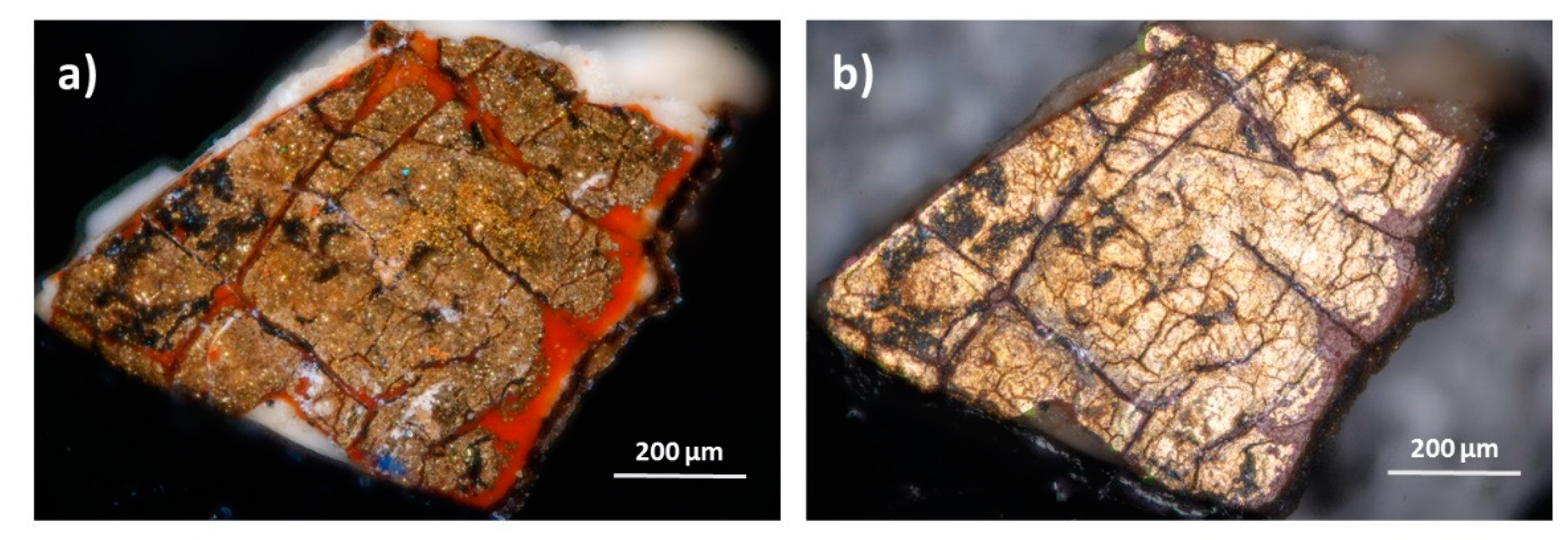
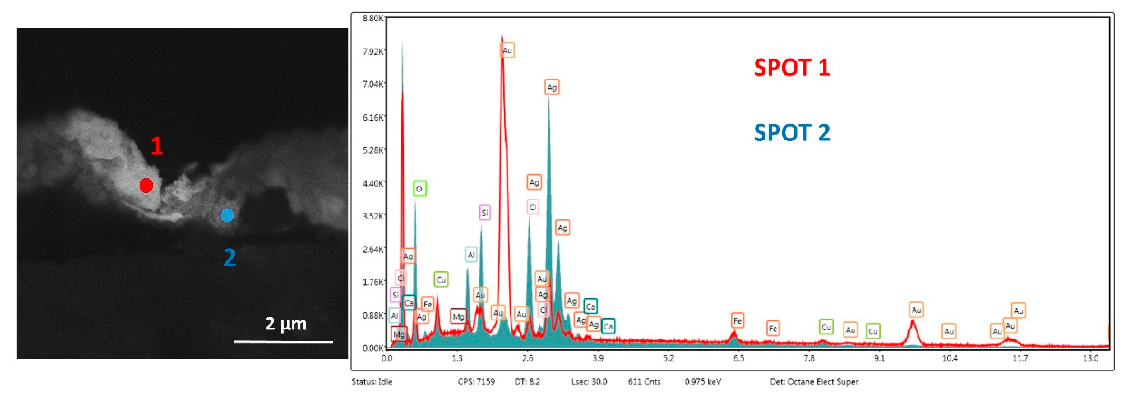
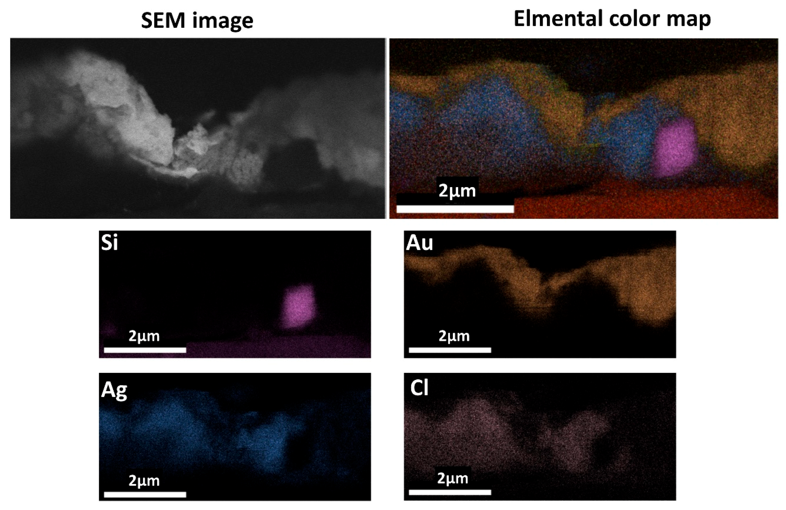
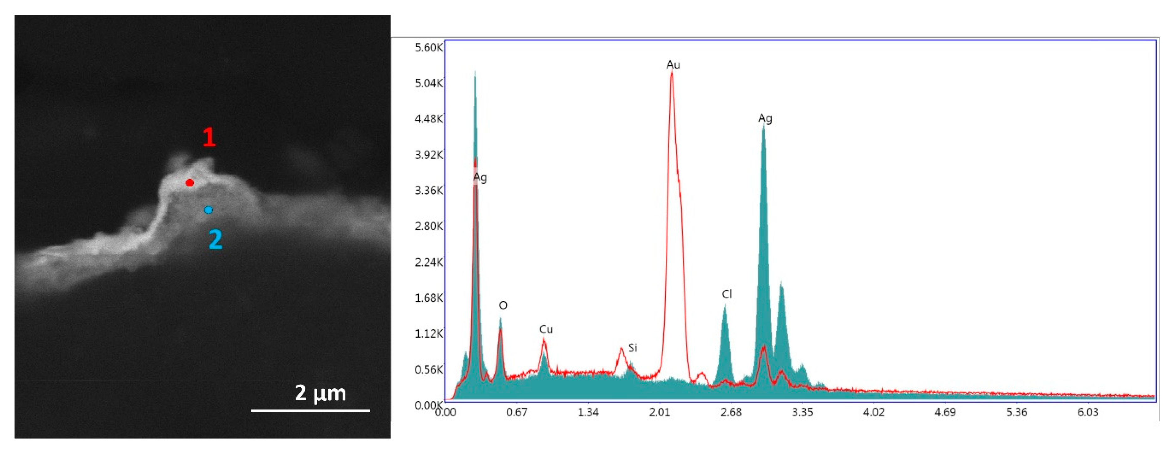
In this work, the observation carried out by means of both a polarized light optical microscope (OM) and an Ultra-High Resolution Scanning Electron Microscope (UHR-SEM) have confirmed, as far as we know for the first time, the presence of two distinct very thin metal foils with different thicknesses in the “oro di metà” gilding. Moreover, Energy Dispersive X-Ray (EDX) analysis has allowed for characterizing the composition of the gold and silver layers which have been found out to be separated and overlapped as well as identifying the degradation products of the silver layer responsible for the blackening process. Finally, complementary vibrational spectroscopic techniques such as micro-ATR-FTIR and micro-Raman spectroscopy were used in order to characterize the molecular composition of the “bole” as well as the preparation layers.

History of the Painting

Fragmentary and incomplete is the information regarding the painting of the Master of St. Ivo. However, Bernacchioni and Tartuferi [11,12] have been able to find out some important information to trace its origin and history.
In 1895, the painting was at the collection of the Banca Popolare di Genova and it was auctioned at the Sangiorgi Gallery in Rome. This is the oldest piece of information on the provenance of this painting. Then, it was divided into three pieces.
However, the inscription on the base of the S. Peter throne says that the wood panel painting was commissioned by Piero di Giovanni Ringli in 1438 when he was the castellan assigned by the Sforza as captain of the garrison to defend and control the Fortress of Avenza, a small village in Lunigiana (Tuscany, Italy), from Genova’s power. In support of this, the presence of the coat of arms of the diocese of Luni with the horns of the moon pointing upwards, refers to Lunigiana. The historical and political context of that period suggests the church of Avenza as a possible place of provenance of the painting. In particular, from 1437 to 1441, before returning to Genoa’s control, Avenza was under the control of the Republic of Florence, thus explaining the presence of a painting made by the Florentine painter the Master of St. Ivo in that region.
The painting underwent only one restoration intervention in the early twentieth century, consisting of the separation of the triptych into three separated panels, their framing as well as the pictorial recovery of the draperies. Moreover, the painting was in a private American collection from the early twentieth century until 1993 and subsequently was put up for auction twice without undergoing further restorations.
Thus, this painting represents a very exceptional case in terms of conservation because it is devoid of previous interventions of cleaning so much that the original varnish is still present on the surface.

2. Sampling, Samples Preparation and Methodology

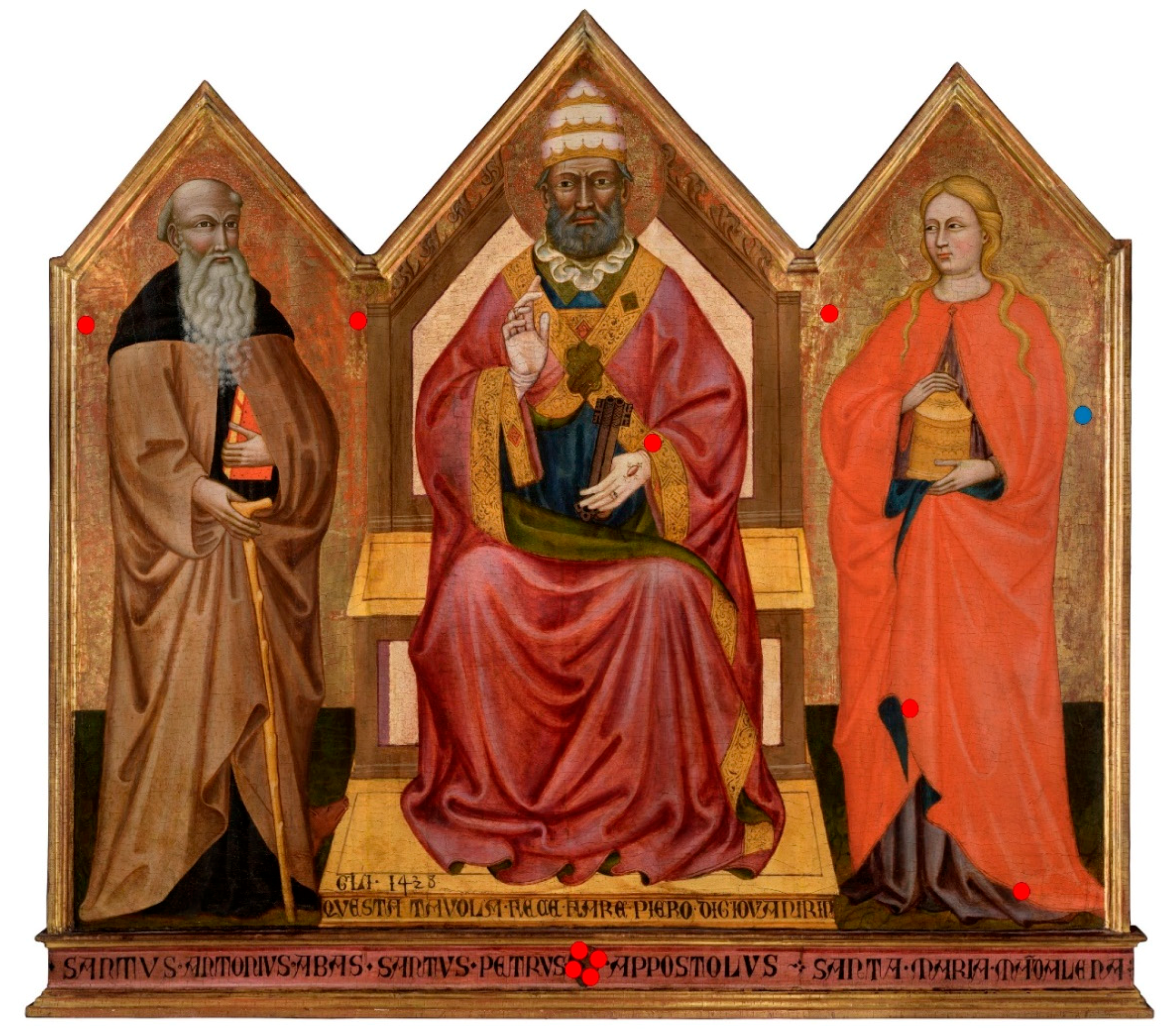
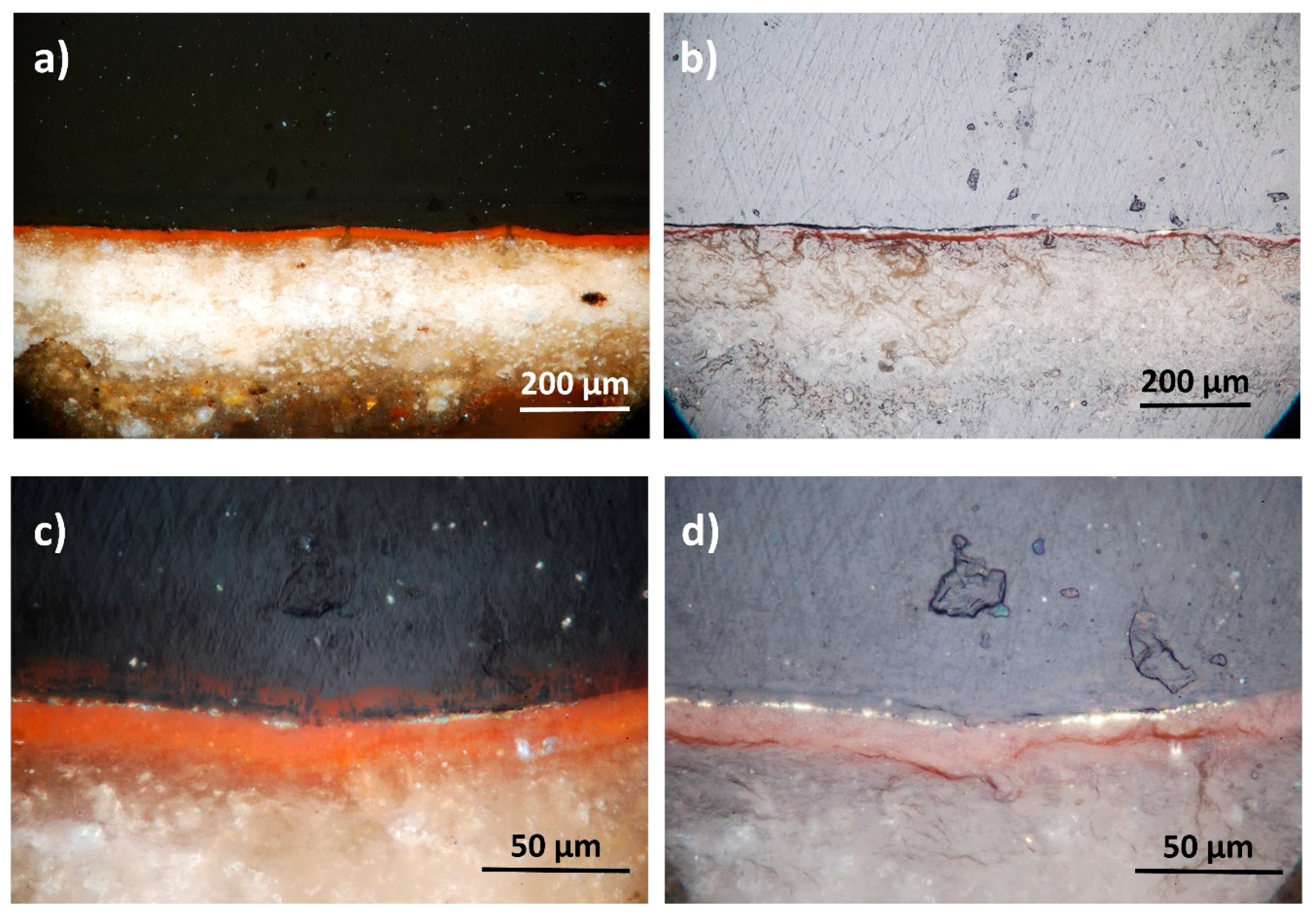
Eleven samples were taken from the painting by using a scalpel in order to gather information on many issues about the technical execution and composition of gildings, painting layers, and the ground (preparation layer). The fragments were cast in epoxy-resin, dried and finely polished for the cross-sections preparation. In particular, polishing was carefully carried out using discs at different mesh (800-1000-1200-4000) in order to perform the process very delicately. The stratigraphies were then observed by reflected polarized light OM and by an UHR-SEM. The elemental composition of different layers was provided by means of EDX spectroscopy; whereas, the molecular composition was provided by using micro-ATR-FTIR and micro-Raman spectroscopy.
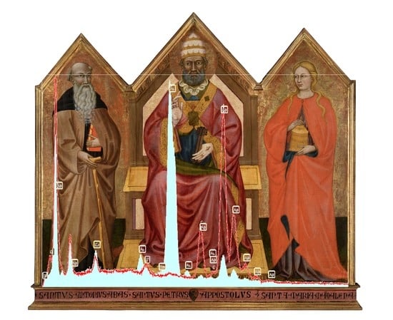
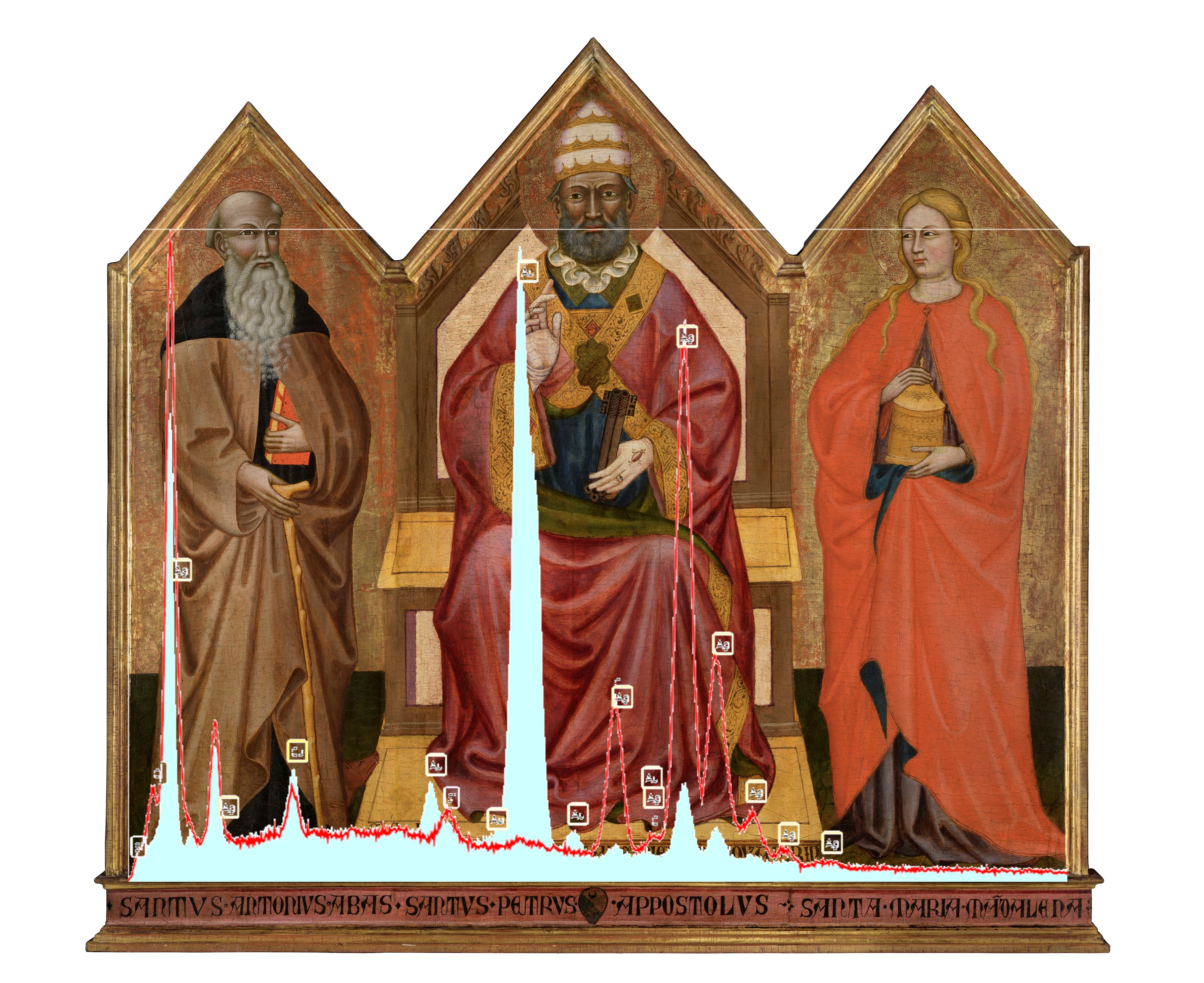
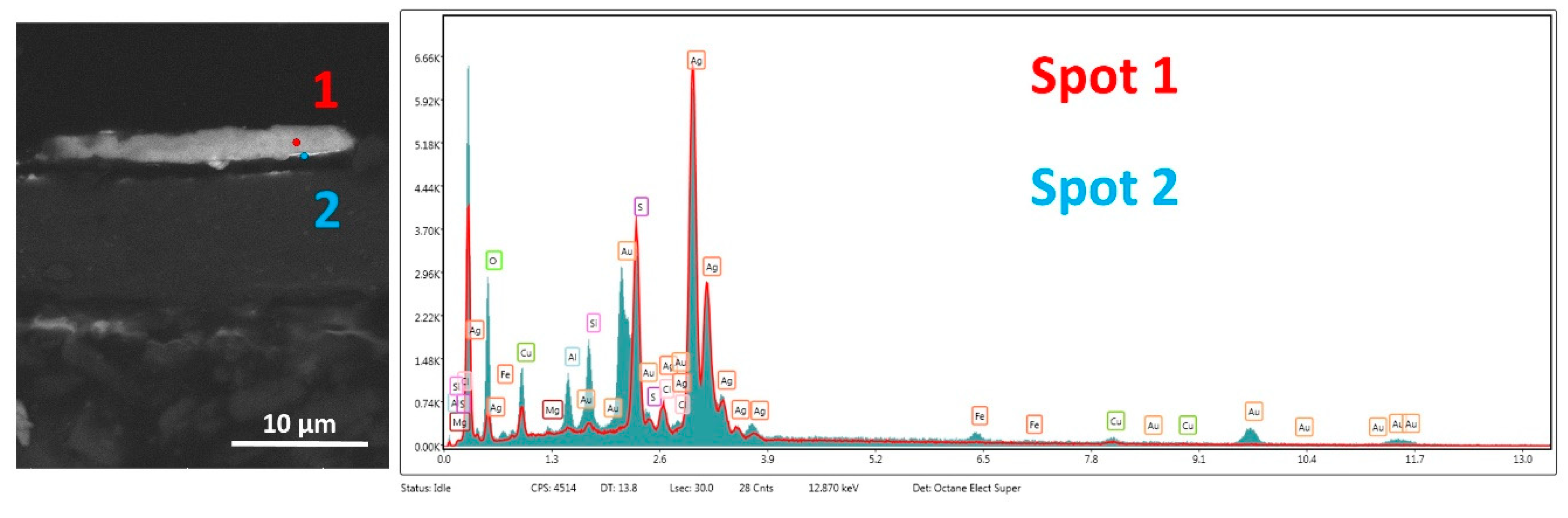
Figure 1 shows a panoramic view of the entire painting along with the sampling points. The cross-section described in this work was taken from sampling area highlighted in blue colour. After microscopic observation, this sample was considered crucial for the interpretation and the assessment of the gilding technical execution with respect to other samples where gildings were either in a bad conservation state or completely gone.
Optical microscopy observation was carried out using the polarised light microscope Eclipse 150 by Nikon provided with five different magnification objectives (5×, 10×, 20×, 50× and 100×).
SEM-EDX observation and measurements were carried out at the Center of Electronic Microscopies “Laura Bonzi” (Ce.M.E-CNR) by using an UHR-SEM Gaia 3 FIB/SEM by Tescan which represents a step forward in the field of advanced electron microscopy. The electronic beam of this microscope enables the possibility of imaging at the “nano” scale level (up to 0.7 nm at 15 keV), even with samples sensitive to electron beams such as the majority of cultural heritage materials. The limit of detection of the EDX semi-quantitative analysis falls into the range of 0.2–1 w% depending on the specific elements.
FT-IR spectra of the red sample were collected with a FT-IR spectrometer Agilent Technologies Cary 660 coupled with Cary 620 Microscope, equipped with a MCT detector. The spectra were acquired in ATR mode with Germanium crystal, collecting 64 scans, with a resolution of 4 cm−1 in the 4000–400 cm−1 range. Spectra were processed using Agilent Resolutions Pro software.
Raman measurements were performed under an XPlora Horiba micro-Raman instrumentation equipped with three laser excitation sources. In particular, spectra of the ground layer were carried out using a 785 nm laser wavelength, a 100× objective, and diffraction grating of 1200 g/mm. The spectra were collected using an accumulation time of 20 s.

3. Results and Discussion

3.1. OM results

Figure 2 shows polarized OM images of the gilded surface of sample before the cross-section preparation.
The presence of gold on the surface is clearly observable along with cracks which allow the view of both a greyish layer underneath the gold (the silver layer) as well as a red layer (the “bole”) further below. Moreover, on the surface, black spots are observable probably due to the degradation of the silver layer.
Figure 3 shows the microscope images of the cross-section at two different magnifications (20× and 100× objectives). The white-coloured preparation layer with a thin (circa 30–40 µm) “bole” layer on top is clearly visible. The gilded layer is very thin (1–2-micrometers) and shows a lack of uniformity due to either the degradation itself or to the polishing process. Where the gilded layer is very fragile, polishing could clean it away, although the process was carried out in a very delicate manner. The part of the cross-section which gave the clearest view of the gilded layer is shown in the image acquired using the highest magnification (100×). This part was observed at SEM and analysed by EDX spectroscopy. For this purpose, the sample was coated with a very thin layer of graphite.

3.2. SEM-EDX results

Back-Scattered Electrons (BSE)-SEM images (Figure 4) confirmed the presence of a very thin layer of metal (of about 2 µm of thickness). The metal layer appears to be constituted by two different elements (Figure 4c): the uppermost layer with a greater atomic number (brighter at SEM) and a second layer, below the former one, with a lower atomic number. Moreover, in some parts, the distribution of the uppermost layer was not straight but folded covering the lower layer (Figure 4b).
In order to identify the composition of both the layers, EDX analysis was performed. The image of the two measuring points as well as the results of the compositional analysis are shown in Figure 5. The results of the analysis highlighted that “spot 1” (red colour) is constituted mainly by gold (Au: 59 w%); whereas in “spot 2” (blue colour), silver (Ag: 42 w%) is the major component. The quantification of carbon (C) and oxygen (O) might be affected by the resin and coating. However, the higher percentage of oxygen in the silver layer (28 w%) with respect to the gold (10 w%) might be due to silver oxidation. Copper (Cu) is present as an impurity in the gold layer [13]. Moreover, the presence of iron (Fe) is ascribable to the “bole” composition, whereas elements as silicon (Si), magnesium (Mg), calcium (Ca) and aluminium (Al) come from the ground. The results of the semi-quantitative elemental analysis are summarized in Table 1.
In order to extend our knowledge of the distribution of chemical elements in a more extended area, a EDX compositional map was performed. Figure 6 shows differences in the distribution of gold, which is mainly distributed on the surface (see Au elemental map in Figure 6), and of silver, which appears to be underneath the gilded layer (see Ag elemental map in Figure 6). The “violet” squared grain is ascribable to the presence of quartz given that it is constituted mainly by silicon. Finally, we particularly focused our attention on the presence of chlorine (Cl) in correspondence with silver. This can be reasonably ascribed to corrosion effects of silver due to sea breeze since the painting resided for a long time in Genova.
The combination of SEM images with EDX analysis confirmed that the “oro di metà” gilding under study is constituted by two divided layers of silver and gold. However, in order to improve the representation and reliability of the results obtained, another point of the gilding was analyzed. In this respect, Figure 7 show the BSE-SEM image and correlated EDX elemental analysis of another region of the gilding. In this case, not only the presence of two divided layers is further confirmed, but some considerations regarding the thickness can be made as well. As indeed documented by Merzenich [9], the fine gold leaf at that time should have had a thickness of 0.25 µm and that of silver three times as much. These dimensions and proportions seem to be confirmed in this case and it would be the first time that scientific evidence on this issue is provided.
Once again, EDX elemental analysis results confirmed the presence of gold and silver in the two layers. In particular, in “spot 1”, weight percentages of gold and silver of respectively 76 and 15 versus percentages in “spot 2” of 81 for silver and virtually “zero” for gold were achieved, further demonstrating the theory of the divided layers.
Finally, this study wanted to contribute to bringing about more clarity regarding the mechanisms of degradation of the silver layer and the corrosion products forming in the tarnishing process mentioned by Cennini. In this respect, the BSE-SEM image of Figure 8 shows a large homogeneous mass growth over the very thin gold layer.
EDX spectroscopic analysis performed in the body of the mass (spot 1) shows very intense peaks of silver and sulphur (S) which suggests the presence of silver–sulphide (probably Ag2S), commonly found as degradation production of silver. As was observed by Selwyn, sulphides grow through pores and produce dark spots that can spread over the gold layer [14]. Moreover, Ag2S is black in colour, explaining the presence of dark stains on the surface of the gilding as shown in Figure 2a. It is worth noticing that the EDX spectrum in “spot 1” did not show peaks characteristic of gold which were instead reasonably observed and quantified (19 w%) in the spectrum acquired in “spot 2” given that the spot of analysis was taken over the thin gold layer. This again confirms that the “oro di metà” gilding is not an alloy.

3.3. Molecular spectroscopy results

The characterisation and identification of the molecular composition of the “bole” and the ground layer of the cross-section were achieved using micro-ATR-FTIR and micro-Raman spectroscopy, respectively. The analyses were carried out on a small flake of the sample without any preparation. For the “bole” analysis, ATR-FTIR spectroscopy was preferred to Raman in order to avoid the strong fluorescence background probably due to either the resin or the layer itself. ATR-FTIR and Raman spectra are shown in Figure 9.
The micro-ATR FTIR punctual analysis of the red layer (Figure 9a) suggested the presence of a clayey pigment, such as ochre rich in kaolinite. Indeed, the typical hydroxyl ion bands of aluminosilicate kaolinite were found at 3697, 3650 and 3620 cm−1, while the Si–O–Si and Si–O–Al bands were visible at 1039 cm−1 and 1010 cm−1, respectively [15].
In addition, calcium sulphate dihydrate was detected, which is consistent with the presence of gypsum in the ground (O-H stretching bands at 3540 and 3401 cm−1, bending vibration of water at 1625 cm−1 and vibration of sulphate at 1117 cm−1). The 1650 and 1553 cm−1 bands of amide I and amide II, respectively, suggest the presence of a proteinaceous substance which could be egg white since it was commonly used at that time for the preparation of the “bole” [16]. The absence of fluorescence (insets in Figure 9a) supports the use of clay.
Differently, Raman spectrum of the ground layer (Figure 9b) shows characteristic bands of Gypsum at 412, 492, 617, 670, 1007 and 1131 cm−1.

4. Conclusions

In this work, the divided structure of gold and silver layers in the “oro di metà” gilding of the fifteenth-century was scientifically demonstrated for the first time. The use of last-generation SEM-EDX instrumentation was crucial for this approach allowing the imaging and analysis of very thin (under micron) layers separately. Elemental colour mapping of the gilding was also performed confirming the divided structure of the metal layers. Moreover, from BSE-SEM images, an estimation of the different thickness of gold and silver was provided along with the confirmation, for the first time, of their hypothetical use in relation 1/3.
The advantage of overlapping two separated layers of gold and silver with respect to an alloy was essentially aesthetic. As soon as it was made, this gilding used to be so bright that it could have been mistaken for pure gold. On the contrary, the gold/silver alloy, used since ancient times (in minting for example), did not have the same effect but a cold color instead.
Besides, in this work, considerations regarding the tarnishing and degradation mechanism of the silver layer were made. The transformation of silver in silver–sulphide as a corrosion product was also demonstrated. Finally, the use of vibrational spectroscopies at the micro level such as Raman and ATR-FTIR allowed the molecular identification of the mineral phases constituting the “bole” as well as the ground layer.

Author Contributions

Conceptualization: I.O. and L.G.; Methodology: I.O.; Investigation: I.O., M.B., P.M., L.C., A.L. and B.S.; Resources I.O., L.G.; M.B., P.M., L.C., A.L., B.S. and S.S.; Writing—Original Draft Preparation: I.O.; Writing—Review & Editing, I.O., L.G., L.C., M.B., P.M. and B.S.; Visualization: I.O.; Supervision: I.O. and L.G.; Project Administration: I.O. and L.G.; Funding Acquisition: S.S.

Funding

This research was funded by European project IPERION-CH “Integrated Platform for the European Research Infrastructure on Cultural Heritage” (H2020-INFRAIA-2014-2015, Grant Agreement n. 654028.

Acknowledgments

A special thank you to Matteo Salamon (Salamon Gallery, Milan, Italy) for his courtesy and support in this work.

Conflicts of Interest

The authors declare no conflict of interest.

References

  1. Sandu, I.C.A.; Afonso, L.U.; Murta, E.; de Sa, M.H. Gildings techniques in religious art between east and west, 14th–18th centuries. Int. J. Conserv. Sci. 2010, 1, 47–62. [Google Scholar]
  2. Cennini, C. Il Libro dell’Arte a cura di Franco Brunello; Pozza, N., Ed.; Vicenza, Italy, 1971. [Google Scholar]
  3. Ciasca, R. Statuti Dell’arte dei Medici e Speziali Nella Storia e nel Commercio Fiorentino; Stabilimenti Grafici Editoriali di Attilio Vallecchi: Firenze, Italy, 1922; p. 82. [Google Scholar]
  4. Gallo, L. Osservazioni e studi effettuati sulle opere appartenenti alla Collezione Alana di New York. Private Archive of Gallo Restauro Studio, 2005–2019. [Google Scholar]
  5. Santi, B. Neri di Bicci; Le Ricordanze: Pisa, Italy, 1976. [Google Scholar]
  6. Doren, A. Le arti Fiorentine; Firenze, Italy, 1940. [Google Scholar]
  7. Bomford, D.; Dunkerton, J.; Gordon, D.; Roy, A. Art in the Making, Italian Painting before 1400; National Gallery: London, UK, 1990. [Google Scholar]
  8. Skaug, E. Cenniniana: Notes on Cennino Cennini and His Treatise; Arte Cristiana: Milan, Italy, 1993; pp. 16–21. [Google Scholar]
  9. Merzenich, C. Dorature e policromie delle parti architettoniche nelle tavole d’altare toscane fra Trecento e Quattrocento. Kermes 1996, 26, 51–71. [Google Scholar]
  10. Skaug, E. Contributions to Giotto’s Workshop. JSTOR 1971, 15, 141–160. [Google Scholar]
  11. Bernacchioni, A. The Ringli Triptych—A New Discovery by the Master of St. Ivo; Salamon, C., Ed.; Milan, Italy, 2019; pp. 24–30. [Google Scholar]
  12. Tartuferi, A. The Ringli Triptych—A New Discovery by the Master of St. Ivo; Salamon, C., Ed.; Milan, Italy, 2019; pp. 9–23. [Google Scholar]
  13. Rizzo, L. Manuale delle Tecniche di Restauro- le Dorature e le Lacche; EDUP: Roma, Italy, 2005. [Google Scholar]
  14. Selwyn, L. Corrosion Chemistry of Gilded Silver and Copper; OSG Postprints: Miami, FL, USA, 2015. [Google Scholar]
  15. Genestar, C.; Pons, C. Earth pigments in painting: Characterisation and differentiation by means of FTIR spectroscopy and SEM-EDS microanalysis. Anal. Bioanal. Chem. 2005, 382, 269–274. [Google Scholar] [CrossRef] [PubMed]
  16. Meilunas, R.J.; Bentsen, J.G.; Steinberg, A. Analysis of Aged Paint Binders by FTIR Spectroscopy. Stud. Conserv. 1990, 35, 33–51. [Google Scholar]
Figure 1. Panoramic view of the painting by the Master of St. Ivo and the sampling points. Dimensions of the painting: 133.8 × 149.2 cm. The cross-section described in this work was taken from the sampling area highlighted in blue colour.
Heritage 02 00076 g001
Figure 2. Polarized OM images (10× magnification) of the surface sample with cross (a) and plane (b) polarized light. Figure 2a was acquired with cross polarized light in order to highlight the red color of the “bole” underneath the gilded layer; whereas, Figure 2b was acquired with plane polarized light in order to highlight the gilded surface.
Heritage 02 00076 g002
Figure 3. OM view of the cross-section at different magnification objectives: (a) 20× with cross polarization; (b) 20× with plane polarization; (c) 100× with cross polarization; and (d) 100× with plane polarization.
Heritage 02 00076 g003
Figure 4. (ac) are the BSE-SEM images of the gilding at different magnifications. In some parts, the distribution of the uppermost layer was not straight but folded covering the lower layer as it can be observed more clearly in the high-magnified figures b and c.
Heritage 02 00076 g004
Figure 5. BSE-SEM image of the gilding and EDX spot analysis on two different layers.
Heritage 02 00076 g005
Figure 6. EDX elemental map of the gilding.
Heritage 02 00076 g006
Figure 7. BSE-SEM image of the gilding and EDX spot analysis on two different layers.
Heritage 02 00076 g007
Figure 8. BSE-SEM image and EDX spot analysis on corrosion products grown over the thin gold layer.
Heritage 02 00076 g008
Figure 9. (a) micro-ATR-FTIR spectrum of the “bole”; (b) micro-Raman spectrum of the ground layer.
Heritage 02 00076 g009
Table 1. Semi-quantitative EDX elemental analysis of the gilding layers.
ElementShellWeight%
Figure 5Figure 7Figure 8
Spot 1
Gold Layer
Spot 2
Silver Layer
Spot 1
Gold Layer
Spot 2
Silver Layer
Spot 1
Silver Degradation
Spot 2
Gold Underneath Silver
OK102856921
MgK11//<1<1
AlK15//12
SiK27/1/3
ClK39/821
AgL194215817242
CaK11////
FeK33///1
CuK124311
AuL59376//19
S/ //169

