
Multi-Modal, Non-Invasive Investigation of Modern Colorants on Three Early Modern Prints by Maria Sibylla Merian


Abstract
:1. Introduction
2. Materials and Methods
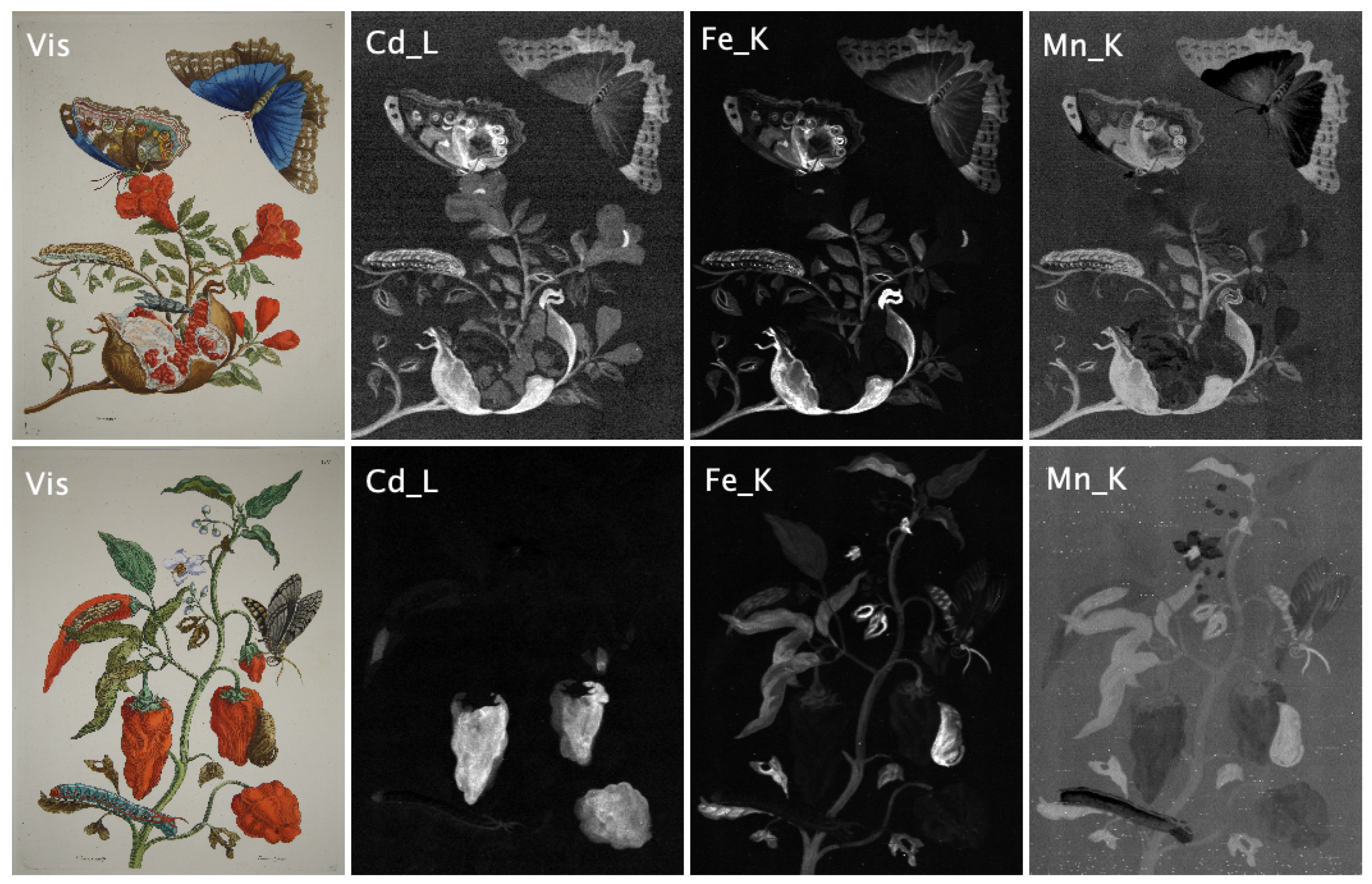
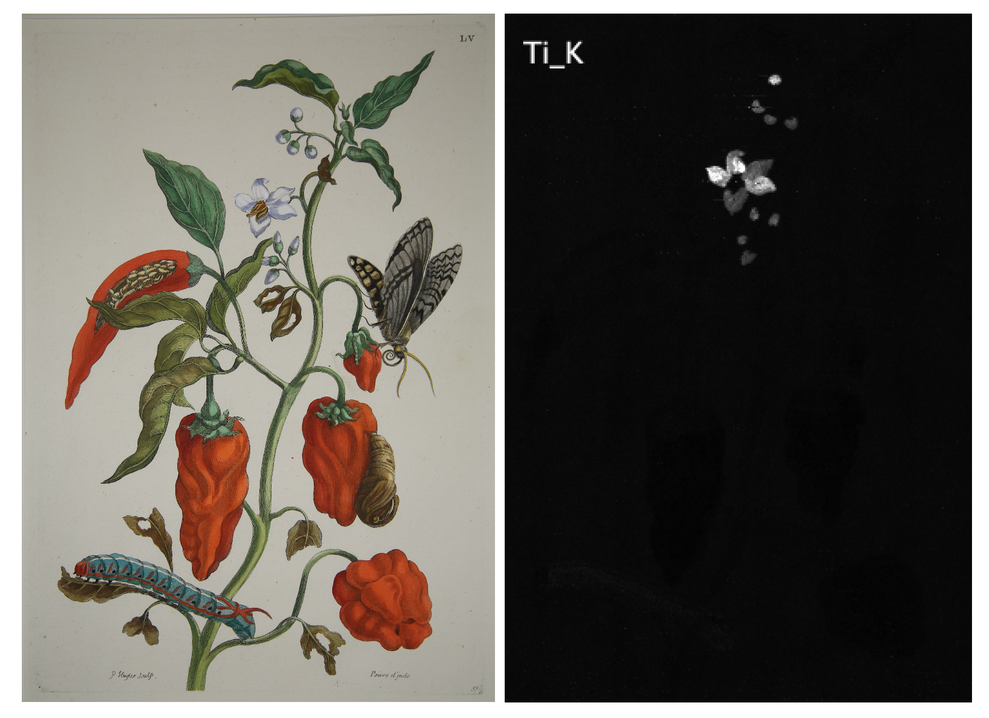
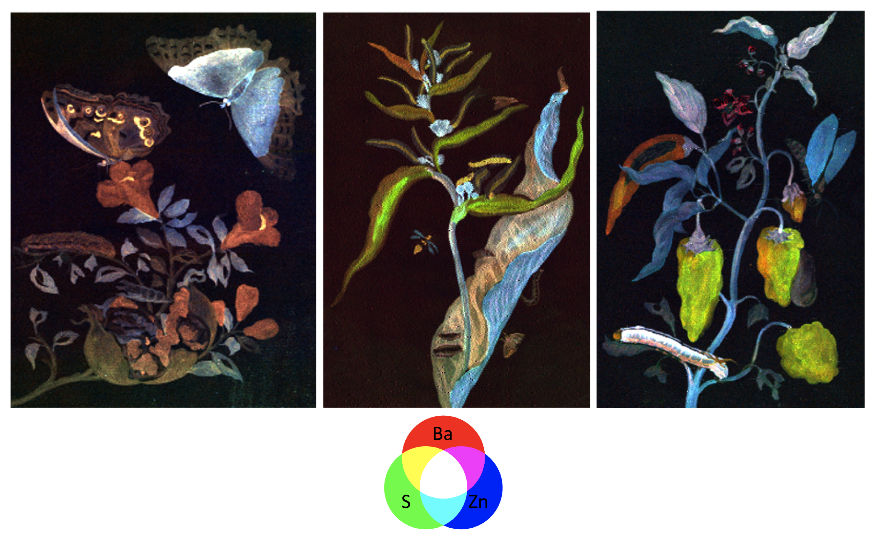
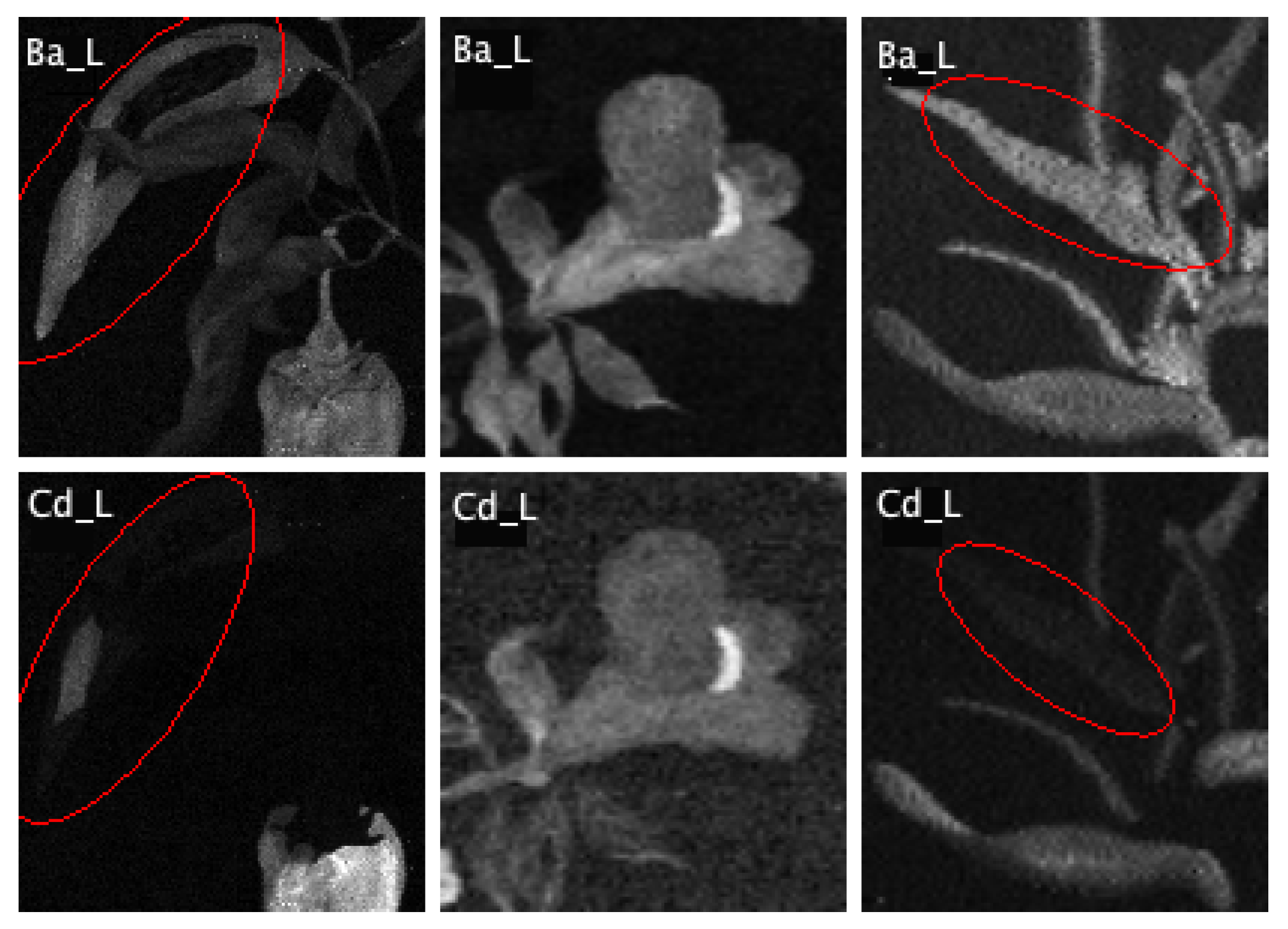
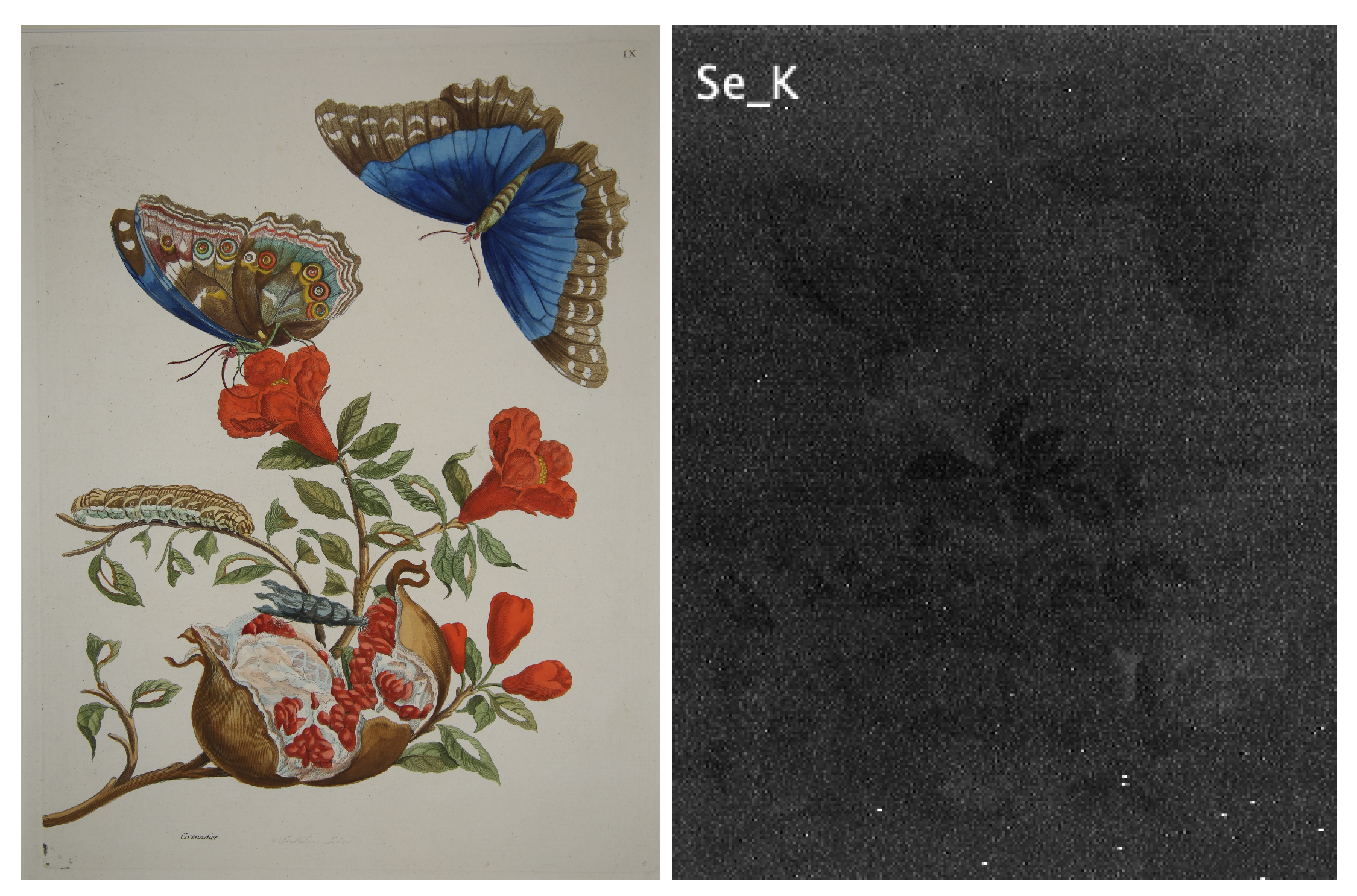
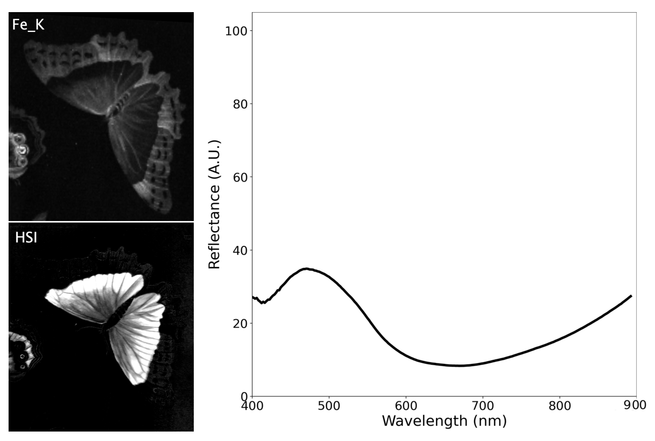
2.1. X-ray Fluorescence Spectroscopy
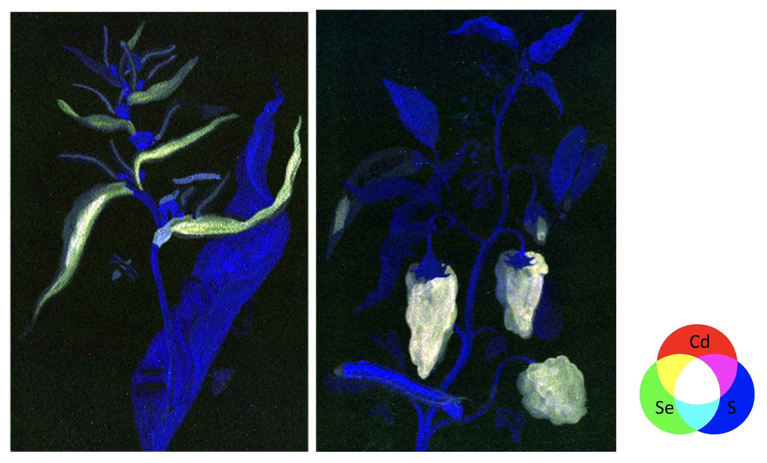
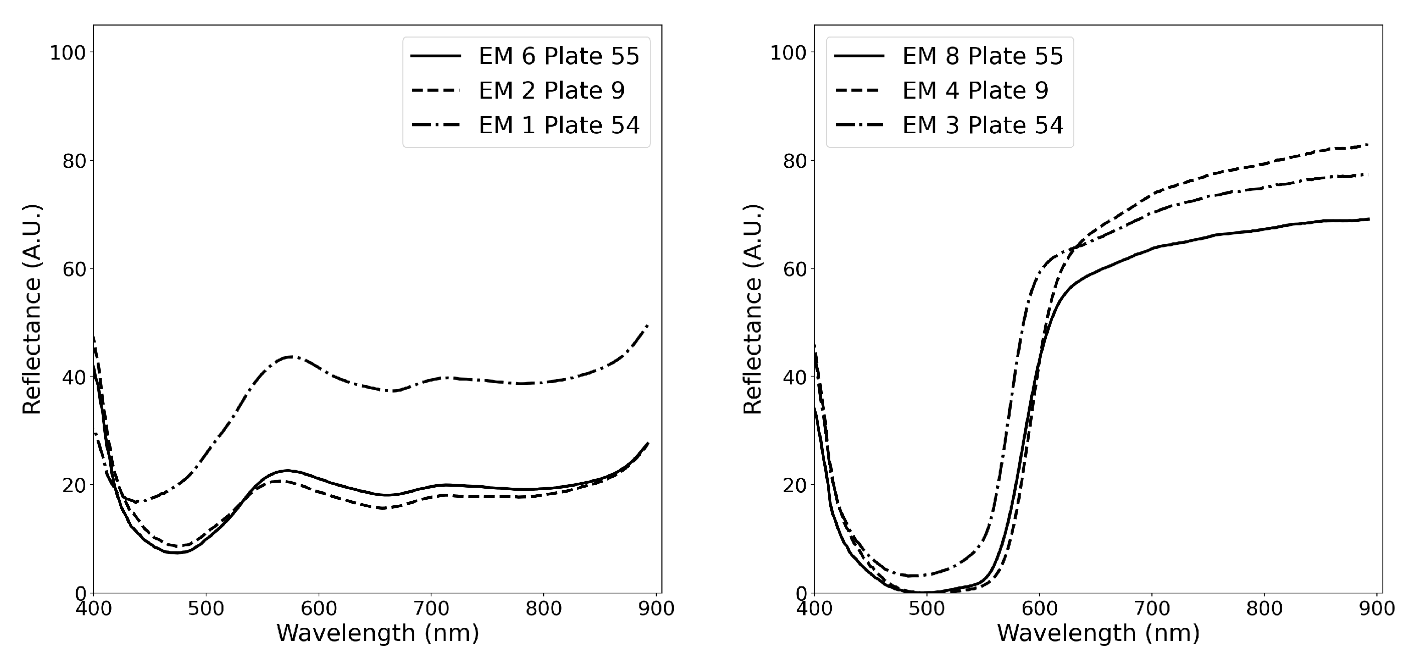
2.2. Hyperspectral Imaging (HSI)
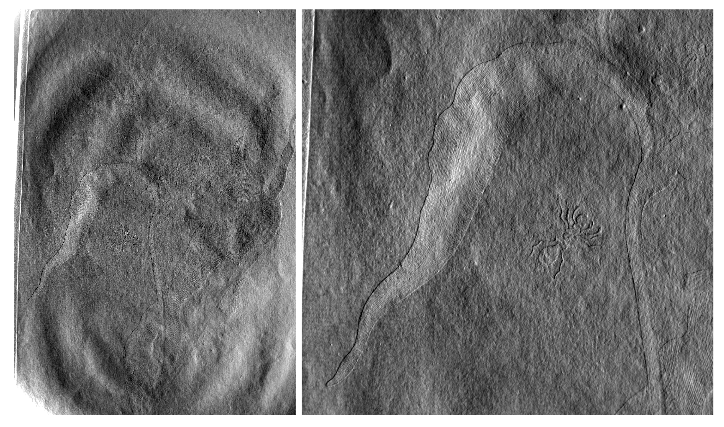
2.3. Photometric Stereo Imaging
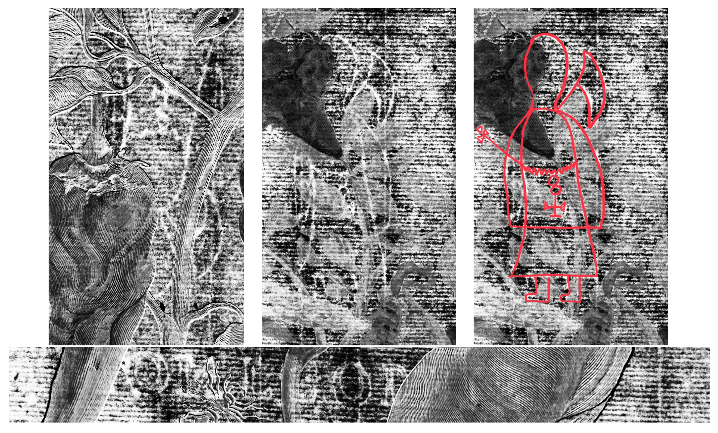
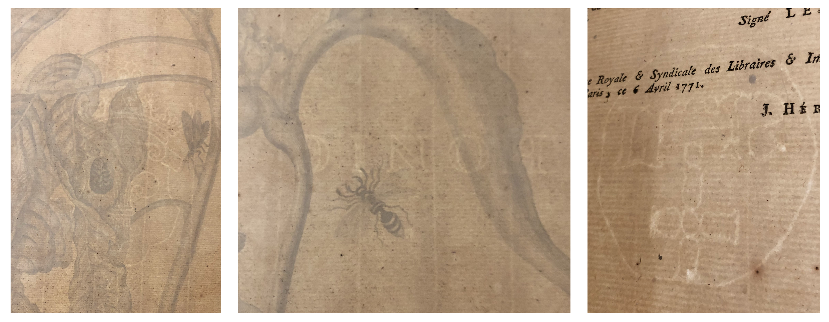
2.4. Transmitted Light Imaging
3. Results and Discussion
3.1. Pigments
3.1.1. Brown
3.1.2. White
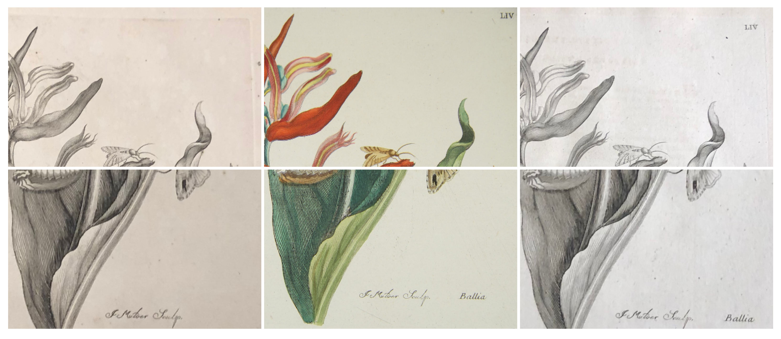
3.1.3. Red
3.1.4. Blue
3.1.5. Green and Yellow
3.1.6. Comparisons between Prints
3.2. Printed Lines
3.2.1. Photometric Stereo Normal Maps
3.2.2. Comparison with Other Editions
3.3. Paper
4. Conclusions
Supplementary Materials
Author Contributions
Funding
Institutional Review Board Statement
Informed Consent Statement
Data Availability Statement
Acknowledgments
Conflicts of Interest
Appendix A. Proposed Pigment Identifications and Corresponding Elements

| Color | Elements Present | Possible Pigment |
|---|---|---|
| Red | Ba, Cd, S, Si, Zn | Organic red (inferred) with barium substrate, or cadmium-barium red |
| Yellow | Ba, Cd, Fe, Mn, S, Si, Zn | Brown umber and/or cadmium yellow, lithopone |
| Green | Ba, Fe, Mn, S, Si, Zn | Organic green (inferred) or Prussian blue mixture, lithopone |
| Green | Ba, Fe, S, Si, Ti, Zn | Organic green (inferred), or Prussian blue or iron oxide mixture, titanium white, lithopone |
| Mint Green | Ba, S, Ti, Zn | Organic green (inferred), titanium white, lithopone |
| Blue | Ba, Fe, Mn, S, Si, Zn | Prussian blue possibly with iron oxide, lithopone |
| Brown | Ba, Ca, Cd, Fe, Mn, S, Si, Zn | Brown umber, cadmium yellow, lithopone |
| Paper | As, Ba, Co, Cu, K, Mn, Ni, S, Si | Paper |
| Mat | Ca, Fe, Ti | Mat |
| Color | Elements Present | Possible Pigment |
|---|---|---|
| Red 1 | Ba, Cd, S, Se, Si, Ti, Zn | Cadmium red, lithopone |
| Red 2 | Ba, Cd, S, Si, Zn | Cadmium red, and/or organic red (inferred) with barium substrate |
| Pink | Ba, S, Zn | Organic red (inferred), zinc white or lithopone |
| Yellow 1 | Ba, S, Zn | organic yellow (inferred), lithopone |
| Yellow 2 | Ba, Cd, Fe, S, Ti, Zn | titanium white, cadmium-based yellow, lithopone |
| Light Green | Ba, Fe, S, Ti, Zn | organic green (inferred), titanium white, possibly Prussian blue mixture, lithopone |
| Green | Ba, Ca, Fe, S, Si, Ti, Zn | Titanium white, organic green(inferred) or possibly Prussian blue mixture, lithopone |
| Brown | Ca, Fe, Mn, S, Zn | Brown umber |
| Paper | As, Co, Cu, Fe, K, Mn, Ni, S, Si | Paper |
| Mat | Ca, Fe, Ti | Mat |
| Color | Elements Present | Possible Pigment |
|---|---|---|
| Red 1 | Ba, Cd, S, Se, Si, Zn | Cadmium red, lithopone |
| Red 2 | Ba, Cd, K P, Si, | Organic red (inferred) with barium substrate and/or cadmium red |
| Yellow | Fe | Iron Oxide |
| Dark Green | Ba, Fe, Mn, S, Si, Zn | Organic green (inferred), or mixture with Prussian blue, lithopone |
| Light Blue | S, Si, Ti, Zn, | Organic Blue (inferred) mixed with titanium White |
| Brown | Ba, Fe, Mn, S, Si, Zn | Brown umber, lithopone |
| White | Si, Ti | Titanium white |
| Paper | As, Cu, K, Mn, S | Paper |
| Mat | Ca, Fe, P, Ti | Mat |
References
- Reitsma, E. Maria Sibylla Merian & Daughters: Women of Art and Science; Rembrandt House Museum: Amsterdam, The Netherlands; JPaul Getty Museum: Los Angeles, CA, USA; ZwolleWaanders: Zwolle, The Netherlands, 2008; pp. 184–204. [Google Scholar]
- Freedberg, D. The Failure of Colour. In Sight & Insight: Essays on Art and Culture in Honour of E.H. Gombrich at 85; Onians, J., Ed.; Phaidon Press: London, UK, 1994; pp. 245–263. [Google Scholar]
- Lack, H.W.; Ibáñez, V. Recording colour in late eighteenth century botanical drawings: Sydney Parkinson, Ferdinand Bauer and Thaddäus Haenke. Curtis’s Bot. Mag. 1997, 14. [Google Scholar] [CrossRef]
- Baker, T.; Dupré, S.; Kusukawa, S.; Leonhard, K. (Eds.) Early Modern Color Worlds; Brill: Leiden, The Netherlands, 2016. [Google Scholar]
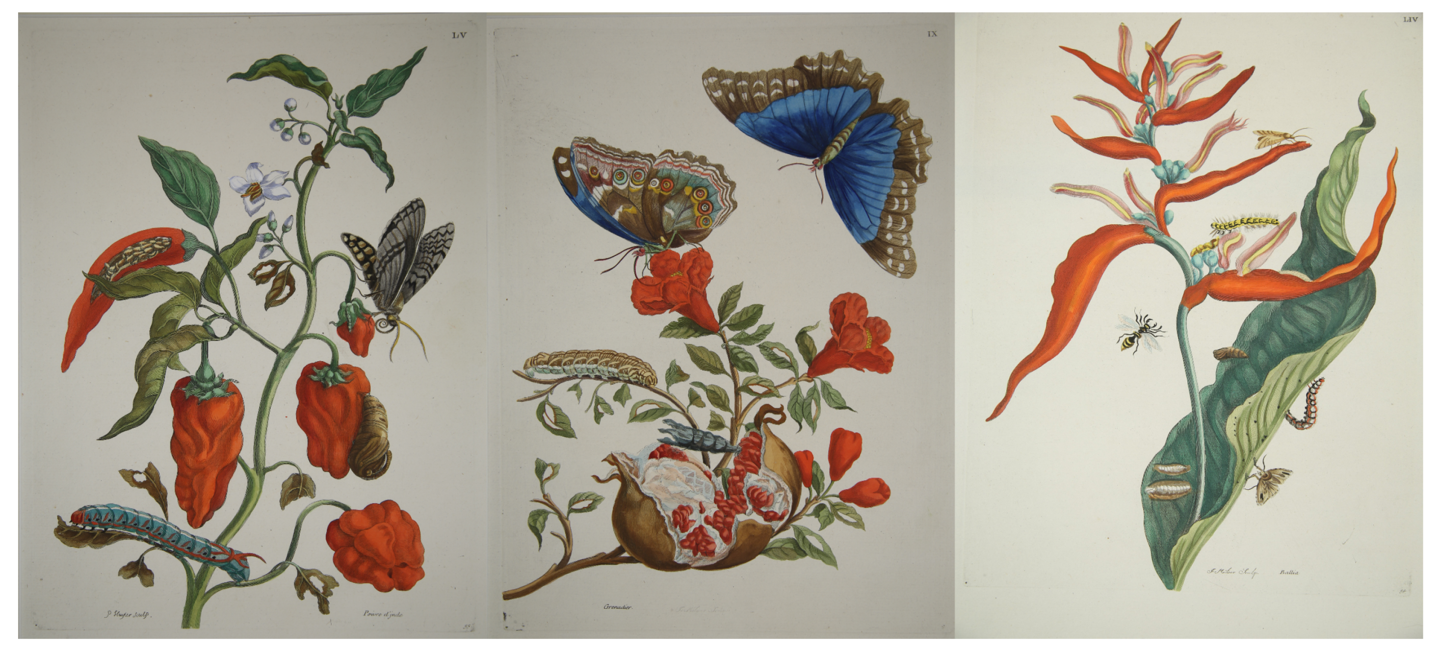
- Merian, M.S. Metamorphosis insectorum Surinamensium, ofte Verandering der Surinaamsche Insecten, Waar in de Surinaamsche Rupsen en Wormen Met Alle Des zelfs Veranderingen na Het Leven Afgebeeld en Beschreven Worden...; Gerard Valck: Amsterdam, The Netherlands, 1705; pp. 9, 49, 53. [Google Scholar]
- Dackerman, S. Painted Prints: The Revelation of Color in Northern Renaissance & Baroque Engravings, Etchings, & Woodcuts; Baltimore Museum of Art: Baltimore, MD, USA; Pennsylvania State University Press: University Park, PA, USA, 2002; pp. 1–9, 49–80. [Google Scholar]
- Schmidt, S.K.K. Altered and Adorned: Using Renaissance Prints in Daily Life, 1st ed.; Art Institute of Chicago: Chicago, IL, USA, 2011. [Google Scholar]
- Stillo, S.E. Putting the World in Its “Proper Colour”: Exploring Hand-Coloring in Early Modern Maps. J. Map Geogr. Libr. 2016, 12, 158–186. [Google Scholar] [CrossRef] [Green Version]
- Dackerman, S.; Primeau, T. The History and Technology of Renaissance and Baroque Hand-couloured prints. In The Broad Spectrum: Studies in the Materials, Techniques, and Conservations of Color on Paper; Salvesen, B., Stratis, H.K., Webber, P., Wheeler, M., Eds.; Archetype: London, UK, 2002; p. 78. [Google Scholar]
- Schmidt-Loske, K. Die Tierwelt der Maria Sibylla Merian (1647–1717): Arten, Beschreibungen und Illustrationen; Number 10 in Acta Biohistorica; Basilisken-Presse: Marburg, Germany, 2007; pp. 128–130. [Google Scholar]
- Hahn, O.; Oltrogge, D.; Bevers, H. Coloured Prints of the 16th Century: Non-Destructive Analyses on Coloured Engravings from Albrecht DÜrer and Contemporary Artists. Archaeometry 2004, 46. [Google Scholar] [CrossRef]
- Zannini, P.; Baraldi, P.; Aceto, M.; Agostino, A.; Fenoglio, G.; Bersani, D.; Canobbio, E.; Schiavon, E.; Zanichelli, G.; De Pasquale, A. Identification of colorants on XVIII century scientific hand-coloured print volumes. J. Raman Spectrosc. 2012, 43. [Google Scholar] [CrossRef]
- Ricciardi, P.; Legrand, S.; Bertolotti, G.; Janssens, K. Macro X-ray fluorescence (MA-XRF) scanning of illuminated manuscript fragments: Potentialities and challenges. Microchem. J. 2016, 124, 785–791. [Google Scholar] [CrossRef]
- Schrader, S.; Turner, N.; Yocco, N. Naturalism under the Microscope: A Technical Study of Maria Sibylla Merian’s “Metamorphosis of the Insects of Surinam”. Getty Res. J. 2012, 4. [Google Scholar] [CrossRef]
- Merian, M.S. Red Maize-Like Flower, Possibly a Banana Plant, from an Album of 91 Drawings Entitled ‘Merian’s Drawings of Surinam Insects’; Reference No. SL,5275.54; British Museum: London, UK, 1701. [Google Scholar]
- Merian, M.S. Sweet Pepper with Examples of a Butterfly, Chryslais and Caterpillar, from an Album of 91 Drawings Entitled ‘Merian’s Drawings of Surinam Insects’; Reference No. SL,5275.55; British Museum: London, UK, 1701. [Google Scholar]
- Merian, M.S. Two Blue Butterflies and a Caterpillar, from an Album of 91 Drawings Entitled ‘Merian’s Drawings of Surinam Insects’; Reference No. SL,5275.9; British Museum: London, UK, 1701. [Google Scholar]
- Casadio, F.; Daher, C.; Bellot-Gurlet, L. Raman Spectroscopy of cultural heritage Materials: Overview of Applications and New Frontiers in Instrumentation, Sampling Modalities, and Data Processing. In Analytical Chemistry for Cultural Heritage; Mazzeo, R., Ed.; Topics in Current Chemistry Collections; Springer International Publishing: Cham, Switzerland, 2017; pp. 161–211. [Google Scholar] [CrossRef] [Green Version]
- Bersani, D.; Madariaga, J.M. Applications of Raman spectroscopy in art and archaeology. J. Raman Spectrosc. 2012, 43. [Google Scholar] [CrossRef]
- Saverwyns, S. Russian avant-garde… or not? A micro-Raman spectroscopy study of six paintings attributed to Liubov Popova. J. Raman Spectrosc. 2010, 41. [Google Scholar] [CrossRef]
- Fremout, W.; Saverwyns, S. Identification of synthetic organic pigments: The role of a comprehensive digital Raman spectral library. J. Raman Spectrosc. 2012, 43. [Google Scholar] [CrossRef]
- Prati, S.; Sciutto, G.; Bonacini, I.; Mazzeo, R. New Frontiers in Application of FTIR Microscopy for Characterization of Cultural Heritage Materials. Top. Curr. Chem. 2016, 374, 26. [Google Scholar] [CrossRef]
- Pouyet, E.; Barbi, N.; Chopp, H.; Healy, O.; Katsaggelos, A.; Moak, S.; Mott, R.; Vermeulen, M.; Walton, M. Development of a highly mobile and versatile large MA-XRF scanner for in situ analyses of painted work of arts. X-ray Spectrom. 2020. [Google Scholar] [CrossRef]
- Solé, V.A.; Papillon, E.; Cotte, M.; Walter, P.; Susini, J. A multiplatform code for the analysis of energy-dispersive X-ray fluorescence spectra. Spectrochim. Acta Part At. Spectrosc. 2007, 62, 63–68. [Google Scholar] [CrossRef]
- Vermeulen, M.; Smith, K.; Eremin, K.; Rayner, G.; Walton, M. Application of Uniform Manifold Approximation and Projection (UMAP) in Spectral Imaging of Artworks. Spectrochim. Acta Part Mol. Biomol. Spectrosc. 2021, 252. [Google Scholar] [CrossRef]
- Huang, X.; Walton, M.; Bearman, G.; Cossairt, O. Near light correction for image relighting and 3D shape recovery. In Proceedings of the 2015 Digital Heritage, Granada, Spain, 28 September–2 October 2015; pp. 215–222. [Google Scholar] [CrossRef]
- Ruiz, P.; Raju, G.; Dill, O.; Cossairt, O.; Walton, M.; Katsaggelos, A. Visible Transmission Imaging of Watermarks by Suppression of Occluding Text or Drawings. Digit. Appl. Archaeol. Cult. Herit. 2017, 15, e00121. [Google Scholar] [CrossRef]
- Boyle, R.D.; Hiary, H. Watermark location via back-lighting and recto removal. Int. J. Doc. Anal. Recognit. 2009, 12. [Google Scholar] [CrossRef]
- Berie, B.H. Prussian Blue. In Artists’ Pigments: A Handbook of Their History and Characteristics; Fitzhugh, E.W., Ed.; National Gallery of Art: Washington, DC, USA, 1986; Volume 3, pp. 191–219. [Google Scholar]
- Fiedler, I.; Bayard, M. Cadmium Yellows, Oranges, and Reds. In Artists’ Pigments: A Handbook of Their History and Characteristics; Feller, R.L., Ed.; National Gallery of Art: Washington, DC, USA, 1986; Volume 1, pp. 65–109. [Google Scholar]
- Barium Oxide—CAMEO. Available online: http://cameo.mfa.org/wiki/Barium_oxide (accessed on 15 May 2021).
- Capua, R. The Obscure History of a Ubiquitous Pigment: Phosphorescent Lithopone and Its Appearance on Drawings by John La Farge. J. Am. Inst. Conserv. 2014, 53. [Google Scholar] [CrossRef]
- Laver, M. Titanium Dioxide Whites. In Artists’ Pigments: A Handbook of Their History and Characteristics; Fitzhugh, E.W., Ed.; National Gallery of Art: Washington, DC, USA, 1986; Volume 3, pp. 295–357. [Google Scholar]
- D’Elia, E.; Buscaglia, P.; Piccirillo, A.; Picollo, M.; Casini, A.; Cucci, C.; Stefani, L.; Romano, F.P.; Caliri, C.; Gulmini, M. Macro X-ray fluorescence and VNIR hyperspectral imaging in the investigation of two panels by Marco d’Oggiono. Microchem. J. 2020, 154, 104541. [Google Scholar] [CrossRef]
- Helwig, K. Iron Oxide Pigments; natural and synthetic. In Artists’ Pigments: A Handbook of Their History and Characteristics; Anderson, B.H., Ed.; National Gallery of Art: Washington, DC, USA, 1986; Volume 4. [Google Scholar]
- Laclavetine, K.; Boust, C.; Clivet, L.; Le Hô, A.S.; Laval, E.; Mathis, R.; Menu, M.; Pagliano, E.; Salmon, X.; Selbach, V.; et al. Non-invasive study of 16th century Northern European chiaroscuro woodcuts: First insights. Microchem. J. 2019, 144, 419–430. [Google Scholar] [CrossRef]
- Duran, A.; Franquelo, M.L.; Centeno, M.A.; Espejo, T.; Perez-Rodriguez, J.L. Forgery detection on an Arabic illuminated manuscript by micro-Raman and X-ray fluorescence spectroscopy. J. Raman Spectrosc. 2011, 42. [Google Scholar] [CrossRef]
- Pronti, L.; Felici, A.C.; Ménager, M.; Vieillescazes, C.; Piacentini, M. Spectral Behavior of White Pigment Mixtures Using Reflectance, Ultraviolet—Fluorescence Spectroscopy, and Multispectral Imaging. Appl. Spectrosc. 2017, 71. [Google Scholar] [CrossRef] [Green Version]
- Picollo, M.; Bacci, M.; Magrini, D.; Trumpy, G.; Tsukada, M.; Kunzelman, D. Modern White Pigments: Their Identification by Means of non Invasive Ultraviolet, Visible and Infrared Fiber Optic Reflectance Spectroscopy. In Modern Paints Uncovered, Proceedings from the Modern Paints Uncovered Symposium, Tate Modern, London, UK, 16–19 May 2006; Tate Modern: London, UK, 2007; pp. 129–139. [Google Scholar]
- Burnstock, A.; Reissner, E.; Richardson, C. Analysis of Inorganic Materials from Paintings and Watercolours by Paul Cézanne from the Courtauld Gallery Using Two Methods of Non-Invasive Portable XRF with Light Microscopy and SEM/EDX Spectroscopy. In Proceedings of the 9th International Conference on NDT of Art, Jerusalem, Israel, 25–30 May 2008; p. 10. [Google Scholar]
- Paulus, J.; Knuutinen, U. Cadmium colours: Composition and properties. Appl. Phys. A 2004, 79, 397–400. [Google Scholar] [CrossRef]
- Rogge, C.E.; Epley, B.A. Behind the Bocour Label: Identification of Pigments and Binders in Historic Bocour Oil and Acrylic Paints. J. Am. Inst. Conserv. 2017, 56, 15–42. [Google Scholar] [CrossRef]
- Biron, C.; Mounier, A.; Bourdon, G.L.; Servant, L.; Chapoulie, R.; Daniel, F. A blue can conceal another! Noninvasive multispectroscopic analyses of mixtures of indigo and Prussian blue. Color Res. Appl. 2020, 45. [Google Scholar] [CrossRef]
- Vermeulen, M.; Leona, M. Evidence of early amorphous arsenic sulfide production and use in Edo period Japanese woodblock prints by Hokusai and Kunisada. Herit. Sci. 2019, 7, 73. [Google Scholar] [CrossRef] [Green Version]
- Vermeulen, M.; Muller, E.; Leona, M. Non-Invasive Study of the Evolution of Pigments and Colourants Use in 19th-Century Ukiyo-e. Arts Asia 2020, 50, 103. [Google Scholar]
- Glinsman, L.D. The practical application of air-path X-ray fluorescence spectrometry in the analysis of museum objects. Stud. Conserv. 2005, 50. [Google Scholar] [CrossRef]
- Aceto, M.; Agostino, A.; Fenoglio, G.; Idone, A.; Gulmini, M.; Picollo, M.; Ricciardi, P.; Delaney, J.K. Characterisation of colourants on illuminated manuscripts by portable fibre optic UV-visible-NIR reflectance spectrophotometry. Anal. Methods 2014, 6. [Google Scholar] [CrossRef]
- Hunter, D. Papermaking: The History and Technique of an Ancient Craft; Dover: New York, NY, USA, 1978. [Google Scholar]


Publisher’s Note: MDPI stays neutral with regard to jurisdictional claims in published maps and institutional affiliations. |
© 2021 by the authors. Licensee MDPI, Basel, Switzerland. This article is an open access article distributed under the terms and conditions of the Creative Commons Attribution (CC BY) license (https://creativecommons.org/licenses/by/4.0/).
Share and Cite
Dill, O.; Vermeulen, M.; McGeachy, A.; Walton, M. Multi-Modal, Non-Invasive Investigation of Modern Colorants on Three Early Modern Prints by Maria Sibylla Merian. Heritage 2021, 4, 1590-1604. https://0-doi-org.brum.beds.ac.uk/10.3390/heritage4030088
Dill O, Vermeulen M, McGeachy A, Walton M. Multi-Modal, Non-Invasive Investigation of Modern Colorants on Three Early Modern Prints by Maria Sibylla Merian. Heritage. 2021; 4(3):1590-1604. https://0-doi-org.brum.beds.ac.uk/10.3390/heritage4030088
Chicago/Turabian StyleDill, Olivia, Marc Vermeulen, Alicia McGeachy, and Marc Walton. 2021. "Multi-Modal, Non-Invasive Investigation of Modern Colorants on Three Early Modern Prints by Maria Sibylla Merian" Heritage 4, no. 3: 1590-1604. https://0-doi-org.brum.beds.ac.uk/10.3390/heritage4030088