Case Report

Geochemical-Microscopical Characterization of the Deterioration of Stone Surfaces in the Cloister of Santa Maria in Vado (Ferrara, Italy)

1
Department of Chemistry, Pharmaceutical and Agricultural Sciences, University of Ferrara, Via L. Borsari 46, 44121 Ferrara, Italy
2
ENEA, Italian National Agency for New Technologies, Energy and Sustainable Economic Development Fusion and Technology for Nuclear Safety and Security Department Nuclear Safety, Security and Sustainability Division, Via Martiri di Monte Sole 4, 40129 Bologna, Italy
3
Department of Life Science and Biotechnologies, University of Ferrara, Via L. Borsari 46, 44121 Ferrara, Italy
4
Department of Physics and Earth Sciences, University of Ferrara, Via Saragat 1, 44121 Ferrara, Italy
*
Author to whom correspondence should be addressed.
Submission received: 6 September 2021 / Revised: 27 September 2021 / Accepted: 28 September 2021 / Published: 1 October 2021
(This article belongs to the Section Geoheritage and Geo-Conservation)

Abstract

:
Santa Maria in Vado is a monument in the rich artistic heritage of the city of Ferrara (north of Italy). In this paper we want to investigate the state of conservation of tombstones, cloister and the entrance to the basilica, in order to keep them in the best possible state for the future generations. From the chemical characterization, the state of conservation was determined focusing on the biodeteriogenic and non-biodeteriogenic factors, which determine a series of unwanted changes in the physical, mechanical and above all aesthetic properties of the material, often closely connected with the environment and conservation conditions. On the macroscopic observation, the state of conservation of the tombstones appeared to be very deteriorated through aesthetic and structural damage. In detail, the stereo microscope observation of samples collected from the tombstones show the presence of efflorescence probably caused by the abundant of water that bring the salts present inside the rock into solution. Relating the columns, μ-XRF analysis confirm the carbonate composition of samples and presence of iron and sulfur. Finally, SEM observation highlighted the presence of black crust on arch samples and the presence of pollen on the black crust and spheroidal particles probably related to atmospheric pollution.

1. Introduction

The Framework Convention on the Value of Cultural Heritage for Society (art. 2a) [1] stated that cultural heritage is “a group of resources inherited from the past which people identify, independently of ownership, as a reflection and expression of their constantly evolving values, beliefs, knowledge and traditions”. Taking into account these statements, the historic center of Ferrara has been included in the list of world heritage sites by UNESCO since 1995, as an example of city designed in the Renaissance, which preserves its historic center still intact with its value and traditions [2].
The city of Ferrara (10 m a.s.l.) developed around a ford on the Po river in about the 7th cent. A.D., and is one of the few Italian cities whose original layout was not based on the Roman tradition. The Este family ruled Ferrara from the second half of the 13th cent. A.D., and under its control the city rose to a significant position within the Italian states [3,4]. Therefore, it played a key role in the political life inside as well as outside the peninsula. In the 15th and 16th centuries A.D., Ferrara became an intellectual and artistic center which attracted the greatest minds of the Italian Renaissance [5].
Today, Ferrara is famous for its historical center, which is extraordinarily well-preserved: it is a city where overall harmony predominates due to an accurate urban project and together with the artistic urban plans of the Este family [6,7]; it is a city that preserves and projects the beauty of the Middle Ages and the Renaissance in the days of today, an intelligently modern city with deep-rooted and lively cultural, artistic and environmental traditions.
Among all the ancient buildings of Ferrara, Guarini [8] identified the very ancient origins of the church of Santa Maria in Vado in the main center of the city, which had this name from a small pond on the shore where it was first built [9]. Tradition told that on the place where the church now stands, a devoted Greek image placed on a capital was venerated [10]. Over time, as the village grew from year to year, the need was felt to thank the hypothetical benefactor by erecting a church in her honor.
On March 28, 1171 a miracle occurred. Blood came out of the host and sprinkled the entire vault of the chapel confirming both the real presence of Christ in the consecrated host and that the holy mass was the renewal of the divine sacrifice. This took place in a particularly delicate moment in the life of the Church, in which there were many heretics that denied the dogma of the transmutation [11]. The vault of the small apse of the church, stained by the blood of the miracle, became a place of worship [12].
Nevertheless, during the years, the church went into decline, but fortunately, several restoration interventions (1981–2000) for some parts of the basilica began [13] under the supervision of Superintendence of Archaeology, Fine Arts and Landscape for the metropolitan city of Bologna.
The speed of some reactions involved in the degradation of monuments might be increased by the presence of numerous compounds with the action of catalysts, such as heavy metals and carbonaceous particles related to pollution, especially atmospheric pollution [14,15,16]. The latter were probably the cause of the acceleration of the process of formation and detachment of black crusts on the surface of the stones of the site analyzed [17,18].
The aim of this work was to characterize the parts of the main cloister of the basilica that appeared more deteriorated respect to other part. For this reason, some columns, the entrance arch and some tombstones contained therein were selected to verify the state of conservation. This knowledge could be able to provide a series of suggestions and indications for a better management over time, in order to ensure its usability for future generations.
As a matter of fact, the conservation of cultural heritage for future generations depends on the ability of a wide number of citizens to perceive, understand, and appreciate its value [19]. It means that the survival of cultural heritage is related to the use people make of it, from public enjoyment to “the construction of a peaceful and democratic society, and in the processes of sustainable development and the promotion of cultural diversity” [1].
For this reason, in this work, great importance has been given to the study of the black crusts on the stones of the main entrance portal to the cloister, because it is subject to traffic pollution which could be a major cause of deterioration [20,21]. It should be remembered that the city of Ferrara is one of the cities of the Emilia Romagna region which, especially in recent years, has been subjected to exceeding the limit value allowed by the law for the emission of atmospheric particulate matter of anthropogenic sources, especially in the winter months, which may contain metal particles that could create severe damage to the monuments [22,23,24].
Air pollution plays an important role in the deterioration of many materials used in buildings and cultural monuments causing an inestimable damage. There is a lot of study regarding the mechanisms of deterioration which can be caused by, for example, air pollution or meteorological conditions [25] or by biological organism that could affect the stone surface of the buildings [26]. However, some problem related to air pollution persists especially in Northern and Southern Italy; in particular, particulate matter are considered the main responsible for stone corrosion [24]. In addition, temperature, precipitation or relative humidity could create serious damage, for example, corrosion on the surface of stone materials [25]. Moreover, biological agents could form microbial biofilms that consequently could create aesthetic and physico-chemical deterioration of stone [26].
As confirmed by the Special Eurobarometer on Cultural Heritage, most Europeans live close to historic monuments or sites (more than 73%), and think cultural heritage is important to them personally, as well to their community, region, country and the EU as a whole (more than 80%) [27]. However, it is not easy to preserve the historical and artistic heritage with the increase of pollution due to anthropogenic activities. With this work, it is hoped that the community of Ferrara and the institutions will also be able to take steps to safeguard the historical and artistic heritage of this wonderful Renaissance city.

2. Materials and Methods

Different kind of samples were collected inside the cloister of the basilica of Santa Maria in Vado, which is located in the city center of Ferrara.
During the sampling it was possible to observe that the tombstones and the column remakes were breakable and easily to remove. For this reason, small quantities of the stone surfaces of the tombstones on the walls of the cloister and of the columns inside the cloister (Figure 1) to evaluate their degradation were collected. In addition, different samples of the entrance portal to the cloister were collected to characterize the presence of the degradation and the black crust. The samples collected and analyzed by different technologies at the laboratories of the Department of Physics and Earth Sciences, University of Ferrara were better described in Table 1.
An optical stereomicroscope (90× total magnification) was used for microscopic observations on the samples. The stereomicroscope used was an Optika SZM-2 (Opto-Lab, Modena, Italy) equipped with MOTICAM 2500 5.0 M pixel webcam. The software Motic Images Plus 2.0 ML software was used for reflected light observation on all the samples to define grain size and texture, dimensional and morphological aspect of the clasts and state of conservation [28,29].
The samples related to the columns were analyzed using a μ-XRF spectrometer at the Department of Physics and Earth Sciences, Ferrara University. The instrument was a portable ARTAX TM 200 from Bruker AXS Microanalysis GmbH. The instrument consisted of an X-ray tube equipped with a Mo target placed at 6° and a Be window. The measurements were performed at 50 kV and 700 μA for 120 s through a collimator with a diameter of 0.65 mm. An SSD Peltier-cooled detector (10 mm2 active area and resolution of <155 eV at 10 kcps) was used for the detection of the secondary fluorescent X-rays [30,31,32].
The surface of the samples collected on the arch that decorates the entrance portal to the cloister was analyzed by a Scanning Electron Microscope (SEM) Zeiss model EVO® 40 Basic Instrument (Carl Zeiss AG, Oberkochen, Germany) for morphological and chemical characterization [33,34,35,36]. SEM instrument worked with a magnification range between 7–1,000,000× and with a working voltage of 20 kV, corresponding to a detection limit = 1 μm particle size [37,38]. SEM images were obtained using the SmartSEM software (Zeiss). A piece not metalized of each sample was firmed on a SEM stub utilizing double-sided conductive adhesive tape.
Not being able to use EDS source of the instrument for the chemical characterization because during the analysis it had problem, the analysis of the columns was carried out with μ-XRF spectrometer.

3. Results and Discussion

Each sample was first subjected to a careful macroscopic analysis to discriminate colors and shapes.

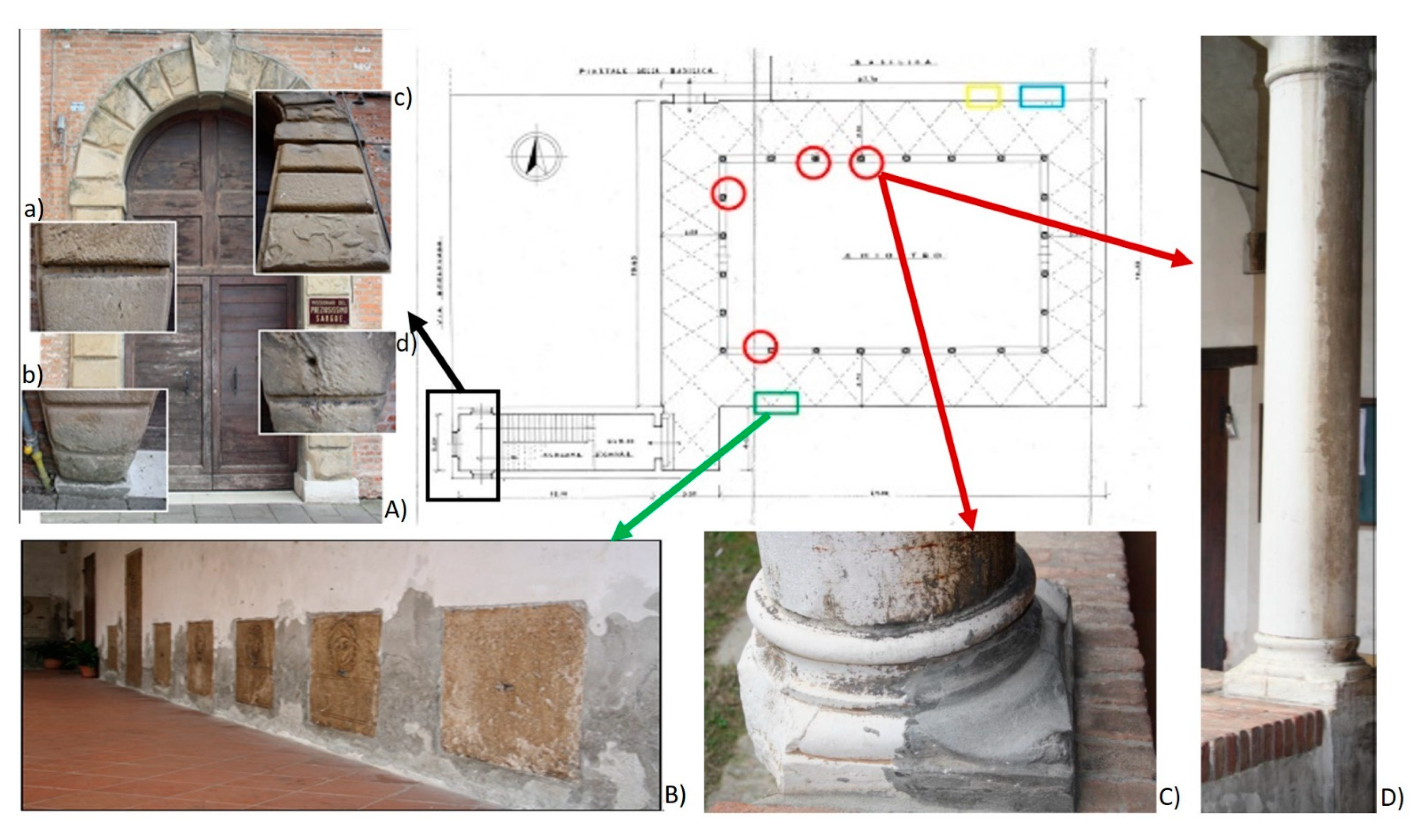
3.1. Macroscopic Characterization

The state of conservation of all 40 tombstones, walled in the internal walls of the cloister, appeared to be very deteriorated through aesthetic and structural damage (Figure 1B). All the tombstones were placed at 15 cm from the ground, but this height was too low to guarantee a good state of conservation. In fact, all the tombstones were affected by the capillary moisture rising from the floor: the picture in Figure 1B shows the plaster swollen (grey) in correspondence with the moisture bubbles (white). As a consequence, the tombstones were also affected by this humidity: in the points where the humidity was observed, they were darker in color than the original. In addition, the sampling area was subject to atmospheric particulate matter pollution, that could be the responsible for the blackening processes occurring on the surfaces of the stone, and their quantification in the crusts could provide information on the contribution of atmospheric pollution sources to the degradation products formation [39,40]. Other important phenomena observed on the samples collected were the presence of colonies of biodeteriogenic agents (as for example musk or green and black film) [41,42,43] with the consequent formation of more or less thick black crusts [44,45].
The white columns surrounding the cloister (Figure 1C,D) were glossy in the half facing inside of the colonnade and opaque in the half facing outwards (Figure 1D). These patinas could have been caused by oxalate films [46] or by a particular protective varnish that has been preserved on the inside, while on the outside it has been washed out, thus giving the column the opacity characteristic. Some columns have been restored in the basement, adding a grey concrete base to bring it closer to the color of the column (Figure 1C) rich in black crust.
In this first analysis of the state of conservation of the cloister with the tombstones and the columns, the arch that decorates the entrance portal to the cloister was also included. The arch was built with a yellow sandstone block cladding (Figure 1A). Inside these sandstone blocks, there were rounded clasts larger than a grain of sand. The state of conservation of the sandstone arch was not good and was diversified. It was in fact possible to divide it into two vertical parts: the one closest to the wall of the facade of the church (left side) showed a fairly good state of conservation characterized by black crusts and a slight erosion (Figure 1(Ab)) with respect to the right side of the same which was more damaged. On the right side, there was deep exfoliation of the sedimentation planes (Figure 1(Ac)); black crusts were also included especially in those areas protected from the washing away (Figure 1(Aa,d)) in which dirty water and also pollutants could stagnate and facilitate their formation (as described in [47]). This different deterioration was probably due to the presence of the cornice (Figure 1A on the top), located on the wall of the facade of the church, which protects the left side of the arch and which, for this reason, was less exposed to the action of water than the right side. The structure of the black crusts was very compact and with a thickness of a few mm.

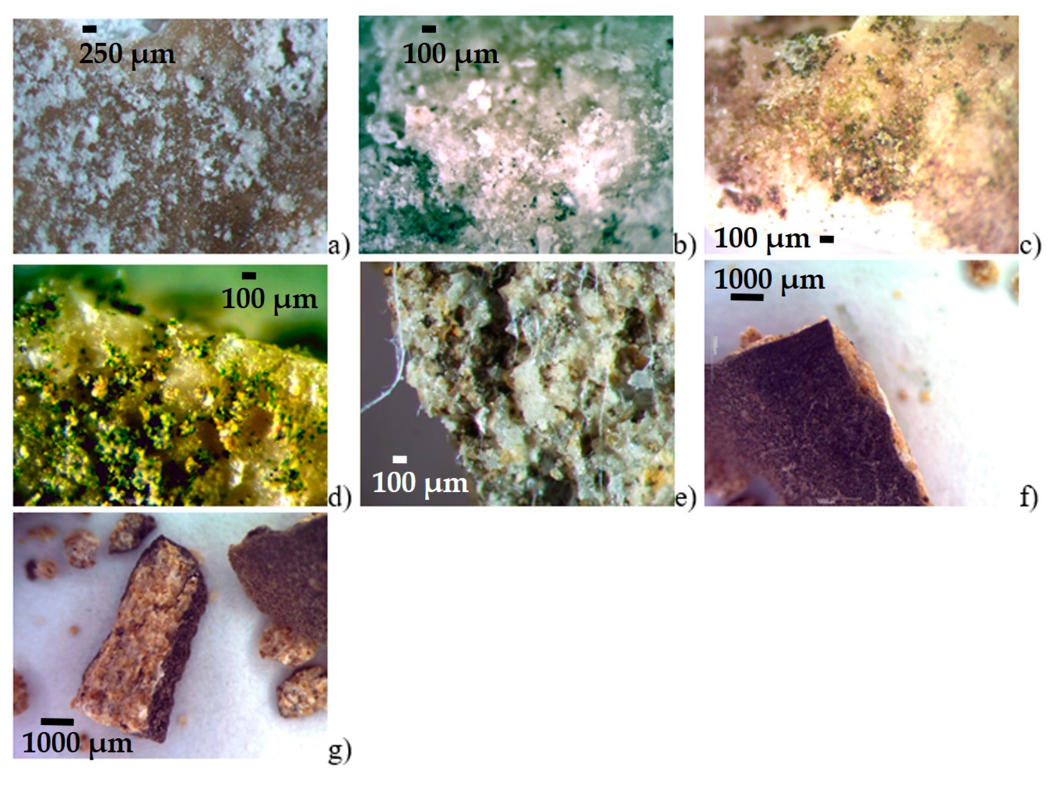
3.2. Microscopic Characterization

The stereo microscope observation of samples collected from all the tombstones analyzed show white efflorescence (Figure 2a) probably caused by the abundant presence of water that brings the salts present inside the rock into solution, as better described in Ergün Hatır [48] and Martínez-Martínez [49].
The investigations carried out on the glossy patina of the columns, on the other hand, did not reveal the evident presence of particular substances used as protective agents. The analyzed samples show the presence of black crusts, already observed macroscopically also on the base of the column itself, recognizable by the particles of atmospheric particulate matter of dark color with abundant saline efflorescence (Figure 2b), as in [50]. The microbial biofilms favor the adherence of airborne particles (dust, pollen, spores, carbonaceous particles from combustion of oil and coal), giving rise to hard crusts and patinas.
On the base of the column, a small portion of a particular yellow patina was collected which, observed by stereomicroscope, gave interesting results: the small fragments of crust appeared externally (Figure 2c) characterized by rounded dark-colored particles with a dark black/green border, while internally (Figure 2d) covered with a layer of green algae. Probably it represented a type of algae which found a habitat conducive to their development. It could be assumed that it belonged to Gloeocapsa algae [51,52], together with other epileptic chlorophytes, such as Trentepohlia, which appeared in yellowish color. The presence of these algal biofilms had an important role in the disfigurement of columns producing variously colored patinas.
From the sandstone images, it was possible to observe the presence of a dense and intricate layer of biofilm and/or fungal hyphae in the interior of the sandstone (Figure 2e). Their presence, from a conservative point of view, could create a lot of problems: these heterotrophic organisms (destroyers or consumers) could cause both mechanical damage, such as surface detachment, superficial losses, or penetration and increased porosity, and chemical alterations [53,54], also using the substrate as a source of nourishment, enhancing soil formation and water retention The sandstone was therefore not in good condition. In some of the deteriorate samples, it was also observed sulphation process growing up in several stages: the black crust tended to thicken over time (Figure 2f,g) (varying from 1000 μm to 3000 μm) to harden more and more and to became less porous, as observed in most of the cultural heritage built [55,56]. In this way, the difference in mechanical and thermal behavior between the black crust and the underlying stone increased.

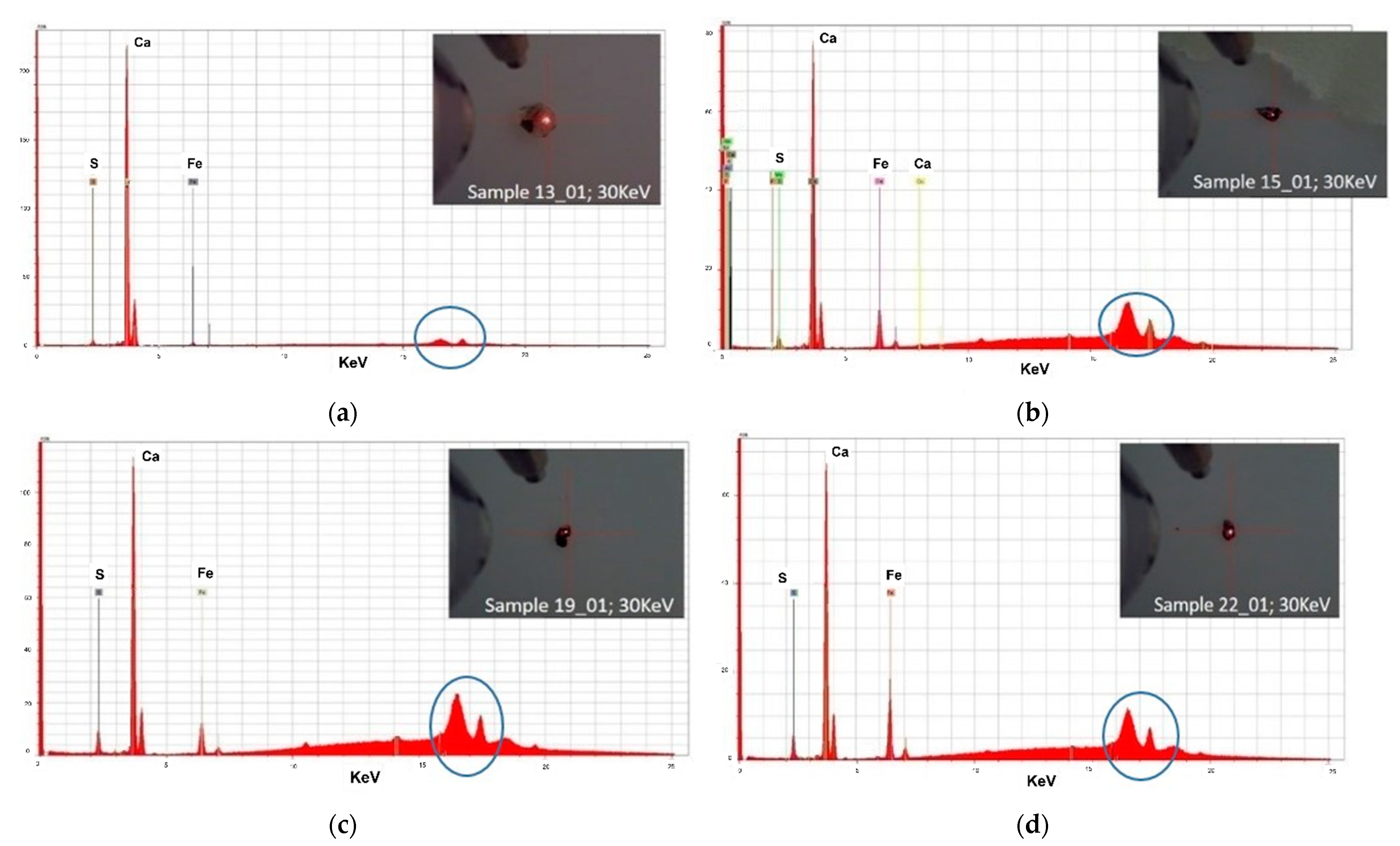
3.3. μ-XRF Analysis on the Yellowish Patina Samples Collected on the Columns

The samples collected on the deteriorated part of the columns of the cloister were analyzed by μ-XRF in order to better characterize the chemical composition of the samples [57]. For each samples we analyzed different area and in the Figure 3 we report the spectra representative of each samples.
Three spectra over four are very similar to each other (Figure 3a,c,d). All the spectra show a high peak of Ca, related to the carbonate composition of the natural stone used to build the cloister. The second major peak is Fe, the presence of this element can be due to its natural occurrence in carbonate rocks and to the presence of Fe hydroxide [58,59,60].
The presence of S can be related to the presence of gypsum in the black crust. The sulphate efflorescence could be due by sulphation of atmospheric dust particles containing gypsum in dust [61].

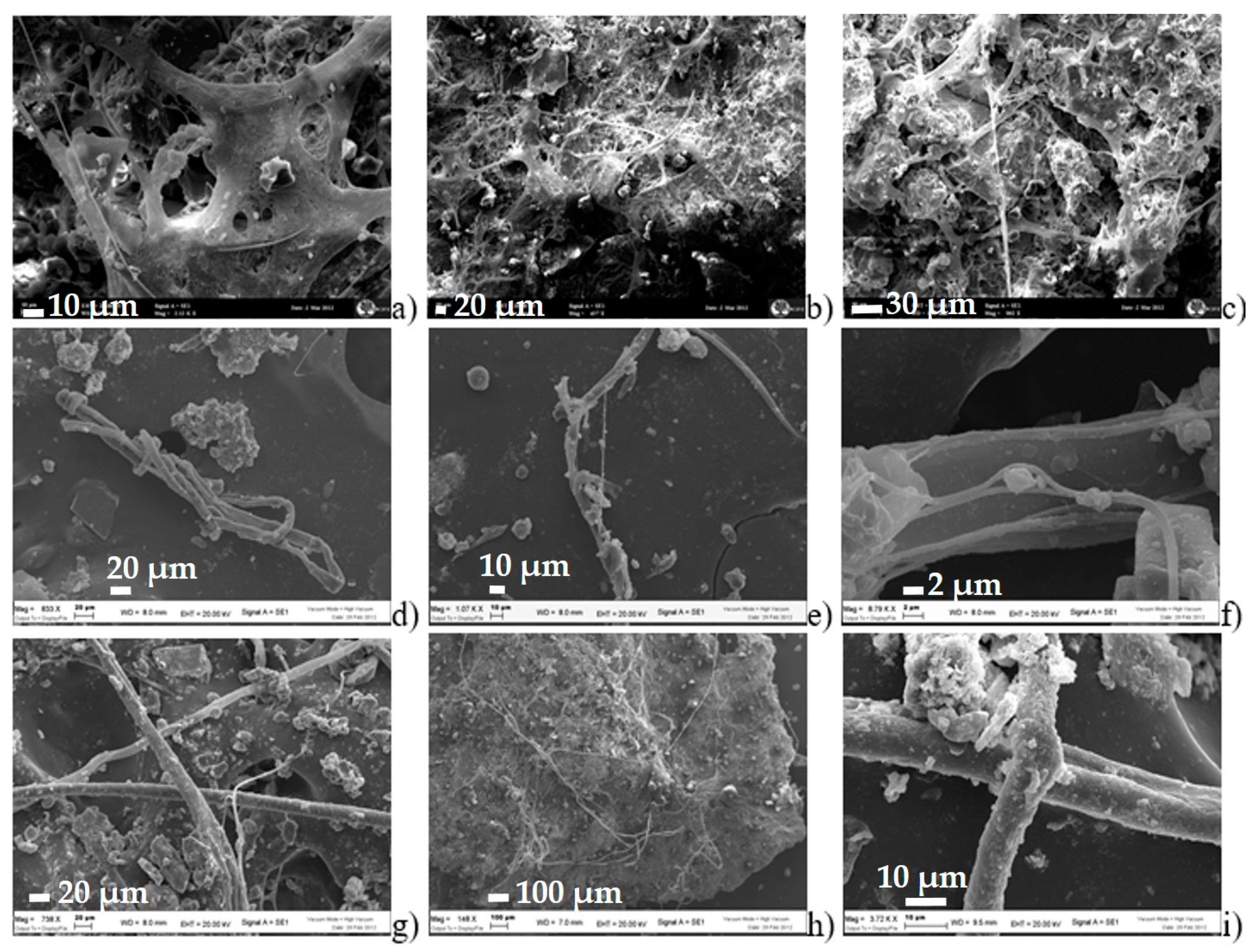
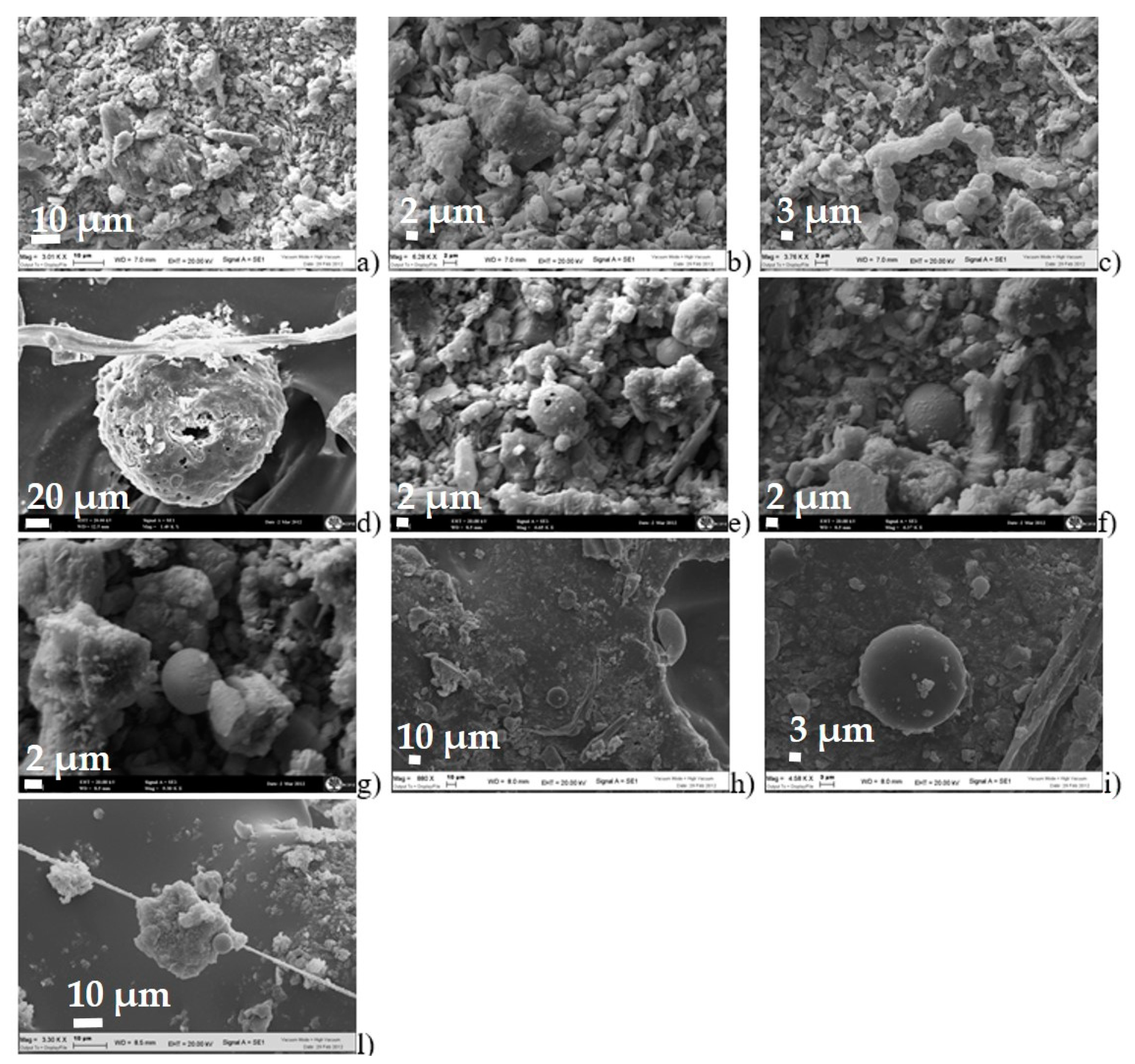
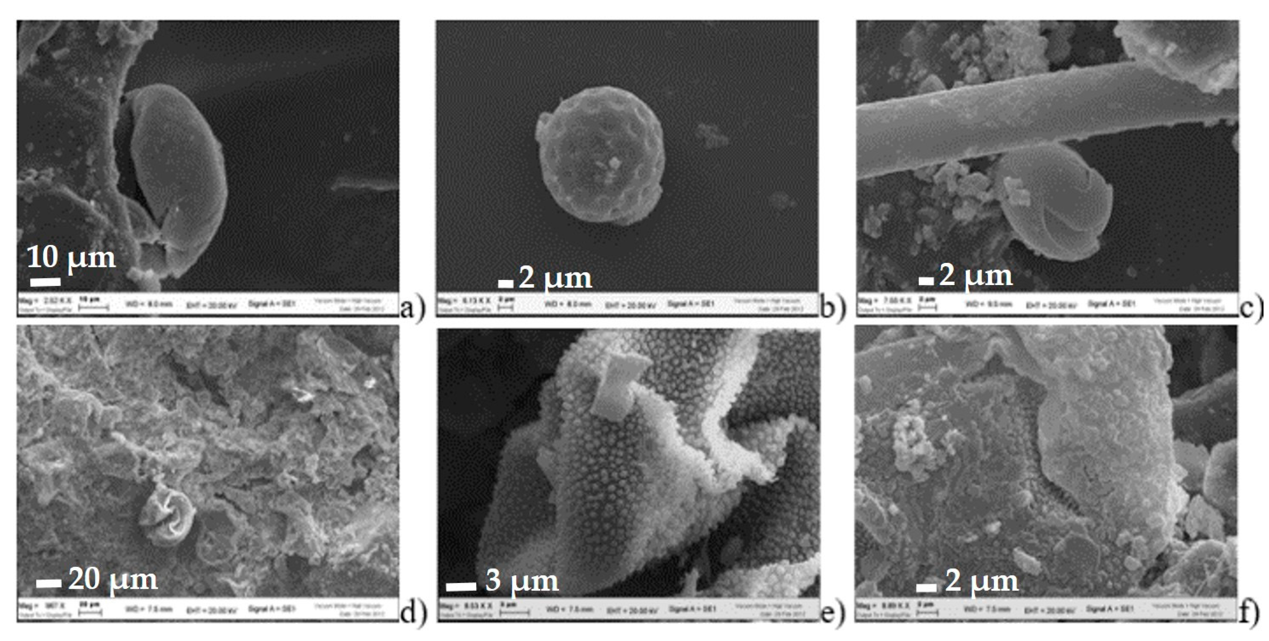
3.4. SEM Observations on the Black Crust Samples Collected into the Arch

SEM analyses of the yellow sandstone collected on the arch that decorates the entrance portal to the cloister allowed to identify the presence of hyphae that appear twisted on themselves (especially in Figure 4d), incorporating sandstone and/or other particles (as shown in Figure 4f), creating a dense layer between the clasts [62,63]. All the four samples collected on the arch (as shown in Figure 1I) observed by SEM show the same observations.
The black crusts of the sandstone samples appeared composed by particles with different size, rounded and mostly globular in shape as can also be seen from the images in Figure 5. The matrix of these black crusts appeared chalky.
The aging and the degradation of the materials could be amplified by the atmospheric pollutants that are represented in Figure 5 by spherical particles [64].
In addition, it was possible to recognize some pollen grains probably coming from native plant species [65]. In particular, a pollen grain with the typical shape of the Chenopodiaceae (Figure 6a), pollen grain typical of arid areas (Figure 6b,c) that could be trapped inside the crust during the summer periods (exploded pollen) [66]. In detail, Figure 6a, c shown pollen grains with spherical or oval shape with smooth and crushed surface. Rounded pollen grains were also observed in Figure 6b but probably belonging to different family of pollen. Figure 6e shows spore with surface ornamentation with irregular verrucous tubercles. Also the pollen in Figure 6f shows pollen with irregular shape and surface with fractures too.

4. Conclusions

The stone monuments from tombstones and columns investigated on the cloister of the Basilica of Santa Maria in Vado in Ferrara highlighted the effects of physical, chemical and biological deteriorating factors.
The investigations have revealed that microbial biofilms, favoring the adherence of airborne particles, gave rise to hard crusts and patinas. The analyzed samples from columns shown the presence of black crusts with saline efflorescence; on the base of the column, a yellow patina an algal biofilm that could play an important role in the disfigurement of columns. In addition, the presence of sulphate efflorescence is confirmed by µ-XRF analysis.
The tombstones analyzed are affected by degradation of stone material, stored in outdoor environments and in unsuitable conditions that the previous restoration of 1981 could have improved in order to preserve them [11].
As regards the fractures on the tombstones, it would be useful to investigate them better to understand if these cracks were limited to the surface only or if they also proceed in the innermost layers of the wall structure. In this case it would be useful to operate on these problems, before they also affect the interior of the church.
The lack of treatments caused widespread alterations which, today, allowed us to collect samples of various constituent elements of the cloister, tombstones, columns and portal, but which were all comparable in terms of level and type of decay.
The obtained results could represent a basic knowledge for better managing future restoration operations in order to leave these historical and monumental complexes in the best possible state for the future generations. Additionally, suitable restoration procedures will make it possible to increase the resistance of stone materials against the degradation phenomena.
Moreover, to gain a better understanding of the role of these microorganisms it will be useful an interdisciplinary work that should focus on ecological and physiological studies of specific species of in stone colonization and biodeterioration processes.

Author Contributions

Conceptualization: E.M., C.T., M.L. and C.V.; methodology: E.M., C.T., M.L. and C.V.; validation; E.M., M.L. and C.V.; formal analysis, E.M., M.L. and C.V.; investigation: E.M., C.T., M.L. and C.V.; resources: M.L. and C.V.; data curation: C.T. and E.M.; writing—original draft preparation: C.T. and E.M.; visualization: E.M. and C.T.; supervision: M.L. and C.V.; project administration: M.L. and C.V. All authors have read and agreed to the published version of the manuscript.

Funding

This research received no external funding.

Conflicts of Interest

The authors declare no conflict of interest.

References

  1. Council of Europe. Council of Europe Framework Convention on the Value of Cultural Heritage for Society, Council of Europe Treaty Series—No. 199 Faro, art. 1d. 27 October 2005. Available online: https://rm.coe.int/1680083746 (accessed on 25 August 2021).
  2. UNESCO Website. Available online: http://www.unesco.it/it/PatrimonioMondiale/Detail/112 (accessed on 25 August 2021).
  3. Dean, T. Land and Power in Late Medieval Ferrara: The Rule of the Este, 1350–1450; Cambridge Studies in Medieval Life & Thought; Cambridge University Press: Cambridge, UK, 2002. [Google Scholar]
  4. Bandini Mazzanti, M.; Bosi, G.; Guarnieri, C. The useful plants of the city of Ferrara (Late Medieval/Renaissance) based on archaeobotanical records from middens and historical/culinary/ethnobotanical documentation. In Plants and Culture: Seeds of the cultural heritage of Europe; Edipuglia s.r.l.: Bari, Italy, 2009; pp. 93–106. [Google Scholar]
  5. Bosi, G.; Mercuri, A.M.; Bandini Mazzanti, M. Plants and Man in urban environment: The history of the city of Ferrara (10th–16th cent. A.D.) through its archaeobotanical records. Bocconea 2009, 23, 285–300. [Google Scholar]
  6. Folin, M. Ferrara: 1385-1505. All’ombra del Principe, in Fabbriche, Piazze, Mercati. La Città Italiana nel Rinascimento; Calabi, D., Ed.; Officina: Roma, Italy, 1997; pp. 354–388. [Google Scholar]
  7. Rosenberg, C.M. The Este Monuments and Urban Development in Renaissance Ferrara; Cambridge University Press: Cambridge, UK, 1997; pp. 1–329. [Google Scholar]
  8. Guarini, M.A. Compendio Historico Dell’origine, Accrescimento e Prerogative Delle Chiese e Luoghi pii Della Diocesi di Ferrara e Delle Memorie di què Personaggi di Pregio Che in Esse Sono Seppelliti; Forgotten Books: Ferrara, Italy, 1621; p. 301. [Google Scholar]
  9. Canonici Fachini, G. Due Giorni in Ferrara; Istruzione per Agevolmente Pervenire Alla Cognizione Delle Opere Tutte Letterarie e di Belle Arti Quivi Raccolte: Corredata di Molte Cognizioni Utili Egualmente al culto Viaggiatore, Che al Cittadino Ferrarese; Company’ Tipi di Gaetano Bresciani: Ferrara, Italy, 1819; p. 90. [Google Scholar]
  10. Cimatti, E. Cenni Storici Intorno al Sangue Miracoloso che si Venera Nella Parrocchiale Basilica di S. Maria del Vado in Ferrara; Tipografia Governativa Taddei: Ferrara, Italy, 1857; Volume 1, p. 5. [Google Scholar]
  11. Scalabrini, G.A. Guida per la città e i Borghi di Ferrara in Cinque Giornate, ca. 1755. Trascrizione a cura di Frongia, C., 1997. I quaderni del Liceo Ariosto, n. 6; Tipo-Litografia Artigiana: Ferrara, Italy, 1997; p. 90. [Google Scholar]
  12. Cavallini, G. Omaggio al Sangue Miracoloso che si Venera Nella Basilica Parrocchiale di Santa Maria del Vado in Ferrara; Silvestri & Taddei: Ferrara, Italy, 1878; p. 247. [Google Scholar]
  13. Di Francesco, C. La Basilica di Santa Maria in Vado a Ferrara; Eds Fondazione Cassa di Risparmio di Ferrara: Ferrara, Italy, 2001. [Google Scholar]
  14. Comite, V.; Fermo, P. The Damage Induced by Atmospheric Pollution on Stone Surfaces: The Chemical Characterization of Black Crusts. In Mathematical Modeling in Cultural Heritage; Bonetti, E., Cavaterra, C., Natalini, R., Solci, M., Eds.; Springer INdAM Series, 41; Springer: Cham, Switzerland, 2021. [Google Scholar]
  15. Comite, V.; Miani, A.; Ricca, M.; La Russa, M.; Pulimeno, M.; Fermo, P. The impact of atmospheric pollution on outdoor cultural heritage: An analytic methodology for the characterization of the carbonaceous fraction in black crusts present on stone surfaces. Environ. Res. 2021, 201, 111565. [Google Scholar] [CrossRef] [PubMed]
  16. Spezzano, P. Mapping the susceptibility of UNESCO World Cultural Heritage sites in Europe to ambient (outdoor) air pollution. Sci. Total Environ. 2021, 754, 142345. [Google Scholar] [CrossRef]
  17. Pozo-Antonio, S.; Cardell, C.; Comite, V.; Fermo, P. Characterization of black crusts developed on historic stones with diverse mineralogy under different air quality environments. Environ. Sci. Pollut. Res. 2021. [Google Scholar] [CrossRef]
  18. Wilhelm, K.; Longman, J.; Orr, S.A.; Viles, H. Stone-built heritage as a proxy archive for long-term historical air quality: A study of weathering crusts on three generations of stone sculptures on Broad Street, Oxford. Sci. Total Environ. 2021, 759, 143916. [Google Scholar] [CrossRef]
  19. Montella, M. Valore e Valorizzazione del Patrimonio Culturale Storico; Mondadori Electa: Milan, Italy, 2009. [Google Scholar]
  20. Vidorni, G.; Sardella, A.; De Nuntiis, P.; Volpi, F.; Dinoi, A.; Contini, D.; Comite, V.; Vaccaro, C.; Fermo, P.; Bonazza, A. Air pollution impact on carbonate building stones in Italian urban sites. Eur. Phys. J. Plus 2019, 134, 439. [Google Scholar] [CrossRef]
  21. Natarajan, N.; Vasudevan, M.; Dineshkumar, S.K.; Nandhini, S.S.; Balaganesch, P. Effects of air pollution on monumental buildings in India: An overview. Environ. Sci. Pollut. Res. 2021. [Google Scholar] [CrossRef]
  22. Carotta, M.C.; Ferrari, E.; Gherardi, S.; Malagù, C.; Piga, M.; Vaccaro, C. A multidisciplinary study on stone monuments damage. Sens. Microsyst. 2005, 145–150. [Google Scholar] [CrossRef]
  23. Modena, C.; Cagliotti, B.; Cescatti, E. Monument of Ludovico Ariosto in Ferrara, Italy: Conservation of architectural surfaces and structural consolidation. WIT Trans. Built Environ. 2019, 191, 151–161. [Google Scholar]
  24. Nava, S.; Becherini, F.; Bernardi, A.; Bonazza, A.; Chiari, M.; García-Orellana, I.; Lucarelli, F.; Ludwig, N.; Migliori, A.; Sabbioni, C.; et al. An integrated approach to assess air pollution threats to cultural heritage in a semi-confined environment: The case study of Michelozzo’s Courtyard in Florence (Italy). Sci. Total Environ. 2010, 408, 1403–1413. [Google Scholar] [CrossRef] [PubMed]
  25. Monforti, F.; Bellasio, R.; Bianconi, R.; Clai, G.; Zanini, G. An evaluation of particle deposition fluxes to cultural heritage sites in Florence, Italy. Sci. Total Environ. 2004, 334–335, 61–72. [Google Scholar] [CrossRef]
  26. Favero-Longo, S.E.; Viles, H.A. A review of the nature, role and control of lithobionts on stone cultural heritage: Weighing-up and managing biodeterioration and bioprotection. World J. Microbiol. Biotechnol. 2020, 36, 100. [Google Scholar] [CrossRef] [PubMed]
  27. European Commission. Special Eurobarometer 466: Cultural Heritage, Fieldwork September–October 2017. Available online: http://data.europa.eu/euodp/en/data/dataset/S2150_88_1_466_ENG (accessed on 25 August 2021).
  28. Marrocchino, E.; Telloli, C.; Novara, P.; Meletti, V.; Vaccaro, C. Petro-archaeometric characterization of historical mortars in the city of Ravenna (Italy). In Proceedings of the IMEKO TC-4 International Conference on Metrology for Archaeology and Cultural Heritage, Trento, Italy, 22–24 October 2020. [Google Scholar]
  29. Marrocchino, E.; Telloli, C.; Pedrini, M.; Vaccaro, C. Natural stones used in the Orsi-Marconi palace façade (Bologna): A petro-mineralogical characterization. Heritage 2020, 3, 1109–1124. [Google Scholar] [CrossRef]
  30. Holakooei, P.; Ahmadi, M.; Volpe, L.; Vaccaro, C. Early Opacifiers In The Glaze Industry Of First Millennium bc Persia: Persepolis And Tepe Rabat. Archaeometry 2017, 59, 239–254. [Google Scholar] [CrossRef]
  31. Holakooei, P.; de Lapérouse, J.F.; Carò, F.; Röhrs, S.; Franke, U.; Müller-Wiener, M.; Reiche, I. Non-invasive scientific studies on the provenance and technology of early Islamic ceramics from Afrasiyab and Nishapur. J. Archaeol. Sci. Rep. 2019, 24, 759–772. [Google Scholar] [CrossRef]
  32. Pessanha, S.; Samouco, A.; Adão, R.; Carvalho, M.L.; Santos, J.P.; Amaro, P. Detection limits evaluation of a portable energy dispersive X-ray fluorescence setup using different filter combinations. X-ray Spectrom. 2017, 46, 102–106. [Google Scholar] [CrossRef]
  33. Marrocchino, E.; Telloli, C.; Caraccio, S.; Guarnieri, C.; Vaccaro, C. Medieval Glassworks in the City of Ferrara (North Eastern Italy): The Case Study of Piazza Municipale. Heritage 2020, 3, 819–837. [Google Scholar] [CrossRef]
  34. Marrocchino, E.; Telloli, C.; Cesarano, M.; Montuori, M. Geochemical and Petrographic Characterization of Bricks and Mortars of the Parish Church SANTA Maria in Padovetere (Comacchio, Ferrara, Italy). Minerals 2021, 11, 530. [Google Scholar] [CrossRef]
  35. Morillas, H.; Maguregui, M.; García-Florentino, C.; Carrero, J.A.; Salcedo, I.; Madariaga, J.M. The cauliflower-like black crusts on sandstones: A natural passive sampler to evaluate the surrounding environmental pollution. Environ. Res. 2016, 147, 218–232. [Google Scholar] [CrossRef]
  36. Telloli, C.; Fazzini, M.; Tassinari, R.; Marrocchino, E.; Vaccaro, C. Monitoring of solid particulate airborne samples from mountain snow in some sites of the Alps, Italy. Int. J. Geosci. 2013, 4, 711–723. [Google Scholar] [CrossRef] [Green Version]
  37. Marrocchino, E.; Telloli, C.; Rizzo, A. Chemical Characterization of Particulate Matter in the Renaissance City of Ferrara. Geosciences 2021, 11, 227. [Google Scholar] [CrossRef]
  38. Telloli, C.; Malaguti, A.; Mircea, M.; Tassinari, R.; Vaccaro, C.; Berico, M. Properties of agricultural aerosol released during wheat harvest threshing, plowing and sowing. J. Enviorn. Sci. 2014, 26, 1903–1912. [Google Scholar] [CrossRef]
  39. De Marco, A.; Screpanti, A.; Mircea, M.; Piersanti, A.; Proietti, C.; Fornasier, M.F. High resolution estimates of the corrosion risk for cultural heritage in Italy. Environ. Pollut. 2017, 226, 260–267. [Google Scholar] [CrossRef]
  40. Patil, S.M.; Kasthurba, A.K. Weathering of stone monuments: Damage assessment of basalt and laterite. Mater. Today Proc. 2021, 43, 1647–1658. [Google Scholar] [CrossRef]
  41. Caldeira, A.T.; Schiavon, N.; Mauran, G.; Salvador, C.; Rosado, T.; Mirão, J.; Candeias, A. On the Biodiversity and Biodeteriogenic Activity of Microbial Communities Present in the Hypogenic Environment of the Escoural Cave, Alentejo, Portugal. Coatings 2021, 11, 209. [Google Scholar] [CrossRef]
  42. Hosseini, Z.; Caneva, G. Evaluating hazard conditions of plant colonization in Pasargadae World Heritage Site (Iran) as a tool of biodeterioration assessment. Int. Biodeterior. Biodegrad. 2021, 160, 105216. [Google Scholar] [CrossRef]
  43. Isola, D.; Zucconi, L.; Cecchini, A.; Caneva, G. Dark-pigmented biodeteriogenic fungi in etruscan hypogeal tombs: New data on their culture-dependent diversity, favouring conditions, and resistance to biocidal treatments. Fungal Biol. 2021, 125, 609–620. [Google Scholar] [CrossRef] [PubMed]
  44. Randazzo, L.; Collina, M.; Ricca, M.; Barbieri, L.; Bruno, F.; Arcudi, A.; La Russa, M.F. Damage Indices and Photogrammetry for Decay Assessment of Stone-Built Cultural Heritage: The Case Study of the San Domenico Church Main Entrance Portal (South Calabria, Italy). Sustainability 2020, 12, 5198. [Google Scholar] [CrossRef]
  45. Nowicka-Krawczyk, P.; Komar, M.; Gutarowska, B. Towards understanding the link between the deterioration of building materials and the nature of aerophytic green algae. Sci. Total Environ. 2022, 802, 149856. [Google Scholar] [CrossRef]
  46. Perez-Rodriguez, J.L.; Duran, A.; Centeno, M.A.; Martinez-Blanes, J.M.; Robador, M.D. Thermal analysis of monument patina containing hydrated calcium oxalates. Thermochim. Acta 2011, 512, 5–12. [Google Scholar] [CrossRef]
  47. Comite, V.; Pozo-Antonio, J.S.; Cardell, C.; Rivas, T.; Randazzo, L.; La Russa, M.F.; Fermo, P. Metals distributions within black crusts sampled on the facade of an historical monument: The case study of the Cathedral of Monza (Milan, Italy). In Proceedings of the 2019 IMEKO TC-4 International Conference on Metrology for Archaeology and Cultural Heritage, Florence, Italy, 4–6 December 2019. [Google Scholar]
  48. Ergün Hatır, M.; İnce, I.; Korkanç, M. Intelligent detection of deterioration in cultural stone heritage. J. Build. Eng. 2021, 44, 102690. [Google Scholar] [CrossRef]
  49. Martínez-Martínez, J.; Torrero, E.; Sanz, D.; Navarro, V. Salt crystallization dynamics in indoor environments: Stone weathering in the Muñoz Chapel of the Cathedral of Santa María (Cuenca, central Spain). J. Cult. Herit. 2021, 47, 123–132. [Google Scholar] [CrossRef]
  50. Unković, N.; Ljaljević Grbić, M.; Subakov-Simić, G.; Stupar, M.; Vukojević, J.; Jelikić, A.; Stanojević, D. Biodeteriogenic and toxigenic agents on 17th century mural paintings and façade of the old church of the Holy Ascension (Veliki Krčimir, Serbia). Indoor Built Environ. 2016, 25, 826–837. [Google Scholar] [CrossRef]
  51. Becerra, J.; Mateo, M.; Ortiz, P.; Nicolas, G.; Zaderenko, A.P. Evaluation of the applicability of nano-biocide treatments on limestones used in cultural heritage. J. Cult. Herit. 2019, 38, 126–135. [Google Scholar] [CrossRef]
  52. Ortega-Morales, O.; Montero-Muños, J.L.; Baptista Neto, J.A.; Beech, I.B.; Sunner, J.; Gaylarde, C. Deterioration and microbial colonization of cultural heritage stone buildings in polluted and unpolluted tropical and subtropical climates: A meta-analysis. Int. Biodeterior. Biodegrad. 2019, 143, 104734. [Google Scholar] [CrossRef]
  53. Kakakhel, M.A.; Wu, F.; Gu, J.D.; Feng, H.; Shah, K.; Wang, W. Controlling biodeterioration of cultural heritage objects with biocides: A review. Int. Biodeterior. Biodegrad. 2019, 143, 104721. [Google Scholar] [CrossRef]
  54. Pinheiro, A.C.; Mesquita, N.; Trovao, J.; Soares, F.; Tiago, I.; Coelho, C.; de Carvalho, H.P.; Gil, F.; Catarino, L.; Piñar, G.; et al. Limestone biodeterioration: A review on the Portuguese cultural heritage scenario. J. Cult. Herit. 2019, 36, 275–285. [Google Scholar] [CrossRef]
  55. Pozo-Antonio, J.S.; Rivas, T.; Lopez, A.J.; Fiorucci, M.P.; Ramil, A. Effectiveness of granite cleaning procedures in cultural heritage: A review. Sci. Total Environ. 2016, 571, 1017–1028. [Google Scholar] [CrossRef] [PubMed]
  56. Lamhasni, T.; El-Marjaoui, H.; El Bakkali, A.; Lyazidi, S.A.; Haddad, M.; Ben-Ncer, A.; Benyaich, F.; Bonazza, A.; Tahri, M. Air pollution impact on architectural heritage of Morocco: Combination of synchronous fluorescence and ATR-FTIR spectroscopies for the analyses of black crusts deposits. Chemosphere 2019, 225, 517–523. [Google Scholar] [CrossRef] [PubMed]
  57. Stoean, C.; Ionescu, L.; Stoean, R.; Boicea, M.; Atencia, M.; Joya, G. A Convolutional Neural Network as a Proxy for the XRF Approximation of the Chemical Composition of Archaeological Artefacts in the Presence of Inter-microscope Variability. In Lecture Notes in Computer Science; Rojas, I., Joya, G., Catala, A., Eds.; Advances in Computational Intelligence; IWANN Springer: Cham, Switzerland, 2021; Volume 12862. [Google Scholar]
  58. Vazquez-Calvo, C.; Alvarez de Buergo, M.; Fort, R.; Varas, M.J. Characterization of patinas by means of microscopic techniques. Mater. Charact. 2007, 58, 1119–1132. [Google Scholar] [CrossRef]
  59. Vazquez-Calvo, C.; Gómez Tubío, B.; Alvarez de Buergo, M.; Ortega Feliu, I.; Fort, R.; Respaldiza, M.A. The use of a portable energy dispersive x-ray fluorescence spectrometer for the characterization of patinas from the architectural heritage of the Iberian peninsula. X-ray Spectrom. 2008, 37, 399–409. [Google Scholar] [CrossRef] [Green Version]
  60. Liritzis, I.; Zacharias, N. Portable XRF of Archaeological Artifacts: Current Research, Potentials and Limitations. In X-ray Fluorescence Spectrometry (XRF) in Geoarchaeology; Shackley, M., Ed.; Springer: New York, NY, USA, 2011. [Google Scholar]
  61. Přikryl, R.; Svobodová, J.; Zák, K.; Hradil, D. Anthropogenic origin of salt crusts on sandstone sculptures of Prague’s Charles Bridge (Czech Republic): Evidence of mineralogy and stable isotope geochemistry. Eur. J. Miner. 2004, 16, 609–617. [Google Scholar] [CrossRef]
  62. Ding, Y.; Salvador, C.S.C.; Caldeira, A.T.; Angelini, E.; Schiavon, N. Biodegradation and Microbial Contamination of Limestone Surfaces: An Experimental Study from Batalha Monastery, Portugal. Corros. Mater. Degrad. 2021, 2, 31–45. [Google Scholar] [CrossRef]
  63. Petraretti, M.; Duffy, K.J.; Del Mondo, A.; Pollio, A.; De Natale, A. Community Composition and Ex Situ Cultivation of Fungi Associated with UNESCO Heritage Monuments in the Bay of Naples. Appl. Sci. 2021, 11, 4327. [Google Scholar] [CrossRef]
  64. Longoria-Rodríguez, F.E.; González, L.T.; Mancilla, Y.; Acuña-Askar, K.; Arizpe-Zapata, J.A.; González, J.; Kharissova, O.V.; Mendoza, A. Sequential SEM-EDS, PLM, and MRS Microanalysis of Individual Atmospheric Particles: A Useful Tool for Assigning Emission Sources. Toxics 2021, 9, 37. [Google Scholar] [CrossRef] [PubMed]
  65. Telloli, C.; Chicca, M.; Leis, M.; Vaccaro, C. Fungal spores and pollen in particulate matter collected during agricultural activities in the Po Valley (Italy). J. Environ. Sci. 2016, 46, 229–240. [Google Scholar] [CrossRef]
  66. Aronson, J.K. Meyler’s Side Effects of Drugs, 6th ed.; Elsevier: Amsterdam, The Netherlands, 2016; p. 225. [Google Scholar]
Figure 1. Map of the cloister of the church of Santa Maria in Vado in the city of Ferrara: (A) photo of the arch of the entrance to the cloister represented by a black rectangle in the map (in the photo: (a) black crusts in the left side; (b) erosion in the left side; (c) exfoliation of the sedimentation planes in the right side; (d) black crusts in the right side); (B) photo of the degraded tombstones represented by different rectangle in the map (green = tombstone n.1; blue = tombstone n.2; yellow = tombstone n.3); (C) photo of a degraded base of a column; (D) photo of a column degraded, represented by red circle in the map.
Heritage 04 00167 g001
Figure 2. Stereomicroscope images: (a) white efflorescence in tombstones; (b) detail patina observed in the columns; (c) external rind/yellowish patina detail; (d) internal rind/yellowish patina detail; e) sandstone detail with black crust, fungal hyphae and cobwebs; (f) black crust surface; (g) black crust thickness.
Heritage 04 00167 g002
Figure 3. XRF spectrum and photo imaging of the samples collected on the columns of the cloister and analyzed at a high voltage of 30 keV and 700 µA: (a) sample 13; (b) sample 15; (c) sample 19; (d) sample 22. Circles indicate Nd and Mo peaks related to the XRF source.
Heritage 04 00167 g003
Figure 4. SEM imaging of hyphae in the sandstones analyzed: (ac) accumulation of hypae (df) hyphae appearing twisted on themselves; (gl) hyphae incorporating sandstone and/or other particles.
Heritage 04 00167 g004
Figure 5. SEM imaging on samples collected on the arch of the basilica: (a,b) black crusts; (c) family of spherical particles; (di) spherical particles from atmospheric pollution; (l) spherical particle on the sandstone samples.
Heritage 04 00167 g005
Figure 6. SEM imaging of the pollen observed on the sandstone samples. (a) a pollen grain with the typical shape of the Chenopodiaceae; (bc) rounded pollen grain of typical of arid areas; (e) spore with surface ornamentation with irregular verrucous tubercles; (f) pollen grain with irregular shape and surface with fractures.
Heritage 04 00167 g006
Table 1. List of the samples collected in the cloister of the church of Santa Maria in Vado in Ferrara city, describing the type of sample, the provenance and the analysis carried out on each type of sample.
Sample NameSample TypeSample ProvenanceAnalysis Carried Out
Sample aBlack crustArch (Figure 1(Aa))Stereomicroscope, SEM observation
Sample bBlack crustArch (Figure 1(Ab))Stereomicroscope, SEM observation
Sample cBlack crustArch (Figure 1(Ac))Stereomicroscope, SEM observation
Sample dBlack crustArch (Figure 1(Ad))Stereomicroscope, SEM observation
Sample n. 1Fragment degradedTombstone (Figure 1B, green rectangle)Stereomicroscope
Sample n. 2Fragment degradedTombstone (Figure 1B, blue rectangle)Stereomicroscope
Sample n. 3Fragment degradedTombstone (Figure 1B, yellow rectangle)Stereomicroscope
Sample n. 13Yellowish patinaColumnStereomicroscope, μ-XRF analysis
Sample n. 15Yellowish patinaColumnStereomicroscope, μ-XRF analysis
Sample n. 19Yellowish patinaColumnStereomicroscope, μ-XRF analysis
Sample n. 22Yellowish patinaColumnStereomicroscope, μ-XRF analysis
Publisher’s Note: MDPI stays neutral with regard to jurisdictional claims in published maps and institutional affiliations.

