
XRF Imaging (MA-XRF) as a Valuable Method in the Analysis of Nonhomogeneous Structures of Grisaille Paint Layers

Faculty of Conservation and Restoration of Works of Art, Jan Matejko Academy of Fine Arts in Kraków, Pl. Matejki 13, 31-157 Kraków, Poland
*
Author to whom correspondence should be addressed.
Heritage 2021, 4(4), 3193-3207; https://0-doi-org.brum.beds.ac.uk/10.3390/heritage4040179
Submission received: 1 September 2021
/
Revised: 23 September 2021
/
Accepted: 5 October 2021
/
Published: 9 October 2021
(This article belongs to the Special Issue New Advances in Stained Glass Research: Materials, Production Techniques and Conservation)
Abstract
:Stained glass paint layers made with vitreous paints can be a challenging subject for analyses. Their heterogenic structure requires proper experimental methodology in order to obtain valuable data. The main goal of this paper is to present the advantages of macro-XRF scanning (MA-XRF) in the non-destructive investigation of historical grisaille paint layers on the basis of research conducted on seven stained glass panels from the Dominican Monastery in Kraków, the Diocesan Museum in Kielce and the National Museum in Poznań (Poland). The obtained results showed the capabilities of MA-XRF scanning in technology recognition, the legibility of damaged fragments of painted depictions, as well as with distinguishing the elemental composition between vitreous paints in different colours. Additionally, SEM-EDS measurements are presented so as to acquire quantitative results and additional information concerning light elements.
1. Introduction
Stained glass studies are mainly focused on analysing technology and the deterioration of glass. Relatively little attention has been paid to depictions painted with vitreous paints. Presumably, the lack of interest in the study of grisaille paint layers results from the difficulties of analysing nonhomogeneous structures. The second reason may be that it is often not possible to collect a representative paint layer sample from a valuable stained glass window. Interesting reviews on the characterisation of historical grisailles [1] and regarding the technology of the vitreous paints [2] were recently published.
The characterisation of components of grisaille paints is crucial to determine the possible origin and age of painted stained glass. Historically, grisaille paints were made by mixing finely ground metallic oxide (as pigment) with lead glass (called flux). Afterwards, the powder was diluted in solvent, such as water, vinegar, gum arabic, and sugar. Painted glass panes were fired in temperatures ranging between 600 and 750 °C [3,4,5]. The quality of grinding had an impact on the size of the grains in prepared paint. The manual mixing of the ingredients and painting techniques were responsible for the final proportion of ingredients and also the nonhomogeneous distribution of paint particles on painted glass surfaces.
The source of flux was primarily highly fusible lead glass, as mentioned above. In the 16th and 17th centuries, rocaille became a popular source of flux, obtained by melting one part SiO2 and three parts Pb3O4 [4]. From the 18th century, borax (sodium tetraborate) began to be added to form borosilicate and boron glass [6]. The addition of borax lowered the melting point of the vitreous paint, enabling firing at lower temperatures.
The choice of pigments added to the paint varied throughout the history of stained glass. In the Middle Ages, the pigments used were relatively simple, i.e., copper and/or iron oxides. In the 16th and 17th centuries, manganese oxide was introduced. A general conclusion can be drawn that during the development of stained glass, iron, copper and manganese oxides were the basic pigments, used alone or mixed with each other in various proportions. The type of pigment used determined the shade of the grisaille paint. Copper oxide alone gave a black paint, or dark brown when mixed with iron or manganese oxide. A greater addition of iron and/or manganese oxides (or the use of only these two oxides) led to red-brown shades [7]. From the 19th century, a significant diversification of pigments was observed and, thus, the colours of vitreous paints. Since then, black cobalt (II, III) oxide (Co3O4), green chromium (III) oxide (Cr2O3), ochre (ZnFe2O4), antimony yellow (Pb3(SbO4)2), as well as chromium (PbCrO4) and cobalt blue (CoAl2O4). This allowed the production of paints of lighter colours than before. At that time, a typical mixture leading to paints from brown to black was the combination of manganese, iron and cobalt oxides in various proportions [8].
SEM-EDS is commonly used for the analysis of paint layers [9,10,11,12,13]. It provides valuable results, allows for the obtainment of quantitative elemental compositions, and helps determine the ratios between components in the studied area, among other things. Equally important are images obtained by means of a scanning electron microscope, which give an insight into the morphology and structure of the grisaille paint layers [5,11,12,13]. Despite the possibilities of acquiring undoubtedly valuable data, the basic limitation in its use is the necessity to collect a sample of glass to prepare a cross-section. Moreover, the analysed material represents only a small fragment of the entire stained glass panel. Therefore, in the case of heterogeneous paint layers, the obtained results should be interpreted with caution.
One of the non-invasive techniques that could be employed in the study of stained glass, including vitreous paints, is X-ray fluorescence (XRF) spectroscopy [14]. Macro-XRF scanning has been introduced relatively recently to the research of stained glass. It is used primarily for the initial analysis of glass composition, the recognition of chromophores, or the recognition of the types of glass in a stained glass window [15,16,17].
This article will discuss the research on the grisaille paint layers from seven historical stained glass panels, investigated within different conservation projects. Macro-XRF scanning was applied to study well-preserved, as well as deteriorated, medieval grisaille paint layers. Additionally, modern paint layers were investigated. In the case of well-preserved medieval paint layers, the basic goal was to initially determine the elemental composition, along with documenting the painting techniques used to paint the compositions. For corroded glass panes with deteriorated depictions, the obtained maps were analysed in order to recognise painted details. Modern grisaille paint layers were studied in order to determine the technological aspects, such as elemental composition, and afterwards, to compare different colours of vitreous paints used by one of the stained glass workshops in Kraków. Thus, the obtained results constituted an introduction to the workshop’s research.
2. Materials and Methods
X-ray fluorescence (XRF) spectroscopy allows for the non-invasive analysis of elemental compositions. Although the obtained results are considered preliminary information on the elemental composition of paint layers and substrate glass, the examples presented below will show how, undoubtedly, this data can be valuable for both conservators and researchers, especially if sampling and quantitative analyses are restricted or not possible.
Macro-XRF scanning was performed using the Bruker M6 JETSTREAM macro-XRF scanner equipped with a rhodium X-ray tube, polycapillary optics, and a 30 mm2 SDD detector. In general, it enables the scanning of areas up to 80 × 60 cm2. Measurements were conducted with a voltage of 50 kV and a current of 0.6 mA. No X-ray tube filter was used. The distance between the measurement head and the object analysed ranged between 0.8 and 1 cm. The spot size was between 25 and 320 μm depending on the measurements. Pixel time was between 30 and 50 ms per pixel.
The obtained data were analysed in the form of maps showing the distribution of elements and fluorescence spectra. Presented below, the results contain elements detected in grisailles and the distribution of calcium for better comparison between substrate glass and paint layers, as well as to show the presence of any deposits.
As mentioned in the previous section, an analysis with scanning electron microscopy coupled with energy dispersive spectroscopy (SEM-EDS) was also conducted. Before any measurement, glass samples were inundated with an epoxy resin. Cylinders of resin with glass samples inside were grinded using diamond grinding discs and polished with diamond suspensions.
The discussed panels were the subjects of different projects and, thus, several scanning microscopes were used. In total, four scanning electron microscopes were used: a JEOL 5500 LV coupled with the EDX detector from IXRF Systems, a TESCAN Vega 3 LMU scanning electron microscope equipped with a EDX Oxford Instruments analyser, a Nova 200 NanoLab SEM (FEI Europe Company) with an EDS/EDAX analyser, and an FEI Quanta 250 FEG ESEM. Depending on the measurement, specific parameters were slightly different. The accelerating potential varied between 18 and 20 kV, and the live time varied between 20 and 100 s. The EDS data presented below were normalised.
Sample Selection
During the several research and conservation projects from 2015 to 2020, the X-ray fluorescence macro scanner was used to analyse the grisaille paint layers. A total of seven Polish stained glass windows made between the end of the 14th century and the beginning of the 16th century were examined. One stained glass panel from the Diocesan Museum in Kielce (1385–1395) was examined, five panels from the Dominican Monastery in Kraków (around 1440), and one panel from the National Museum in Poznań (around 1500). In total, the scanning of the stained glass panels in lead matrix and 27 glass panes disassembled from the lead cames were performed. Additionally, SEM-EDS of around 20 glass samples with medieval and modern grisailles was carried out.
For a better introduction to the glass panes discussed in the next sections, it is worth briefly introducing the historical background of the studied objects. The oldest panel examined was the stained glass window depicting St. Stanislaus from the Diocesan Museum in Kielce (the Świętokrzyskie Voivodeship). This panel is the only remnant of the 14th-century glazing of the parish church in Szaniec (the Świętokrzyskie Voivodeship). At the end of the 15th century, it was transferred to a new church built in its place. It was probably removed from the window in 1914, when the building was thoroughly renovated and geometric glazing was installed. In 2019, the panel was placed in the Diocesan Museum in Kielce [18,19].
The most numerous group of the studied objects were five panels from the Dominican Monastery in Krakow (Małopolska, the Lesser Poland). They were probably made around 1440 and originally glazed the windows of the chancel and chapels of the Church of St. Trinity in Kraków [20]. The panels depict the Holy Trinity (the Throne of Grace), the Annunciation (two panels) and the coronation of the Virgin (two panels). Among the events affecting the state of preservation of the stained glass panels, the most important ones include the transfer from church windows to the cloister windows, and two world wars. Any damage of the glass panes was repaired by introducing additional lead profiles or replacing the entire glass panes. The result of these practices was the progressive loss of legibility of the composition. This situation only changed in the first years of the 20th century, when new infills and borders were proposed by Stanisław Wyspiański (a Polish Art Nouveau artist). Projects designed by Wyspiański were made after 1902 by the local stained glass workshop of Teodor Zajdzikowski [21,22,23].
The youngest studied object was the stained glass panel depicting the angel with the coat of arms from the National Museum in Poznań (Wielkopolska, the Greater Poland). It was probably made around 1500 by one of the local workshops. A more detailed history of the panel remains unknown. The stained glass panel was kept in the collection of the Society of Friends of Science (TPN) until World War II, and then it was placed in the collection of the National Museum in Poznań [19,24].
In the case of stained glass panels from the Diocesan Museum in Kielce and the National Museum in Poznań, the research focused on the analysis of glass panes considered original or infills made in a similar period of time. The glass panes from the stained glass panels from the Dominican Monastery in Kraków were divided into two main chronological groups. Thus, original glass panes from around 1440 and glass panes added at the beginning of the 20th century were analysed separately.
Glass panes from each panel were selected on the basis of analysis with the naked eye, the scanning of entire objects and the results of other analyses (mainly SEM-EDS). The names and numbers of the glass panes referred to by the authors in the text correspond to the adopted numbering of the individual stained glass windows in the workshop that performed their conservation.
3. Results
As the research questions for the medieval and 20th century grisailles were different, the results presented below are divided into two groups: glass panes from 1385 to 1500 and glass panes after 1902.
3.1. Glass Panes from 1385 to 1500
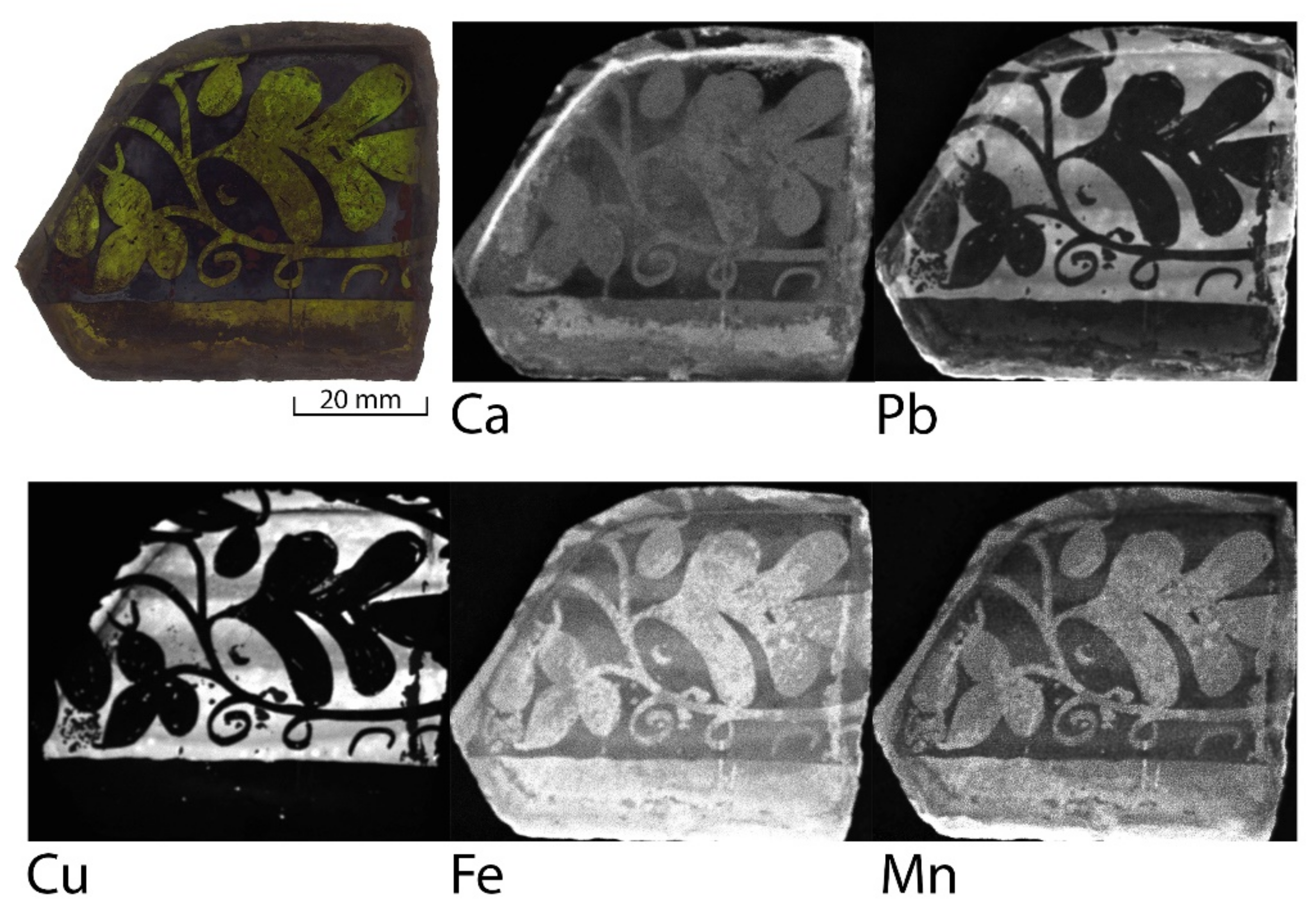
In the black grisaille paint that was visible on the glass pane from the oldest stained glass window that was analysed, mainly lead and copper were detected. Results also suggest the addition of iron and manganese (Figure 1). Moreover, this example shows the lack of homogeneity of the paint layer very well. Both in reflected and transmitted light, it seems that the paint layers, in which the ornament was etched, are opaque and evenly applied. However, the maps of the lead and copper distribution show areas of stronger and weaker signals from these elements, suggesting areas of higher and lower concentration of paint grains.
Grisaille paint layers in a bad state of preservation were a little more difficult and challenging for analysis. Stained glass panels from the Dominican Monastery presented the worst condition of paint layers among the objects examined. Glass corrosion and numerous losses of the grisailles negatively affected the legibility of the painted depictions, and significantly distorted the compositions. For these reason, apart from a preliminary analysis of the technology, attempts were made to make the deteriorated fragments more readable.
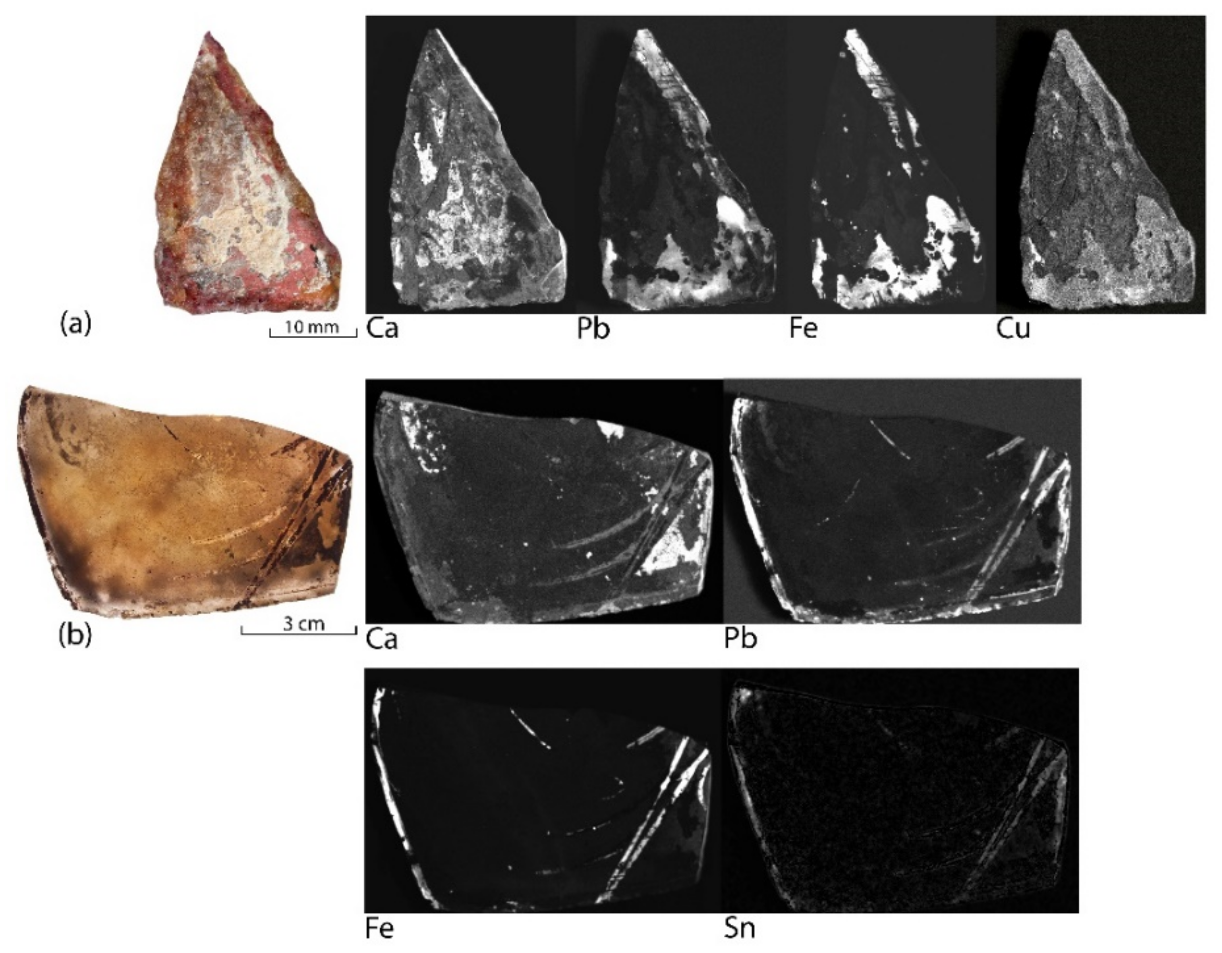
The most significant measurements were obtained for the basic components of the vitreous paint used, mainly iron and lead. For the K-M 87 glass pane, the signals of these elements clearly coincide with the thinly applied shading paint (Figure 2b). Unfortunately, in the case of very damaged fragments, it was not possible to detect even trace elements from the lost paint layers (Figure 2a and Figure 3a,b). However, as is visible on Figure 3a, in spite of the severe deterioration of grisaille and substrate glass, the heterogeneity of vitreous paints is still noticeable. Moreover, a high resolution of that measurement showed the presence of copper in the analysed paint layer.
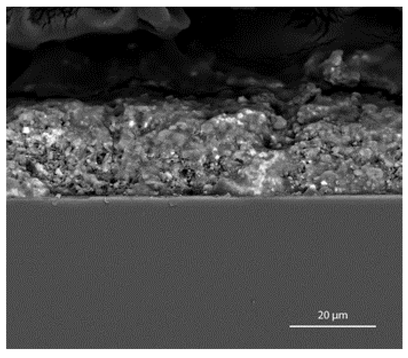
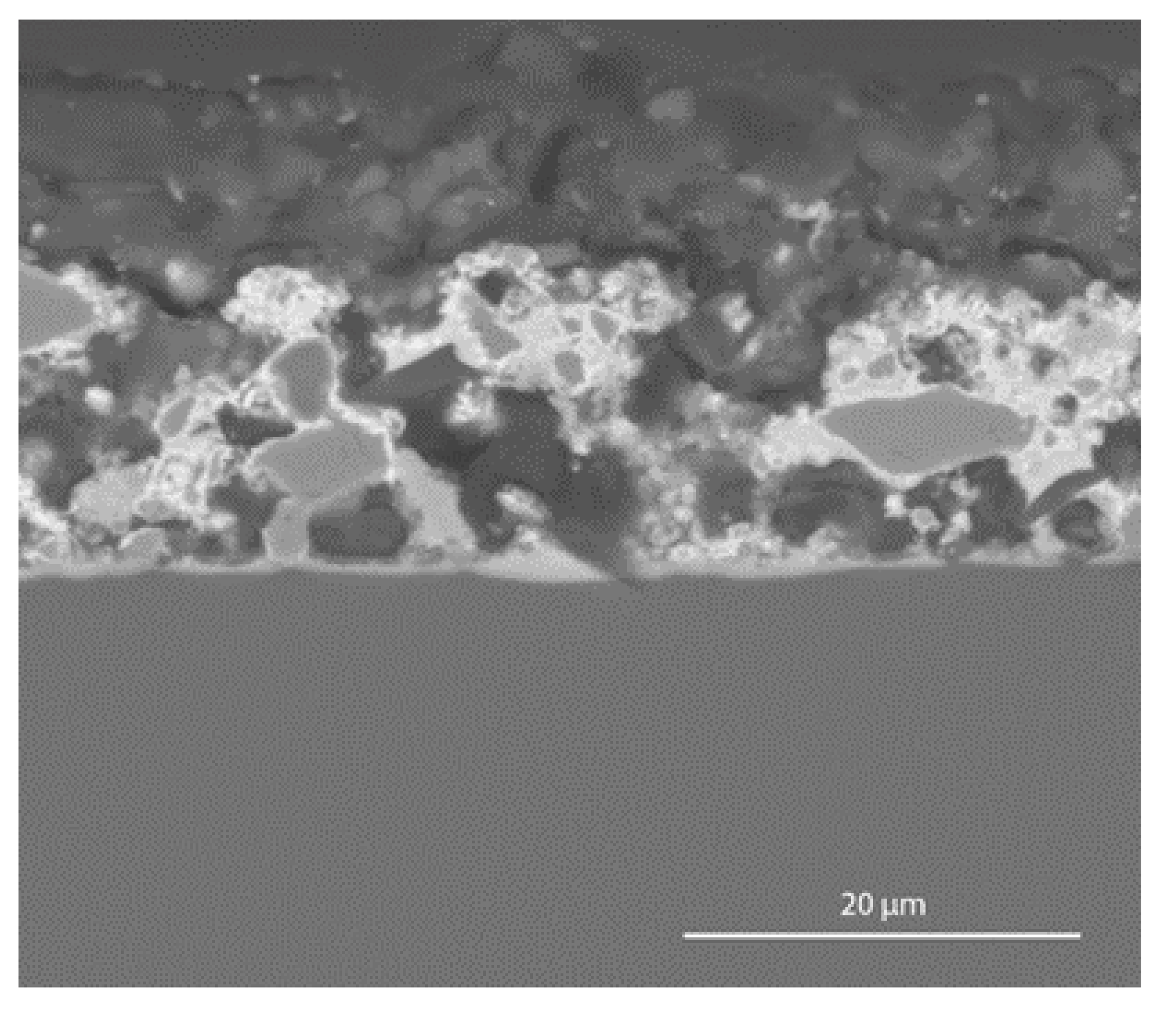
The chemical composition of grisaille obtained due to SEM-EDS analysis for the sample of glass pane Z 12 (Table 1) corresponds with MA-XRF results. Both techniques detected the most important elements for analyses of grisailles. Valuable information was also obtained from the SEM-BSE images of the cross-sections. Figure 4 shows an example of one of the images of grisailles from the Dominican Monastery in Krakow. The analysed paint layers were granular, with clearly visible grains of colouring agent distributed in flux. In the presented example, microcracks in the paint layer were noticed, but the substrate glass was not corroded. Nevertheless, in most samples, the glass under the paint layer was significantly deteriorated.
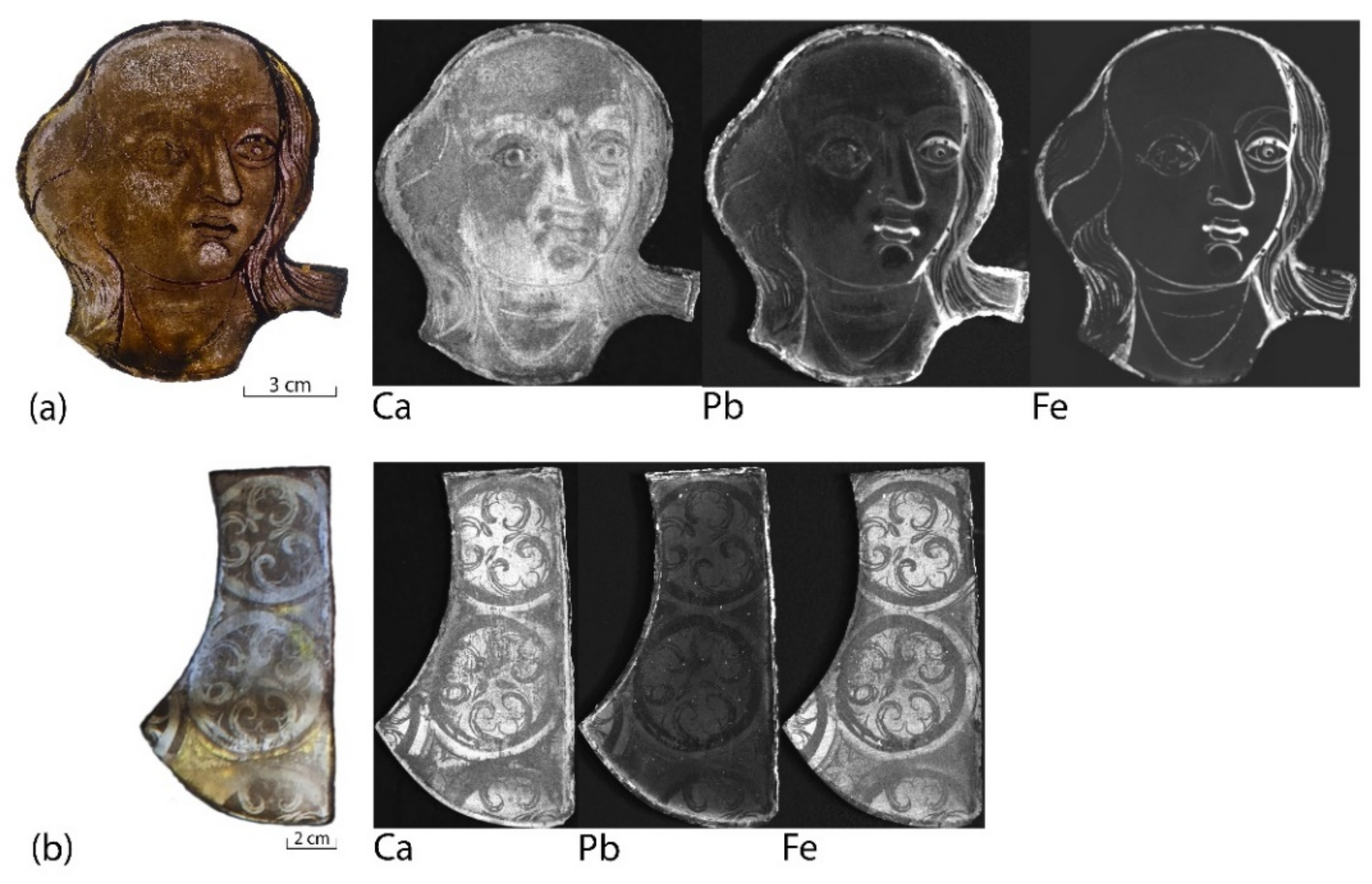
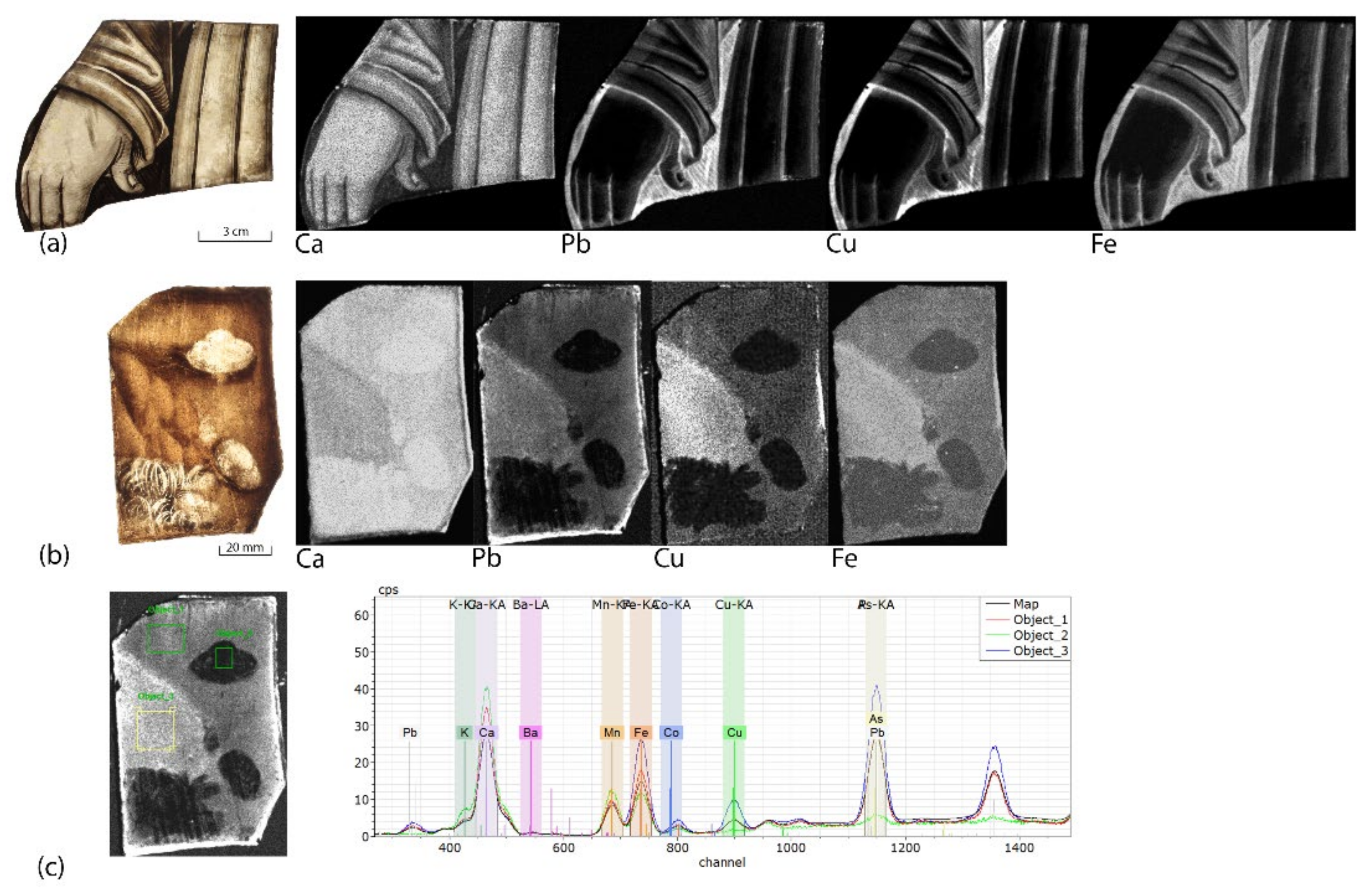
The analysis of well-preserved paint layers is shown in the example of two glass panes from about 1500 from the National Museum in Poznań visible in Figure 5, which present relatively easy to observe technological features. Lead, copper and iron were detected in all brown grisailles used to paint the composition, possibly in different proportions. The distribution of particular elements highlights individual brush strokes (Figure 5a) (e.g., a brush used to remove the shading paint used for modelling folds of robes or a thin brush used for hatching), significantly enriching the painting techniques observed in that panel.
Moreover, the presence of shading paints in several shades of brown is characteristic for that object. Figure 5b,c present an example where two shades of brown shading paint are painted on the outer surface of one of the glass panes. This measurement showed not only the apparent tonal difference of the paints used, but also the composition differences between the layers.
The possibility of collecting several glass samples for SEM-EDS analysis gave better insight into the technological aspects of the examined paint layers. Figure 6 shows the SEM-BSE image of a glass sample covered with contour paint. This image shows the morphological features of the layer, such as grain size and porosity. The conducted EDS analysis (Table 2) presents the chemical composition of the examined layer in more detail. Among the colourants, apart from iron and copper, small amounts of manganese and cobalt oxides were also detected.
3.2. Glass Panes after 1902
Fragments added at the beginning of the 20th century to the medieval stained glass panels from the Dominican Monastery in Krakow were painted in a variety of techniques: from glass panes painted with only one layer of contour paint, to panes with several layers of different colours of grisailles. Among the presented results, the glass panes painted with only contour paint were omitted, as they were not a major research problem. The authors decided to show examples of more complex glass panes painted with several layers of different vitreous paints in order to discuss the advantages and disadvantages of the XRF macro scanner on this basis.
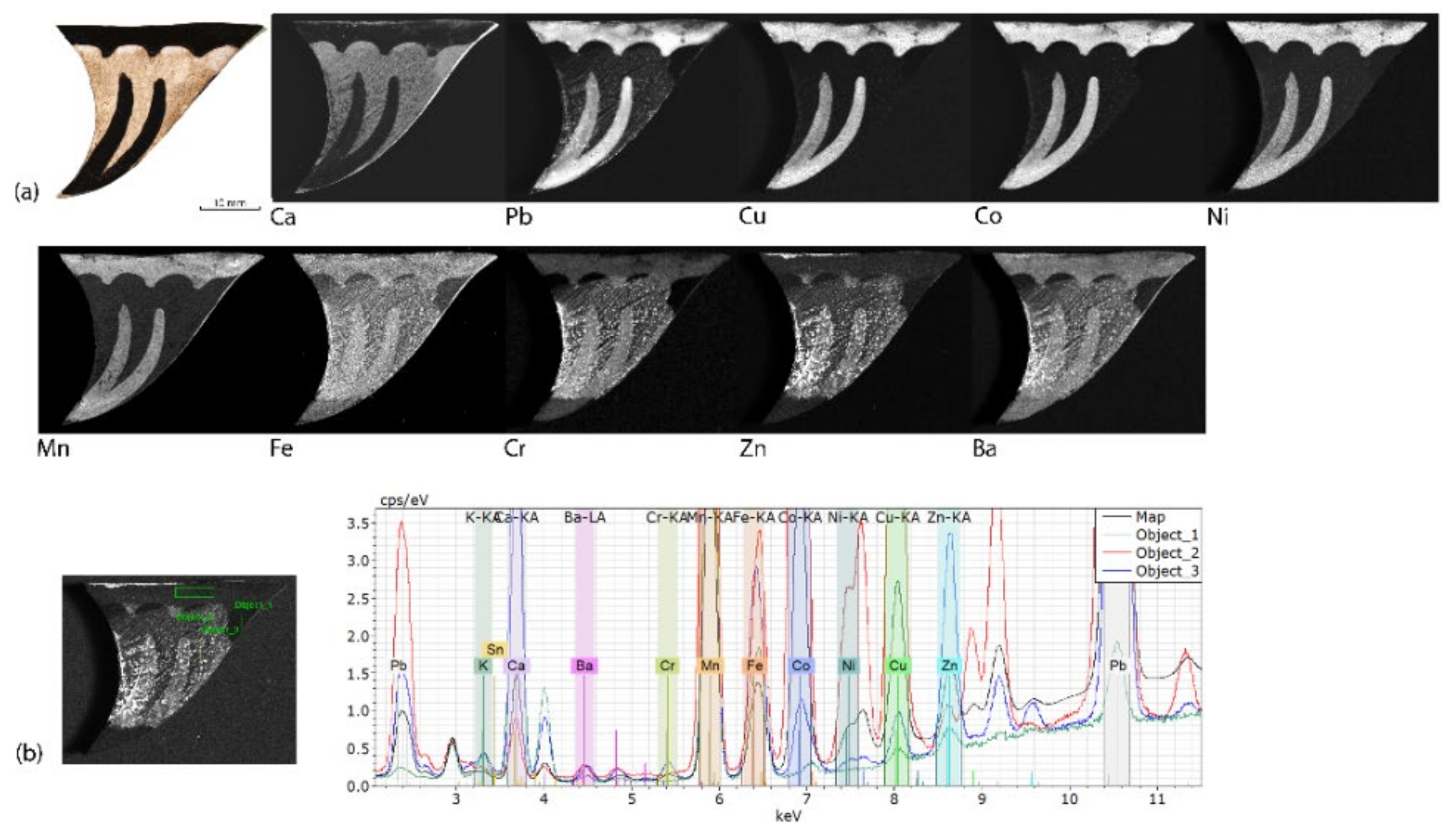
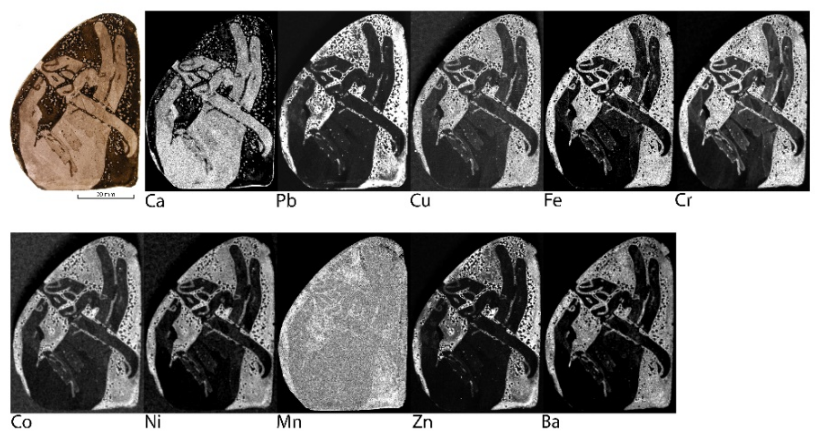
The obtained results showed the initial elemental composition of grisailles from the beginning of the 20th century. In the red vitreous paint (Figure 7) used to paint the feather contour, mainly lead, iron, barium and zinc were detected. However, in the black shading paint, cobalt and nickel were also noticed.
The complexity of the analysed layers was clearly visible in the results for the K 98 glass pane (Figure 8). Glass painted with only one layer of patina and one layer of contour paint clearly shows the differences in the elemental composition between the two paints used. Both, black grisaille and brown shading paint contain all the elements placed on the maps. However, in patina the signals from copper, cobalt, manganese and nickel are definitely weaker than in the contour paint.
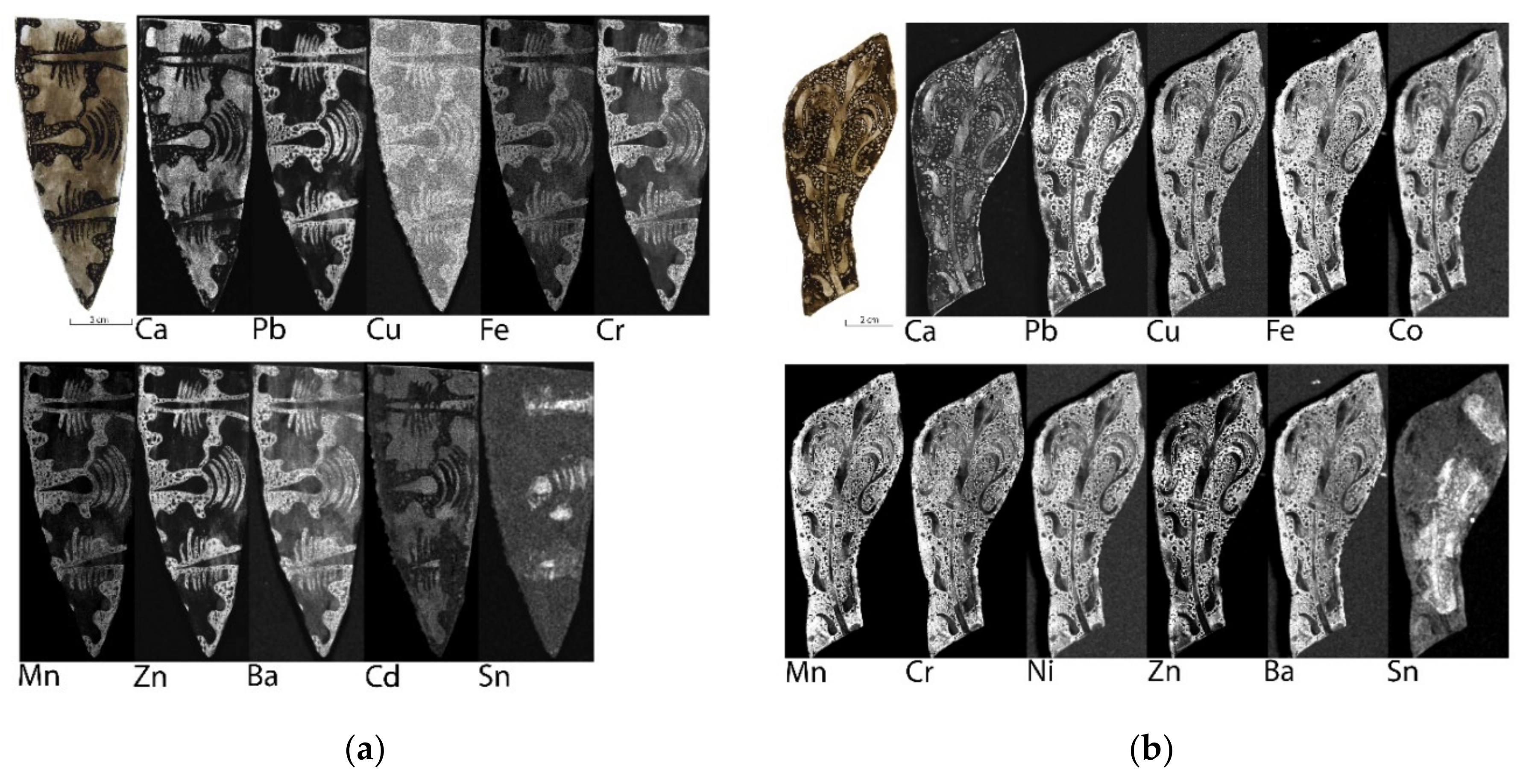
The results for the brown and black shades of vitreous paints were less obvious. As shown in Figure 9 and Figure 10, there are only slight nuances between the signal intensities of the elements between shading and contour paints, even if there is a difference in the colour of the paint used. In Figure 9, the tin signal is clearly visible, coming from a yellowish paint applied only locally.
Due to the mentioned complexity of the grisailles in black and brown shades, Table 3 presents the obtained EDS results of several samples. As it shows, there are quantitative differences between samples. Samples are named by the colours of grisailles and, thus, three samples of black contour paints, two of brown-black contour paints and one brown shading paint were analysed. Differences in qualitative analysis between EDS and MA-XRF were noticed. Observations are discussed in the next section.
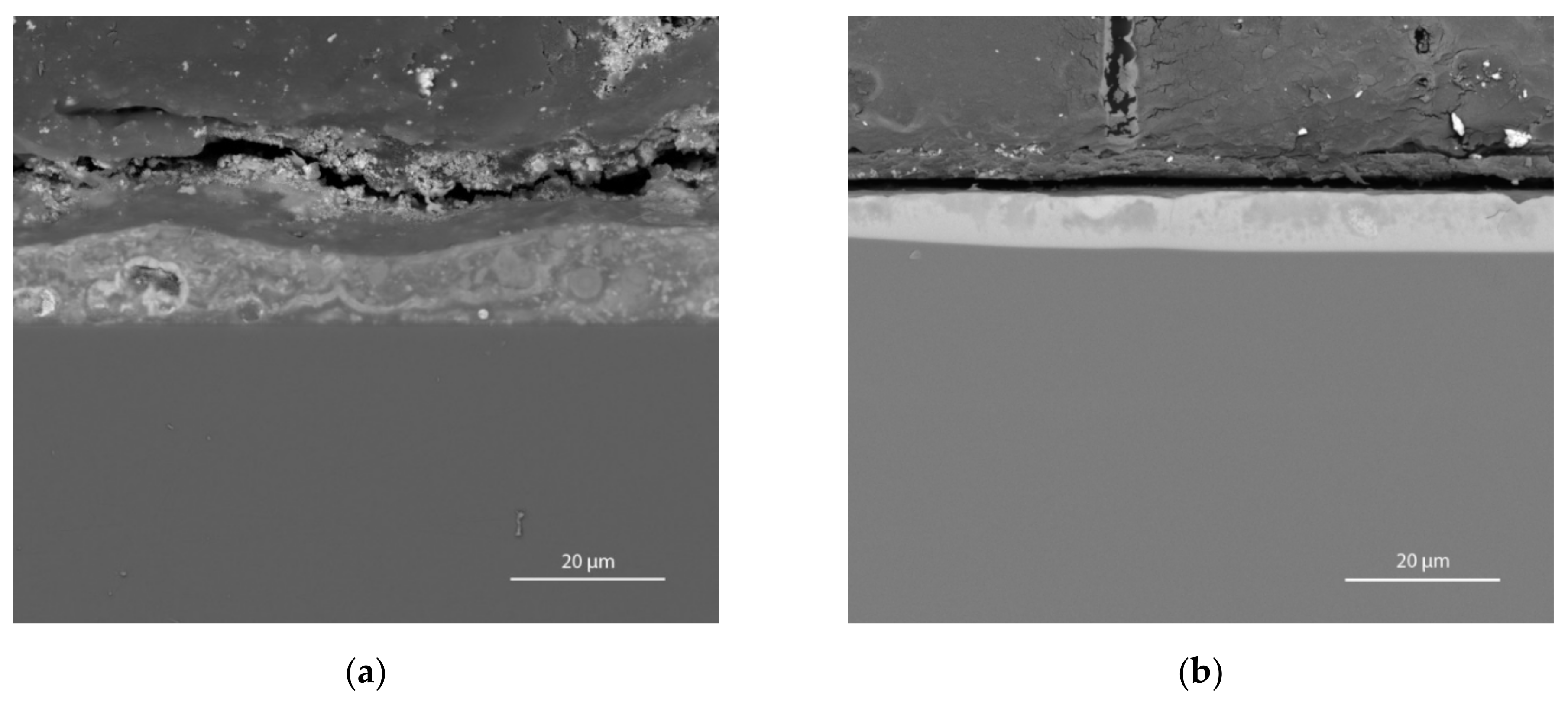
The differences in modern grisaille paint layers are also visible in the SEM-BSE images. The two paint layers shown in Figure 11 represent samples taken from places painted with a contour paint (Figure 11a) and shading paint (Figure 11b). These images show that within the glass panes painted by one stained glass workshop at the beginning of the 20th century, there are more homogeneous, well-vitrified layers (Figure 11b) and more granular, heterogeneous layers (Figure 11a).
4. Discussion
4.1. Glass Panes from 1385 to 1500
According to the obtained results, grisaille paint layers on medieval glass panes have a typical elemental composition [3,6]. Regardless of the origin and dating of the stained glass panel, lead as a component of flux was detected. Only the elements included in the pigments were differentiated. In the case of the oldest vitreous paint, this was copper by itself. In the case of the vitreous paint from the 15th century, this was mainly iron, with a small addition of copper and tin. Additionally, in the paint from the youngest stained glass, it was copper, with additions of iron, manganese and cobalt. According to local literature and preserved examples, the oldest preserved Polish stained glass windows were painted with black grisailles (e.g., the 13th-century panels depicting St. Augustine, Bishop St. Stanislaus, and the crucifixion from the Dominican Monastery in Kraków [25]), and red-brown grisailles were used only from the 15th century [26]. Nevertheless, comparisons of the elemental composition between medieval Polish stained glass is difficult due to the small number of studied objects. Among the most valuable and oldest preserved stained glass collections, only two groups have been researched so far. The first are stained glass windows from St. Mary’s Church in Krakow, and the second are the panels described in this article. Out of 23 panels from the Dominican Monastery in Kraków alone, only five were examined.
The main aim of the article is to discuss the possibilities of macro-XRF scanning in the analysis of paint layers. As this technique does not provide quantitative data, it cannot be used to estimate the flux to colourants ratio or compare the proportions between different stained glass windows. The obtained EDS results provided more comparative data. Among the stained glass windows of medieval workshops in Lesser Poland, only those from St. Mary’s Church in Kraków were studied. However, the fourteenth century stained glass windows are placed between the panels discussed in this article.
Stained glass windows from St. Mary’s Church were thoroughly studied in 1997. According to Karaszkiewicz (Table 4), black grisailles used in stained glass panels were made with lead glass mixed with colouring agents, mainly copper oxide [27]. Unfortunately, the author does not describe any morphological features of the analysed paint layers. However, it is worth noting that similar elements were detected in the analysis of the stained glass panel from the Diocesan Museum in Kielce, also dating back to the 14th century. The obtained MA-XRF results suggest copper as the main colouring agent, but the presence of manganese and iron was also recognised (Figure 1).
Additionally, it is worth mentioning the comparison of stained glass studies from neighbouring regions. Generally, stained glass in medieval Polish territories was influenced by German, Austrian and Bohemian techniques and stylistics [28]. As a result, the composition of the grisaille paint layers from stained glass windows considered to be made in Poland can be related to the available data obtained for panels of foreign origin. Examples of stained glass windows analysed with Macro-XRF scanning are panels of Austrian origin, known as the stained glass windows from the Grodziec Collection. These stained glass windows, which are ten to fifteen years younger than the studied panels from the Dominican Monastery in Kraków, show interesting research results. Contrary to the stained glass panels described in the article (painted with only one type of vitreous paints), the composition of at least two grisailles were distinguished in the objects from the Grodziec Collection, one copper-based and the other iron-based [29]. Taking into account only the available MA-XRF results, a more adequate comparison of grisailles cannot be made.
As the obtained elemental composition of medieval grisailles was expected, the maps of elemental distribution were also analysed in order to present the technological aspects of grisaille paint layers and to recognise the more deteriorated parts of the depictions. Thin layers of shading paint or slightly deteriorated areas were getting more visible in the MA-XRF maps, compared to the photography (Figure 4a). Unfortunately, in the case of very deteriorated glass panes covered with thick layers of corrosion crust, it was not possible to detect even traces of grisaille paint layers.
4.2. Glass Panes after 1902
The greatest potential of MA-XRF was seen in the analysis of twentieth-century paint layers. According to preliminary information provided in the first part of the article, from the 19th century, the composition of vitreous paints became more complex than in previous centuries. The examples of the obtained results presented above allowed for a preliminary differentiation of the elemental composition of grisailles used by one of the stained glass workshops in Kraków at the turn of the 19th and 20th centuries. On the basis of the obtained results, the elemental composition of grisaille paint layers was established.
Unfortunately, in the case of the Krakow workshop that made the analysed fragments of the panels as well as the Polish stained glass from the beginning of the 20th century in general, no results from the studies on the composition of grisailles are available. Thus, the results obtained represent the first insight into the vitreous paints from that time. Perhaps examining the remaining Dominican stained glass windows and a collective discussion of the results will show a better overview of this workshop.
There were only a few stained glass workshops in Krakow at the end of the 19th century. Nonetheless, Teodor Zajdzikowski’s workshop is considered the one that brought back techniques of painting on glass using grisailles [30]. This workshop was the subject of historical research [30,31]; however, it has not yet been subject to technological analyses. Therefore, it is difficult to find any comparative data relevant to the studied glass panes.
Although it is not possible to refer the obtained results to the literature, the analysis can be focused on comparing the obtained EDS to the MA-XRF results. The comparison made for grisailles in shades of brown and black shows differences in the detection of some elements. For example, the EDS measurement of the K 98 sample did not reveal the content of iron, zinc and barium oxides, the weak signals of which were observed in the MA-XRF elemental distribution maps (Figure 8, Table 3 s EDS measurement). Moreover, only in one sample analysed by means of SEM-EDS was the content of nickel oxide detected. That element was easily identified in most MA-XRF measurements. These examples showed the advantages of macro-XRF scanning in detecting even weak signals from colouring agents in grisailles, as well as the necessity of conducting research using complementary techniques.
5. Conclusions
In the case of medieval paint layers, the macro-XRF scanning can be considered a preliminary analysis of the grisaille paint layers. The obtained data shows the qualitative composition and distribution of individual elements. However, in the case of the deteriorated fragments of the compositions, the chosen technique did not give satisfactory results. Moreover, only SEM images and EDS analysis provide an insight into the morphology of grisailles, as well as the proportions of its components. Only this information makes it possible to perform a reliable comparative analysis with other panels made in different times or workshops.
As shown in the article’s results, marco-XRF scanning turned out to be a valuable instrumental technique, which allowed for the recognition of the characteristics of paint layers made with more modern vitreous paints. However, this study can still only be considered a preliminary investigation, and more research is needed to fully understand complex paint layers and to better distinguish the technological aspects between the different colours of grisailles (considering, especially, the slight differences between similar shades of vitreous paints). Despite the lack of a quantitative analysis, acquiring qualitative information along with determining the distribution of individual elements is undoubtedly a valuable achievement. Nevertheless, only when complemented with SEM images and EDS analyses, can macro-XRF scanning provide reliable and comparative data for further research.
Author Contributions
Conceptualization, E.B.; methodology, E.B and M.W.; performing analyses, M.G. and M.W.; writing—original draft preparation, E.B.; writing—review and editing, M.W. and M.G.; visualization, E.B.; funding acquisition, E.B. and M.W. All authors have read and agreed to the published version of the manuscript.
Funding
This research was partially funded by the Polish Ministry of Science and Higher Education, Diamond Grant (project no. 0164/DIA/20015/44).
Acknowledgments
The authors wish to thank the Dominican Monastery in Krakow, the Diocesan Museum in Kielce and the National Museum in Poznań for providing access to stained glass panels and all necessary materials for this study.
Conflicts of Interest
The authors declare no conflict of interest. The funders had no role in the design of the study; in the collection, analyses, or interpretation of data; in the writing of the manuscript; or in the decision to publish the results.
References
- Machado, C.; Vilarigues, M.; Palomar, T. Historical grisailles characterisation: A literature review. J. Cult. Herit. 2021, 49, 239–249. [Google Scholar] [CrossRef]
- Machado, C.; Machado, A.; Palomar, T.; Vilarigues, M. Grisaille in Historical Written Sources. J. Glass Stud. 2019, 61, 71–86. [Google Scholar]
- Davison, S. Conservation and Restoration of Glass, 2nd ed.; Butterworth Heinemann: Oxford, UK, 2003; pp. 131–133. [Google Scholar]
- Schlam, O.; Jansens, K.; Caen, J. Characterization of the main causes of deterioration of grisaille paint layers in 19th century stained-glass windows by J.-B. Carponnier. Spectrochim. Acta B 2003, 58, 589–607. [Google Scholar] [CrossRef]
- Pradell, T.; Molina, G.; Murcia, S.; Ibáñez, R.; Liu, C.; Molera, J.; Shortland, A. Materials, techniques and conservation of historic stained glass grisailles. Int. J. Appl. Glass Sci. 2016, 7, 41–58. [Google Scholar] [CrossRef] [Green Version]
- Reboulleau, M.E.F. Nouveau Manuel Complet de la Peinture sur Verre, sur Porcelaine et sur Émail par m. M. E. F. Reboulleau; Librairie Encyclopédique de Roret: Paris, France, 1844. [Google Scholar]
- Caen, J. The Production of Stained Glass in the Country of Flanders and the Duchy of Brabant from the XVth to the XVIIIth Centuries: Materials and Techniques; Brepols: Antwerpen, Belgium, 2009. [Google Scholar]
- Schalm, O. Characterization of Paint Layers in Stained-Glass Windows: Main Causes of the Degradation of Nineteenth Century Grisaille Paint Layers; Universiteit Antwerpen: Antwerp, Belgium, 2000. [Google Scholar]
- Dungworth, D.; Bower, H.; Gilchrist, A.; Wilkes, R. The West Window, Beverley Minster, Beverley, East Yorkshire: Chemical Analysis of the Window Glass and Paint. In Research Department Report Series; English Heritage: Portsmouth, UK, 2010. [Google Scholar]
- Gilchrist, A. The Tears Wept by Our Windows’: Severe Paint Loss from Stained Glass Windows of the Mid-Nineteenth Century. Master’s Thesis, Department of History of Art, University of York, York, UK, 2010. Available online: http://www.cvma.ac.uk (accessed on 29 May 2021).
- Alberta, S.; Gianmario, M.; Valentina, P. The stained glass window of the southern transept of St. Anthony’s Basilica (Padova, Italy): Study of glasses and grisaille paint layers. Spectrochim. Acta Part B 2011, 66, 81–87. [Google Scholar] [CrossRef]
- Machado, A.; Wolf, S.; Alves, L.C.; Katona-Serneels, I.; Serneels, V.; Trümpler, S.; Vilarigues, M. Swiss Stained-Glass panels: An Analytical Study. Microsc. Microanal. 2017, 23, 878–890. [Google Scholar] [CrossRef] [PubMed] [Green Version]
- Veritá, M. Composition, structure et mécanisme de détérioration des grisailles. In Grisaille, Jaune d’argent, Sanguine, Email et Peinture à Froid, Dossier de la Commission Royale des Monuments, Sites et Fouilles; Barlet, J., Ed.; Commission Royale des Monuments, Sites et Fouilles de la Région Wallonne: Liège, Belgium, 1996; pp. 61–68. [Google Scholar]
- Janssens, K. (Ed.) Modern Methods for Analysing Archaeological and Historical Glass; John Wiley & Sons, Ltd.: Chichester, UK, 2013. [Google Scholar]
- Van der Snickt, G.; Legrand, S.; Caen, J.; Vanmeert, F.; Alfeld, M.; Janssens, K. Chemical imaging of stained-glass windows by means of macro X-ray fluorescence (MA-XRF) scanning. Microchem. J. 2016, 124, 615–622. [Google Scholar] [CrossRef]
- Bernady, E.; Kamińska, M.; Płotek, M.; Walczak, M. The investigation of 15th century paint layers on two stained glass windows from the Dominican Monastery in Krakow, Poland. Eur. J. Glass Sci. Technol. A 2018, 59, 46–53. [Google Scholar] [CrossRef]
- Legrand, S.; Van der Snickt, G.; Cagno, S.; Caen, J.; Janssens, K. MA-XRF imaging as a tool to characterize the 16th century heraldic stained-glass panels in Ghent Saint Bavo Cathedral. J. Cult. Herit. 2019, 40, 163–168. [Google Scholar] [CrossRef]
- Archives of the Diocesan Curia in Kielce.
- Horzela, D. (Ed.) Cud światła. Witraże średniowieczne w Polsce [Miracle of Light. Medieval Stained Glass in Poland]; Muzeum Narodowe w Krakowie: Kraków, Poland, 2020. [Google Scholar]
- Horzela, D. Piętnastowieczny witrażowy cykl maryjny i jego miejsce w wystroju kościoła Dominikanów w Krakowie [A fifteenth-century Marian stained glass cycle and its place in the decoration of the Dominican church in Cracow]. In Claritas et Consonantia. Funkcje, Formy i Znaczenia w Sztuce Średniowiecza; Jakubek-Raczkowska, M., Raczkowski, I.J., Eds.; Wydział Sztuk Pięknych Uniwersytetu Mikołaja Kopernika w Toruniu: Toruń, Poland, 2017; pp. 149–174. [Google Scholar]
- Pieńkowska, H. Konserwacja witraży dominikańskich w Krakowie [Conservation of stained glass from Dominican Monastery in Kraków]. Ochrona Zabytków 1949, 3, 182–189. [Google Scholar]
- Czapczyńska-Kleszczyńska, D.; Szybisty, T. Korpus Witraży z lat 1800–1945 w Kościołach Rzymskokatolickich Metropolii Krakowskiej i Przemyskiej; v. 1, Corpus Vitrearum Polska; Imedius Agencja Reklamowa: Kraków, Poland, 2014. [Google Scholar]
- Kamińska, M.; Bernady, E.; Płotek, M.; Kaszowska, Z.; Walczak, M. ‘The Throne of Grace’—The history and conservation strategy fora medieval stained-glass panel from the Dominican Monastery in Kraków, Poland. In Recent Advances in Glass and Ceramics Conservation 2016; Roemich, H., Fair, L., Eds.; International Council of Museums—Committee for Conservation (ICOM-CC): Paris, France, 2016; pp. 43–51. [Google Scholar]
- Pajzderski, N. Śląskie witraże średniowieczne w zbiorach poznańskich [Medieval Silesian stained glass in collections form Poznań]. Dawna Sztuka 1938, 1, 241–246. [Google Scholar]
- Kalinowski, L. Die ältesten Glasgemälde der Dominikanerkirche in Krakau. In Ars Vitrea. Collected Writings on Mediaeval Stained Glass; Kalinowski, L., Małkiewiczówna, I.H., Eds.; PAU: Kraków, Poland, 2016; pp. 127–138. [Google Scholar]
- Małkiewiczówna, H. Stan badań nad średniowiecznym malarstwem witrażowym w Małopolsce [The state of research on medieval stained glass in Lesser Poland], In Dziedzictwo Polskiej Sztuki Witrażowej; Pawłowska, K., Budyn-Kamykowska, I.J., Eds.; Stowarzyszenie Miłośników Witraży: Kraków, Poland, 2000; pp. 9–20. [Google Scholar]
- Karaszkiewicz, P. Badania średniowiecznych witraży kościoła Mariackiego w Krakowie [Investigation of the technology, technique and state of preservation of the stained-glass panels of St Mary’s Church in Cracow]. In Średniowieczne witraże kościoła Mariackiego w Krakowie. Studia i Materiały Wydziału Konserwacji i Restauracji Dzieł Sztuki Akademii Sztuk Pięknych w Krakowie, 7; Kalinowski, L., Małkiewiczówna, H., Heine, L., Karaszkiewicz, I.P., Eds.; Wydawnictwo Akademii Sztuk Pięknych im. Jana Matejki w Krakowie: Kraków, Poland, 1997; pp. 101–137. [Google Scholar]
- Kalinowski, L. Mediaval stained glass in Poland. In Ars Vitrea. Collected Writings on Mediaeval Stained Glass; Kalinowski, L., Małkiewiczówna, I.H., Eds.; PAU: Kraków, Poland, 2016; pp. 69–125. [Google Scholar]
- Walczak, M.; Kamińska, M.; Sobczyk, J.; Płotek, M.; Horzela, D.; Sylwestrzak, M.; Targowski, P. The application of non-invasive analytical techniques in the investigation and documentation of medieval stained-glass windows from the Grodziec collection. In Recent Advances in Glass and Ceramics Conservation 2016; Roemich, H., Fair, L., Eds.; International Council of Museums—Committee for Conservation (ICOM-CC): Paris, France, 2016; pp. 21–30. [Google Scholar]
- Czapczyńska-Kleszczyńska, D. Witraże w Krakowie. Dzieła i twórcy. [Stained glass in Cracow. Works and creators]. In Krakowska Teka Konserwatorska, 5; Urząd Miasta Krakowa, Wydział Kultury i Dziedzictwa Narodowego, Oddział Ochrony Zabytków: Kraków, Poland, 2005. [Google Scholar]
- Czapczyńska-Kleszczyńska, D. Teodor Andrzej Zajdzikowski (1840–1907) Pionier Krakowskich Witrażowników [Teodor Andrzej Zajdzikowski (1840–1907) Trailblazer of the Cracow stained glass makers]. Rocznik Krakowski 2003, 69, 151–170. [Google Scholar]
Figure 1.
Glass pane S 37 (Diocesan Museum in Kielce), MA-XRF images showing distribution of calcium, lead, copper, iron and manganese.
Figure 1.
Glass pane S 37 (Diocesan Museum in Kielce), MA-XRF images showing distribution of calcium, lead, copper, iron and manganese.

Figure 2.
(a) Glass pane K-M 85 (Dominican Monastery in Kraków), MA-XRF images showing distribution of calcium, lead and iron; (b) Glass pane K-M 87 (Dominican Monastery in Kraków), MA-XRF images showing distribution of calcium, lead and iron.
Figure 2.
(a) Glass pane K-M 85 (Dominican Monastery in Kraków), MA-XRF images showing distribution of calcium, lead and iron; (b) Glass pane K-M 87 (Dominican Monastery in Kraków), MA-XRF images showing distribution of calcium, lead and iron.

Figure 3.
(a) Glass pane Z 5a (Dominican Monastery in Kraków), MA-XRF images showing distribution of calcium, lead, iron and copper; (b) Glass pane Z 12 (Dominican Monastery in Kraków), MA-XRF images showing distribution of calcium, lead, iron and tin.
Figure 3.
(a) Glass pane Z 5a (Dominican Monastery in Kraków), MA-XRF images showing distribution of calcium, lead, iron and copper; (b) Glass pane Z 12 (Dominican Monastery in Kraków), MA-XRF images showing distribution of calcium, lead, iron and tin.

Figure 4.
SEM-BSE image of grisaille cross-section (glass pane Z 12) fromthe Dominican Monastery in Kraków.
Figure 4.
SEM-BSE image of grisaille cross-section (glass pane Z 12) fromthe Dominican Monastery in Kraków.

Figure 5.
(a) Glass pane MNP 25 (National Museum in Poznań), MA-XRF images showing distribution of calcium, lead, copper and iron; (b) Glass pane MNP 12 (National Museum in Poznań), MA-XRF images showing distribution of calcium, lead, copper and iron; (c) obtained fluorescence spectra for glass pane MNP 12.

Figure 6.
SEM-BSE image of grisaille cross-section from the National Museum in Poznań.

Figure 7.
Glass pane Z-A 2 (Dominican Monastery in Kraków), MA-XRF images showing distribution of calcium, lead, copper, manganese, iron, barium, cobalt, nickel, and zinc.
Figure 7.
Glass pane Z-A 2 (Dominican Monastery in Kraków), MA-XRF images showing distribution of calcium, lead, copper, manganese, iron, barium, cobalt, nickel, and zinc.

Figure 8.
Glass pane K 98 (Dominican Monastery in Kraków), (a) MA-XRF images showing distribution of calcium, lead, copper, cobalt, nickel, manganese, iron, chromium, zinc, and barium; (b) obtained fluorescence spectra.
Figure 8.
Glass pane K 98 (Dominican Monastery in Kraków), (a) MA-XRF images showing distribution of calcium, lead, copper, cobalt, nickel, manganese, iron, chromium, zinc, and barium; (b) obtained fluorescence spectra.

Figure 9.
(a) Glass pane Z-A 18 (Dominican Monastery in Kraków), MA-XRF images showing distribution of calcium, lead, copper, iron, chromium, manganese, zinc, barium, cadmium, and tin; (b) Glass pane Z-A 29 (Dominican Monastery in Kraków), MA-XRF images showing distribution of calcium, lead, copper, iron, cobalt, manganese, chromium, nickel, zinc, barium, and tin.

Figure 10.
Glass pane Z-A 27 (Dominican Monastery in Kraków), MA-XRF images showing distribution of calcium, lead, copper, iron, chromium, cobalt, nickel, manganese, zinc, and barium.
Figure 10.
Glass pane Z-A 27 (Dominican Monastery in Kraków), MA-XRF images showing distribution of calcium, lead, copper, iron, chromium, cobalt, nickel, manganese, zinc, and barium.

Figure 11.
(a) SEM-BSE image of granular grisaille cross-section from the Dominican Monastery in Kraków; (b) SEM-BSE image of well-vitrified grisaille cross-section from the Dominican Monastery in Kraków.
Figure 11.
(a) SEM-BSE image of granular grisaille cross-section from the Dominican Monastery in Kraków; (b) SEM-BSE image of well-vitrified grisaille cross-section from the Dominican Monastery in Kraków.


Table 1.
Chemical composition of grisaille on glass pane Z 12 from the Dominican Monastery in Kraków, as analysed by EDS.
Table 1.
Chemical composition of grisaille on glass pane Z 12 from the Dominican Monastery in Kraków, as analysed by EDS.
| Oxides | wt % |
|---|---|
| MgO | 0.26 |
| Al2O3 | 1.03 |
| SiO2 | 26.75 |
| P2O5 | 0.34 |
| Cl™ | 0.40 |
| K2O | 1.21 |
| CaO | 0.99 |
| TiO2 | 0.49 |
| Fe2O3 | 43.74 |
| ZnO | 4.81 |
| SnO2 | 4,85 |
| PbO | 15.15 |
Table 2.
Chemical composition of grisaille paint from the National Museum in Poznań, as analysed by EDS.
Table 2.
Chemical composition of grisaille paint from the National Museum in Poznań, as analysed by EDS.
| Oxides | wt % |
|---|---|
| Na2O | 0.24 |
| MgO | 0.54 |
| Al2O3 | 0.96 |
| SiO2 | 19.02 |
| P2O5 | 0.6 |
| K2O | 0.47 |
| CaO | 1.43 |
| MnO | 0.59 |
| Fe2O3 | 28.52 |
| CoO | 0.78 |
| CuO | 18.82 |
| PbO | 28.04 |
Table 3.
Chemical composition of modern grisailles (black and brown shades) from the Dominican Monastery in Kraków, as analysed by EDS.
Table 3.
Chemical composition of modern grisailles (black and brown shades) from the Dominican Monastery in Kraków, as analysed by EDS.
| Oxides | Black Contour Paint | Black Contour Paint | Black Contour Paint | Brown-Black Contour Paint | Brown-Black Contour Paint | Brown Shading Paint |
|---|---|---|---|---|---|---|
| Na2O | 4.28 | 7.98 | 4.01 | 3.11 | 4.02 | 2.53 |
| MgO | nd | nd | nd | 0.30 | 0.11 | nd |
| Al2O3 | 0.41 | 1.58 | nd | 3.19 | 0.45 | 0.80 |
| SiO2 | 24.55 | 31.30 | 54.10 | 22.11 | 26.89 | 34.76 |
| SO3 | 8.20 | nd | nd | nd | nd | nd |
| Cl− | nd | 0.19 | nd | nd | 0.31 | nd |
| K2O | 0.26 | 0.16 | 4.35 | 0.24 | 0.74 | 0.50 |
| CaO | 0.20 | 1.34 | 4.15 | 1.02 | 1.89 | nd |
| Cr2O3 | nd | nd | 0.25 | 0.44 | 0.82 | nd |
| MnO | 14.60 | 6.29 | 0.30 | 0.84 | 1.61 | 20.34 |
| Fe2O3 | nd | nd | 0.96 | 1.52 | 2.37 | nd |
| CoO | 13.13 | 5.36 | 0.47 | 0.70 | 1.44 | 11.72 |
| NiO | nd | 0.38 | nd | nd | nd | nd |
| CuO | 4.64 | 2.85 | nd | 1.20 | 0.86 | 4.71 |
| ZnO | nd | nd | 3.04 | 5.77 | 5.36 | nd |
| BaO | nd | nd | 0.41 | 1.74 | 1.92 | nd |
| PbO | 29.73 | 42.56 | 27.96 | 56.63 | 48.25 | 24.64 |
| UO2 | nd | nd | nd | 1.18 | 2.96 | nd |
Table 4.
Chemical composition of grisaille paint from St. Mary’s Church in Kraków, as analysed by EDS [27].
Table 4.
Chemical composition of grisaille paint from St. Mary’s Church in Kraków, as analysed by EDS [27].
| Oxides | 1 | 3 | 3’ | 18 |
|---|---|---|---|---|
| Na2O | nd | 1.2 | 0.6 | nd |
| Al2O3 | nd | nd | nd | 0.2 |
| SiO2 | 12.6 | 10.9 | 17.3 | 15.3 |
| SO3 | nd | nd | nd | 7.4 |
| P2O5 | nd | nd | nd | 5.1 |
| Cl− | nd | nd | nd | 1.6 |
| K2O | 1.2 | 3 | 4.1 | 1.4 |
| CaO | 3.9 | 4.9 | 3.1 | 16.9 |
| MnO | nd | 0.3 | 0.3 | nd |
| FeO | 2.9 | 0.5 | 0.3 | 0.5 |
| CuO | 62.5 | 44.1 | 33.5 | 5.1 |
| PbO | 16.9 | 35.1 | 40.6 | 46.5 |
Publisher’s Note: MDPI stays neutral with regard to jurisdictional claims in published maps and institutional affiliations. |
© 2021 by the authors. Licensee MDPI, Basel, Switzerland. This article is an open access article distributed under the terms and conditions of the Creative Commons Attribution (CC BY) license (https://creativecommons.org/licenses/by/4.0/).
Share and Cite
MDPI and ACS Style
Bernady, E.; Goryl, M.; Walczak, M. XRF Imaging (MA-XRF) as a Valuable Method in the Analysis of Nonhomogeneous Structures of Grisaille Paint Layers. Heritage 2021, 4, 3193-3207. https://0-doi-org.brum.beds.ac.uk/10.3390/heritage4040179
AMA Style
Bernady E, Goryl M, Walczak M. XRF Imaging (MA-XRF) as a Valuable Method in the Analysis of Nonhomogeneous Structures of Grisaille Paint Layers. Heritage. 2021; 4(4):3193-3207. https://0-doi-org.brum.beds.ac.uk/10.3390/heritage4040179
Chicago/Turabian StyleBernady, Edyta, Maria Goryl, and Małgorzata Walczak. 2021. "XRF Imaging (MA-XRF) as a Valuable Method in the Analysis of Nonhomogeneous Structures of Grisaille Paint Layers" Heritage 4, no. 4: 3193-3207. https://0-doi-org.brum.beds.ac.uk/10.3390/heritage4040179