
State-of-the-Art Review of Aliphatic Polyesters and Polyolefins Biodeterioration by Microorganisms: From Mechanism to Characterization

Abstract
:1. Introduction
- The following five key phrases were searched on Google Scholar and Scopus: “polymer biodegradation”, “polymer biodeterioration”, “polymeric blend biodegradation”, “degradation of polyolefin by microorganisms”, and “degradation of aliphatic polyesters by microorganisms”.
- The pre-screening process was carried out in order to ascertain the pertinence of the search results. A comprehensive analysis of the articles published after the year 1990 resulted in a total of 229 articles that were deemed principally relevant within the scope of this review article.
- The selected publications were categorized into five distinct groups: “review articles and book chapters”, “original research”, “polyolefins”, “aliphatic polyesters”, and “polymeric blends and composites”.
- The original research publications pertaining to each distinct polymeric family were categorized based on the individual polymer within each family (e.g., polycaprolactone (PCL), polylactic acid (PLA), etc., for polyesters) and the degrading agent involved (e.g., bacteria, fungi).
- The selected original articles underwent a secondary screening process to determine the primary experimental methodology employed for characterization and the key outcomes. This process resulted in the selection of 187 publications for inclusion in this manuscript.
- The review articles, book chapters, and original research articles that were not specifically related to the biodeterioration of selected polymers for Section 6 but that presented results that highlighted specific outcomes for the biodeterioration of polymers via microorganisms, such as the effect of influencing parameters or experimental methods used in a creative or critical manner, were used in Section 1, Section 2, Section 3 and Section 4 to provide the reader with a clear background.
- Original articles pertaining to the targeted polymers’ biodeterioration were utilized in Section 6.
- A parallel study was carried out using “ASTM International” and the “International Organization for Standardization” to address the standardized procedures for the characterization of the biodegradation of polymers. Due to the shared content of these standards, which is explicitly acknowledged within each standard, only nine of the ASTM International publications were included in this work (Section 5).
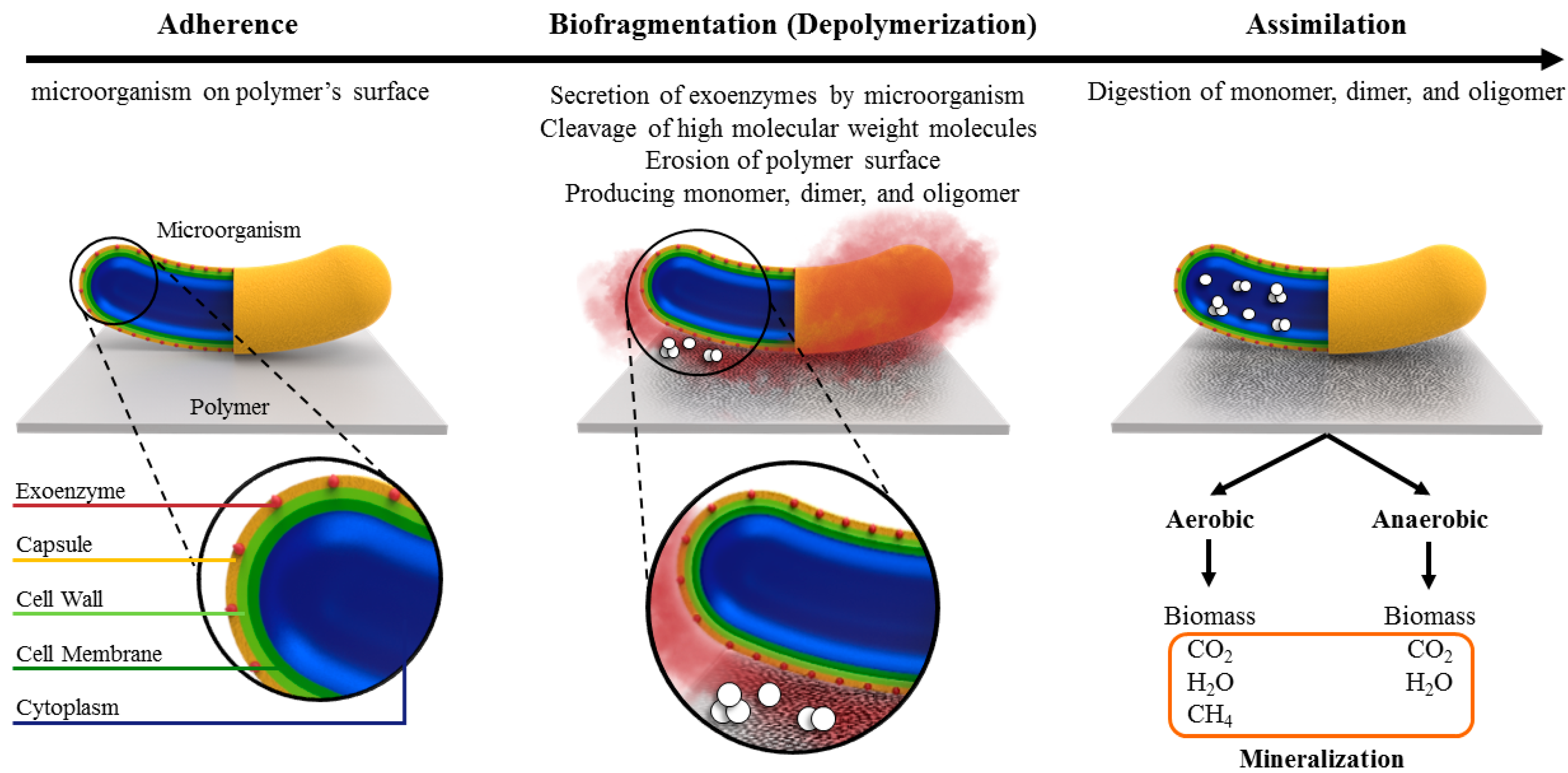
2. Biodeterioration Mechanism
3. Factors Affecting Biodegradation
3.1. Environmental Conditions
3.1.1. Humidity
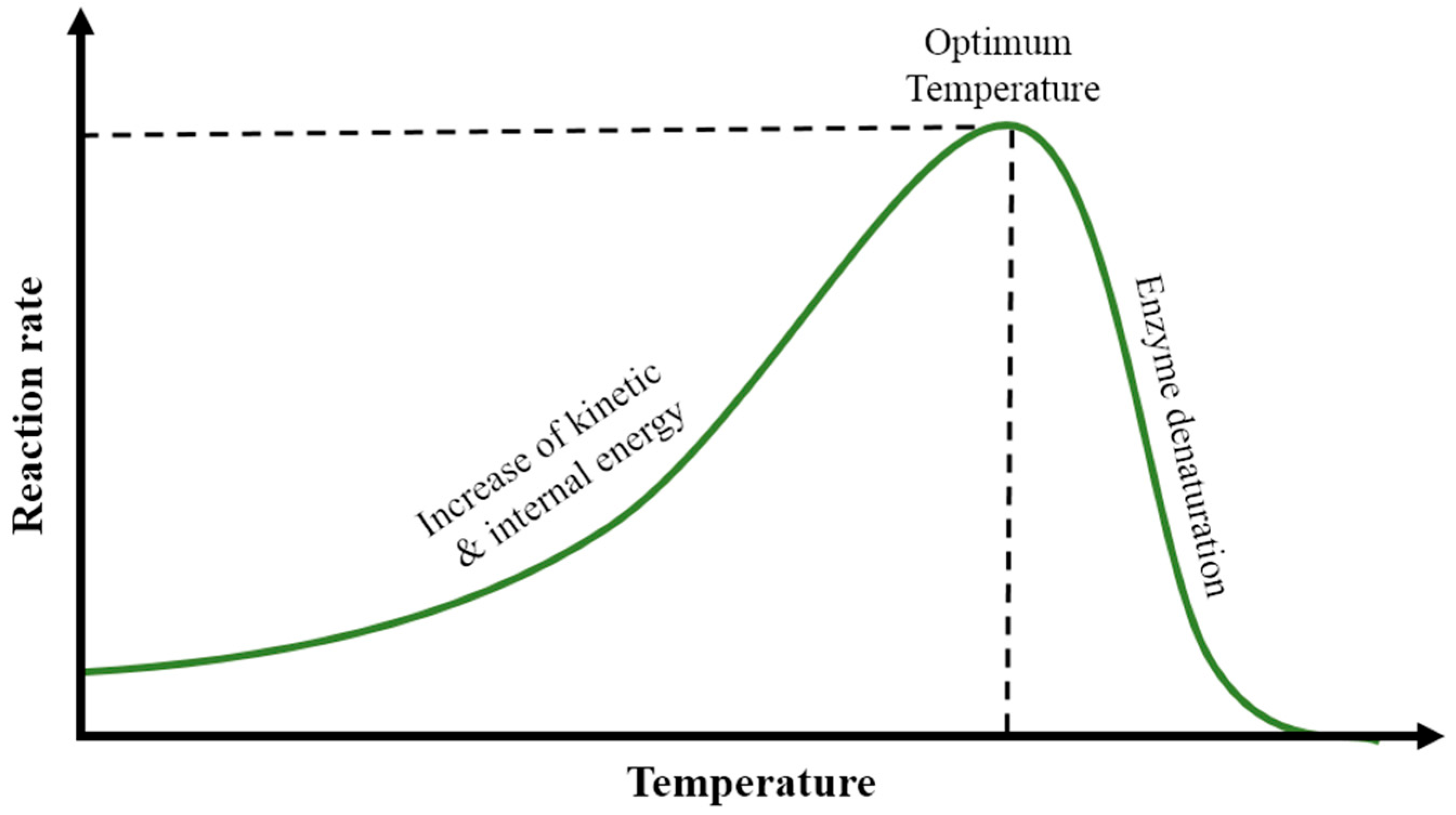
3.1.2. Temperature
3.1.3. pH
3.2. Polymer Properties
3.2.1. Polymers’ Molecular Structures
3.2.2. Crystallinity ()
3.2.3. Molecular Weight ()
3.2.4. Physical Form
4. Experimental Techniques for Characterization (Analysis of Degradation)
4.1. Morphological Analysis
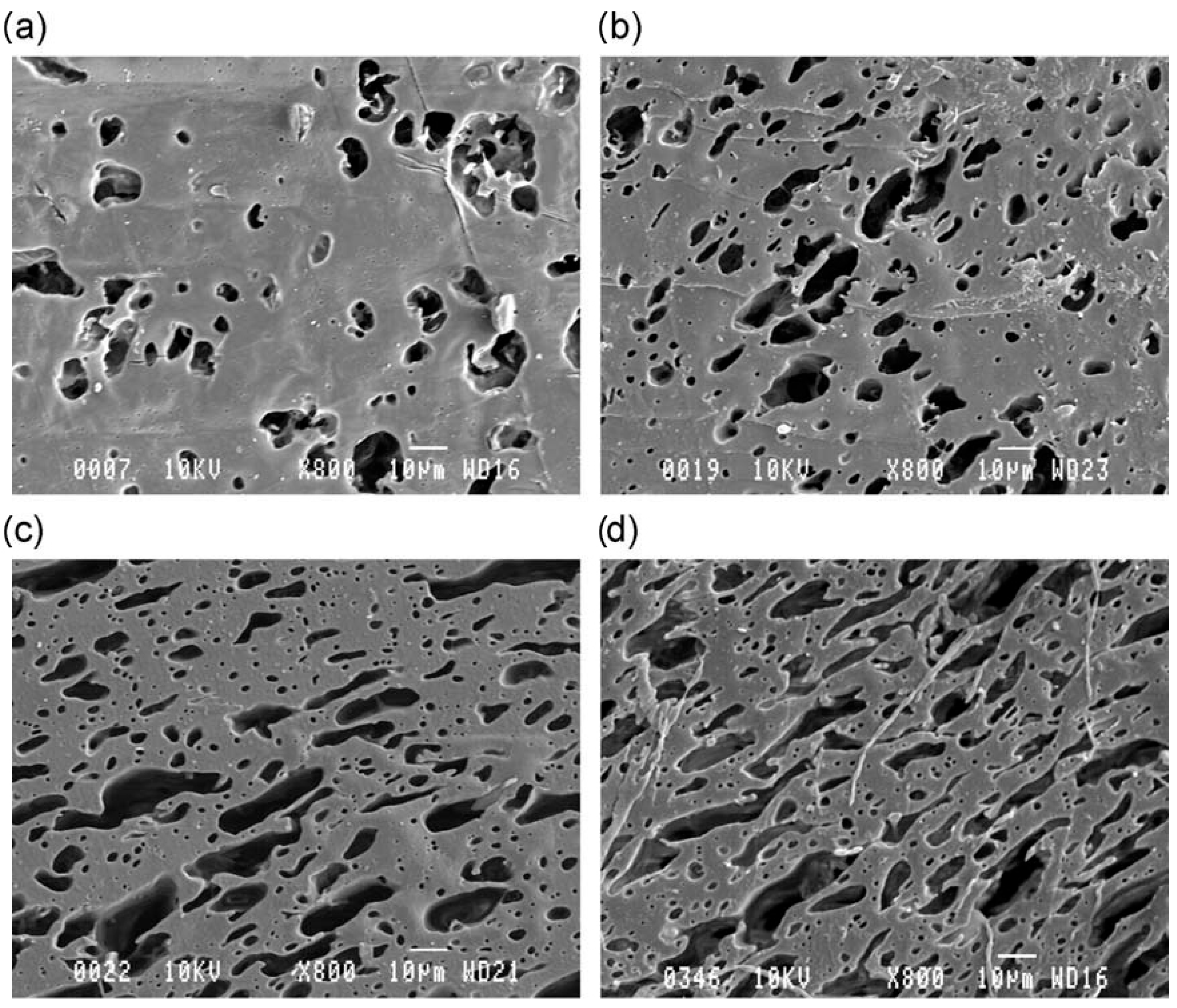
4.1.1. Scanning Electron Microscopy (SEM)
4.1.2. Atomic Force Microscopy (AFM)
4.2. Gravimetric Measurements
4.3. Respirometry Measurement
4.4. Fourier Transform Infrared Spectroscopy (FTIR)
4.5. Thermal Analysis
4.5.1. Differential Scanning Calorimetry (DSC)
4.5.2. Thermogravimetric Analysis (TGA)
4.6. Molecular Mass Characterization
4.6.1. Viscosimetry
4.6.2. Chromatography
4.7. Surface Hydrolysis and pH Level Characterization
4.8. Mechanical Characterization
5. Standardized Protocols
6. Recent Advances in the Biodegradation of Polymers
6.1. Aliphatic Polyesters
6.1.1. Polycaprolactone (PCL)
6.1.2. Polylactic Acid (PLA)
6.1.3. Poly(3-hydroxybutyrate) (PHB)
6.2. Polyolefins
6.2.1. Polyethylene (PE)
6.2.2. Polypropylene (PP)
6.2.3. Polystyrene (PS)
6.3. Polymeric Blends and Composites
6.3.1. Blends of Two Polymers
6.3.2. Blends of Polymeric and Natural Materials
6.3.3. Fiber-Reinforced Composites
7. Conclusions
Funding
Data Availability Statement
Acknowledgments
Conflicts of Interest
References
- Ritchie, H.; Roser, M. Plastic Pollution. World Data 2018. Published online. [Google Scholar]
- Geyer, R.; Jambeck, J.R.; Law, K.L. Production, Use, and Fate of All Plastics Ever Made. Sci. Adv. 2017, 3, e1700782. [Google Scholar] [CrossRef]
- Zheng, Y.; Yanful, E.K.; Bassi, A.S. A Review of Plastic Waste Biodegradation. Crit. Rev. Biotechnol. 2005, 25, 243–250. [Google Scholar] [CrossRef] [PubMed]
- Karamanlioglu, M.; Preziosi, R.; Robson, G.D. Abiotic and Biotic Environmental Degradation of the Bioplastic Polymer Poly(Lactic Acid): A Review. Polym. Degrad. Stab. 2017, 137, 122–130. [Google Scholar] [CrossRef]
- Madhavan Nampoothiri, K.; Nair, N.R.; John, R.P. An Overview of the Recent Developments in Polylactide (PLA) Research. Bioresour. Technol. 2010, 101, 8493–8501. [Google Scholar] [CrossRef] [PubMed]
- Tokiwa, Y.; Calabia, B.; Ugwu, C.; Aiba, S. Biodegradability of Plastics. Int. J. Mol. Sci. 2009, 10, 3722–3742. [Google Scholar] [CrossRef]
- Muthukumar, A.; Veerappapillai, S. Biodegradation of Plastics—A Brief Review. Int. J. Pharm. Sci. Rev. Res. 2015, 31, 204–209. [Google Scholar]
- Ahmed, T.; Shahid, M.; Azeem, F.; Rasul, I.; Shah, A.A.; Noman, M.; Hameed, A.; Manzoor, N.; Manzoor, I.; Muhammad, S. Biodegradation of Plastics: Current Scenario and Future Prospects for Environmental Safety. Environ. Sci. Pollut. Res. 2018, 25, 7287–7298. [Google Scholar] [CrossRef]
- Tokiwa, Y.; Jarerat, A. Biodegradation of Poly(l-Lactide). Biotechnol. Lett. 2004, 26, 771–777. [Google Scholar] [CrossRef] [PubMed]
- Shah, A.A.; Hasan, F.; Hameed, A.; Ahmed, S. Biological Degradation of Plastics: A Comprehensive Review. Biotechnol. Adv. 2008, 26, 246–265. [Google Scholar] [CrossRef]
- Gu, J.-G.; Gu, J.-D. Methods Currently Used in Testing Microbiological Degradation and Deterioration of a Wide Range of Polymeric Materials with Various Degree of Degradability: A Review. J. Polym. Environ. 2005, 13, 65–74. [Google Scholar] [CrossRef]
- Restrepo-Flórez, J.-M.; Bassi, A.; Thompson, M.R. Microbial Degradation and Deterioration of Polyethylene—A Review. Int. Biodeterior. Biodegrad. 2014, 88, 83–90. [Google Scholar] [CrossRef]
- Lucas, N.; Bienaime, C.; Belloy, C.; Queneudec, M.; Silvestre, F.; Nava-Saucedo, J.-E. Polymer Biodegradation: Mechanisms and Estimation Techniques—A Review. Chemosphere 2008, 73, 429–442. [Google Scholar] [CrossRef] [PubMed]
- Soroudi, A.; Jakubowicz, I. Recycling of Bioplastics, Their Blends and Biocomposites: A Review. Eur. Polym. J. 2013, 49, 2839–2858. [Google Scholar] [CrossRef]
- Tokiwa, Y.; Calabia, B.P. Review Degradation of Microbial Polyesters. Biotechnol. Lett. 2004, 26, 1181–1189. [Google Scholar] [CrossRef] [PubMed]
- Ghosh, S.K.; Pal, S.; Ray, S. Study of Microbes Having Potentiality for Biodegradation of Plastics. Environ. Sci. Pollut. Res. 2013, 20, 4339–4355. [Google Scholar] [CrossRef] [PubMed]
- Ashori, A. Wood–Plastic Composites as Promising Green-Composites for Automotive Industries! Bioresour. Technol. 2008, 99, 4661–4667. [Google Scholar] [CrossRef] [PubMed]
- Wambua, P.; Ivens, J.; Verpoest, I. Natural Fibres: Can They Replace Glass in Fibre Reinforced Plastics? Compos. Sci. Technol. 2003, 63, 1259–1264. [Google Scholar] [CrossRef]
- Sudin, R.; Swamy, N. Bamboo and Wood Fibre Cement Composites for Sustainable Infrastructure Regeneration. J. Mater. Sci. 2006, 41, 6917–6924. [Google Scholar] [CrossRef]
- Chandra, R.; Rustgi, R. Biodegradable polymers. Prog. Polym. Sci. 1998, 23, 1273–1335. [Google Scholar] [CrossRef]
- Gu, J.-D.; Ford, T.; Mitton, B.; Mitchell, R. Microbial Degradation of Polymeric Materials. In Uhlig’s Corrosion Handbook; Wiley: London, UK, 2000; pp. 439–460. [Google Scholar]
- Gu, J.-D. Microbiological Deterioration and Degradation of Synthetic Polymeric Materials: Recent Research Advances. Int. Biodeterior. Biodegrad. 2003, 52, 69–91. [Google Scholar] [CrossRef]
- Eggins, H.O.W.; Oxley, T.A. Biodeterioration and Biodegradation. Int. Biodeterior. Biodegrad. 2001, 48, 12–15. [Google Scholar] [CrossRef]
- Hueck, H.J. The Biodeterioration of Materials—An Appraisal (Reprinted). Int. Biodeterior. Biodegrad. 2001, 48, 5–11. [Google Scholar] [CrossRef]
- Lugauskas, A.; Levinskaitė, L.; Pečiulytė, D. Micromycetes as Deterioration Agents of Polymeric Materials. Int. Biodeterior. Biodegrad. 2003, 52, 233–242. [Google Scholar] [CrossRef]
- Ho, K.L.i.G.; Pometto, A.L.I.i.; Hinz, P.N. Effects of Temperature and Relative Humidity on Polylactic Acid Plastic Degradation. J. Environ. Polym. Degrad. 1999, 7, 83–92. [Google Scholar] [CrossRef]
- Peterson, M.E.; Daniel, R.M.; Danson, M.J.; Eisenthal, R. The Dependence of Enzyme Activity on Temperature: Determination and Validation of Parameters. Biochem. J. 2007, 402, 331–337. [Google Scholar] [CrossRef]
- Sukkhum, S.; Tokuyama, S.; Tamura, T.; Kitpreechavanich, V. A Novel Poly (L-Lactide) Degrading Actinomycetes Isolated from Thai Forest Soil, Phylogenic Relationship and the Enzyme Characterization. J. Gen. Appl. Microbiol. 2009, 55, 459–467. [Google Scholar] [CrossRef] [PubMed]
- Nakamura, K.; Tomita, T.; Abe, N.; Kamio, Y. Purification and Characterization of an Extracellular Poly(l-Lactic Acid) Depolymerase from a Soil Isolate, Amycolatopsis sp. Strain K104-1. Appl. Environ. Microbiol. 2001, 67, 345–353. [Google Scholar] [CrossRef]
- Oda, Y.; Asari, H.; Urakami, T.; Tonomura, K. Microbial Degradation of Poly(3-Hydroxybutyrate) and Polycaprolactone by Filamentous Fungi. J. Ferment. Bioeng. 1995, 80, 265–269. [Google Scholar] [CrossRef]
- Santo, M.; Weitsman, R.; Sivan, A. The Role of the Copper-Binding Enzyme—Laccase—in the Biodegradation of Polyethylene by the Actinomycete Rhodococcus Ruber. Int. Biodeterior. Biodegrad. 2013, 84, 204–210. [Google Scholar] [CrossRef]
- Walter, T.; Augusta, J.; Müller, R.-J.; Widdecke, H.; Klein, J. Enzymatic Degradation of a Model Polyester by Lipase from Rhizopus Delemar. Enzym. Microb. Technol. 1995, 17, 218–224. [Google Scholar] [CrossRef]
- Marten, E.; Müller, R.-J.; Deckwer, W.-D. Studies on the Enzymatic Hydrolysis of Polyesters I. Low Molecular Mass Model Esters and Aliphatic Polyesters. Polym. Degrad. Stab. 2003, 80, 485–501. [Google Scholar] [CrossRef]
- Mueller, R.-J. Biological Degradation of Synthetic Polyesters—Enzymes as Potential Catalysts for Polyester Recycling. Process Biochem. 2006, 41, 2124–2128. [Google Scholar] [CrossRef]
- Iwata, T.; Doi, Y. Morphology and Enzymatic Degradation of Poly(l-Lactic Acid) Single Crystals. Macromolecules 1998, 31, 2461–2467. [Google Scholar] [CrossRef]
- Tsuji, H.; Miyauchi, S. Poly(l-Lactide): VI Effects of Crystallinity on Enzymatic Hydrolysis of Poly(l-Lactide) without Free Amorphous Region. Polym. Degrad. Stab. 2001, 71, 415–424. [Google Scholar] [CrossRef]
- Patel, G.N.; Keller, A. Crystallinity and the Effect of Ionizing Radiation in Polyethylene. II. Crosslinking in Chain-Folded Single Crystals. J. Polym. Sci. Polym. Phys. Ed. 1975, 13, 323–331. [Google Scholar] [CrossRef]
- Duong, D.T.; Bell, J.P. Characterization of Selectively Degraded Poly(Ethylene Terephthalate). J. Polym. Sci. Polym. Phys. Ed. 1975, 13, 765–774. [Google Scholar] [CrossRef]
- Williams, T.; Keller, A.; Ward, I.M. Gel Permeation Chromatographic Studies of the Degradation of Polyethylene with Fuming Nitric Acid. II. Bulk Polyethylene. J. Polym. Sci. Part A-2: Polym. Phys. 1968, 6, 1621–1625. [Google Scholar] [CrossRef]
- Tokiwa, Y.; Suzuki, T. Hydrolysis of Polyesters by Rhizopus Delemar Lipase. Agric. Biol. Chem. 1978, 42, 1071–1072. [Google Scholar] [CrossRef]
- Tokiwa, Y.; Suzuki, T. Hydrolysis of Copolyesters Containing Aromatic and Aliphatic Ester Blocks by Lipase. J. Appl. Polym. Sci. 1981, 26, 441–448. [Google Scholar] [CrossRef]
- Tokiwa, Y.; Suzuki, T.; Ando, T. Synthesis of Copolyamide–Esters and Some Aspects Involved in Their Hydrolysis by Lipase. J. Appl. Polym. Sci. 1979, 24, 1701–1711. [Google Scholar] [CrossRef]
- Walker, J.D.; Austin, H.F.; Colwell, R.R. Utilization of Mixed Hydrocarbon Substrate by Petroleum-Degrading Microorganisms. J. Gen. Appl. Microbiol. 1975, 21, 27–39. [Google Scholar] [CrossRef]
- Kim, M.N.; Park, S.T. Degradation of Poly(L-Lactide) by a Mesophilic Bacterium. J. Appl. Polym. Sci. 2010, 117, 67–74. [Google Scholar] [CrossRef]
- Sangeetha Devi, R.; Rajesh Kannan, V.; Nivas, D.; Kannan, K.; Chandru, S.; Robert Antony, A. Biodegradation of HDPE by Aspergillus spp. from Marine Ecosystem of Gulf of Mannar, India. Mar. Pollut. Bull. 2015, 96, 32–40. [Google Scholar] [CrossRef]
- Hadad, D.; Geresh, S.; Sivan, A. Biodegradation of Polyethylene by the Thermophilic Bacterium Brevibacillus Borstelensis. J. Appl. Microbiol. 2005, 98, 1093–1100. [Google Scholar] [CrossRef]
- Yang, J.; Yang, Y.; Wu, W.-M.; Zhao, J.; Jiang, L. Evidence of Polyethylene Biodegradation by Bacterial Strains from the Guts of Plastic-Eating Waxworms. Environ. Sci. Technol. 2014, 48, 13776–13784. [Google Scholar] [CrossRef]
- Oda, Y.; Yonetsu, A.; Urakami, T.; Tonomura, K. Degradation of Polylactide by Commercial Proteases. J. Polym. Environ. 2000, 8, 29–32. [Google Scholar] [CrossRef]
- Mahdi, M.S.; Ameen, R.S.; Ibrahim, H.K. Study on Degradation of Nylon 6 by Thermophilic Bacteria Anoxybacillus Rupiensis Ir3 (JQ912241). Int. J. Adv. Res. Biol. Sci. 2016, 10, 27. [Google Scholar]
- Gebauer, B.; Jendrossek, D. Assay of Poly(3-Hydroxybutyrate) Depolymerase Activity and Product Determination. Appl. Environ. Microbiol. 2006, 72, 6094–6100. [Google Scholar] [CrossRef]
- Arkatkar, A.; Arutchelvi, J.; Bhaduri, S.; Uppara, P.V.; Doble, M. Degradation of Unpretreated and Thermally Pretreated Polypropylene by Soil Consortia. Int. Biodeterior. Biodegrad. 2009, 63, 106–111. [Google Scholar] [CrossRef]
- Li, G.; Sarazin, P.; Orts, W.J.; Imam, S.H.; Favis, B.D. Biodegradation of Thermoplastic Starch and Its Blends with Poly(Lactic Acid) and Polyethylene: Influence of Morphology: Biodegradation of Thermoplastic Starch and Its Blends with Poly(lactic acid) and Polyethylene: Influence of Morphology. Macromol. Chem. Phys. 2011, 212, 1147–1154. [Google Scholar] [CrossRef]
- Ikada, E. Electron Microscope Observation of Biodegradation of Polymers. J. Polym. Environ. 1999, 7, 197–201. [Google Scholar] [CrossRef]
- Cook, W.J.; Cameron, J.A.; Bell, J.P.; Huang, S.J. Scanning Electron Microscopic Visualization of Biodegradation of Polycaprolactones by Fungi. J. Polym. Sci. Polym. Lett. Ed. 1981, 19, 159–165. [Google Scholar] [CrossRef]
- Hrubanova, K.; Voberkova, S.; Hermanova, S.; Krzyzanek, V. Characterization of Polycaprolactone Films Biodeterioration by Scanning Electron Microscopy. Microsc. Microanal. 2014, 20, 1950–1951. [Google Scholar] [CrossRef]
- Bombelli, P.; Howe, C.J.; Bertocchini, F. Polyethylene Bio-Degradation by Caterpillars of the Wax Moth Galleria Mellonella. Curr. Biol. 2017, 27, R292–R293. [Google Scholar] [CrossRef]
- Fernando, S.S.; Christensen, P.A.; Egerton, T.A.; White, J.R. Carbon Dioxide Evolution and Carbonyl Group Development during Photodegradation of Polyethylene and Polypropylene. Polym. Degrad. Stab. 2007, 92, 2163–2172. [Google Scholar] [CrossRef]
- Yang, Y.; Yang, J.; Wu, W.-M.; Zhao, J.; Song, Y.; Gao, L.; Yang, R.; Jiang, L. Biodegradation and Mineralization of Polystyrene by Plastic-Eating Mealworms: Part 1. Chemical and Physical Characterization and Isotopic Tests. Environ. Sci. Technol. 2015, 49, 12080–12086. [Google Scholar] [CrossRef]
- Raghavan, D.; Torma, A.E. DSC and FTIR Characterization of Biodegradation of Polyethylene. Polym. Eng. Sci. 1992, 32, 438–442. [Google Scholar] [CrossRef]
- Fukushima, K.; Abbate, C.; Tabuani, D.; Gennari, M.; Camino, G. Biodegradation of Poly(Lactic Acid) and Its Nanocomposites. Polym. Degrad. Stab. 2009, 94, 1646–1655. [Google Scholar] [CrossRef]
- Fortunati, E.; Puglia, D.; Monti, M.; Santulli, C.; Maniruzzaman, M.; Foresti, M.L.; Vazquez, A.; Kenny, J.M. Okra (Abelmoschus Esculentus) Fibre Based PLA Composites: Mechanical Behaviour and Biodegradation. J. Polym. Environ. 2013, 21, 726–737. [Google Scholar] [CrossRef]
- Selective Enzymatic Degradations of Poly(l-Lactide) and Poly(ε-Caprolactone) Blend Films. Biomacromolecules 2000, 1, 350–359. [CrossRef] [PubMed]
- Lee, S.H.; Kim, I.Y.; Song, W.S. Biodegradation of Polylactic Acid (PLA) Fibers Using Different Enzymes. Macromol. Res. 2014, 22, 657–663. [Google Scholar] [CrossRef]
- Petinakis, E.; Liu, X.; Yu, L.; Way, C.; Sangwan, P.; Dean, K.; Bateman, S.; Edward, G. Biodegradation and Thermal Decomposition of Poly(Lactic Acid)-Based Materials Reinforced by Hydrophilic Fillers. Polym. Degrad. Stab. 2010, 95, 1704–1707. [Google Scholar] [CrossRef]
- Dutta, S.; Karak, N.; Saikia, J.P.; Konwar, B.K. Biodegradation of Epoxy and MF Modified Polyurethane Films Derived from a Sustainable Resource. J. Polym. Environ. 2010, 18, 167–176. [Google Scholar] [CrossRef]
- Shikha, R.; Harshita, N.; Tithi, A.; Mgh, Z.; Reeta, G. Comparative Biodegradation Studies of Cow Dung Modified Epoxy with Epoxy Using an Indigenously Developed Bacterial Consortium. Afr. J. Microbiol. Res. 2015, 9, 1558–1572. [Google Scholar] [CrossRef]
- Negi, H.; Kapri, A.; Zaidi, M.G.H.; Satlewal, A.; Goel, R. Comparative In-Vitro Biodegradation Studies of Epoxy and Its Silicone Blend by Selected Microbial Consortia. Int. Biodeterior. Biodegrad. 2009, 63, 553–558. [Google Scholar] [CrossRef]
- Orhan, Y.; Büyükgüngör, H. Enhancement of Biodegradability of Disposable Polyethylene in Controlled Biological Soil. Int. Biodeterior. Biodegrad. 2000, 45, 49–55. [Google Scholar] [CrossRef]
- Tomita, K.; Ikeda, N.; Ueno, A. Isolation and Characterization of a Thermophilic Bacterium, Geobacillus Thermocatenulatus, Degrading Nylon 12 and Nylon 66. Biotechnol. Lett. 2003, 25, 1743–1746. [Google Scholar] [CrossRef]
- Rae, P.J.; Brown, E.N.; Orler, E.B. The Mechanical Properties of Poly(Ether-Ether-Ketone) (PEEK) with Emphasis on the Large Compressive Strain Response. Polymer 2007, 48, 598–615. [Google Scholar] [CrossRef]
- Deguchi, T.; Kakezawa, M.; Nishida, T. Nylon Biodegradation by Lignin-Degrading Fungi. Appl. Environ. Microbiol. 1997, 63, 329–331. [Google Scholar] [CrossRef]
- Marten, E.; Müller, R.-J.; Deckwer, W.-D. Studies on the Enzymatic Hydrolysis of Polyesters. II. Aliphatic–Aromatic Copolyesters. Polym. Degrad. Stab. 2005, 88, 371–381. [Google Scholar] [CrossRef]
- Das, M.P.; Kumar, S. An Approach to Low-Density Polyethylene Biodegradation by Bacillus Amyloliquefaciens. 3 Biotech 2015, 5, 81–86. [Google Scholar] [CrossRef] [PubMed]
- Deguchi, T.; Kitaoka, Y.; Kakezawa, M.; Nishida, T. Purification and Characterization of a Nylon-Degrading Enzyme. Appl. Environ. Microbiol. 1998, 64, 1366–1371. [Google Scholar] [CrossRef] [PubMed]
- Orhan, Y.; Hrenović, J.; Büyükgüngör, H. Biodegradation of plastic compost bags under controlled soil conditions. Acta Chim. Slov. 2004, 10, 579–588. [Google Scholar]
- D5526-18; Test Method for Determining Anaerobic Biodegradation of Plastic Materials Under Accelerated Landfill Conditions. ASTM International: West Conshohocken, PA, USA, 2018. Available online: https://www.astm.org/d5526-18.html (accessed on 3 October 2023).
- D5988-18; Test Method for Determining Aerobic Biodegradation of Plastic Materials in Soil. ASTM International: West Conshohocken, PA, USA, 2018. Available online: https://www.astm.org/d5988-18.html (accessed on 3 October 2023).
- D5511-18; Test Method for Determining Anaerobic Biodegradation of Plastic Materials Under High-Solids Anaerobic-Digestion Conditions. ASTM International: West Conshohocken, PA, USA, 2018. Available online: https://www.astm.org/d5511-18.html (accessed on 3 October 2023).
- D7991-22; Standard Test Method for Determining Aerobic Biodegradation of Plastics Buried in Sandy Marine Sediment under Controlled Laboratory Conditions. ASTM International: West Conshohocken, PA, USA, 2022. Available online: https://www.astm.org/d7991-22.html (accessed on 3 October 2023).
- D5338-15(2021); Test Method for Determining Aerobic Biodegradation of Plastic Materials in Soil. ASTM International: West Conshohocken, PA, USA, 2021. Available online: https://www.astm.org/d5338-15r21.html (accessed on 15 January 2023).
- D6954-18; Guide for Exposing and Testing Plastics That Degrade in the Environment by a Combination of Oxidation and Biodegradation. ASTM International: West Conshohocken, PA, USA, 2018. Available online: https://www.astm.org/d6954-18.html (accessed on 3 October 2023).
- D7475-20; Test Method for Determining the Aerobic Degradation and Anaerobic Biodegradation of Plastic Materials under Accelerated Bioreactor Landfill Conditions. ASTM International: West Conshohocken, PA, USA, 2018. Available online: https://www.astm.org/d7475-20.html (accessed on 3 October 2023).
- D6400-23; Specification for Labeling of Plastics Designed to Be Aerobically Composted in Municipal or Industrial Facilities. ASTM International: West Conshohocken, PA, USA, 2018. Available online: https://www.astm.org/d6400-23.html (accessed on 3 October 2023).
- D6868-21; Specification for Labeling of End Items That Incorporate Plastics and Polymers as Coatings or Additives with Paper and Other Substrates Designed to Be Aerobically Composted in Municipal or Industrial Facilities. ASTM International: West Conshohocken, PA, USA, 2018. Available online: https://www.astm.org/d6868-21.html (accessed on 3 October 2023).
- Hakkarainen, M. Aliphatic Polyesters: Abiotic and Biotic Degradation and Degradation Products. In Degradable Aliphatic Polyesters; Advances in Polymer Science; Springer: Berlin/Heidelberg, Germany, 2002; pp. 113–138. ISBN 978-3-540-45734-3. [Google Scholar]
- Al Hosni, A.S.; Pittman, J.K.; Robson, G.D. Microbial Degradation of Four Biodegradable Polymers in Soil and Compost Demonstrating Polycaprolactone as an Ideal Compostable Plastic. Waste Manag. 2019, 97, 105–114. [Google Scholar] [CrossRef]
- Tokiwa, Y.; Suzuki, T. Hydrolysis of Polyesters by Lipases. Nature 1977, 270, 76–78. [Google Scholar] [CrossRef]
- Kobayashi, T.; Sugiyama, A.; Kawase, Y.; Saito, T.; Mergaert, J.; Swings, J. Biochemical and Genetic Characterization of an Extracellular Poly(3-Hydroxybutyrate) Depolymerase from Acidovorax sp. Strain TP4. J. Polym. Environ. 1999, 7, 9–18. [Google Scholar] [CrossRef]
- Jarerat, A.; Tokiwa, Y. Degradation of Poly(Tetramethylene Succinate) by Thermophilic Actinomycetes. Biotechnol. Lett. 2001, 23, 647–651. [Google Scholar] [CrossRef]
- Jung, H.-W.; Yang, M.-K.; Su, R.-C. Purification, Characterization, and Gene Cloning of an Aspergillus Fumigatus Polyhydroxybutyrate Depolymerase Used for Degradation of Polyhydroxybutyrate, Polyethylene Succinate, and Polybutylene Succinate. Polym. Degrad. Stab. 2018, 154, 186–194. [Google Scholar] [CrossRef]
- Tansengco, M.L.; Tokiwa, Y. Thermophilic Microbial Degradation of Polyethylene Succinate. World J. Microbiol. Biotechnol. 1997, 1, 133–138. [Google Scholar] [CrossRef]
- Tezuka, Y.; Ishii, N.; Kasuya, K.-I.; Mitomo, H. Degradation of Poly(Ethylene Succinate) by Mesophilic Bacteria. Polym. Degrad. Stab. 2004, 84, 115–121. [Google Scholar] [CrossRef]
- Ramanujam, R.; Sundaram, B.; Janarthanan, G.; Devendran, E.; Venkadasalam, M.; Milton, M.C.J. Biodegradable Polycaprolactone Nanoparticles Based Drug Delivery Systems: A Short Review. Biosci. Biotechnol. Res. Asia 2018, 15, 679–685. [Google Scholar] [CrossRef]
- Richert, A.; Dąbrowska, G.B. Enzymatic Degradation and Biofilm Formation during Biodegradation of Polylactide and Polycaprolactone Polymers in Various Environments. Int. J. Biol. Macromol. 2021, 176, 226–232. [Google Scholar] [CrossRef] [PubMed]
- Zhao, Q.; Tao, J.; Yam, R.C.M.; Mok, A.C.K.; Li, R.K.Y.; Song, C. Biodegradation Behavior of Polycaprolactone/Rice Husk Ecocomposites in Simulated Soil Medium. Polym. Degrad. Stab. 2008, 93, 1571–1576. [Google Scholar] [CrossRef]
- Rutkowska, M.; Jastrzębska, M.; Janik, H. Biodegradation of Polycaprolactone in Sea Water. React. Funct. Polym. 1998, 38, 27–30. [Google Scholar] [CrossRef]
- Chen, D.R.; Bei, J.Z.; Wang, S.G. Polycaprolactone Microparticles and Their Biodegradation. Polym. Degrad. Stab. 2000, 67, 455–459. [Google Scholar] [CrossRef]
- Castilla-Cortázar, I.; Más-Estellés, J.; Meseguer-Dueñas, J.M.; Escobar Ivirico, J.L.; Marí, B.; Vidaurre, A. Hydrolytic and Enzymatic Degradation of a Poly(ε-Caprolactone) Network. Polym. Degrad. Stab. 2012, 97, 1241–1248. [Google Scholar] [CrossRef]
- Benedict, C.V.; Cameron, J.A.; Huang, S.J. Polycaprolactone Degradation by Mixed and Pure Cultures of Bacteria and a Yeast. J. Appl. Polym. Sci. 1983, 28, 335–342. [Google Scholar] [CrossRef]
- Benedict, C.V.; Cook, W.J.; Jarrett, P.; Cameron, J.A.; Huang, S.J.; Bell, J.P. Fungal Degradation of Polycaprolactones. J. Appl. Polym. Sci. 1983, 28, 327–334. [Google Scholar] [CrossRef]
- Khan, I.; Ray Dutta, J.; Ganesan, R. Lactobacillus sps. Lipase Mediated Poly (ε-Caprolactone) Degradation. Int. J. Biol. Macromol. 2017, 95, 126–131. [Google Scholar] [CrossRef]
- Ma, Q.; Shi, K.; Su, T.; Wang, Z. Biodegradation of Polycaprolactone (PCL) with Different Molecular Weights by Candida Antarctica Lipase. J. Polym. Environ. 2020, 28, 2947–2955. [Google Scholar] [CrossRef]
- Funabashi, M.; Ninomiya, F.; Kunioka, M. Biodegradation of Polycaprolactone Powders Proposed as Reference Test Materials for International Standard of Biodegradation Evaluation Method. J. Polym. Environ. 2007, 15, 7–17. [Google Scholar] [CrossRef]
- Lefèvre, C.; Tidjani, A.; Vander Wauven, C.; David, C. The Interaction Mechanism between Microorganisms and Substrate in the Biodegradation of Polycaprolactone. J. Appl. Polym. Sci. 2002, 83, 1334–1340. [Google Scholar] [CrossRef]
- Shah, A.A.; Nawaz, A.; Kanwal, L.; Hasan, F.; Khan, S.; Badshah, M. Degradation of Poly(ε-Caprolactone) by a Thermophilic Bacterium Ralstonia sp. Strain MRL-TL Isolated from Hot Spring. Int. Biodeterior. Biodegrad. 2015, 98, 35–42. [Google Scholar] [CrossRef]
- Shi, K.; Jing, J.; Song, L.; Su, T.; Wang, Z. Enzymatic Hydrolysis of Polyester: Degradation of Poly(ε-Caprolactone) by Candida Antarctica Lipase and Fusarium Solani Cutinase. Int. J. Biol. Macromol. 2020, 144, 183–189. [Google Scholar] [CrossRef]
- Fields, R.D.; Rodriguez, F.; Finn, R.K. Microbial Degradation of Polyesters: Polycaprolactone Degraded by P. Pullulans. J. Appl. Polym. Sci. 1974, 18, 3571–3579. [Google Scholar] [CrossRef]
- Sanchez, J.G.; Tsuchii, A.; Tokiwa, Y. Degradation of Polycaprolactone at 50 °C by a Thermotolerant Aspergillus sp. Biotechnol. Lett. 2000, 22, 849–853. [Google Scholar] [CrossRef]
- Jarrett, P.; Benedict, C.V.; Bell, J.P.; Cameron, J.A.; Huang, S.J. Mechanism of the Biodegradation of Polycaprolactone. In Polymers as Biomaterials; Shalaby, S.W., Hoffman, A.S., Ratner, B.D., Horbett, T.A., Eds.; Springer: Boston, MA, USA, 1984; pp. 181–192. ISBN 978-1-4613-2433-1. [Google Scholar]
- Stepczyńska, M.; Rytlewski, P. Enzymatic Degradation of Flax-Fibers Reinforced Polylactide. Int. Biodeterior. Biodegrad. 2018, 126, 160–166. [Google Scholar] [CrossRef]
- De Rezende, M.L. Enzymatic Hydrolysis of Poly (Lactic Acid) (PLA) and Thermoplastic Starch (TPS). Appl. Biochem. Biotechnol. 2017, 4, 5. [Google Scholar]
- Williams, D.F. Enzymic Hydrolysis of Polylactic Acid. Eng. Med. 1981, 10, 5–7. [Google Scholar] [CrossRef]
- Tokiwa, Y.; Konno, M.; Nishida, H. Isolation of Silk Degrading Microorganisms and Its Poly(L-Lactide) Degradability. Chem. Lett. 1999, 24, 355–356. [Google Scholar] [CrossRef]
- Ikura, Y.; Kudo, T. Isolation of a Microorganism Capable of Degrading Poly-(L-Lactide). J. Gen. Appl. Microbiol. 1999, 45, 247–251. [Google Scholar] [CrossRef] [PubMed]
- Auras, R.; Harte, B.; Selke, S. An Overview of Polylactides as Packaging Materials. Macromol. Biosci. 2004, 4, 835–864. [Google Scholar] [CrossRef] [PubMed]
- Sangwan, P.; Wu, D.Y. New Insights into Polylactide Biodegradation from Molecular Ecological Techniques. Macromol. Biosci. 2008, 8, 304–315. [Google Scholar] [CrossRef] [PubMed]
- Sangwan, P.; Way, C.; Wu, D.-Y. New Insight into Biodegradation of Polylactide (PLA)/Clay Nanocomposites Using Molecular Ecological Techniques. Macromol. Biosci. 2009, 9, 677–686. [Google Scholar] [CrossRef]
- Pranamuda, H.; Tokiwa, Y. Degradation of Poly(L-Lactide) by Strains Belonging to Genus Amycolatopsis. Biotechnol. Lett. 1999, 21, 901–905. [Google Scholar] [CrossRef]
- Torres, A.; Li, S.M.; Roussos, S.; Vert, M. Screening of Microorganisms for Biodegradation of Poly(Lactic-Acid) and Lactic Acid-Containing Polymers. Appl. Environ. Microbiol. 1996, 62, 2393–2397. [Google Scholar] [CrossRef] [PubMed]
- Pranamuda, H.; Tokiwa, Y.; Tanaka, H. Polylactide Degradation by an Amycolatopsis sp. Appl. Environ. Microbiol. 1997, 63, 1637–1640. [Google Scholar] [CrossRef] [PubMed]
- Pranamuda, H.; Tsuchii, A.; Tokiwa, Y. Poly (L-Lactide)-Degrading Enzyme Produced by Amycolatopsis sp. Macromol. Biosci. 2001, 1, 25–29. [Google Scholar] [CrossRef]
- Jarerat, A.; Pranamuda, H.; Tokiwa, Y. Poly(L-Lactide)-Degrading Activity in Various Actinomycetes. Macromol. Biosci. 2002, 2, 420–428. [Google Scholar] [CrossRef]
- Jarerat, A.; Tokiwa, Y. Poly(L-Lactide) Degradation by Saccharothrix Waywayandensis. Biotechnol. Lett. 2003, 25, 401–404. [Google Scholar] [CrossRef] [PubMed]
- Jarerat, A.; Tokiwa, Y.; Tanaka, H. Poly(L-Lactide) Degradation by Kibdelosporangium Aridum. Biotechnol. Lett. 2003, 25, 2035–2038. [Google Scholar] [CrossRef]
- Tomita, K.; Kuroki, Y.; Nagai, K. Isolation of Thermophiles Degrading Poly(l-Lactic Acid). J. Biosci. Bioeng. 1999, 87, 752–755. [Google Scholar] [CrossRef] [PubMed]
- Tomita, K.; Tsuji, H.; Nakajima, T.; Kikuchi, Y.; Ikarashi, K.; Ikeda, N. Degradation of Poly(d-Lactic Acid) by a Thermophile. Polym. Degrad. Stab. 2003, 81, 167–171. [Google Scholar] [CrossRef]
- Tomita, K.; Nakajima, T.; Kikuchi, Y.; Miwa, N. Degradation of Poly(l-Lactic Acid) by a Newly Isolated Thermophile. Polymer Degrad. Stab. 2004, 84, 433–438. [Google Scholar] [CrossRef]
- Ghorpade, V.M.; Gennadios, A.; Hanna, M.A. Laboratory Composting of Extruded Poly(Lactic Acid) Sheets. Bioresour. Technol. 2001, 76, 57–61. [Google Scholar] [CrossRef]
- Jeon, H.J.; Kim, M.N. Biodegradation of Poly(l-Lactide) (PLA) Exposed to UV Irradiation by a Mesophilic Bacterium. Int. Biodeterior. Biodegrad. 2013, 85, 289–293. [Google Scholar] [CrossRef]
- Husárová, L.; Pekařová, S.; Stloukal, P.; Kucharzcyk, P.; Verney, V.; Commereuc, S.; Ramone, A.; Koutny, M. Identification of Important Abiotic and Biotic Factors in the Biodegradation of Poly(l-Lactic Acid). Int. J. Biol. Macromol. 2014, 71, 155–162. [Google Scholar] [CrossRef]
- Apinya, T.; Sombatsompop, N.; Prapagdee, B. Selection of a Pseudonocardia sp. RM423 That Accelerates the Biodegradation of Poly(Lactic) Acid in Submerged Cultures and in Soil Microcosms. Int. Biodeterior. Biodegrad. 2015, 99, 23–30. [Google Scholar] [CrossRef]
- Jarerat, A.; Tokiwa, Y. Degradation of Poly(L-Lactide) by a Fungus. Macromol. Biosci. 2001, 1, 136–140. [Google Scholar] [CrossRef]
- Karamanlioglu, M.; Houlden, A.; Robson, G.D. Isolation and Characterisation of Fungal Communities Associated with Degradation and Growth on the Surface of Poly(Lactic) Acid (PLA) in Soil and Compost. Int. Biodeterior. Biodegrad. 2014, 95, 301–310. [Google Scholar] [CrossRef]
- Lipsa, R.; Tudorachi, N.; Darie-Nita, R.N.; Oprică, L.; Vasile, C.; Chiriac, A. Biodegradation of Poly(Lactic Acid) and Some of Its Based Systems with Trichoderma Viride. Int. J. Biol. Macromol. 2016, 88, 515–526. [Google Scholar] [CrossRef] [PubMed]
- Uğur, A.; Şahin, N.; Beyatli, Y. Accumulation of Poly-\beta-Hydroxybutyrate in Streptomyces Species During Growth with Different Nitrogen Sources. Turk. J. Biol. 2002, 26, 171–174. [Google Scholar]
- Kawaguchi, Y.; Doi, Y. Kinetics and Mechanism of Synthesis and Degradation of Poly(3-Hydroxybutyrate) in Alcaligenes Eutrophus. Macromolecules 1992, 25, 2324–2329. [Google Scholar] [CrossRef]
- Kunioka, M.; Kawaguchi, Y.; Doi, Y. Production of Biodegradable Copolyesters of 3-Hydroxybutyrate and 4-Hydroxybutyrate by Alcaligenes Eutrophus. Appl. Microbiol. Biotechnol. 1989, 30, 569–573. [Google Scholar] [CrossRef]
- Foster, L.J.R.; Zervas, S.J.; Lenz, R.W.; Fuller, R.C. The Biodegradation of Poly-3-Hydroxyalkanoates, PHAs, with Long Alkyl Substitutents ByPseudomonas Maculicola. Biodegradation 1995, 6, 67–73. [Google Scholar] [CrossRef]
- Doi, Y.; Kanesawa, Y.; Kunioka, M.; Saito, T. Biodegradation of Microbial Copolyesters: Poly(3-Hydroxybutyrate-Co-3-Hydroxyvalerate) and Poly(3-Hydroxybutyrate-Co-4-Hydroxybutyrate). Macromolecules 1990, 23, 26–31. [Google Scholar] [CrossRef]
- Shah, A.A.; Hasan, F.; Hameed, A. Degradation of Poly(3-Hydroxybutyrate-Co-3-Hydroxyvalerate) by a Newly Isolated Actinomadura sp. AF-555, from Soil. Int. Biodeterior. Biodegrad. 2010, 64, 281–285. [Google Scholar] [CrossRef]
- Calabia, B.P.; Tokiwa, Y. A Novel PHB Depolymerase from a Thermophilic Streptomyces sp. Biotechnol. Lett. 2006, 28, 383–388. [Google Scholar] [CrossRef]
- Lee, K.-M.; Gimore, D.F.; Huss, M.J. Fungal Degradation of the Bioplastic PHB (Poly-3-Hydroxy-Butyric Acid). J. Polym. Environ. 2005, 13, 213–219. [Google Scholar] [CrossRef]
- Altaee, N.; El-Hiti, G.A.; Fahdil, A.; Sudesh, K.; Yousif, E. Biodegradation of Different Formulations of Polyhydroxybutyrate Films in Soil. SpringerPlus 2016, 5, 762. [Google Scholar] [CrossRef]
- Cho, J.Y.; Kim, S.H.; Jung, H.J.; Cho, D.H.; Kim, B.C.; Bhatia, S.K.; Ahn, J.; Jeon, J.-M.; Yoon, J.-J.; Lee, J.; et al. Finding a Benign Plasticizer to Enhance the Microbial Degradation of Polyhydroxybutyrate (PHB) Evaluated by PHB Degrader Microbulbifer sp. SOL66. Polymers 2022, 14, 3625. [Google Scholar] [CrossRef] [PubMed]
- Park, S.L.; Cho, J.Y.; Kim, S.H.; Bhatia, S.K.; Gurav, R.; Park, S.-H.; Park, K.; Yang, Y.-H. Isolation of Microbulbifer sp. SOL66 with High Polyhydroxyalkanoate-Degrading Activity from the Marine Environment. Polymers 2021, 13, 4257. [Google Scholar] [CrossRef] [PubMed]
- Calabia, B.P.; Tokiwa, Y. Microbial Degradation of Poly(d-3-Hydroxybutyrate) by a New Thermophilic Streptomyces Isolate. Biotechnol. Lett. 2004, 26, 15–19. [Google Scholar] [CrossRef] [PubMed]
- Arkatkar, A.; Arutchelvi, J.; Sudhakar, M.; Bhaduri, S.; Uppara, P.V.; Doble, M. Approaches to Enhance the Biodegradation of Polyolefins. TOENVIEJ 2009, 2, 68–80. [Google Scholar] [CrossRef]
- Zhang, N.; Ding, M.; Yuan, Y. Current Advances in Biodegradation of Polyolefins. Microorganisms 2022, 10, 1537. [Google Scholar] [CrossRef]
- Arutchelvi, J.; Sudhakar, M.; Arkatkar, A.; Doble, M.; Bhaduri, S.; Uppara, P.V. Biodegradation of Polyethylene and Polypropylene. Indian J. Biotechnol. 2008, 7, 1. [Google Scholar]
- Yang, S.-S.; Ding, M.-Q.; He, L.; Zhang, C.-H.; Li, Q.-X.; Xing, D.-F.; Cao, G.-L.; Zhao, L.; Ding, J.; Ren, N.-Q.; et al. Biodegradation of Polypropylene by Yellow Mealworms (Tenebrio Molitor) and Superworms (Zophobas Atratus) via Gut-Microbe-Dependent Depolymerization. Sci. Total Environ. 2021, 756, 144087. [Google Scholar] [CrossRef]
- Peng, B.-Y.; Li, Y.; Fan, R.; Chen, Z.; Chen, J.; Brandon, A.M.; Criddle, C.S.; Zhang, Y.; Wu, W.-M. Biodegradation of Low-Density Polyethylene and Polystyrene in Superworms, Larvae of Zophobas Atratus (Coleoptera: Tenebrionidae): Broad and Limited Extent Depolymerization. Environ. Pollut. 2020, 266, 115206. [Google Scholar] [CrossRef]
- Kumari, K.; Aanad, R.C.; Narula, N. Microbial Degradation of Polyethylene (PE). S. Pac. J. Nat. App. Sci. 2009, 27, 66. [Google Scholar] [CrossRef]
- Kumar Sen, S.; Raut, S. Microbial Degradation of Low Density Polyethylene (LDPE): A Review. J. Environ. Chem. Eng. 2015, 3, 462–473. [Google Scholar] [CrossRef]
- Karlsson, S.; Ljungquist, O.; Albertsson, A.-C. Biodegradation of Polyethylene and the Influence of Surfactants. Polym. Degrad. Stab. 1988, 21, 237–250. [Google Scholar] [CrossRef]
- Volke-Sepúlveda, T.; Saucedo-Castañeda, G.; Gutiérrez-Rojas, M.; Manzur, A.; Favela-Torres, E. Thermally Treated Low Density Polyethylene Biodegradation by Penicillium Pinophilum and Aspergillus Niger. J. Appl. Polym. Sci. 2002, 83, 305–314. [Google Scholar] [CrossRef]
- Fontanella, S.; Bonhomme, S.; Koutny, M.; Husarova, L.; Brusson, J.-M.; Courdavault, J.-P.; Pitteri, S.; Samuel, G.; Pichon, G.; Lemaire, J.; et al. Comparison of the Biodegradability of Various Polyethylene Films Containing Pro-Oxidant Additives. Polym. Degrad. Stab. 2010, 95, 1011–1021. [Google Scholar] [CrossRef]
- Peixoto, J.; Silva, L.P.; Krüger, R.H. Brazilian Cerrado Soil Reveals an Untapped Microbial Potential for Unpretreated Polyethylene Biodegradation. J. Hazard. Mater. 2017, 324, 634–644. [Google Scholar] [CrossRef] [PubMed]
- Muhonja, C.N.; Makonde, H.; Magoma, G.; Imbuga, M. Biodegradability of Polyethylene by Bacteria and Fungi from Dandora Dumpsite Nairobi-Kenya. PLoS ONE 2018, 13, e0198446. [Google Scholar] [CrossRef]
- El-Shafei, H.A.; Abd El-Nasser, N.H.; Kansoh, A.L.; Ali, A.M. Biodegradation of Disposable Polyethylene by Fungi and Streptomyces Species. Polym. Degrad. Stab. 1998, 62, 361–365. [Google Scholar] [CrossRef]
- Sivan, A.; Szanto, M.; Pavlov, V. Biofilm Development of the Polyethylene-Degrading Bacterium Rhodococcus Ruber. Appl. Microbiol. Biotechnol. 2006, 72, 346–352. [Google Scholar] [CrossRef] [PubMed]
- Orr, I.G.; Hadar, Y.; Sivan, A. Colonization, Biofilm Formation and Biodegradation of Polyethylene by a Strain of Rhodococcus Ruber. Appl. Microbiol. Biotechnol. 2004, 65, 97–104. [Google Scholar] [CrossRef]
- Divyalakshmi, S. Screening and Isolation of Polyethylene Degrading Bacteria from Various Soil Environments. Int. Res. J. Environ. Sci. 2016, 2, 1–7. [Google Scholar]
- Azeko, S.T.; Etuk-Udo, G.A.; Odusanya, O.S.; Malatesta, K.; Anuku, N.; Soboyejo, W.O. Biodegradation of Linear Low Density Polyethylene by Serratia Marcescens subsp. Marcescens and Its Cell Free Extracts. Waste Biomass. Valor. 2015, 6, 1047–1057. [Google Scholar] [CrossRef]
- Kyaw, B.M.; Champakalakshmi, R.; Sakharkar, M.K.; Lim, C.S.; Sakharkar, K.R. Biodegradation of Low Density Polythene (LDPE) by Pseudomonas Species. Indian J. Microbiol. 2012, 52, 411–419. [Google Scholar] [CrossRef] [PubMed]
- Mehmood, C.T.; Qazi, I.A.; Hashmi, I.; Bhargava, S.; Deepa, S. Biodegradation of Low Density Polyethylene (LDPE) Modified with Dye Sensitized Titania and Starch Blend Using Stenotrophomonas Pavanii. Int. Biodeterior. Biodegrad. 2016, 113, 276–286. [Google Scholar] [CrossRef]
- Auta, H.S.; Emenike, C.U.; Fauziah, S.H. Screening of Bacillus Strains Isolated from Mangrove Ecosystems in Peninsular Malaysia for Microplastic Degradation. Environ. Pollut. 2017, 231, 1552–1559. [Google Scholar] [CrossRef] [PubMed]
- Raaman, N.; Rajitha, N.; Jayshree, A.; Jegadeesh, R. Biodegradation of Plastic by Aspergillus spp. Isolated from Polythene Polluted Sites around Chennai. Eng. Biol. 2012, 1, 4. [Google Scholar]
- Manzur, A.; Limón-González, M.; Favela-Torres, E. Biodegradation of Physicochemically Treated LDPE by a Consortium of Filamentous Fungi. J. Appl. Polym. Sci. 2004, 92, 265–271. [Google Scholar] [CrossRef]
- Alariqi, S.A.S.; Pratheep Kumar, A.; Rao, B.S.M.; Singh, R.P. Biodegradation of γ-Sterilised Biomedical Polyolefins under Composting and Fungal Culture Environments. Polym. Degrad. Stab. 2006, 91, 1105–1116. [Google Scholar] [CrossRef]
- Yamada-Onodera, K.; Mukumoto, H.; Katsuyaya, Y.; Saiganji, A.; Tani, Y. Degradation of Polyethylene by a Fungus, Penicillium Simplicissimum YK. Polym. Degrad. Stab. 2001, 72, 323–327. [Google Scholar] [CrossRef]
- Gajendiran, A.; Krishnamoorthy, S.; Abraham, J. Microbial Degradation of Low-Density Polyethylene (LDPE) by Aspergillus Clavatus Strain JASK1 Isolated from Landfill Soil. 3 Biotech 2016, 6, 52. [Google Scholar] [CrossRef]
- Cacciari, I.; Quatrini, P.; Zirletta, G.; Mincione, E.; Vinciguerra, V.; Lupattelli, P.; Giovannozzi Sermanni, G. Isotactic Polypropylene Biodegradation by a Microbial Community: Physicochemical Characterization of Metabolites Produced. Appl. Environ. Microbiol. 1993, 59, 3695–3700. [Google Scholar] [CrossRef]
- Jeon, H.J.; Kim, M.N. Isolation of Mesophilic Bacterium for Biodegradation of Polypropylene. Int. Biodeterior. Biodegrad. 2016, 115, 244–249. [Google Scholar] [CrossRef]
- Arkatkar, A.; Juwarkar, A.A.; Bhaduri, S.; Uppara, P.V.; Doble, M. Growth of Pseudomonas and Bacillus Biofilms on Pretreated Polypropylene Surface. Int. Biodeterior. Biodegrad. 2010, 64, 530–536. [Google Scholar] [CrossRef]
- Jeyakumar, D.; Chirsteen, J.; Doble, M. Synergistic Effects of Pretreatment and Blending on Fungi Mediated Biodegradation of Polypropylenes. Bioresour. Technol. 2013, 148, 78–85. [Google Scholar] [CrossRef]
- Tsuchii, A.; Suzuki, T.; Takahara, Y. Microbial Degradation of Styrene Oligomer. Agric. Biol. Chem. 1977, 41, 2417–2421. [Google Scholar] [CrossRef]
- Peng, B.-Y.; Su, Y.; Chen, Z.; Chen, J.; Zhou, X.; Benbow, M.E.; Criddle, C.S.; Wu, W.-M.; Zhang, Y. Biodegradation of Polystyrene by Dark (Tenebrio Obscurus) and Yellow (Tenebrio Molitor) Mealworms (Coleoptera: Tenebrionidae). Environ. Sci. Technol. 2019, 53, 5256–5265. [Google Scholar] [CrossRef] [PubMed]
- Yang, Y.; Yang, J.; Wu, W.-M.; Zhao, J.; Song, Y.; Gao, L.; Yang, R.; Jiang, L. Biodegradation and Mineralization of Polystyrene by Plastic-Eating Mealworms: Part 2. Role of Gut Microorganisms. Environ. Sci. Technol. 2015, 49, 12087–12093. [Google Scholar] [CrossRef] [PubMed]
- Motta, O.; Proto, A.; De Carlo, F.; De Caro, F.; Santoro, E.; Brunetti, L.; Capunzo, M. Utilization of Chemically Oxidized Polystyrene as Co-Substrate by Filamentous Fungi. Int. J. Hyg. Environ. Health 2009, 212, 61–66. [Google Scholar] [CrossRef]
- Mor, R.; Sivan, A. Biofilm Formation and Partial Biodegradation of Polystyrene by the Actinomycete Rhodococcus Ruber: Biodegradation of Polystyrene. Biodegradation 2008, 19, 851–858. [Google Scholar] [CrossRef]
- Mohan, A.J.; Sekhar, V.C.; Bhaskar, T.; Nampoothiri, K.M. Microbial Assisted High Impact Polystyrene (HIPS) Degradation. Bioresour. Technol. 2016, 213, 204–207. [Google Scholar] [CrossRef]
- Sekhar, V.C.; Nampoothiri, K.M.; Mohan, A.J.; Nair, N.R.; Bhaskar, T.; Pandey, A. Microbial Degradation of High Impact Polystyrene (HIPS), an e-Plastic with Decabromodiphenyl Oxide and Antimony Trioxide. J. Hazard. Mater. 2016, 318, 347–354. [Google Scholar] [CrossRef]
- Iwamoto, A.; Tokiwa, Y. Enzymatic Degradation of Plastics Containing Polycaprolactone. Polym. Degrad. Stab. 1994, 45, 205–213. [Google Scholar] [CrossRef]
- Iwamoto, A.; Tokiwa, Y. Effect of the Phase Structure on Biodegradability of Polypropylene/Poly(ε-Caprolactone) Blends. J. Appl. Polym. Sci. 1994, 52, 1357–1360. [Google Scholar] [CrossRef]
- Jain, K.; Bhunia, H.; Sudhakara Reddy, M. Degradation of Polypropylene–Poly-L-Lactide Blend by Bacteria Isolated from Compost. Bioremediation J. 2018, 22, 73–90. [Google Scholar] [CrossRef]
- Bastioli, C.; Cerutti, A.; Guanella, I.; Romano, G.C.; Tosin, M. Physical State and Biodegradation Behavior of Starch-Polycaprolactone Systems. J. Environ. Polym. Degr. 1995, 3, 81–95. [Google Scholar] [CrossRef]
- Yavuz, H.; Babaç, C. Preparation and Biodegradation of Starch/Polycaprolactone Films. J. Polym. Environ. 2003, 11, 107–113. [Google Scholar] [CrossRef]
- Pranamuda, H.; Tokiwa, Y.; Tanaka, H. Physical Properties and Biodegradability of Blends Containing Poly(ε-Caprolactone) and Tropical Starches. J. Environ. Polym. Degr. 1996, 4, 1–7. [Google Scholar] [CrossRef]
- Kalita, N.K.; Bhasney, S.M.; Mudenur, C.; Kalamdhad, A.; Katiyar, V. End-of-Life Evaluation and Biodegradation of Poly(Lactic Acid) (PLA)/Polycaprolactone (PCL)/Microcrystalline Cellulose (MCC) Polyblends under Composting Conditions. Chemosphere 2020, 247, 125875. [Google Scholar] [CrossRef]
- Griffin, G.J.L. Biodegradable Synthetic Resin Sheet Material Containing Starch and a Fatty Material. US Patent Apllication No. 4,016,117, 5 April 1977. [Google Scholar]
- Lee, B.; Pometto, A.L.; Fratzke, A.; Bailey, T.B. Biodegradation of Degradable Plastic Polyethylene by Phanerochaete and Streptomyces Species. Appl. Environ. Microbiol. 1991, 57, 678–685. [Google Scholar] [CrossRef]
- Albertsson, A.-C.; Karlsson, S. Aspects of Biodeterioration of Inert and Degradable Polymers. Int. Biodeterior. Biodegrad. 1993, 31, 161–170. [Google Scholar] [CrossRef]
- Cornell, J.H.; Kaplan, A.M.; Rogers, M.R. Biodegradability of Photooxidized Polyalkylenes. J. Appl. Polym. Sci. 1984, 29, 2581–2597. [Google Scholar] [CrossRef]
- Jang, W.; Shin, B.; Lee, T.; Narayan, R. Thermal Properties and Morphology of Biodegradable PLA/Starch Compatibilized Blends. J. Ind. Eng. Chem. 2007, 13, 457–464. [Google Scholar]
- Noomhorm, C.; Tokiwa, Y. Effect of Poly(Dioxolane) as Compatibilizer in Poly(ɛ-Caprolactone)/Tapioca Starch Blends. J. Polym. Environ. 2006, 14, 149–156. [Google Scholar] [CrossRef]
- Bayerl, T.; Geith, M.; Somashekar, A.A.; Bhattacharyya, D. Influence of Fibre Architecture on the Biodegradability of FLAX/PLA Composites. Int. Biodeterior. Biodegrad. 2014, 96, 18–25. [Google Scholar] [CrossRef]


| Standards | Scope | Condition | Analyzed Parameters | Ref. |
|---|---|---|---|---|
| D5526-18 | Anaerobic biodegradability of plastic materials under accelerated landfill conditions | 30–300 days in anaerobic condition Mesophilic temperatures (35 ± 2 °C) pH between 7.5 and 8.5 Decomposition under dry (more than 30% total solids) and static non-mixed conditions Pretreated household waste exposed to a methanogenic inoculum derived from anaerobic digesters operating | CH4 evolution CO2 evolution Wet-weight loss | [76] |
| D5988-18 | Aerobic biodegradation of plastic materials in soil | Equivalent to ISO 17556 120–180 days in aerobic condition Mesophilic temperatures: 20 to 28 °C ± 2 °C pH between 6 and 8 Natural, fertile soil collected from the surface layers of fields and forests (at least three diverse locations) | CO2 evolution O2 consumption | [77] |
| D5511-18 | Anaerobic biodegradation of plastic materials under high solids | Equivalent to ISO 15985 15–30 days in anaerobic condition Temperature: 37 ± 2 °C or 52 ± 2 °C pH between 7.5 and 8.5 Methanogenic inoculum derived from anaerobic digesters operating only on pretreated household waste Decomposition under high solids (more than 30% total solids) and static non-mixed conditions. | CH4 evolution CO2 evolution | [78] |
| D7991-22 | Aerobic biodegradation of plastics buried in sandy marine sediment | [79] | ||
| D5338-15 (2021) | Aerobic biodegradation of plastic materials under controlled composting conditions | Equivalent to ISO 14855 45 days in aerobic condition Thermophilic temperatures (58 ± 2 °C) pH between 7 and 8.2 Inoculum compost from municipal solid waste | CO2 evolution Visual assessment Weight loss | [80] |
| D6954-18 | Plastics that degrade in the environment by a combination of oxidation and biodegradation | Decomposition in soil, landfill, and compost in which thermal oxidation occurs Degree of physical property losses by thermal and photo-oxidation processes and biodegradation Temperatures for decomposition in soil (20 to 30 °C), landfill (20 to 35 °C), and composting facilities (30 to 65 °C). | DSC () Molar weight loss Polydispersity index Tensile strength loss Weight loss CO2 evolution | [81] |
| D7475-20 | Aerobic degradation and anaerobic biodegradation of plastic materials under accelerated bioreactor landfill conditions | Simulate change from aerobic to anaerobic condition over time as landfill depth increases Material is mixed with household waste, then pretreated and stabilized aerobically in the presence of air; exposed to a methanogenic inoculum derived from anaerobic digesters operating only on pretreated household waste Aerobic incubation 30 ± 10 °C for 4 weeks Anaerobic incubation 35 ± 2 °C for 4 months | O2 consumption CO2 evolution CH4 evolution Tensile strength loss Molar weight loss | [82] |
| D6400-23 | Plastics designed to be aerobically composted in municipal or industrial facilities | Equivalent to ISO 17088 180 days in aerobic condition Thermophilic temperatures Municipal and industrial aerobic composting facilities | CO2 evolution Weight loss | [83] |
| D6868-21 | Biodegradation of polymers as coatings to be aerobically composted in municipal or industrial facilities | Thermophilic temperatures (58 ± 2 °C) 180 days in aerobic conditions Municipal and industrial composting facilities | CO2 evolution Weight loss | [84] |
| Organism | Characterization | Time (Days) | Degrad. % | Ref. | ||||||||||
|---|---|---|---|---|---|---|---|---|---|---|---|---|---|---|
| SEM | Grav. * | CO2 | DSC/ TGA | NMR | pH | XRD. | Mech | Mw * | FTIR | |||||
| Bacterial | Pseudomonas | X | X | X | X | X | 98 | 20 | [98] | |||||
| Lactobacillus brevis | X | X | X | X | 10 | 10 | [101] | |||||||
| Lactobacillus plantarum | X | X | X | X | 10 | 60 | [101] | |||||||
| Amano Lipase P. Cepacia, | X | X | X | 47 | 90 | [103] | ||||||||
| Household refuse (strain 2.2) | X | X | X | X | 18 | 100 | [104] | |||||||
| Ralstonia sp. strain MRL-TL | X | X | X | 40 | 64 | [105] | ||||||||
| Fungal | Candida antarctica Lipase | X | X | X | X | X | X | X | X | 1 | 85 | [102] | ||
| X | X | X | X | X | 3 | 87.6 | [106] | |||||||
| Fusarium solani cutinase | X | X | X | X | X | 3 | 80.8 | [106] | ||||||
| Pullularia pullulans | X | X | X | 42 | [107] | |||||||||
| Penicillium lilacinus D218 | X | 10 | 10 | [30] | ||||||||||
| Aspergillus sp. strain ST-01 | X | X | X | 6 | 100 | [108] | ||||||||
| Cryptococcus laurentii | X | X | X | X | X | 30 | 100 | [109] | ||||||
| Fusarium | X | X | X | X | X | 30 | 100 | [109] | ||||||
| Organism | Characterization | Time (Days) | Degrad. % | Ref. | ||||||||||
|---|---|---|---|---|---|---|---|---|---|---|---|---|---|---|
| Clear Zone | SEM | Grav. * | CO2 | DSC/ TGA | NMR | pH | Mech. | Mw * | TOC * | |||||
| Bacterial | Bordetella petrii PLA-3 | X | X | X | X | X | X | 40 | 4 | [44] | ||||
| Amycolatopsis sp. HT 32 | X | X | X | X | X | 14 | 60 | [120] | ||||||
| Amycolatopsis sp. KT-s-9 | X | X | X | X | X | 37 | 86.1 | [113] | ||||||
| Amycolatopsis sp. 3118 | X | 14 | 100 | [114] | ||||||||||
| Amycolatopsis sp. K104-1 | X | X | X | X | 8 | >90 | [29] | |||||||
| Amycolatopsis sp. 41 | X | X | X | X | [121] | |||||||||
| Amycolatopsis orientalis subsp. orientalis IFO 12362 | X | X | X | X | X | 14 | 46 | [122] | ||||||
| Saccharothrix waywayandensis JCM 9114 | X | X | X | X | X | 14 | 44 | [122] | ||||||
| Saccharothrix waywayandensis | X | X | X | X | 7 | 15 | [123] | |||||||
| X | X | X | X | 7 | 95 | [123] | ||||||||
| Kibdelosporangium aridum | X | X | X | X | X | 14 | 97 | [124] | ||||||
| Bacillus brevis | X | X | X | 20 | ≈20 | [125] | ||||||||
| Bacillus stearothermophilus | X | X | X | X | 20 | 30 | [126] | |||||||
| Geobacillus thermocatenulatus | X | X | X | X | 20 | ≈85 | [127] | |||||||
| Thermomonospora sp. | X | X | 28 | [128] | ||||||||||
| Stenotrophomonas maltophilia LB 2-3. | X | X | X | X | 40 | 50 | [129] | |||||||
| Thermopolyspora flexuosa | X | X | X | 100 | [130] | |||||||||
| Pseudonocardia sp. RM423 | X | X | X | 28 | 70.9 | [131] | ||||||||
| Fungal | Fusarium moniliforme | X | X | X | X | 7 | 100 | [119] | ||||||
| Penicillium roqueforti | X | X | X | X | 7 | 100 | [119] | |||||||
| Tritirachium album ATCC 22563 | X | X | X | X | 14 | 76 | [132] | |||||||
| Eurotiomycetes species | X | X | X | X | 60 | 21–27 | [117] | |||||||
| Aspergillus fumigatus | X | X | 56 | 100 | [133] | |||||||||
| Thermomyces lanuginosus | X | X | 56 | 100 | [133] | |||||||||
| Trichoderma viride | X | X | X | X | 21 | ≈18 | [134] | |||||||
| Organism | Characterization | Time (Days) | Degrad. % | Ref. | ||||||||||
|---|---|---|---|---|---|---|---|---|---|---|---|---|---|---|
| Clear Zone | SEM | Grav. * | DSC/ TGA | pH | XRD. | Mech | Mw * | FTIR | TOC | |||||
| Bacterial | Alcaligenes faecalis | X | X | X | 1 | 68 | [139] | |||||||
| Microbulbifer sp. SOL66 | X | X | X | X | X | X | 2 | 95 | [144] | |||||
| X | X | X | X | X | 7 | 100 | [145] | |||||||
| Streptomyces sp. strain MG | X | X | X | X | 3 | 100 | [146] | |||||||
| Actinomadura sp. AF-555 | X | X | X | 30 | [140] | |||||||||
| Organism | Characterization | Time (Days) | Degrad. % | Ref. | ||||||||||
|---|---|---|---|---|---|---|---|---|---|---|---|---|---|---|
| Clear Zone | SEM | Grav. * | Hyd. * | DSC/ TGA | CO2 | FTIR | Mech. | Mw * | BV * | |||||
| Bacterial | Rhodococcus rhodochrous ATCC 29672 | X | X | X | X | X | 180 | [156] | ||||||
| Rhodococcus ruber strain C208 | X | X | X | X | 60 | 7.5 | [160] | |||||||
| X | X | X | X | X | 30 | 8 | [161] | |||||||
| X | X | X | X | 30 | 2.5 | [31] | ||||||||
| Staphylococcus arlettae | X | X | X | 30 | 13.6 | [162] | ||||||||
| Serratia marcescens | X | X | X | X | 70 | 36 | [163] | |||||||
| Pseudomonas aeruginosa PAO1 | X | X | X | X | X | 120 | 20 | [164] | ||||||
| Pseudomonas aeruginosa | 11 | |||||||||||||
| Pseudomonas putida | 9 | |||||||||||||
| Pseudomonas syringae | 11.3 | |||||||||||||
| Stenotrophomonas pavanii | X | X | X | X | X | X | X | 56 | 25 | [165] | ||||
| Bacillus amyloliquefaciens | X | X | X | X | 60 | 16 | [73] | |||||||
| Bacillus cereus | X | X | X | 40 | 7.4 | [166] | ||||||||
| Bacillus gottheilii | 5.8 | |||||||||||||
| Brevibacillus borstelensis | X | X | 20 | 21 | [46] | |||||||||
| Larvae | Plodia interpunctella Enterobacter asburiae YT1 | X | X | X | X | X | X | X | 60 | 6.1 | [47] | |||
| Plodia interpunctella Bacillus sp. YP1 | 10.7 | |||||||||||||
| Zophobas atratus | X | X | X | X | X | 90 | 73 | [151] | ||||||
| Fungal | Aspergillus niger | X | X | X | 140 | [59] | ||||||||
| X | X | X | 30 | 8 | [167] | |||||||||
| X | X | X | X | 270 | [168] | |||||||||
| X | X | X | X | 180 | 40 | [169] | ||||||||
| Aspergillus japonicus | X | X | X | X | 30 | 12 | [167] | |||||||
| Penicillium simplicissimum | X | X | X | X | 90 | [170] | ||||||||
| Penicillium pinophilum | X | X | X | X | 270 | [168] | ||||||||
| Gliocladium virens | ||||||||||||||
| Phanerochaete chrysosporium | ||||||||||||||
| Aspergillus tubingensis | X | X | X | X | X | 30 | 8.5 | [45] | ||||||
| Aspergillus clavatus JASK1 | X | X | X | 90 | 35 | [171] | ||||||||
| Organism | Characterization | Time (Days) | Degrad. % | Ref. | ||||||||||
|---|---|---|---|---|---|---|---|---|---|---|---|---|---|---|
| Clear Zone | SEM | Grav. * | Hyd. * | DSC/ TGA | NMR | FTIR | Mech. | Mw * | BV * | |||||
| Bacterial | Bacillus flexus | X | X | X | X | X | X | X | 365 | 10.7 | [51] | |||
| Pseudomonas azotoformans | X | X | X | X | X | 365 | 0.55 | [174] | ||||||
| Pseudomonas stutzeri | 1.2 | |||||||||||||
| Bacillus subtilis | 1.5 | |||||||||||||
| Bacillus flexus | 2.57 | |||||||||||||
| Bacillus gottheilii | X | X | X | 40 | 3.6 | [166] | ||||||||
| Larvae | Tenebrio molitor Kluyvera sp. | X | X | X | X | X | X | 35 | [150] | |||||
| Zophobas atratus Citrobacter sp. | ||||||||||||||
| Zophobas atratus Enterobacter sp. | ||||||||||||||
| Fungal | Aspergillus niger | X | X | X | X | 180 | 76 | [169] | ||||||
| Phanerochaete chrysosporium NCIM 1170 (F1) | X | X | X | X | 365 | 10 | [175] | |||||||
| Engyodontium album MTP091 (F2) | 9 | |||||||||||||
| Organism | Characterization | Time (Days) | Degrad. % | Ref. | ||||||||||
|---|---|---|---|---|---|---|---|---|---|---|---|---|---|---|
| Clear Zone | SEM | Grav. * | Hyd. * | DSC/ TGA | CO2 | FTIR | NMR | Mw * | BV * | |||||
| Bacterial | Rhodococcus ruber strain C208 | X | X | X | X | 30 | 0.8 | [180] | ||||||
| Bacillus cereus | X | X | X | 40 | 7.4 | [166] | ||||||||
| Bacillus gottheilii | 5.8 | |||||||||||||
| Pseudomonas spp. | X | X | X | X | 30 | 10 | [181] | |||||||
| Bacillus | 23 | |||||||||||||
| Enterobacter sp. | X | X | X | X | X | 30 | 12.4 | [182] | ||||||
| Larvae | Zophobas atratus | X | X | X | X | X | 90 | 38 | [151] | |||||
| Tenebrio obscurus | X | X | X | X | X | 31 | 55.4 | [177] | ||||||
| Tenebrio molitor | 41.5 | |||||||||||||
| Tenebrio molitor Linnaeus Exiguobacterium sp. YT2 | X | X | X | X | X | X | 60 | 7.4 | [178] | |||||
| Tenebrio molitor Linnaeus | X | X | X | X | X | X | X | 16 | 97.4 | [58] | ||||
Disclaimer/Publisher’s Note: The statements, opinions and data contained in all publications are solely those of the individual author(s) and contributor(s) and not of MDPI and/or the editor(s). MDPI and/or the editor(s) disclaim responsibility for any injury to people or property resulting from any ideas, methods, instructions or products referred to in the content. |
© 2023 by the author. Licensee MDPI, Basel, Switzerland. This article is an open access article distributed under the terms and conditions of the Creative Commons Attribution (CC BY) license (https://creativecommons.org/licenses/by/4.0/).
Share and Cite
Khoshtinat, S. State-of-the-Art Review of Aliphatic Polyesters and Polyolefins Biodeterioration by Microorganisms: From Mechanism to Characterization. Corros. Mater. Degrad. 2023, 4, 542-572. https://0-doi-org.brum.beds.ac.uk/10.3390/cmd4040029
Khoshtinat S. State-of-the-Art Review of Aliphatic Polyesters and Polyolefins Biodeterioration by Microorganisms: From Mechanism to Characterization. Corrosion and Materials Degradation. 2023; 4(4):542-572. https://0-doi-org.brum.beds.ac.uk/10.3390/cmd4040029
Chicago/Turabian StyleKhoshtinat, Shiva. 2023. "State-of-the-Art Review of Aliphatic Polyesters and Polyolefins Biodeterioration by Microorganisms: From Mechanism to Characterization" Corrosion and Materials Degradation 4, no. 4: 542-572. https://0-doi-org.brum.beds.ac.uk/10.3390/cmd4040029