Article

Characteristics and Clinical Outcomes of Individuals at High Risk for Pancreatic Cancer: A Descriptive Analysis from a Comprehensive Cancer Center

1
Department of Cancer Epidemiology, H. Lee Moffitt Cancer Center and Research Institute, Tampa, FL 33612, USA
2
Department of Breast Oncology, Moffitt Cancer Center, Tampa, FL 33612, USA
3
Department of Gastrointestinal Oncology, Moffitt Cancer Center and Research Institute, Tampa, FL 33612, USA
*
Author to whom correspondence should be addressed.
Gastrointest. Disord. 2019, 1(1), 106-119; https://0-doi-org.brum.beds.ac.uk/10.3390/gidisord1010008
Submission received: 20 September 2018 / Revised: 19 October 2018 / Accepted: 26 October 2018 / Published: 1 November 2018

Abstract

:
Pancreatic cancer (PC), a leading cause of cancer-related deaths in the United States, is typically diagnosed at an advanced stage. To improve survival, there is an unmet need to detect pre-malignant lesions and early invasive disease. Prime populations to study for early detection efforts include cohorts of high risk individuals (HRI): those with increased risk to develop pre-malignant pancreatic cysts and PC because of a familial or hereditary predisposition to the disease and those in the general population of sporadic cases who are incidentally found to harbor a pre-malignant pancreatic cyst. The objective of this study was to describe the characteristics and clinical outcomes of cohorts of HRI identified at Moffitt Cancer Center. We set out to determine the uptake of screening, the prevalence and characteristics of solid and cystic pancreatic lesions detected via screening or as incidental findings, and the age at which lesions were detected. Of a total of 329 HRI, roughly one-third were found to have pancreatic lesions, most of which constituted pre-malignant cysts known as intraductal papillary mucinous neoplasms. Individuals with the highest genetic risk for PC were found to have smaller cysts at a much earlier age than sporadic cases with incidental findings; however, many individuals at high genetic risk did not have abdominal imaging reports on file. We also identified a subset of HRI at moderate genetic risk for PC that were found to have cystic and solid pancreatic lesions as part of a diagnostic work-up rather than a screening protocol. These findings suggest the pancreatic research community should consider expanding criteria for who should be offered screening. We also emphasize the importance of continuity of care between cancer genetics and gastrointestinal oncology clinics so that HRI are made aware of the opportunities related to genetic counseling, genetic testing, and screening.

1. Introduction

Pancreatic ductal adenocarcinoma (PDAC), commonly known as PC, is currently the third leading cause of cancer-related deaths in the United States and has a 5-year relative survival rate of 8–9%, the lowest of any malignancy [1,2,3,4]. In 2018, approximately 55,440 new cases of PC will be diagnosed and 44,330 patients will die from PC [1]. Unfortunately, 80–85% of patients are diagnosed with late stage, inoperable disease because symptoms did not appear until the disease had metastasized [5]. Symptoms include jaundice, abdominal pain, and weight loss, whereas earlier stages are typically asymptomatic [6]. To improve PC survival, there is a dire need to develop screening approaches to detect early stage, operable malignancies. Given that the lifetime risk for developing PC is only 1.5% [7], screening the general population is impractical; however, selectively screening individuals at increased risk for PC can enable detection of early-stage malignancies and even pre-malignant lesions and prolong survival [8].

1.1. Risk Factors for PC

It is estimated that approximately 25% of PDAC cases can be attributed to environmental and lifestyle risk factors [9]. In addition to age, the most established risk factors for PC are tobacco exposure, heavy alcohol use (>60 mL ethanol/day), and a personal history of obesity, pancreatitis, and/or diabetes [9,10]. Approximately 10% of PC cases develop because of a familial or hereditary predisposition, which place them at a 1.8- to 132-fold higher risk than individuals in the general population [11,12,13]. Thus, genetically high-risk individuals (HRI) are a prime population for early detection efforts [11,12,14,15,16,17,18]. HRI can be defined as having “familial PC” when they have two or more first degree relatives (FDR) with the disease or have one FDR and at least two affected second degree relatives (SDR) [7]. The lifetime risk for developing PC increases with the number of FDR with the disease, and spans from 3% with one affected FDR, 8–12% with two affected FDR, and up to a 40% lifetime risk with three affected FDR [7,12]. As summarized in Table 1, deleterious mutations in genes associated with a hereditary cancer syndrome or chronic inflammation of the pancreas also pose significant risks for PC development [16,19]. Of PDACs associated with hereditary PC syndromes, most (around 5–19%) are attributed to mutations in BRCA2 [17] which confer a 3.5–10-fold increase in risk [20,21,22].

1.2. Pre-Malignant PC Precursors Exist and Can Be Detected via Imaging

It is now established that PC can develop from three main precursor lesions: pancreatic intraepithelial neoplasia (PanIN), intraductal papillary mucinous neoplasms (IPMN), and mucinous cystic neoplasms (MCN) [32]. While PanINs are microscopic and are typically only viewed pathologically, IPMNs and MCNs are macroscopic lesions that can be detected via radiologic imaging with endoscopic ultrasound (EUS), magnetic resonance imaging (MRI), or computed tomography (CT) [33,34,35,36,37,38]. Most IPMNs and MCNs are detected incidentally among individuals in the general population who undergo cross-sectional imaging for reasons unrelated to their pancreas and are often thought to be ‘sporadic’ or non-familial. However, PC precursors are also commonly detected in familial kindreds and as part of hereditary cancer syndromes (Table 1) [2,14,19,23,24,25,26,28,29,30,31,34,39]. For example, in a small Israeli study involving 51 patients with IPMNs who underwent genetic testing, 25% of cases with a family history of PC were found to have BRCA2 mutations [23]. In a study of 79 P16/CDKN2A carriers who underwent MRIs in the Netherlands, 11% (n = 7) primarily had side branch duct IPMNs detected [25]. Most IPMNs with side branch duct involvement have a lower risk of malignant transformation than main pancreatic duct IPMNs, with mean frequencies of high-grade disease or invasive carcinoma of 31% and 62%, respectively [40]. Those with main pancreatic duct involvement are typically recommended for surgical removal because of their significant malignant potential, whereas side-branch duct IPMNs are typically observed unless they occur with concerning radiologic features [14,41]
Based on recommendations from the International Cancer of the Pancreas Screening (CAPS) consortium, HRI with a 5–10% lifetime risk for developing PC should be offered screening for pancreatic masses as part of a research study using EUS, MRI, or both. CT is not opportune for screening unaffected individuals because of exposure to radiation [7,42]. Current CAPS guidelines recommend screening those with at least two affected FDRs; patients with Peutz-Jeghers syndrome (PJS); and P16, BRCA2 and hereditary non-polyposis colorectal cancer (HNPCC) mutation carriers with ≥1 affected FDR [7]. The proper age for screening, however, remains a topic of debate. Although experts from CAPS do not make specific recommendations regarding the proper age to initiate screening, some studies have recommended screening at 55 years of age or 10 years younger than the closest relative with PDAC [7,43], while others have shown diagnostic yield is highest in individuals >65 years [7,44]. It is noteworthy that HRI with PJS are prone to developing PDAC at a much younger age and require earlier screening [16].
The objective of the current study was to describe characteristics and clinical outcomes of cohorts of HRI identified at H. Lee Moffitt Cancer Center and Research Institute (Moffitt), the only National Cancer Institute-designated Comprehensive Cancer Center based in the state of Florida. Specifically, we set out to determine the uptake of screening, the prevalence and characteristics of solid and cystic lesions detected via screening or as incidental findings, and the age at which lesions are detected.

2. Results

2.1. Characteristics of the Study Cohort

We identified 329 unique individuals at risk for PC who were seen at Moffitt and consented to one of three IRB-approved protocols leveraged for this analysis (see Methods). Individuals were classified into three groups of HRI: (1) Those meeting the 5–10% lifetime risk of PC mentioned in CAPS guidelines (n = 105): including 45 mutation carriers who reported a FDR with PC and 60 individuals with familial PC and no known mutation; (2) those not meeting the CAPS guidelines threshold (n = 158): including 11 individuals who have one affected FDR and 147 individuals with a known deleterious mutation and no family history of PC; and (3) individuals with incidental findings of pancreatic lesions that had no known family history of PC or a hereditary syndrome (“sporadic cases”, n = 66) (Supplementary Figure S1). Characteristics of the entire study population, stratified by the HRI group, are included in Table 2. Nearly three-quarters of the study population were female (n = 243, 73.9%), primarily due to a much higher number of women seeking genetic counseling and testing for BRCA1/2 mutations. The majority of our population was Non-Hispanic, White (n = 277, 84.2%). Almost 59% of individuals had never smoked (n = 192) and 18.5% (n = 61) had a personal history of cancer, pancreatitis, or diabetes.
Almost half of the individuals (n = 157, 47.7%) did not have evidence of abdominal imaging in their electronic medical record. Of 172 individuals that had abdominal imaging reports on file, 107 (62%) had cystic or solid pancreatic lesions detected, most of which were IPMNs. The mean age at lesion diagnosis was significantly different across the three groups (p-value = 0.0165); on average, sporadic cases were older (67.6 years) than individuals meeting CAPS criteria (62.6 years) and those at elevated risk who did not meet CAPS criteria (60.7 years) (Table 2). Lesion size was significantly different between HRI groups (p < 0.0001), with the greatest lesion size observed for sporadic cases (2.62 cm) followed by individuals not meeting CAPS guidelines (1.95 cm) and those meeting CAPS guidelines (0.72 cm) (Table 2). Not surprisingly, because of their small size, approximately 40% of the lesions identified during surveillance of HRI meeting CAPS guidelines were an unclassified type.

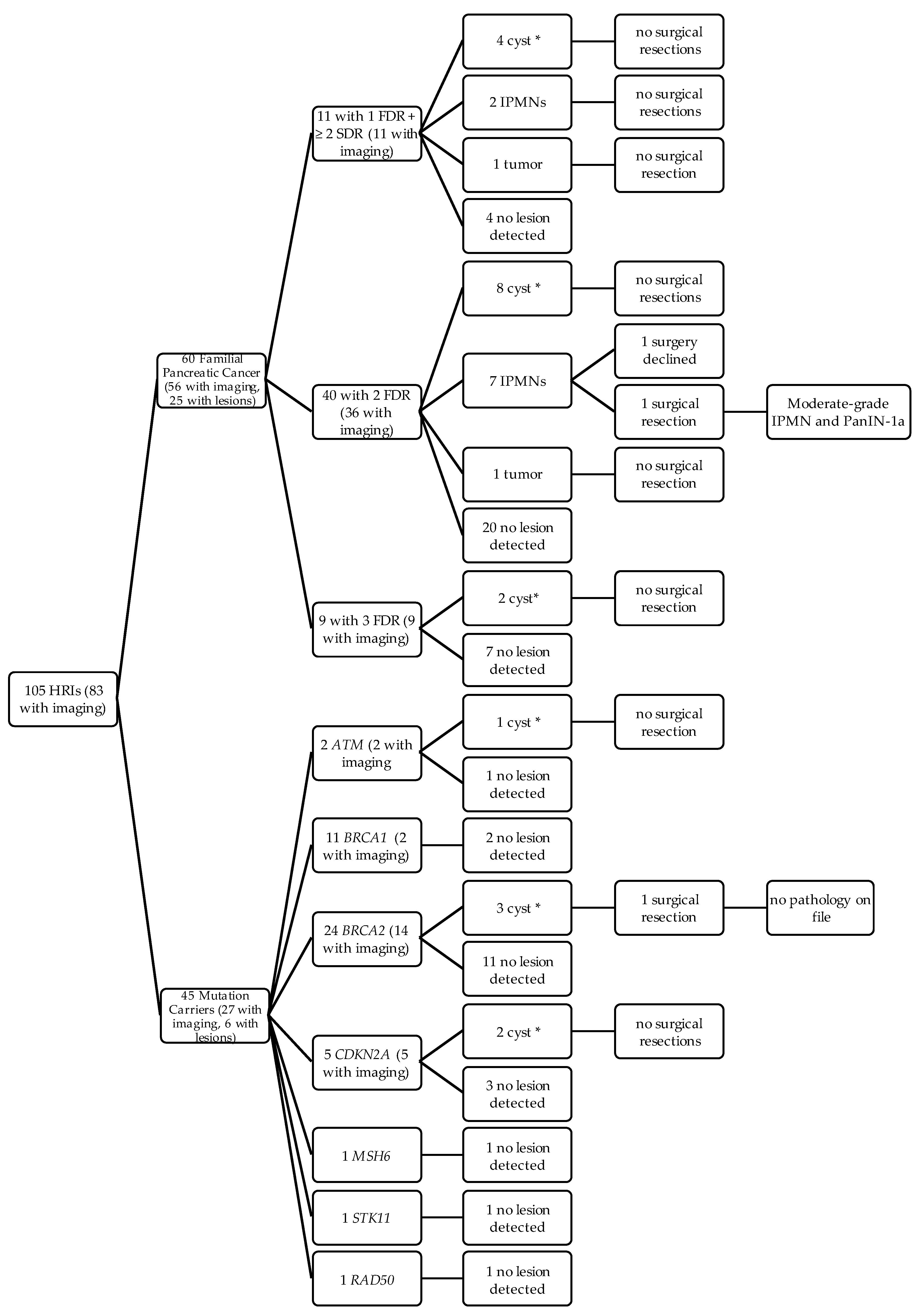
2.2. Outcomes of HRI that met CAPS Guidelines

Of the 105 HRI that met CAPS guidelines, most (n = 83, 79.1%) underwent imaging of the pancreas at Moffitt or had outside imaging on file. Thirty-one of these 83 individuals with imaging (37.4%) were found to have pancreatic lesions (Figure 1). Of the 60 familial cases, nearly all (n = 56 or 93.3%) had abdominal imaging reports on file. Twenty-five of the 56 familial cases (44.6%) had lesions detected, including two cancerous tumors and 9 IPMNs, one of which was resected and found to harbor moderate-grade disease and PANIN-1A (Figure 1). On the other hand, only 27 (60.0%) of the 45 mutation carriers meeting CAPS criteria had abdominal imaging on file. Six of the 27 mutation carriers (22%) had pancreatic cysts of unknown type detected, including 1 ATM carrier, 3 BRCA2 carriers, and 2 CDKN2A carriers. To our knowledge, all but one of the 105 HRI in this category are still living; the one deceased participant did not have a cystic or solid pancreatic lesion.

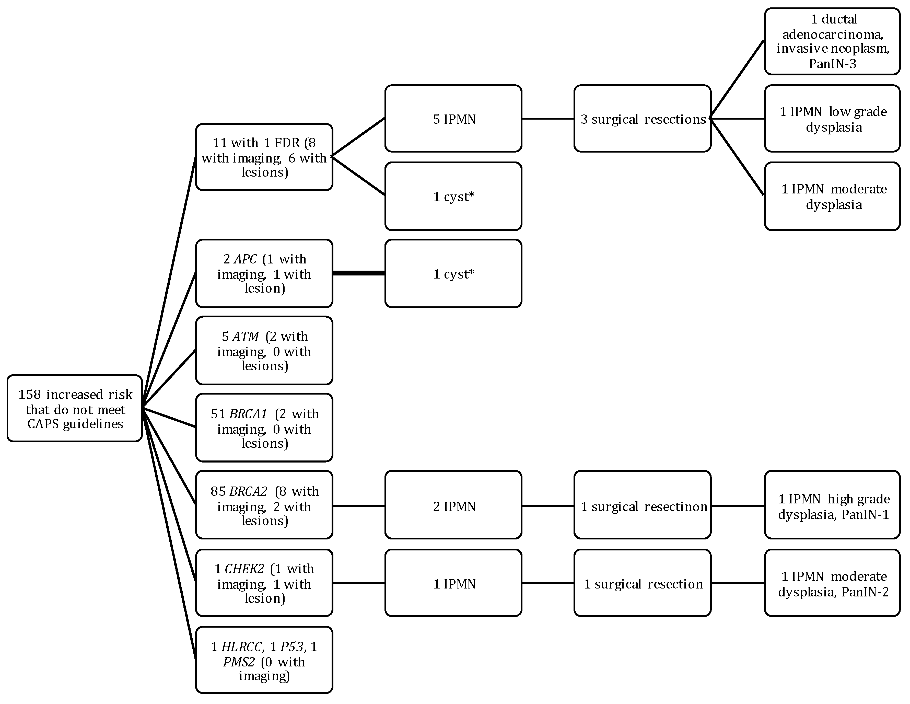
2.3. Outcomes of HRI That Did Not Meet CAPS Guidelines

Of the 158 patients who were at increased risk for PC but did not meet CAPS guidelines for screening (Figure 2), 11 had one affected FDR and 147 were known mutation carriers with no known family history of PC. Of the 11 individuals with one affected FDR, eight had imaging on file. Six of these eight individuals had lesions, five of which were IPMNs. Three of these individuals underwent surgical resection, resulting in one adenocarcinoma with PanIN-3, one IPMN with low-grade dysplasia, and one IPMN with moderate-grade dysplasia. Of the 147 mutation carriers, only 23 (15.6%) had abdominal imaging on file and five of these patients were found to carry a 6174 delT BRCA2 mutation which provides a 10% lifetime risk for PC, nearly double the lifetime risk required for CAPS screening [7,45]. Four of the mutation carriers with imaging on file had lesions; three had IPMNs and one had a cyst of unknown type. Two of the IPMN patients (a CHEK2 and a BRCA2 mutation carrier) underwent surgical resection. The IPMN found in the BRCA2 carrier had main duct involvement seen on imaging. Pathology reports after surgical resection reported that the patient with a CHEK2 mutation had an IPMN with moderate grade dysplasia, and the patient with a BRCA2 mutation had an IPMN with high-grade dysplasia and PanIN-1. These lesions were not detected as part of Moffitt’s high-risk surveillance protocol because the participants did not meet CAPS guidelines. Instead, they were detected incidentally among participants recruited as part of our team’s Florida Pancreas Collaborative (FPC) Study (see Methods).

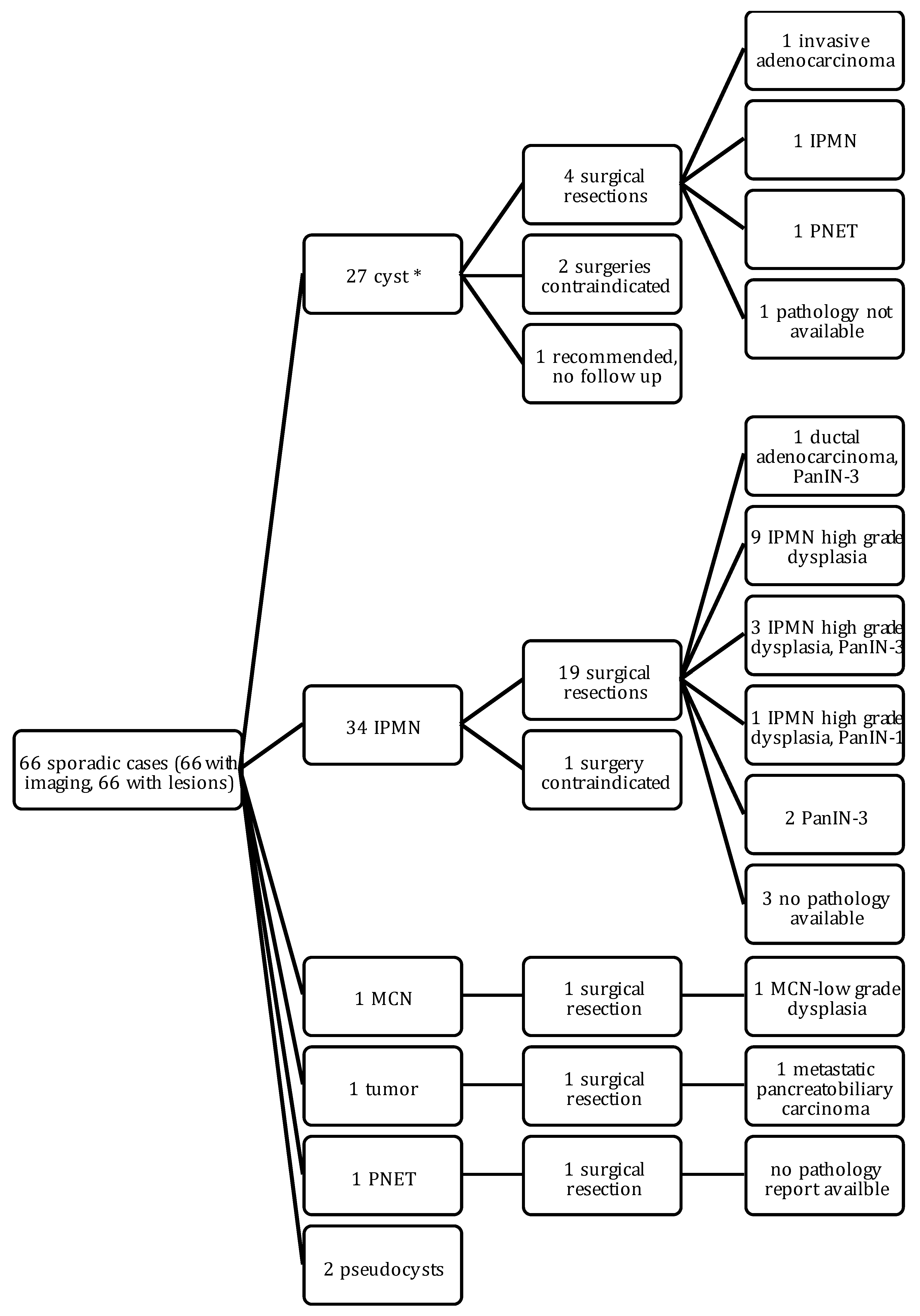
2.4. Outcomes of Sporadic Cases

Sixty-six of the 329 HRI in our cohort (20.0%) were considered to be “sporadic” cases, meaning they had no known family history of PC or hereditary syndrome (Figure 3). Most of these cases were recruited through our Florida Pancreas Collaborative Protocol; approximately 10.5% presented with asymptomatic, incidental pancreatic lesions on imaging and the remainder presented with symptoms such as abdominal pain and jaundice. Most of these cases had IPMNs (n = 34, 51.5%), and 19 of the IPMNs were surgically resected. This yielded one ductal adenocarcinoma with PanIN-3, 13 high-grade IPMNs (three of which had an associated PanIN-3 and nine of which had main duct involvement), and two PanIN-3s. Six of the 66 sporadic cases are deceased; three had an IPMN, one with high-grade dysplasia, and three had cysts of unknown types.

3. Discussion

Advancements in the early detection and prevention of pancreatic cancer requires strategies to detect and treat pre-malignant lesions and early invasive disease among individuals at high risk to develop this malignancy. We performed a descriptive analysis of the characteristics and clinical outcomes of three groups of high risk individuals (HRI) identified at our cancer center in order to better understand the uptake of screening, the prevalence and characteristics of solid and cystic pancreatic lesions detected via screening or as incidental findings, and the age at which lesions were detected.
As expected, individuals meeting the 5–10% lifetime risk of PC recommended by CAPS had the highest uptake of screening, primarily with a previously-described EUS-only High-risk Surveillance protocol [6]. Nearly 40% of the patients meeting CAPS criteria with abdominal imaging on file were found to have pancreatic lesions, primarily unclassifiable cysts due to their small size, which is consistent with findings from other studies [46]. This suggests that proactive screening efforts for high risk individuals are effective in identifying a high prevalence of smaller pancreatic lesions that should be monitored. Of the 22 individuals for whom we do not have a report of abdominal imaging on file, 18 were at least 50 years old and therefore should be recommended for a screening protocol. Moreover, 11 of these individuals without a report of pancreas screening on file harbored a deleterious BRCA1 or BRCA2 mutation and a family history of pancreatic cancer, including one individual with six second degree relatives with the disease. It is possible that these HRI received imaging elsewhere, but our observation highlights the importance of continuity of care between cancer genetics and gastrointestinal oncology clinics so that HRI are made aware of opportunities for screening studies.
Among those at increased moderate genetic risk for PC who did not meet the CAPS guidelines threshold for screening and therefore were not part of a surveillance study at our institution, we identified 10 individuals who had abdominal imaging on file and for whom cystic and solid pancreatic lesions were detected as part of a diagnostic work-up. This is likely an underestimate since most of these patients did not have abdominal imaging on file. We observed an earlier mean age of lesion diagnosis (60.7 years) in this group of HRI compared to those that met CAPs guidelines (62.61 years), and also observed a significantly larger lesion size (1.95 cm, p < 0.0001). Although this cohort is small, the earlier age, the larger size of lesions detected, and the detection of one invasive tumor and several high-grade IPMNs seems relevant when considering expanding current screening guidelines to include patients at an increased risk for disease that are on the cusp of reaching current CAPS guidelines. Moreover, a personalized approach that also examines individual patient characteristics such as hereditary cancer disorders, medical conditions such as pancreatitis, diabetes and obesity, and environmental factors such as smoking, and drinking histories can help to determine whether these people are at a 5% lifetime risk and would benefit from screening.
We also reported on 66 individuals not known to have a familial or hereditary predisposition to PC who presented with incidental findings of pre-malignant pancreatic cysts. This group of “sporadic” HRI had a later age at diagnosis, which may have led to larger lesions than the other two groups, in line with other studies [47,48]. Moreover, this group had the highest percentage (45.5%) of patients requiring surgical resection who had significant pathology. The National Comprehensive Cancer Network (version 1.2018) recently started recommending that genetic counseling and germline testing should be considered for patients with a personal or family history of PC or those with a clinical suspicion of inherited susceptibility. Data from a recent case-control study support this recommendation, as germline mutations in one of six genes (CDKN2A, TP53, MLH1, BRCA2, ATM, and BRCA1) were identified in 5.5% of all PC patients, including 7.9% of patients with a family history of PC and 5.2% of patients without a family history of PC [49]. Taken together, this group with sporadic findings of pre-malignant pancreatic cysts may also represent a population for whom germline genetic testing for cancer predisposition genes should be offered.
Our study is limited because it is based on a retrospective cohort of individuals from a single institution. Despite this, our cohort is unique because it includes patients from two protocols that recruited participants based on their family history and/or presence of a known germline mutation and another protocol that recruited participants unselected for family history or genetic profile. In doing so, we included a diverse cohort of HRI from our comprehensive cancer center. Additionally, it is very possible that a subset of HRI from this cohort has been followed for their medical management or received genetic counseling and testing at other centers and that our information is incomplete. Thus, further follow-up of this cohort is warranted to update their medical and screening history. Moreover, results of this follow-up could inform a prospective study that could serve to verify this retrospective review. Despite these limitations, our study identifies a number of areas in which we can work to optimize guidelines for who is offered genetic counseling and testing and screening. Large multi-center trials are needed to validate our findings and better define the optimal screening regimen for various groups of HRI, with an overarching goal of promoting the detection and treatment of pre-malignant lesions and early, operable disease.

4. Methods

Data was gathered from three IRB-approved protocols based at Moffitt Cancer Center: the Inherited Cancer Registry (ICARE) Initiative (MCC 12347, PI: C. Laronga), the High Risk Surveillance Protocol (MCC 14882, PI: J. Klapman) [6], and the Florida Pancreas Collaborative (MCC 18336, PI: J. Permuth) [50]. The ICARE initiative aims to establish a registry of individuals interested in participating in studies of the genetic causes of cancer and seeks to evaluate the roles of genetic and environmental risk factors in the development of tumors or related conditions. Eligible individuals joined ICARE between July 2010 and March 2017 at Moffitt Cancer Center. The High Risk Surveillance Protocol provides annual EUS (with fine needle aspiration if possible) for individuals meeting CAPs guidelines and took place between June 2007 and December 2017 [7]. To be eligible, individuals must have two or more relatives with PDAC with at least one FDR affected, and be at least 40 years old or 10 years younger than the youngest affected family member, have Peutz-Jeghers Syndrome and be at least 30 years old, have hereditary pancreatitis, have familial atypical multiple mole melanoma syndrome, or have a BRCA2 mutation and at least one FDR or SDR with documented PC [6]. The Florida Pancreas Collaborative is an ongoing biorepository that was established in September of 2015 and includes individuals newly-diagnosed with a breadth of pancreatic conditions ranging from early and late-stage PDAC; benign, pre-malignant, and malignant pancreatic cysts; and pancreatitis [50].
Eligible HRI were identified from the aforementioned protocols and data was collected from the electronic medical record on variables including: age, gender, race/ethnicity, abdominal imaging modalities used and at what age, type of lesion detected, age at which lesion was detected, surgical pathology, and vital status. Data was also collected on personal medical history, body mass index, and history of tobacco exposure and alcohol use. When recording the type of lesion detected, the diagnosis was abstracted from the first imaging report on which the lesion was found or an ambulatory care note. Comparisons were made across different at-risk groups using chi-squared test and Fisher’s exact test (where the cell size was <5) for categorical variables and ANOVA for continuous variables.

Supplementary Materials

Supplementary materials can be found at https://0-www-mdpi-com.brum.beds.ac.uk/2624-5647/1/1/8/s1.

Author Contributions

Study conceptualization/design: J.B.P., G.P.J.M., T.H., M.N., K.N.A.; Data abstraction: G.P.J.M., D.A.; Formal analysis/investigation: G.P.J.M., J.B.P., S.V.; Manuscript writing: G.P.J.M., J.B.P., Manuscript review/approval: G.P.J.M., J.B.P., K.N.A., T.H., S.V., M.N., D.A., C.L., J.K.

Acknowledgments

This investigation was supported in part by: The State of Florida and the Florida Academic Cancer Center Alliance (FACCA) and the Total Cancer Care™ Protocol at the H. Lee Moffitt Cancer Center & Research Institute, an NCI designated Comprehensive Cancer Center (P30-CA076292).

Conflicts of Interest

We declare no conflicts of interest.

References

  1. Noone, A.M.H.N.; Krapcho, M.; Brest, A.; Yu, M.; Ruhl, J.; Tatalovich, Z.; Mariotto, A.; Lewis, D.R.; Chen, H.S.; Feuer, E.J.; et al. SEER Cancer Statistics Review, 1975–2015. Available online: https://seer.cancer.gov/csr/1975_2015/ (accessed on 20 July 2018).
  2. Shi, C.; Hruban, R.H.; Klein, A.P. Familial pancreatic cancer. Arch. Pathol. Lab. Med. 2009, 133, 365–374. [Google Scholar] [CrossRef] [PubMed]
  3. Siegel, R.L.; Miller, K.D.; Jemal, A. Cancer statistics, 2017. CA A Cancer J. Clin. 2017, 67, 7–30. [Google Scholar] [CrossRef] [PubMed]
  4. Rahib, L.; Smith, B.D.; Aizenberg, R.; Rosenzweig, A.B.; Fleshman, J.M.; Matrisian, L.M. Projecting Cancer Incidence and Deaths to 2030: The Unexpected Burden of Thyroid, Liver, and Pancreas Cancers in the United States. Cancer Res. 2014. [Google Scholar] [CrossRef] [PubMed]
  5. Vincent, A.; Herman, J.; Schulick, R.; Hruban, R.H.; Goggins, M. Pancreatic cancer. Lancet 2011, 378, 607–620. [Google Scholar] [CrossRef]
  6. Gangi, A.; Malafa, M.; Klapman, J. Endoscopic Ultrasound–Based Pancreatic Cancer Screening of High-Risk Individuals: A Prospective Observational Trial. Pancreas 2018, 47, 586–591. [Google Scholar] [CrossRef] [PubMed]
  7. Canto, M.I.; Harinck, F.; Hruban, R.H.; Offerhaus, G.J.; Poley, J.-W.; Kamel, I.; Nio, Y.; Schulick, R.S.; Bassi, C.; Kluijt, I.; et al. International Cancer of the Pancreas Screening (CAPS) Consortium summit on the management of patients with increased risk for familial pancreatic cancer. Gut 2013, 62, 339–347. [Google Scholar] [CrossRef] [PubMed]
  8. Lu, C.; Xu, C.-F.; Wan, X.-Y.; Zhu, H.-T.; Yu, C.-H.; Li, Y.-M. Screening for pancreatic cancer in familial high-risk individuals: A systematic review. World J. Gastroenterol. WJG 2015, 21, 8678–8686. [Google Scholar] [CrossRef] [PubMed]
  9. Islami, F.; Goding Sauer, A.; Miller, K.D.; Siegel, R.L.; Fedewa, S.A.; Jacobs, E.J.; McCullough, M.L.; Patel, A.V.; Ma, J.; Soerjomataram, I.; et al. Proportion and number of cancer cases and deaths attributable to potentially modifiable risk factors in the United States. CA A Cancer J. Clin. 2018, 68, 31–54. [Google Scholar] [CrossRef] [PubMed]
  10. Hassan, M.M.; Bondy, M.L.; Wolff, R.A.; Abbruzzese, J.L.; Vauthey, J.-N.; Pisters, P.W.; Evans, D.B.; Khan, R.; Chou, T.-H.; Lenzi, R.; et al. Risk Factors for Pancreatic Cancer: Case-Control Study. Am. J. Gastroenterol. 2007, 102, 2696. [Google Scholar] [CrossRef] [PubMed]
  11. Permuth-Wey, J.; Egan, K.M. Family history is a significant risk factor for pancreatic cancer: Results from a systematic review and meta-analysis. Fam. Cancer 2009, 8, 109–117. [Google Scholar] [CrossRef] [PubMed]
  12. Klein, A.P.; Brune, K.A.; Petersen, G.M.; Goggins, M.; Tersmette, A.C.; Offerhaus, G.J.A.; Griffin, C.; Cameron, J.L.; Yeo, C.J.; Kern, S.; et al. Prospective Risk of Pancreatic Cancer in Familial Pancreatic Cancer Kindreds. Cancer Res. 2004, 64, 2634–2638. [Google Scholar] [CrossRef] [PubMed] [Green Version]
  13. Giardiello, F.M.; Brensinger, J.D.; Tersmette, A.C.; Goodman, S.N.; Petersen, G.M.; Booker, S.V.; Cruz-Correa, M.; Offerhaus, J.A. Very high risk of cancer in familial Peutz-Jeghers syndrome. Gastroenterology 2000, 119, 1447–1453. [Google Scholar] [CrossRef] [PubMed]
  14. Bartsch, D.K.; Gress, T.M.; Langer, P. Familial pancreatic cancer—Current knowledge. Nat. Rev. Gastroenterol. Hepatol. 2012, 9, 445–453. [Google Scholar] [CrossRef] [PubMed]
  15. Shi, C.; Klein, A.P.; Goggins, M.; Maitra, A.; Canto, M.; Ali, S.; Schulick, R.; Palmisano, E.; Hruban, R.H. Increased Prevalence of Precursor Lesions in Familial Pancreatic Cancer Patients. Clin. Cancer Res. 2009, 15, 7737–7743. [Google Scholar] [CrossRef] [PubMed] [Green Version]
  16. Bartsch, D.K.; Slater, E.P.; Carrato, A.; Ibrahim, I.S.; Guillen-Ponce, C.; Vasen, H.F.; Matthai, E.; Earl, J.; Jendryschek, F.S.; Figiel, J.; et al. Refinement of screening for familial pancreatic cancer. Gut 2016, 65, 1314–1321. [Google Scholar] [CrossRef] [PubMed]
  17. Midha, S.; Chawla, S.; Garg, P.K. Modifiable and non-modifiable risk factors for pancreatic cancer: A review. Cancer Lett. 2016, 381, 269–277. [Google Scholar] [CrossRef] [PubMed]
  18. Thompson, D.; Easton, D.F.; The Breast Cancer Linkage Consortium. Cancer Incidence in BRCA1 Mutation Carriers. JNCI J. Natl. Cancer Inst. 2002, 94, 1358–1365. [Google Scholar] [CrossRef] [PubMed] [Green Version]
  19. Potjer, T.P.; Schot, I.; Langer, P.; Heverhagen, J.T.; Wasser, M.N.J.M.; Slater, E.P.; Klöppel, G.; Morreau, H.M.; Bonsing, B.A.; de Vos tot Nederveen Cappel, W.H.; et al. Variation in Precursor Lesions of Pancreatic Cancer among High-Risk Groups. Clin. Cancer Res. 2013, 19, 442–449. [Google Scholar] [CrossRef] [PubMed]
  20. Lucas, A.L.; Shakya, R.; Lipsyc, M.D.; Mitchel, E.B.; Kumar, S.; Hwang, C.; Deng, L.; Devoe, C.; Chabot, J.A.; Szabolcs, M.; et al. High Prevalence of BRCA1 and BRCA2 Germline Mutations with Loss of Heterozygosity in a Series of Resected Pancreatic Adenocarcinoma and Other Neoplastic Lesions. Clin. Cancer Res. 2013, 19, 3396–3403. [Google Scholar] [CrossRef] [PubMed]
  21. The Breast Cancer Linkage Consortium. Cancer Risks in BRCA2 Mutation Carriers. JNCI J. Natl. Cancer Inst. 1999, 91, 1310–1316. [Google Scholar] [CrossRef] [Green Version]
  22. Van Asperen, C.J.; Brohet, R.M.; Meijers-Heijboer, E.J.; Hoogerbrugge, N.; Verhoef, S.; Vasen, H.F.A.; Ausems, M.G.E.M.; Menko, F.H.; Gomez Garcia, E.B.; Klijn, J.G.M.; et al. Cancer risks in BRCA2 families: Estimates for sites other than breast and ovary. J. Med. Genet. 2005, 42, 711–719. [Google Scholar] [CrossRef] [PubMed] [Green Version]
  23. Lubezky, N.; Ben-Haim, M.; Lahat, G.; Marmor, S.; Solar, I.; Brazowski, E.; Nackache, R.; Klausner, J.M. Intraductal papillary mucinous neoplasm of the pancreas: Associated cancers, family history, genetic predisposition? Surgery 2012, 151, 70–75. [Google Scholar] [CrossRef] [PubMed]
  24. Sato, N.; Rosty, C.; Jansen, M.; Fukushima, N.; Ueki, T.; Yeo, C.J.; Cameron, J.L.; Iacobuzio-Donahue, C.A.; Hruban, R.H.; Goggins, M. STK11/LKB1 Peutz-Jeghers Gene Inactivation in Intraductal Papillary-Mucinous Neoplasms of the Pancreas. Am. J. Pathol. 2001, 159, 2017–2022. [Google Scholar] [CrossRef] [Green Version]
  25. Vasen, H.F.A.; Wasser, M.; van Mil, A.; Tollenaar, R.A.; Konstantinovski, M.; Gruis, N.A.; Bergman, W.; Hes, F.J.; Hommes, D.W.; Offerhaus, G.J.A.; et al. Magnetic Resonance Imaging Surveillance Detects Early-Stage Pancreatic Cancer in Carriers of a p16-Leiden Mutation. Gastroenterology 2011, 140, 850–856. [Google Scholar] [CrossRef] [PubMed]
  26. Sparr, J.A.; Bandipalliam, P.; Redston, M.S.; Syngal, S. Intraductal papillary mucinous neoplasm of the pancreas with loss of mismatch repair in a patient with Lynch syndrome. Am. J. Surg. Pathol. 2009, 33, 309. [Google Scholar] [CrossRef] [PubMed]
  27. Xiao, S.-Y. Intraductal Papillary Mucinous Neoplasm of the Pancreas: An Update. Scientifica 2012, 2012, 20. [Google Scholar] [CrossRef] [PubMed]
  28. Chetty, R.; Salahshor, S.; Bapat, B.; Berk, T.; Croitoru, M.; Gallinger, S. Intraductal papillary mucinous neoplasm of the pancreas in a patient with attenuated familial adenomatous polyposis. J. Clin. Pathol. 2005, 58, 97–101. [Google Scholar] [CrossRef] [PubMed] [Green Version]
  29. Sessa, F.; Solcia, E.; Capella, C.; Bonato, M.; Scarpa, A.; Zamboni, G.; Pellegata, N.S.; Ranzani, G.N.; Rickaert, F.; Klöppel, G. Intraductal papillary-mucinous tumours represent a distinct group of pancreatic neoplasms: An investigation of tumour cell differentiation and K-ras, p53 and c-erbB-2 abnormalities in 26 patients. Virchows Arch. 1994, 425, 357–367. [Google Scholar] [CrossRef] [PubMed]
  30. Talamini, G.; Zamboni, G.; Salvia, R.; Capelli, P.; Sartori, N.; Casetti, L.; Bovo, P.; Vaona, B.; Falconi, M.; Bassi, C. Intraductal papillary mucinous neoplasms and chronic pancreatitis. Pancreatology 2006, 6, 626–634. [Google Scholar] [CrossRef] [PubMed]
  31. Pagliari, D.; Saviano, A.; Serricchio, M.; Dal Lago, A.; Brizi, M.; Manfredi, R.; Costamagna, G.; Attili, F.J.E. The association of pancreatic cystosis and IPMN in cystic fibrosis: Case report and literature review. Eur. Rev. Med. Pharmacol. Sci. 2017, 21, 5179–5184. [Google Scholar] [PubMed]
  32. Yonezawa, S.; Higashi, M.; Yamada, N.; Goto, M. Precursor lesions of pancreatic cancer. Gut Liver 2008, 2, 137. [Google Scholar] [CrossRef] [PubMed]
  33. Hruban, R.H.; Adsay, N.V.; Albores-Saavedra, J.; Compton, C.; Garrett, E.S.; Goodman, S.N.; Kern, S.E.; Klimstra, D.S.; Kloppel, G.; Longnecker, D.S.; et al. Pancreatic intraepithelial neoplasia: A new nomenclature and classification system for pancreatic duct lesions. Am. J. Surg. Pathol. 2001, 25, 579–586. [Google Scholar] [CrossRef] [PubMed]
  34. Hruban, R.H.; Maitra, A.; Kern, S.E.; Goggins, M. Precursors to Pancreatic Cancer. Gastroenterol. Clin. N. Am. 2007, 36, 831–849. [Google Scholar] [CrossRef] [PubMed] [Green Version]
  35. Hruban, R.H.; Takaori, K.; Klimstra, D.S.; Adsay, N.V.; Albores-Saavedra, J.; Biankin, A.V.; Biankin, S.A.; Compton, C.; Fukushima, N.; Furukawa, T.; et al. An illustrated consensus on the classification of pancreatic intraepithelial neoplasia and intraductal papillary mucinous neoplasms. Am. J. Surg. Pathol. 2004, 28, 977–987. [Google Scholar] [CrossRef] [PubMed]
  36. Matthaei, H.; Schulick, R.D.; Hruban, R.H.; Maitra, A. Cystic precursors to invasive pancreatic cancer. Nat. Rev. Gastroenterol. Hepatol. 2011, 8, 141. [Google Scholar] [CrossRef] [PubMed]
  37. Zamboni, G.; Hirabayashi, K.; Castelli, P.; Lennon, A.M. Precancerous lesions of the pancreas. Best Pract. Res. Clin. Gastroenterol. 2013, 27, 299–322. [Google Scholar] [CrossRef] [PubMed]
  38. Canto, M.I.; Goggins, M.; Hruban, R.H.; Petersen, G.M.; Giardiello, F.M.; Yeo, C.; Fishman, E.K.; Brune, K.; Axilbund, J.; Griffin, C.; et al. Screening for early pancreatic neoplasia in high-risk individuals: A prospective controlled study. Clin. Gastroenterol. Hepatol. 2006, 4, 766–781. [Google Scholar] [CrossRef] [PubMed]
  39. Torphy, R.J.; Schulick, R.D. Screening of Patients at Risk for Familial Pancreatic Cancer. Surg. Clin. 2018, 98, 25–35. [Google Scholar] [CrossRef] [PubMed]
  40. Tanaka, M.; Fernandez-Del Castillo, C.; Kamisawa, T.; Jang, J.Y.; Levy, P.; Ohtsuka, T.; Salvia, R.; Shimizu, Y.; Tada, M.; Wolfgang, C.L. Revisions of international consensus Fukuoka guidelines for the management of IPMN of the pancreas. Pancreatology 2017, 17, 738–753. [Google Scholar] [CrossRef] [PubMed]
  41. Verna, E.C.; Hwang, C.; Stevens, P.D.; Rotterdam, H.; Stavropoulos, S.N.; Sy, C.D.; Prince, M.A.; Chung, W.K.; Fine, R.L.; Chabot, J.A.; et al. Pancreatic Cancer Screening in a Prospective Cohort of High-Risk Patients: A Comprehensive Strategy of Imaging and Genetics. Clin. Cancer Res. 2010, 16, 5028–5037. [Google Scholar] [CrossRef] [PubMed] [Green Version]
  42. Brand, R.E.; Lerch, M.M.; Rubinstein, W.S.; Neoptolemos, J.P.; Whitcomb, D.C.; Hruban, R.H.; Brentnall, T.A.; Lynch, H.T.; Canto, M.I. Advances in counselling and surveillance of patients at risk for pancreatic cancer. Gut 2007, 56, 1460–1469. [Google Scholar] [CrossRef] [PubMed] [Green Version]
  43. Canto, M.I.; Almario, J.A.; Schulick, R.D.; Yeo, C.J.; Klein, A.; Blackford, A.; Shin, E.J.; Sanyal, A.; Yenokyan, G.; Lennon, A.M.; et al. Risk of Neoplastic Progression in Individuals at High Risk for Pancreatic Cancer Undergoing Long-term Surveillance. Gastroenterology 2018. [Google Scholar] [CrossRef] [PubMed]
  44. Kimmey, M.B.; Bronner, M.P.; Byrd, D.R.; Brentnall, T.A. Screening and surveillance for hereditary pancreatic cancer. Gastrointest. Endosc. 2002, 56, S82–S86. [Google Scholar] [CrossRef]
  45. Özcelik, H.; Schmocker, B.; Nicola, N.D.; Shi, X.-H.; Langer, B.; Moore, M.; Taylor, B.R.; Narod, S.A.; Darlington, G.; Andrulis, I.L.; et al. Germline BRCA2 6174delT mutations in Ashkenazi Jewish pancreatic cancer patients. Nat. Genet. 1997, 16, 17. [Google Scholar] [CrossRef] [PubMed]
  46. Canto, M.I.; Hruban, R.H.; Fishman, E.K.; Kamel, I.R.; Schulick, R.; Zhang, Z.; Topazian, M.; Takahashi, N.; Fletcher, J.; Petersen, G.; et al. Frequent Detection of Pancreatic Lesions in Asymptomatic High-Risk Individuals. Gastroenterology 2012, 142, 796–804. [Google Scholar] [CrossRef] [PubMed] [Green Version]
  47. Lee, K.S.; Sekhar, A.; Rofsky, N.M.; Pedrosa, I. Prevalence of Incidental Pancreatic Cysts in the Adult Population on MR Imaging. Am. J. Gastroenterol. 2010, 105, 2079. [Google Scholar] [CrossRef] [PubMed]
  48. Lee, C.J.; Scheiman, J.; Anderson, M.A.; Hines, O.J.; Reber, H.A.; Farrell, J.; Kochman, M.L.; Foley, P.J.; Drebin, J.; Oh, Y.S.; et al. Risk of Malignancy in Resected Cystic Tumors of the Pancreas ≤3 cm in Size: Is it Safe to Observe Asymptomatic Patients? A Multi-institutional Report. J. Gastrointest. Surg. 2008, 12, 234–242. [Google Scholar] [CrossRef] [PubMed]
  49. Hu, C.; Hart, S.N.; Bamlet, W.R.; Moore, R.M.; Nandakumar, K.; Eckloff, B.W.; Lee, Y.K.; Petersen, G.M.; McWilliams, R.R.; Couch, F.J.J.C.E.; et al. Prevalence of pathogenic mutations in cancer predisposition genes among pancreatic cancer patients. Cancer Epidemiol. Prev. Biomark. 2016, 25, 207–211. [Google Scholar] [CrossRef] [PubMed]
  50. Permuth, J.B.; Trevino, J.; Merchant, N.; Malafa, M.; Florida Pancreas Collaborative. Partnering to advance early detection and prevention efforts for pancreatic cancer: The Florida Pancreas Collaborative. Future Med. 2016, 12, 997–1000. [Google Scholar] [CrossRef] [PubMed]
Figure 1. Breakdown of high risk individuals meeting CAPs guidelines and the yield of pancreatic lesions detected. * cyst type not specified.
Gastrointestdisord 01 00008 g001
Figure 2. Breakdown of high risk individuals that did not meet CAPS guidelines and the yield of pancreatic lesions detected. These individuals were at increased risk for pancreatic cancer because they had only one first degree relative with pancreatic cancer or a known deleterious mutation (with no family history of pancreatic cancer). * cyst type not specified.
Gastrointestdisord 01 00008 g002
Figure 3. Sporadic cases and pancreatic lesion findings. Individuals in this cohort were identified through the FPC study and have no family history for pancreatic cancer or known deleterious mutation for a hereditary cancer syndrome. * cyst type not specified.
Gastrointestdisord 01 00008 g003
Table 1. Known hereditary cancer syndromes or syndromes involving chronic inflammation, the relative lifetime risk for PC, and reports of PC precursor lesions among mutation carriers [14]. * Estimates in this column pertain to intraductal papillary mucinous neoplasms (IPMNs), the cystic PC precursor most commonly detected via imaging.
SyndromeGene(s)Risk of PC by Age 70–75Studies Reporting PC Precursor Lesions * among Carriers
Hereditary Breast and Ovarian Cancer (HBOC)BRCA24.5–8%[23]
BRCA1, PALB23.6%
Peutz-Jeghers (PJS)STK11/LKB136%[24]
Familial atypical multiple-mole melanoma (FAMMM)P16/CDKN2A13–17%[25]
Hereditary non-polyposis colorectal cancer (HNPCC)MSH2, MLH1, MSH6, PMS1, PMS23.7%[23,26,27]
Familial adenomatous polyposis (FAP)APC1.7%[28]
Ataxia telangiectasiaATM<5%None identified
Li FraumeniTP53<5%[29]
Hereditary PancreatitisPRSS1, SPINK125–54%[30]
Cystic FibrosisCFTR<5%[31]
Table 2. Characteristics of the high risk cohort, stratified by group. Note: High risk individuals that met CAPS guidelines had either ≥2 FDR or a known hereditary cancer disorder and a relative with pancreatic cancer. Increased risk and sporadic cases did not meet CAPS guidelines for screening. The increased risk group had either 1 FDR or a germline mutation with no family history of pancreatic cancer. Sporadic cases did not have any family history or known genetic mutation.
CharacteristicEntire CohortHigh Risk According to CAPS Guidelines (n = 105)Increased risk that Does Not Meet CAPS Guidelines (n = 158)Sporadic Cases (n = 66)p *
Sex
Male86 (26.1)33 (31.4)21 (13.3)32 (48.5)<0.0001
Female243 (73.9)72 (68.6)137 (86.7)34 (51.5)
Race/ethnicity
Non-Hispanic White277 (84.5)93 (88.5)127 (80.9)57 (86.4)0.86
Non-Hispanic Black18 (5.5)5 (4.8)10 (6.4)3 (4.5)
Hispanic White22 (6.7)5 (4.8)13 (8.3)4 (6.1)
Other11 (3.3)2 (1.9)7(4.4)2 (3.0)
Missing1001
Smoking Status
Current27 (8.2)9 (8.9)14 (10.2)4 (6.3)0.06
Former192 (58.4)23 (22.8)33 (23.9)27 (42.9)
Never83 (25.2)69 (68.3)91 (65.9)32 (50.8)
Missing274203
Personal History of Cancer or Other Conditions
Breast Cancer10 (3.1)5 (4.8)2 (1.3)3 (4.5)
Colorectal cancer3 (0.9)0 (0.0)1(0.6)2 93.0)
¥ Gynecological Cancer3 (0.9)1 (1.0)0 (0.0)2 (3.0)
Prostate Cancer10 (3.1)2 (1.9)0 (0.0)8 (12.1)
¥ Other cancer13 (4.0)1 (1.0)2 (1.3)10 (15.2)
Pancreatitis9 (2.8)2 (1.9)2 (1.3)5 (7.6)
Diabetes9 (2.8)1 (1.0)1 (0.6)7 (10.6)
None270 (82.6)92 (88.5)149 (94.9)29 (43.9)
Missing2110
Pancreatic lesion detected on imaging
Had imaging and a lesion was detected107 (32.5)31 (29.5)10 (6.3)66 (100.0)<0.0001
Had imaging and no lesion was detected65 (19.8)52 (49.5)13 (8.2)0 (0.0)
No imaging on file157 (47.7)22 (21.0)135 (85.4)0 (0.0)
Type of pancreatic lesion detected <0.001
Cyst, type not specified49 (14.9)20 (19.1)2 (1.3)27 (40.9)
Intraductal papillary mucinous neoplasm51 (15.5)9 (8.6)8 (5.1)34 (51.5)
Mucinous cystic neoplasm1 (0.3)0 (0.0))0 (0.0)1 (1.5)
Pseudocyst2 (0.6)0 (0.0)0 (0.0)2 (3.0)
Solid Tumor/Adenocarcinoma3 (0.9)2 (1.9)0 (0.0)1 (1.5)
Pancreatic Neuroendocrine Tumor1 (0.3)0 (0.0)0 (0.0)1 (1.5)
Average age at diagnosis of pancreatic lesion65.562.660.767.6<0.020
Average size of largest lesion detected (cm)20.722.6<0.0001
Body Mass Index (BMI)-28.330.3270.27
* When calculating p-values, missing values were not included. (No percentages were calculated for missing values.); € p value could not be reliably estimated due to the small sample sizes in each group. ¥ Gynecological cancer includes ovarian cancer, uterine cancer, or cervical cancer. Other cancers included melanoma, bladder cancer, lung cancer, anus cancer, lymphoma, throat cancer, thyroid cancer, and tongue cancer. p value estimated by collapsing data into three categories: cyst, type not specified; IPMN; and all others.

