
Cyclic Voltammetry in Biological Samples: A Systematic Review of Methods and Techniques Applicable to Clinical Settings

 , and
, and
Abstract
:1. Introduction
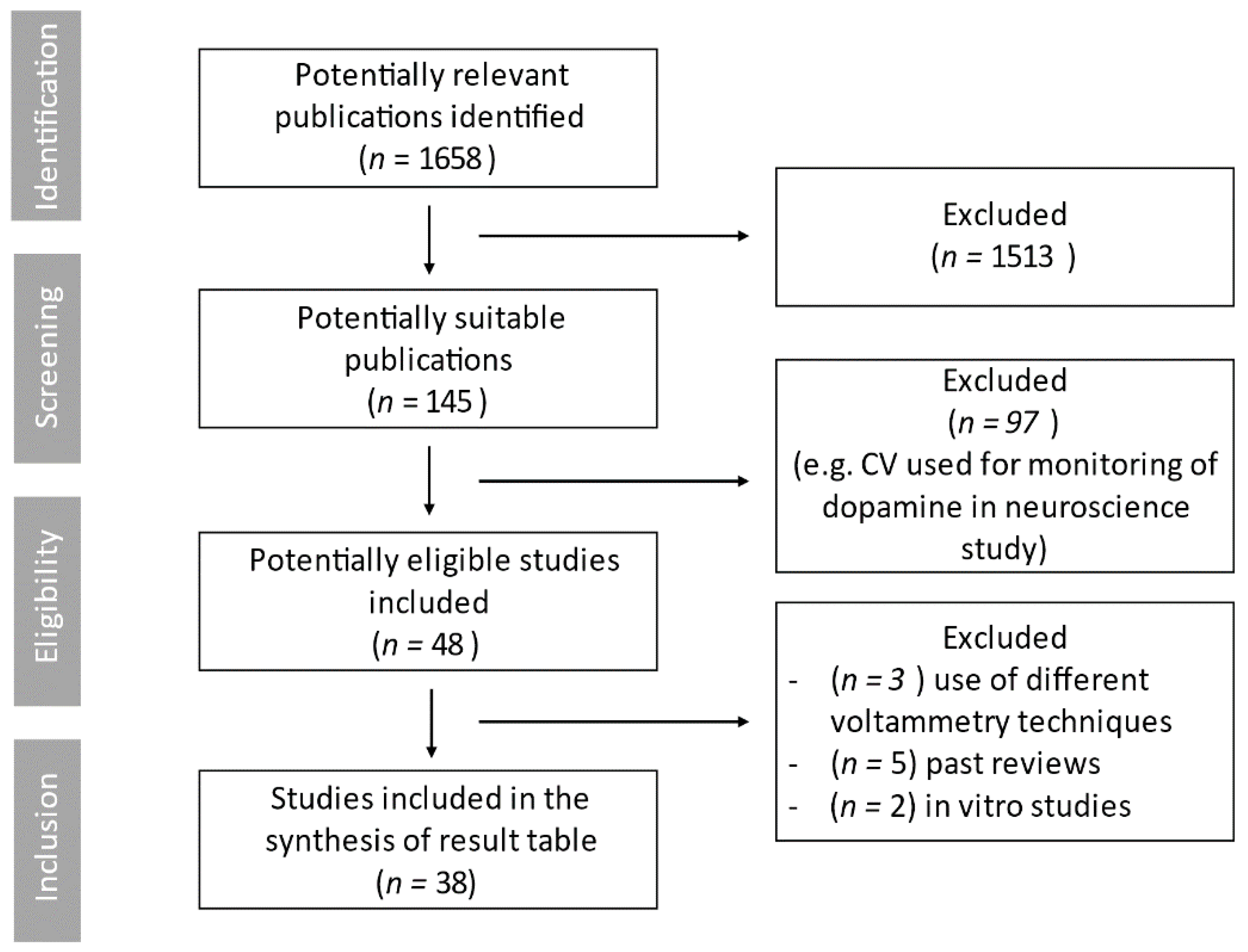
2. Materials and Methods
2.1. Inclusion and Exclusion Criteria
2.2. Data Extraction
3. Results
3.1. Sample Sources, Processing, and Storage
3.1.1. Blood Sampling
3.1.2. Tissue Samples
3.1.3. Lipophilic Low Molecular Weight Antioxidant Extraction
3.1.4. Other Sample Types
3.1.5. Sample Storage
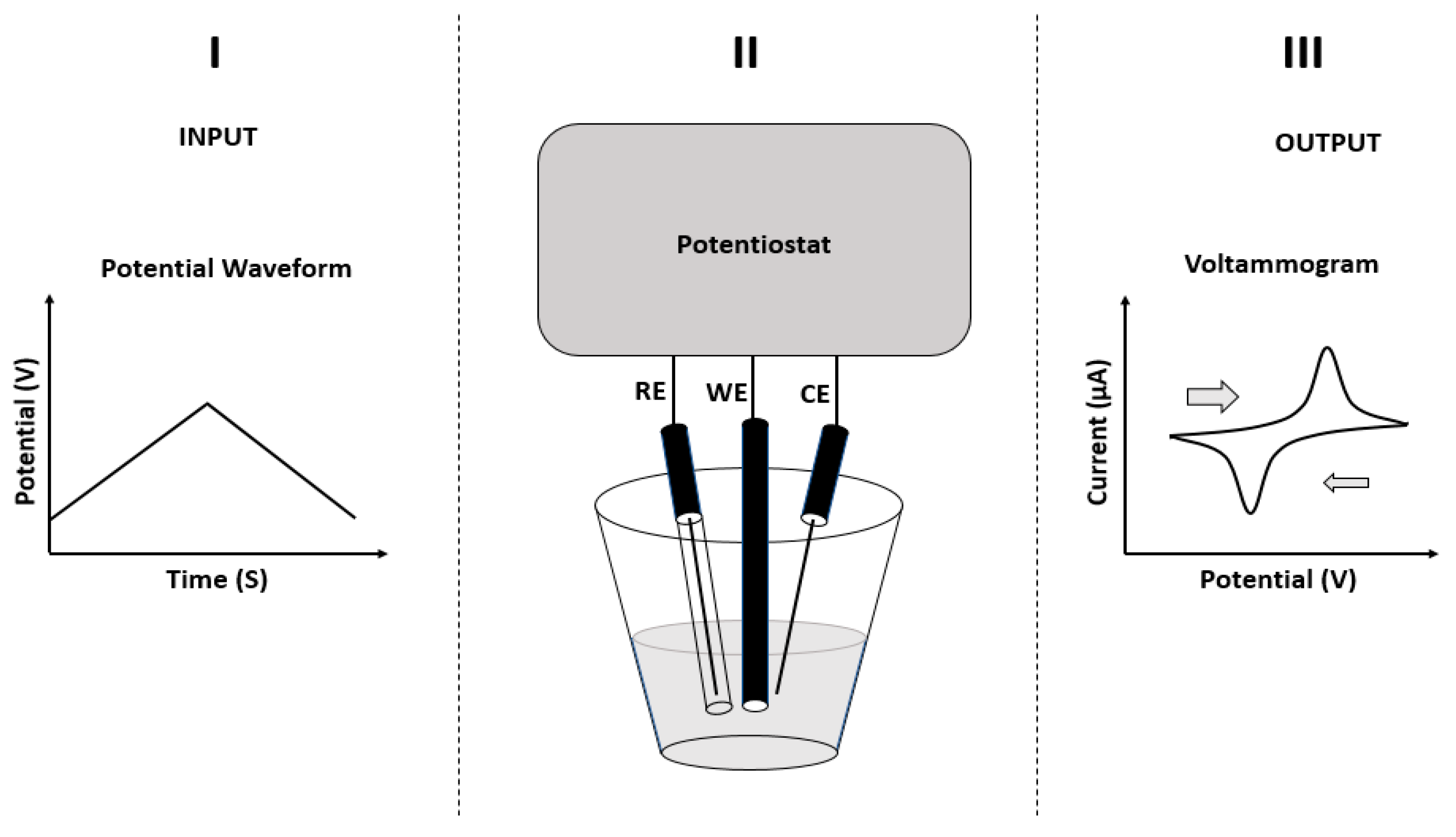
3.2. Voltammetry Equipment, Variables and Setting Parameters
3.2.1. Electrodes and Required Sample Volume
3.2.2. Scan Range
3.2.3. Scan Rate
3.2.4. Temperature and pH Control
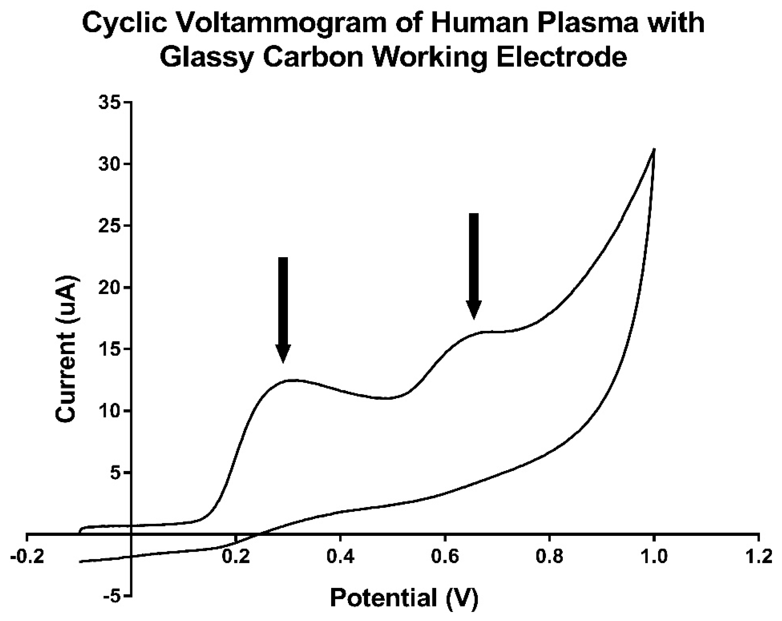
3.3. Determine the Constituents of Peak Potential
4. Discussion
5. Conclusions
Author Contributions
Funding
Institutional Review Board Statement
Informed Consent Statement
Data Availability Statement
Conflicts of Interest
References
- Pham-Huy, L.; He, H.; Pham-Huy, C. Free Radicals, Antioxidants in Disease and Health. Int. J. Biomed. Sci. 2008, 4, 89–96. [Google Scholar]
- Bar-Or, D.; Bar-Or, R.; Rael, L.T.; Brody, E.N. Oxidative stress in severe acute illness. Redox Biol. 2015, 4, 340–345. [Google Scholar] [CrossRef] [Green Version]
- Manzanares, W.; Dhaliwal, R.; Jiang, X.; Murch, L.; Heyland, D.K. Antioxidant micronutrients in the critically ill: A systematic review and meta-analysis. Critical Care 2012, 16, R66. [Google Scholar] [CrossRef] [Green Version]
- Siriwardena, A.K.; Fau, M.J.; Balachandra, S.F.; Bagul, A.F.; Galloway, S.F.; Formela, L.; Hardman, J.G.; Jamdar, S. Randomised, double blind, placebo controlled trial of intravenous antioxidant (n-acetylcysteine, selenium, vitamin C) therapy in severe acute pancreatitis. Gut 2007, 56, 1439–1444. [Google Scholar] [CrossRef] [Green Version]
- Tanaka, H.; Matsuda, T.; Miyagantani, Y.; Yukioka, T.; Matsuda, H.; Shimazaki, S. Reduction of resuscitation fluid volumes in severely burned patients using ascorbic acid administration: A randomized, prospective study. Arch. Surg. 2000, 135, 326–331. [Google Scholar] [CrossRef] [Green Version]
- Birben, E.; Sahiner, U.M.; Sackesen, C.; Erzurum, S.; Kalayci, O. Oxidative Stress and Antioxidant Defense. World Allergy Organ. J. 2012, 5, 9–19. [Google Scholar] [CrossRef] [Green Version]
- Valko, M.; Rhodes, C.J.; Moncol, J.; Izakovic, M.; Mazur, M. Free radicals, metals and antioxidants in oxidative stress-induced cancer. Chem. Biol. Interact. 2006, 160, 1–40. [Google Scholar] [CrossRef] [PubMed]
- Valko, M.; Leibfritz, D.; Moncol, J.; Cronin, M.T.D.; Mazur, M.; Telser, J. Free radicals and antioxidants in normal physiological functions and human disease. Int. J. Biochem. Cell Biol. 2007, 39, 44–84. [Google Scholar] [CrossRef]
- Sardesai, V.M. Role of Antioxidants in Health Maintenance. Nutr. Clin. Pract. 1995, 10, 19–25. [Google Scholar] [CrossRef] [PubMed]
- Goode, H.F.; Cowley, H.C.; Walker, B.E.; Howdle, P.D.; Webster, N.R. Decreased antioxidant status and increased lipid peroxidation in patients with septic shock and secondary organ dysfunction. Crit. Care Med. 1995, 23, 46–51. [Google Scholar] [CrossRef]
- Takeda, K.; Shimada, Y.; Amano, M.; Sakai, T.; Okada, T.; Yoshiya, I. Plasma lipid peroxides and alpha-tocopherol in critically ill patients. Crit. Care Med. 1984, 12, 957–959. [Google Scholar] [CrossRef]
- Leff, J.A.; Day, C.E.; McCord, J.M.; Repine, J.E.; Parsons, P.E.; Moore, F.A.; Fritz, H. Serum antioxidants as predictors of adult respiratory distress syndrome in patients with sepsis. Lancet 1993, 341, 777–780. [Google Scholar] [CrossRef] [Green Version]
- Borrelli, E.; Roux-Lombard, P.; Grau, G.E.; Girardin, E.; Ricou, B.; Dayer, J.; Suter, P.M. Plasma concentrations of cytokines, their soluble receptors, and antioxidant vitamins can predict the development of multiple organ failure in patients at risk. Crit. Care Med. 1996, 24, 392–397. [Google Scholar] [CrossRef]
- Abiles, J.; de la Cruz, A.P.; Castano, J.; Rodriguez-Elvira, M.; Aguayo, E.; Moreno-Torres, R.; Planells, E.M. Oxidative stress is increased in critically ill patients according to antioxidant vitamins intake, independent of severity: A cohort study. Crit. Care 2006, 10, R146. [Google Scholar] [CrossRef] [Green Version]
- Asmat, U.; Abad, K.; Ismail, K. Diabetes mellitus and oxidative stress—A concise review. Saudi Pharm. J. 2016, 24, 547–553. [Google Scholar] [CrossRef] [Green Version]
- Holley, A.E.; Cheeseman, K.H. Measuring free radical reactions in vivo. Br. Med. Bull. 1993, 49, 494–505. [Google Scholar] [CrossRef]
- Abu-Zidan, F.; Bonham, M.J.D.; Windsor, J.A. Severity of acute pancreatitis: A multivariate analysis of oxidative stress markers and modified Glasgow criteria. Br. J. Surg. 2000, 87, 1019–1023. [Google Scholar] [CrossRef] [PubMed]
- Schoenberg, M.H.; Buchler, M.; Gaspar, M.; Stinner, A.; Younes, M.; Melzner, I.; Beger, H.G. Oxygen free radicals in acute pancreatitis of the rat. Gut 1990, 31, 1138–1143. [Google Scholar] [CrossRef] [Green Version]
- Kumar, Y.; Singh, G.; Davidson, B.R. Free Radical and Antioxidant Levels in Patients with Secondary Peritonitis and Their Prognostic Significance. Dig. Surg. 2007, 24, 331–337. [Google Scholar] [CrossRef] [PubMed]
- Huang, D.; Ou, B.; Prior, R.L. The Chemistry behind Antioxidant Capacity Assays. J. Agric. Food Chem. 2005, 53, 1841–1856. [Google Scholar] [CrossRef]
- Niki, E. Assessment of Antioxidant Capacity in vitro and in vivo. Free Radic. Biol. Med. 2010, 49, 503–515. [Google Scholar] [CrossRef]
- Carocho, M.; Ferreira, I.C. A review on antioxidants, prooxidants and related controversy: Natural and synthetic compounds, screening and analysis methodologies and future perspectives. Food Chem. Toxicol. 2013, 51, 15–25. [Google Scholar] [CrossRef]
- Ibrahim, M.S. Voltammetric studies of the interaction of nogalamycin antitumor drug with DNA. Analytica Chimica Acta 2001, 443, 63–72. [Google Scholar] [CrossRef]
- Jain, R.; Sharma, R. Voltammetric quantification of anti-hepatitis drug Adefovir in biological matrix and pharmaceutical formulation. J. Pharm. Anal. 2012, 2, 98–104. [Google Scholar] [CrossRef] [Green Version]
- Kilmartin, P.A.; Zou, H.; Waterhouse, A.L.; Waterhouse, A.L. A cyclic voltammetry method suitable for characterizing antioxidant properties of wine and wine phenolics. J. Agric. Food Chem. 2001, 49, 1957–1965. [Google Scholar] [CrossRef] [PubMed]
- Jiang, D.N.; Xiang, G.M.; Liu, C.; Yu, J.C.; Liu, L.L.; Pu, X.Y. Development of a Cyclic Voltammetry Method for DNA Electrochemical Detection on Microfluidic Gene Chip. Int. J. Electrochem. Sci. 2012, 7, 10607–10619. [Google Scholar]
- Aydemir, N.; McArdle, H.; Patel, S.; Whitford, W.; Evans, C.W.; Travas-Sejdic, J.; Williams, D.E. A Label-Free, Sensitive, Real-Time, Semiquantitative Electrochemical Measurement Method for DNA Polymerase Amplification (ePCR). Anal. Chem. 2015, 87, 5189–5197. [Google Scholar] [CrossRef]
- Kohen, R.; Beit-Yannai, E.; Berry, E.M.; Tirosh, O. Overall low molecular weight antioxidant activity of biological fluids and tissues by cyclic voltammetry. Methods Enzymeol. 1999, 300, 285–296. [Google Scholar]
- Chevion, S.; Berry, E.M.; Kitrossky, N.F.; Kohen, R. Evaluation of plasma low molecular weight antioxidant capacity by cyclic voltammetry. Free Radic. Biol. Med. 1997, 22, 411–421. [Google Scholar] [CrossRef]
- Chevion, S.; Chevion, M. Antioxidant status and human health. Use of cyclic voltammetry for the evaluation of the antioxidant capacity of plasma and of edible plants. Ann. N. Y. Acad. Sci. 2000, 889, 308–325. [Google Scholar] [CrossRef]
- Mittal, A.; Flint, R.J.; Fanous, M.; Delahunt, B.; Kilmartin, P.A.; Cooper, G.J.S.; Phillips, A.R. Redox status of acute pancreatitis as measured by cyclic voltammetry: Initial rodent studies to assess disease severity. Crit. Care Med. 2008, 36, 866–872. [Google Scholar] [CrossRef] [PubMed]
- Mittal, A.; Göke, F.; Flint, R.; Loveday, B.P.T.; Thompson, N.; Delahunt, B.; Phillips, A.R. The redox status of experimental hemorrhagic shock as measured by cyclic voltammetry. Shock 2010, 33, 460–466. [Google Scholar] [CrossRef]
- Kohen, R.; Fanberstein, D.F.; Tirosh, O. Reducing equivalents in the aging process. Arch. Gerontol. Geriatr. 1997, 24, 103–123. [Google Scholar] [CrossRef]
- Mantovani, G.; Maccio, A.F.; Madeddu, C.F.; Mura, L.F.; Gramignano, G.F.; Lusso, M.R.; Elsener, B. Quantitative evaluation of oxidative stress, chronic inflammatory indices and leptin in cancer patients: Correlation with stage and performance status. Int. J. Cancer 2002, 98, 84–91. [Google Scholar] [CrossRef] [PubMed] [Green Version]
- Kohen, R.; Vellaichamy, E.F.; Hrbac, J.F.; Gati, I.F.; Tirosh, O. Quantification of the overall reactive oxygen species scavenging capacity of biological fluids and tissues. Free Radic. Biol. Med. 2000, 28, 871–879. [Google Scholar] [CrossRef]
- Moher, D.; Liberati, A.F.; Tetzlaff, J.; Altman, D.G. Preferred reporting items for systematic reviews and meta-analyses: The PRISMA statement. Ann. Intern. Med. 2009, 151, 264–269. [Google Scholar] [CrossRef] [Green Version]
- Koren, E.; Kohen, R.; Lipkin, J.; Klar, A.; Hershkovitz, E.; Ginsburg, I. Total oxidant-scavenging capacities of plasma from glycogen storage disease type Ia patients as measured by cyclic voltammetry, FRAP and luminescence techniques. J. Inherit Metab. Dis. 2009, 32, 651–659. [Google Scholar] [CrossRef]
- Beit-Yannai, E.; Kohen, R.F.; Horowitz, M.F.; Trembovler, V.F.; Shohami, E. Changes of biological reducing activity in rat brain following closed head injury: A cyclic voltammetry study in normal and heat-acclimated rats. J. Cereb. Blood Flow Metab. 1997, 17, 273–279. [Google Scholar] [CrossRef] [PubMed] [Green Version]
- Beit-Yannai, E.; Trembovler, V.; Solomon, A.S. Decrease in reducing power of aqueous humor originating from glaucomatous rabbits. Eye 2007, 21, 658–664. [Google Scholar] [CrossRef] [PubMed] [Green Version]
- Beni, S.M.; Tsenter, J.; Alexandrovich, A.G.; Galron-Krool, N.; Barzilai, A.; Kohen, R.; Shohami, E. CuZn-SOD deficiency, rather than overexpression, is associated with enhanced recovery and attenuated activation of NF-kappaB after brain trauma in mice. J. Cereb. Blood Flow Metab. 2006, 26, 478–490. [Google Scholar] [CrossRef] [Green Version]
- Beni, S.M.; Kohen, R.; Reiter, R.J.; Tan, D.; Shohami, E. Melatonin-induced neuroprotection after closed head injury is associated with increased brain antioxidants and attenuated late-phase activation of NF-kappaB and AP-1. FASEB J. 2004, 18, 149–151. [Google Scholar] [CrossRef] [Green Version]
- Blau, S.; Kohen, R.F.; Bass, P.F.; Rubinstein, A. Relation between colonic inflammation severity and total low-molecular-weight antioxidant profiles in experimental colitis. Dig. Dis. Sci. 2000, 45, 1180–1187. [Google Scholar] [CrossRef]
- Chevion, S.; Hofmann, M.; Ziegler, R.; Chevion, M.; Nawroth, P.P. The antioxidant properties of thioctic acid: Characterization by cyclic voltammetry. Biochem. Mol. Biol. Int. 1997, 41, 317–327. [Google Scholar] [CrossRef] [PubMed]
- Chevion, S.; Or, R.; Berry, E.M. The antioxidant status of patients subjected to total body irradiation. Biochem. Mol. Biol. Int. 1999, 47, 1019–1027. [Google Scholar]
- Dubnov, G.; Kohen, R.; Berry, E.M. Diet restriction in mice causes differential tissue responses in total reducing power and antioxidant compounds. Eur. J. Nutr. 2000, 39, 18–30. [Google Scholar] [CrossRef]
- Elangovan, V.; Shohami, E.F.; Gati, I.F.; Kohen, R. Increased hepatic lipid soluble antioxidant capacity as compared to other organs of streptozotocin-induced diabetic rats: A cyclic voltammetry study. Free Radic. Res. 2000, 32, 125–134. [Google Scholar] [CrossRef] [PubMed]
- Elangovan, V.; Kohen, R.F.; Shohami, E. Neurological recovery from closed head injury is impaired in diabetic rats. J. Neurotrauma 2000, 17, 1013–1027. [Google Scholar] [CrossRef]
- Glantz, L.; Avramovich, A.; Trembovler, V.; Gurvitz, V.; Kohen, R.; Eidelman, L.A.; Shohami, E. Ischemic preconditioning increases antioxidants in the brain and peripheral organs after cerebral ischemia. Exp. Neurol. 2005, 192, 117–124. [Google Scholar] [CrossRef] [PubMed]
- Granot, E.; Elinav, H.F.; Kohen, R. Markers of oxidative stress in cyclosporine-treated and tacrolimus-treated children after liver transplantation. Liver Transpl. 2002, 8, 469–475. [Google Scholar] [CrossRef]
- Granot, E.; Kohen, R. Oxidative stress in abetalipoproteinemia patients receiving long-term vitamin E and vitamin A supplementation. Am. J. Clin. Nutr. 2004, 79, 226–230. [Google Scholar] [CrossRef] [Green Version]
- Granot, E.; Golan, D.; Rivkin, L.; Kohen, R. Oxidative stress in healthy breast fed versus formula fed infants. Nutr. Res. 1999, 19, 869–879. [Google Scholar] [CrossRef]
- Green, P.; Glozman, S.F.; Weiner, L.F.; Yavin, E. Enhanced free radical scavenging and decreased lipid peroxidation in the rat fetal brain after treatment with ethyl docosahexaenoate. Biochim. Biophys. Acta 2001, 1532, 203–212. [Google Scholar] [CrossRef]
- Kohen, R.; Tirosh, O.F.; Gorodetsky, R. The biological reductive capacity of tissues is decreased following exposure to oxidative stress: A cyclic voltammetry study of irradiated rats. Free Radic. Res. Commun. 1992, 17, 239–248. [Google Scholar] [CrossRef] [PubMed]
- Kohen, R.; Tirosh, O.F.; Kopolovich, K. The reductive capacity index of saliva obtained from donors of various ages. Exp. Gerontol. 1992, 27, 161–168. [Google Scholar] [CrossRef]
- Kohen, R.; Oron, M.F.; Zelkowicz, A.F.; Kanevsky, E.F.; Farfouri, S.F.; Wormser, U. Low molecular weight antioxidants released from the skin’s epidermal layers: An age dependent phenomenon in the rat. Exp. Gerontol. 2004, 39, 67–72. [Google Scholar] [CrossRef]
- Ligumsky, M.; Klar, A.F.; Siguencia, J.F.; Arnon, R.F.; Gati, I.F.; Kohen, R. Changes in reducing power profile of gastric juice in patients with active duodenal ulcer. Biomed. Pharmacother. 2005, 59, 345–350. [Google Scholar] [CrossRef]
- Lomnitski, L.; Kohen, R.F.; Chen, Y.F.; Shohami, E.F.; Trembovler, V.F.; Vogel, T.; Michaelson, D.M. Reduced levels of antioxidants in brains of apolipoprotein E-deficient mice following closed head injury. Pharmacol. Biochem. Behav. 1997, 56, 669–673. [Google Scholar] [CrossRef]
- Nitzan, D.W.; Goldfarb, A.F.; Gati, I.F.; Kohen, R. Changes in the reducing power of synovial fluid from temporomandibular joints with “anchored disc phenomenon”. J. Oral Maxillofac. Surg. 2002, 60, 735–740. [Google Scholar] [CrossRef] [PubMed]
- Panikashvili, D.; Mechoulam, R.F.; Trembovler, V.F.; Kohen, R.F.; Alexandrovich, A.F.; Shohami, E. The endocannabinoid 2-AG protects the blood-brain barrier after closed head injury and inhibits mRNA expression of proinflammatory cytokines. Neurobiol. Dis. 2006, 22, 257–264. [Google Scholar] [CrossRef]
- Ryu, S.; Ornoy, A.; Samuni, A.; Zangen, S.; Kohen, R. Oxidative stress in Cohen diabetic rat model by high-sucrose, low-copper diet: Inducing pancreatic damage and diabetes. Metab. Clin. Exp. 2008, 57, 1253–1261. [Google Scholar] [CrossRef] [PubMed]
- Shohami, E.; Gati, I.F.; Beit-Yannai, E.F.; Trembovler, V.F.; Kohen, R. Closed head injury in the rat induces whole body oxidative stress: Overall reducing antioxidant profile. J. Neurotrauma 1999, 16, 365–376. [Google Scholar] [CrossRef]
- Lenchner, I.; Segev, G.; Ben Ari, T.; Kohen, R.; Sirota, R.; Bruchim, Y. Serial evalutaion of serum total reduction power potential bby cyclic voltammetry in 30 dogs with gastric dilatation and volvulus—A randomized controlled (lidocaine vs placebo), clinical trial. Res. Vet. Sci. 2018, 117, 92–96. [Google Scholar] [CrossRef]
- Zaken, V.; Kohen, R.F.; Ornoy, A. Vitamins C and E improve rat embryonic antioxidant defense mechanism in diabetic culture medium. Teratology 2001, 64, 33–44. [Google Scholar] [CrossRef]
- Portugal-Cohen, M.; Soroka, Y.F.; Ma’or, Z.F.; Oron, M.F.; Zioni, T.F.; Brégégère, F.M.; Milner, Y. Protective effects of a cream containing Dead Sea minerals against UVB-induced stress in human skin. Exp. Dermatol. 2009, 18, 781–788. [Google Scholar] [CrossRef] [PubMed]
- Shohami, E.; Beit-Yannai, E.F.; Horowitz, M.F.; Kohen, R. Oxidative stress in closed-head injury: Brain antioxidant capacity as an indicator of functional outcome. J. Cereb. Blood Flow Metab. 1997, 17, 1007–1019. [Google Scholar] [CrossRef] [PubMed] [Green Version]
- Pohanka, M.; Karasova, J.Z.; Musilek, K.; Kuca, K.; Jung, Y.; Kassa, J. Changes of rat plasma total low molecular weight antioxidant level after tabun exposure and consequent treatment by acetylcholinesterase reactivators. J. Enzyme Inhib. Med. Chem. 2011, 26, 93–97. [Google Scholar] [CrossRef] [PubMed]
- Pohanka, M.; Musilek, K.F.; Kuca, K.F.; Kassa, J. Effect of five acetylcholinesterase reactivators on tabun-intoxicated rats: Induction of oxidative stress versus reactivation efficacy. J. Appl. Toxicol. 2009, 29, 483–489. [Google Scholar] [CrossRef]
- Pohanka, M.; Stetina, R. Shift of oxidants and antioxidants levels in rats as a reaction to exposure to sulfur mustard. J. Appl. Toxicol. 2009, 29, 643–647. [Google Scholar] [CrossRef]
- Pohanka, M.; Novotny, L.F.; Misik, J.F.; Kuca, K.F.; Zdarova-Karasova, J.F.; Hrabinova, M. Evaluation of cholinesterase activities during in vivo intoxication using an electrochemical sensor strip—Correlation with intoxication symptoms. Sensors 2009, 9, 3627–3634. [Google Scholar] [CrossRef] [Green Version]
- Bandouchova, H.; Sedlackova, J.; Pohanka, M.; Novotny, L.; Hubalek, M.; Treml, F.; Pikula, J. Tularemia induces different biochemical responses in BALB/c mice and common voles. BMC Infect. Dis. 2009, 9, 101. [Google Scholar] [CrossRef] [Green Version]
- Pohanka, M.; Bandouchova, H.; Sobotka, J.; Sedlackova, J.; Soukupova, I.; Pikula, J. Ferric reducing antioxidant power and square wave voltammetry for assay of low molecular weight antioxidants in blood plasma: Performance and comparison of methods. Sensors 2009, 9, 9094–9103. [Google Scholar] [CrossRef]
- Bandouchova, H.; Pohanka, M.F.; Vlckova, K.F.; Damkova, V.F.; Peckova, L.F.; Sedlackova, J.F.; Pikula, J. Biochemical responses and oxidative stress in Francisella tularensis infection: A European brown hare model. Acta Vet. Scand. 2011, 53, 2. [Google Scholar] [CrossRef] [Green Version]
- Pohanka, M.; Hynek, D.; Kracmarova, A.; Kruseova, J.; Ruttkay-Nedecky, B.; Sochor, J.; Kizek, R. Voltammetry assay for assessment of oxidative stress linked pathologies in brain tumor suffered childhood patients. Int. J. Electrochem. Sci. 2012, 7, 11978–11992. [Google Scholar]
- Osteryoung, J.G.; Osteryoung, R.A. Square wave voltammetry. Anal. Chem. 1985, 57, 101–110. [Google Scholar] [CrossRef]
- Ruffien-Ciszak, A.; Baur, J.; Gros, P.; Questel, E.; Comtat, M. Electrochemical microsensors for cutaneous surface analysis: Application to the determination of pH and the antioxidant properties of stratum corneum. ITBM-RBM 2008, 29, 162–170. [Google Scholar] [CrossRef] [Green Version]
- Psotova, J.; Zahalkova, J.F.; Hrbac, J.F.; Simanek, V.F.; Bartek, J. Determination of total antioxidant capacity in plasma by cyclic voltammetry. Two case reports. Biomed. Pap. Med. Fac. Univ. Palacky Olomouc Czech Repub. 2001, 145, 81–83. [Google Scholar] [CrossRef] [PubMed] [Green Version]
- Devkar, S.; Kandhare, A.; Zanwar, A.; Jagtap, S.; Katyare, S.; Bodhankar, S.; Hegde, M.V. Hepatoprotective effect of withanolide-rich fraction in acetaminophen-intoxicated rat: Decisive role of TNF-α, IL-1β, COX-II and iNOS. Pharm. Biol. 2016, 54, 2394–2403. [Google Scholar] [CrossRef] [Green Version]
- Motchnik, P.A.; Frei, B.; Ames, B.N. Measurement of antioxidants in human blood plasma. Methods Enzymol. 1994, 234, 269–279. [Google Scholar]
- Lippi, G.; Blanckaert, N.; Bonini, P.; Green, S.; Kitchen, S.; Palicka, V.; Plebani, M. Haemolysis: An overview of the leading cause of unsuitable specimens in clinical laboratories. Clin. Chem. Lab. Med. 2008, 46, 764–772. [Google Scholar] [CrossRef]
- Lippi, G.; Salvagno, G.L.; Montagnana, M.; Brocco, G.; Guidi, G.C. Influence of hemolysis on routine clinical chemistry testing. Clin. Chem. Lab. Med. 2006, 44, 2343–2351. [Google Scholar] [CrossRef] [PubMed]
- Mohanty, J.G.; Nagababu, R.; Rifkind, J.M. Red blood cell oxidative stress imparis oxygen delviery and induces red blood cell aging. Front. Physiol. 2014, 5, 84. [Google Scholar] [CrossRef] [Green Version]
- Toh, R.; Peng, W.; Han, J.; Pumera, M. Direct In Vivo electrochemical detection of haemoglobin in Red Blood Cells. Sci. Rep. 2015, 4, 6209. [Google Scholar] [CrossRef] [Green Version]
- Karlsen, A.; Blomhoff, R.; Gundersen, T.E. The stability of whole blood and plasma ascorbic acid. Eur. J. Clin. Nutr. 2007, 61, 1233–1236. [Google Scholar] [CrossRef] [Green Version]
- Lee, P.T.; Compton, R.G. Selective Thiol Detection in Authentic Biological Samples with the Use of Screen-printed Electrodes. Anal. Sci. 2015, 31, 685–691. [Google Scholar] [CrossRef] [Green Version]
- Forster, R.J. Microelectrodes: New dimensions in electrochemistry. Chem. Soc. Rev. 1994, 23, 289–297. [Google Scholar] [CrossRef]
- Hayat, A.; Marty, J.L. Disposable screen printed electrochemical Sensors: Tools for environmental monitroing. Sensors 2014, 14, 10432–10453. [Google Scholar] [CrossRef] [Green Version]
- Smith, T.J.; Stevenson, K.J. Reference electrodes. In Handbook of Electrochemistry; Zoski, C.G., Ed.; Elsevier: London, UK, 2007; pp. 73–110. [Google Scholar]
- Shinwari, M.W.; Zhitomirsky, D.; Deen, I.A.; Selvaganapathy, P.R.; Deen, M.J.; Landheer, D. Microfabricated reference electrodes and their biosensing applications. Sensors 2010, 10, 1670–1715. [Google Scholar] [CrossRef]
- Inzelt, G. Pseudo-reference Electrodes. In Handbook of Reference Electrodes; Inzelt, G., Lewenstam, A., Scholz, F., Eds.; Springer: Berlin, Germany, 2013; pp. 331–332. [Google Scholar]
- Van Benschoten, J.J.; Lewis, J.Y.; Heineman, W.R.; Roston, D.A.; Kissinger, P.T. Cyclic voltammetry experiment. J. Chem. Educ. 1983, 60, 772. [Google Scholar] [CrossRef]
- Nicholson, R.S.; Shain, I. Theory of Stationary Electrode Polarography. Single Scan and Cyclic Methods Applied to Reversible, Irreversible, and Kinetic Systems. Anal. Chem. 1964, 36, 706–723. [Google Scholar] [CrossRef]
- Wang, J. Analytical Electorchemistry, 2nd ed.; Wiley-VCH: New York, NY, USA, 2000. [Google Scholar]
- Lee, P.T.; Lowinsohn, D.; Compton, R.G. The use of screen-printed electrodes in a proof of concept electrochemical estimation of homocysteine and glutathione in the presence of cysteine using catechol. Sensors 2014, 14, 10395–10411. [Google Scholar] [CrossRef] [PubMed]
- Runnels, P.L.; Joseph, J.D.; Logman, M.J.; Wightman, R.M. Effect of pH and Surface Functionalities on the Cyclic Voltammetric Responses of Carbon-Fiber Microelectrodes. Anal. Chem. 1999, 71, 2782–2789. [Google Scholar] [CrossRef]
- Krejci, J.; Sajdlova, Z.; Krejci, J.; Marvanek, T. Voltammetry under a Controlled Temperature Gradient. Sensors 2010, 10, 6821–6835. [Google Scholar] [CrossRef] [Green Version]
- Tanner, E.E.L.; Compton, R.G. How can electrode surface modification benefit electroanalysis? Electroanalysis 2018, 30, 1336–1341. [Google Scholar] [CrossRef]
- Safavi, A.; Maleki, N.; Farjami, E.; Mahyari, F.A. Simultaneous Electrochemical Determination of Glutathione and Glutathione Disulfide at a Nanoscale Copper Hydroxide Composite Carbon Ionic Liquid Electrode. Anal. Chem. 2009, 81, 7538–7543. [Google Scholar] [CrossRef]
- Scott, K. Electrochemical principles and Characeterization of bioelectrochemical systems. In Microbial Electrochemical and Fuel Cells, 1st ed.; Scott, K., Yu, E.H., Eds.; Woodhead Publishing: Edinburgh, UK, 2016; pp. 29–66. [Google Scholar]
- Mirceski, V.; Gulaboski, R.; Lovric, M.; Bogeski, I.; Kappl, R.; Hoth, M. Square-wave voltammtery:a review on the recent progress. Electroanalysis 2013, 25, 2411–2422. [Google Scholar] [CrossRef] [Green Version]
- Mirceski, V.; Guziejewski, D.; Stojanov, L.; Gulaboski, R. Differential Square-Wave Voltammtery. Anal. Chem. 2019, 91, 14904–14910. [Google Scholar] [CrossRef] [PubMed]
- Alshalalfeh, M.M.; Sohail, M.; Saleh, T.A.; Aziz, M.A. Electrochemical investigation of gold nanoparticle-modified glassy carbon electrode and its application in ketoconazole determination. Aust. J. Chem. 2016, 69, 1314–1320. [Google Scholar] [CrossRef]


| Author (Country) (Ref) | Year | Organism | Sample Type | Sample Storage | Working Electrodes | Counter Electrodes | Reference Electrodes | Scan Range | Scan Rate | Sample Size |
|---|---|---|---|---|---|---|---|---|---|---|
| Lenchner (Israel) [62] | 2018 | Dog | Blood plasma | Stored at −80 °C | Glassy carbon disc | Pt wire | Ag/AgCL | #~1.3 V | 100 mV/s | not specified |
| Devkar (India) [77] | 2016 | Rat | Liver | N/A | Glassy Carbon | Pt Wire | Saturated Calomel | −0.3 V–1.3 V | 400 mV/s | 2 mL of liver/PBS homogenates (20% w/v) |
| Pohanka (Czech) [66] | 2011 | Rat | Blood plasma | Fresh sample tested | Graphite * | Graphite | Ag/AgCl | −0.1~1.1 V | 50 mV/s | 20 microliter |
| Mittal (NZ) [32] | 2010 | Rat | Blood serum | Aliquot and stored at −80 °C. | Glassy Carbon | Pt wire | Ag/AgCl | −0.1~1.2 V | 100 mV/s | 4× dilution with PBS to 1 mL |
| Pohanka (Czech) [67] | 2009 | Rat | Blood plasma/brain homogenate | Storage of plasma sample unclear, brain sample stored at −80 °C | Graphite * | Platinum | Ag/AgCl | −0.4~0.9 V | 10 mV/s | 20 microliter |
| Pohanka (Czech) [68] | 2009 | Rat | Blood plasma | not specified | Platinum * | Platinum | Ag/AgCl | −1~1 V | 50 mV/s | 20 microliter |
| Koren (Israel) [37] | 2009 | Human | Blood plasma | Stored at −80 °C | Glassy Carbon | Pt wire | Ag/AgCl | −0.3~1 V | 100 mV/s | Not specified |
| Bandouchova (Czech) [70] | 2009 | Mouse/Vole | Blood plasma | Stored at −20 °C and tested within few days. | Platinum * | Platinum | Ag/AgCl | Not specified | 100 mV/s | 20 microliter |
| Pohanka (Czech) [69] | 2009 | Rat | Blood plasma | no specified | Platinum * | Platinum | Ag/AgCl | −0.5~1.1 V | 50 mV/s | 20 microliter |
| Mittal (NZ) [31] | 2008 | Rat | Blood serum | Stored at −80 °C | Glassy carbon | Pt wire | Ag/AgCl | −0.1~1.2 V | 100 mV/s | 4× dilution with PBS to 1 mL |
| Ruffien-Ciszak (France) [75] | 2008 | Human | Skin | N/A | Gold and Platinum microelectrodes | Pt wire | Saturated Calomel | Gold—0.3~1.5 V Platinum −0.4~1.2 V | 50 mV/s | Direct skin measurement |
| Ryu (Israel) [60] | 2008 | Rat | Brain, lung, liver, heart, pancreas, spleen, kidney homogenate and blood plasma | stored at −80 °C | Glassy Carbon | Pt wire | Ag/AgCl | −0.3~1.3 V | 100 mV/s | Not specified |
| Beit-yannai (Israel) [39] | 2007 | Rabbit | Aqueous Humour | stored at −70 °C | Glassy Carbon | Pt wire | Ag/AgCl | −0.3~1.3 V | 100 mV/s | Not specified |
| Panikashvili (Israel) [59] | 2006 | Rat | Brain homogenate | not specified | Glassy Carbon | Pt wire | Ag/AgCl | 0~1.3 V | 100 mV/s | Not specified |
| Beni (Israel) [40] | 2006 | Rat | Brain cortical, cerebella and liver homogenate | not specified if fresh sample tested or stored. | Glassy Carbon | Pt wire | Ag/AgCl | 0~1.3 V | 100 mV/s | 120 mg of tissue homogenates in PBS 10:1 (W:V) |
| Glantz (Israel) [48] | 2005 | Rat | Brain, heart, liver and lung homogenate | not specified | Not specified | Not specified | Ag/AgCl | 0~1.3 V | 100 mV/s | Not specified |
| Ligumsky (Israel) [56] | 2005 | Human | Gastric juice | stored at −70 °C | Glassy Carbon | Pt wire | Ag/AgCl | not specified | 100 mV/s | 1 mL to 1 mL dilution with PBS |
| Beni (Israel) [41] | 2004 | Mouse | Brain cortex homogenate | not Specified | Glassy Carbon | Pt wire | Ag/AgCL | 0~1.3 V | 100 mV/s | Not specified |
| Granot (Israel) [50] | 2004 | Human | Blood plasma (EDTA) | stored at −70 °C | Glassy Carbon | Pt wire | Ag/AgCl | −0.3~1.3 V | 100 mV/s | Not specified |
| Kohen (Israel) [55] | 2004 | Human/Rat | Skin secretion | Not specified, if only fresh sample tested | Glassy Carbon | Pt wire | Ag/Agcl | 0~1.3 V | 100 mV/s | 0.5 mL |
| Granot (Israel) [49] | 2002 | Human | Blood plasma (EDTA) | stored at −70 °C | Glassy Carbon | Pt wire | Ag/AgCl | −0.3~1.3 V | 100 mV/s | Not specified |
| Mantovani (Italy) [34] | 2002 | Human | Blood plasma | stored at −20 °C | Double Pt wire | Double Pt wire | Ag/AgCl | −0.3~1.3 V | 100 mV/s | 7 mL sample + 1 mL of 0.8 M KCL |
| Nitzan (Israel) [58] | 2002 | Human | Synovial fluid/saline aspirate | Stored at −20 °C | Glassy Carbon | Pt wire | Ag/AgCl | 0~1.3 V | 100 mV/s | Not specified |
| Psotova (Cezch) [76] | 2001 | Human | Blood plasma (EDTA) | not specified | Glassy Carbon | Pt wire | Calomel saturated electrode | −0.4~0.8 V | 200 mV/s | Plasma 0.3 mL + PBS 1.5 mL |
| Green (Israel) [52] | 2001 | Rat | Brain homogenate | not specified | Glassy Carbon | Pt wire | Ag/AgCl | 0~1.3 V | 100 mV/s | 0.5 mL |
| Blau (Israel) [42] | 2000 | Rat | Colonic mucosal homogenate | stored at −70 °C | Glassy Carbon | Pt wire | Ag/AgCl | 0~1.3 V | 100 mV/s | Not specified |
| Dubnov (Israel) [45] | 2000 | Mouse | Brain, heart, lung, spleen, liver, small bowel, kidney, quadriceps muscle homogenates and blood plasma | stored at −70 °C | Glassy Carbon | Pt wire | Ag/AgCl | 0~1.3 V | 100 mV/s | Not specified |
| Elangovan (Israel) [47] | 2000 | Rat | Brain homogenate | stored at −70 °C | Glassy Carbon | Pt wire | Ag/AgCl | 0~1.3 V | 100 mV/s | Not specified |
| Elangovan (Israel) [46] | 2000 | Rat | Brain, liver, heart, kidney homogenate and blood plasma | stored at −70 °C | Glassy Carbon | Pt wire | Ag/AgCl | 0~1.3 V | 100 mV/s | Not specified |
| Shohami (Israel) [61] | 1999 | Rat | Brain, heart, liver, lung, kidney, intestine and skin homogenate | Snap frozen using liquid nitrogen then stored at −70 °C before final processing | Glassy Carbon | Pt wire | Ag/AgCl | 0~1.3 V | 100 mV/s | Not specified |
| Chevion (Israel/Germany) [44] | 1999 | Human | Blood plasma | stored at −80 °C | Glassy carbon | Pt wire | Ag/AgCl | −0.3~1.3 V | 100 mV/s | Not specified |
| Granot (Israel) [51] | 1999 | Human | Blood plasma (EDTA) | Stored at −70 °C in nitrogen gas | Glassy carbon | Pt wire | Ag/AgCl | −0.3~1.3 V | 100 mV/s | Not specified |
| Beit-Yannai (Israel) [38] | 1997 | Rat | Brain and heart homogenate | not Specified | Glassy Carbon | Pt wire | Ag/AgCl | 0~1.3 V | 100 mV/s | 250 microliters |
| Chevion (Israel) [43] | 1997 | Human | Blood plasma | Stored at −80 °C | Glassy Carbon | Pt wire | Ag/AgCl | −0.3~1.3 V | 100 mV/s | low volume cell |
| Kohen (Israel) [33] | 1997 | Rat | Brain, lung, liver, heart, kidneys and skin homogenate | Organ homogenate stored at −20 °C, skin snap froze by liquid nitrogen then stored at −70 °C | Glassy Carbon | Pt wire | Ag/AgCl | −0.2~1.3 V | Not specified | Not specified |
| Lomnitski (Israel) [57] | 1997 | Mouse | Brain homogenate | −70 °C | Glassy Carbon | Pt wire | Ag/AgCl | 0~2 V | Not Specified | 250 microliters |
| Kohen (Israel) [54] | 1992 | Human | saliva | Not specified | Glassy Carbon | Not specified | Ag/AgCl | 0~2 V or −1.5~2 V | 100 mV/s | PBS dilution to 250 microliters |
| Kohen (Israel) [53] | 1992 | Rat | Skin, Brain, intestinal epithelium, kidney, liver, lung homogenate and whole blood | Stored at −20 °C | Glassy Carbon | Pt wire | Ag/AgCl | 0~2.0 V | Not specified | 1:1 sample: PBS dilution to 250 microliters |
| Antioxidants | Reference | Methods of Detection | In Vitro Potential Reported | The Potential Range Where Antioxidant Is Likely to Contribute | Order of Peak on Voltammogram Where Antioxidant Likely to Contribute |
|---|---|---|---|---|---|
| ASCORBIC ACID (H) | Pohanka et al., 2009 [67] | Plasma Spiking | |||
| Bandouchova et al., 2009 [70] | Plasma spiking | 658 mV | 1st | ||
| Mittal el al. 2008 [31] | Correlation with standard curve and calculation of theoretical contribution | 450 mV | 450 mV | 1st | |
| Ruffien-Ciszak et al., 2008 [75] | Sample measurement post AA exposure, in vitro measurement | 200–600 mV | 200–600 mV | N/A | |
| Beit-Yannai et al., 2007 [39] | HPLC | 268.5 +/− 16.29 mV | 1st | ||
| Glantz et al., 2005 [48] | correlation with HPLC measurement | 350 +/− 50 mV | 1st | ||
| Kohen et al., 2004 [55] | HPLC, Ascorbate oxidase to reduce the current height | 476 +/− 49 mV | 1st | ||
| Shohami et al., 1999 [61] | Spiking, HPLC, Ascorbate oxidase cause 85% peak reduction. | 330 mV | 320 mV–370 mV | 1st | |
| Beit-Yannai et al., 1997 [38] | Spiking | 350 +/− 50 mV | 1st | ||
| ALPHA-TOCOPHEROL (L) | Shohami et al., 1999 [61] | Spiking, HPLC | 855 mV | 932 +/− 107 mV | 2nd |
| BETA-CAROTENE (L) | Shohami et al., 1999 [61] | Spiking, HPLC | 340 mV | 240 +/− 43 mV | 1st |
| CARNOSINE (H) | Shohami et al., 1999 [61] | Spiking | 895 mV | 900 +/− 70 mV | 2nd |
| Beit-Yannai et al., 1997 [38] | Spiking | 750 mV | 2nd | ||
| CYSTEINE | Pohanka 2009 [67] | Spiking | 2nd | ||
| GLUTATHIONE | Pohanka 2009 [67] | Plasma spiking, Used for molar equilibration. | |||
| Ruffien-Ciszak et al., 2008 [75] | Measurement post glutathione exposure, in vitro measurement. | 1200 mV | 1200 mV | n/a | |
| LIPOIC ACID (L) | Shohami et al., 1999 [61] | Spiking, HPLC | 1080 mV | 932 +/− 107 mV | 3rd |
| MELATONIN (H) (L) | Shohami et al., 1999 [61] | Spiking, HPLC | 755 mV; 870 mV | 900 +/− 70 mV; 932 +/− 107 mV | 2nd |
| Beit-Yannai et al., 1997 [38] | Spiking | 750 mV | 2nd | ||
| NADH (H) | Shohami et al. [61] | LDH + pyruvic acid leads 2nd peak reduction | 730 mV | 900 +/− 70 mV | 2nd |
| NADPH (H) | Shohami et al. [61] | Spiking | 720 mV | 900 +/− 70 mV | 2nd |
| TRYPTOPHAN (L) | Shohami et al. [61] | Spiking | 870 mV | 900 +/− 70 mV | 2nd |
| Beit-Yannai et al., 1997 [38] | Spiking. | 750 mV | 2nd | ||
| UBIQUINOL-10 (L) | Shohami et al. [61] | Spiking, HPLC-ECD, | 240 +/− 43 mV | 1st | |
| URIC ACID (H) | Mittal et al., 2010 [32] | Correlated with standard UA concentration curve. | 350 mV | 1st | |
| Chevion et al., 1997 [43] | Uricase to reduce peak of the sample and comparison with UA spiking standard curve. | 420 mV | 420 mV | 1st | |
| Mittal et al., 2008 [31] | Correlated with standard curve and calculation of theoretical maximum. | 450 mV | 450 mV | 1st | |
| Ruffien-Ciszak et al., 2008 [75] | Sample measurement post UA exposure, in vitro measurement. | 800–1000 mV | 800–1000 mV | n/a | |
| Beit-Yannai et al., 2007 [39] | HPLC | 268.5 +/− 16.29 mV | 1st | ||
| Glantz et al., 2005 [48] | correlation with HPLC measurement | 350 +/− 50 mV | 1st | ||
| Kohen et al., 2004 [55] | HPLC-ECD, Uricase removal of 1st wave | 476 +/− 49 mV | 1st | ||
| Shohami et al. [61] | HPLC-ECD, Uricase cause of 1st peak reduction, Homogenate spiking | 360 mV | 320–370 mV | 1st | |
| Beit-Yannai et al., 1997 [38] | Spiking | 350 +/− 50 mV | 1st |
Publisher’s Note: MDPI stays neutral with regard to jurisdictional claims in published maps and institutional affiliations. |
© 2021 by the authors. Licensee MDPI, Basel, Switzerland. This article is an open access article distributed under the terms and conditions of the Creative Commons Attribution (CC BY) license (http://creativecommons.org/licenses/by/4.0/).
Share and Cite
Wang, H.-W.; Bringans, C.; Hickey, A.J.R.; Windsor, J.A.; Kilmartin, P.A.; Phillips, A.R.J. Cyclic Voltammetry in Biological Samples: A Systematic Review of Methods and Techniques Applicable to Clinical Settings. Signals 2021, 2, 138-158. https://0-doi-org.brum.beds.ac.uk/10.3390/signals2010012
Wang H-W, Bringans C, Hickey AJR, Windsor JA, Kilmartin PA, Phillips ARJ. Cyclic Voltammetry in Biological Samples: A Systematic Review of Methods and Techniques Applicable to Clinical Settings. Signals. 2021; 2(1):138-158. https://0-doi-org.brum.beds.ac.uk/10.3390/signals2010012
Chicago/Turabian StyleWang, Hsiang-Wei, Cameron Bringans, Anthony J. R. Hickey, John A. Windsor, Paul A. Kilmartin, and Anthony R. J. Phillips. 2021. "Cyclic Voltammetry in Biological Samples: A Systematic Review of Methods and Techniques Applicable to Clinical Settings" Signals 2, no. 1: 138-158. https://0-doi-org.brum.beds.ac.uk/10.3390/signals2010012