
Seroepidemiology of Crimean-Congo Hemorrhagic Fever Virus (CCHFV) in Cattle across Three Livestock Pastoral Regions in Kenya

 ,
,
Abstract
:1. Introduction
2. Materials and Methods
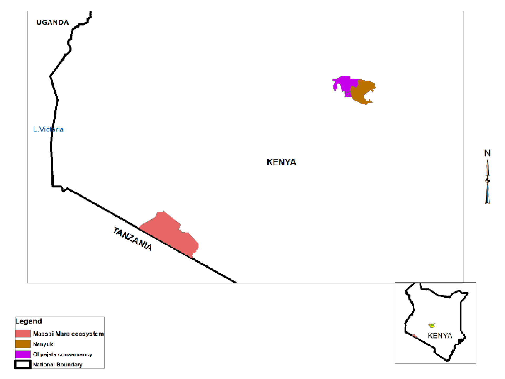
2.1. Study Area
2.2. Sample Collection
2.3. Laboratory Testing Procedures
2.4. Data Analysis
3. Results
4. Discussion
5. Conclusions
Author Contributions
Funding
Institutional Review Board Statement
Informed Consent Statement
Data Availability Statement
Acknowledgments
Conflicts of Interest
References
- Adams, M.J.; Lefkowitz, E.J.; King, A.M.; Harrach, B.; Harrison, R.L.; Knowles, N.J.; Kropinski, A.M.; Krupovic, M.; Kuhn, J.H.; Mushegian, A.R.; et al. Changes to taxonomy and the International Code of Virus Classification and Nomenclature ratified by the International Committee on Taxonomy of Viruses. Arch. Virol. 2017, 162, 2505–2538. [Google Scholar] [CrossRef]
- Bente, D.A.; Forrester, N.L.; Watts, D.M.; McAuley, A.J.; Whitehouse, C.A.; Bray, M. Crimean-Congo hemorrhagic fever: History, epidemiology, pathogenesis, clinical syndrome and genetic diversity. Antivir. Res. 2013, 100, 159–189. [Google Scholar] [CrossRef] [PubMed] [Green Version]
- Mertens, M.; Schmidt, K.; Ozkul, A.; Groschup, M.H. The impact of Crimean-Congo hemorrhagic fever virus on public health. Antivir. Res. 2013, 98, 248–260. [Google Scholar] [CrossRef] [PubMed]
- Sorvillo, T.E.; Rodriguez, S.E.; Hudson, P.; Carey, M.; Rodriguez, L.L.; Spiropoulou, C.F.; Bird, B.H.; Spengler, J.R.; Bente, D.A. Towards a sustainable one health approach to crimean–congo hemorrhagic fever prevention: Focus areas and gaps in knowledge. Trop. Med. Infect. Dis. 2020, 5, 113. [Google Scholar] [CrossRef]
- Mourya, D.T.; Yadav, P.D.; Shete, A.M.; Gurav, Y.K.; Raut, C.G.; Jadi, R.S.; Pawar, S.D.; Nichol, S.T.; Mishra, A.C. Detection, isolation and confirmation of Crimean-Congo hemorrhagic fever virus in human, ticks and animals in Ahmadabad, India, 2010–2011. PLoS Negl. Trop. Dis. 2012, 6, e1653. [Google Scholar] [CrossRef]
- Appannanavar, S.B.; Mishra, B. An update on Crimean Congo hemorrhagic fever. J. Glob. Infect. Dis. 2011, 3, 285. [Google Scholar] [PubMed]
- Vorou, R.; Pierroutsakos, I.N.; Maltezou, H.C. Crimean-Congo hemorrhagic fever. Curr. Opin. Infect. Dis. 2007, 20, 495–500. [Google Scholar] [CrossRef]
- Bell-Sakyi, L.; Kohl, A.; Bente, D.A.; Fazakerley, J.K. Tick cell lines for study of Crimean-Congo hemorrhagic fever virus and other arboviruses. Vector Borne Zoonotic Dis. 2012, 12, 769–781. [Google Scholar] [CrossRef] [Green Version]
- Kajihara, M.; Simuunza, M.; Saasa, N.; Dautu, G.; Mori-Kajihara, A.; Qiu, Y.; Nakao, R.; Eto, Y.; Furumoto, H.; Hang’ombe, B.M.; et al. Serologi and molecular evidence for circulation of Crimean-Congo hemorrhagic fever virus in ticks and cattle in Zambia. PLoS Negl. Trop. Dis. 2021, 15, e0009452. [Google Scholar] [CrossRef]
- Hoogstraal, H. The epidemiology of tick-borne Crimean-Congo hemorrhagic fever in Asia, Europe, and Africa. J. Med. Entomol. 1979, 15, 307–417. [Google Scholar] [CrossRef]
- Smirnova, S.E.; Nepesova, N.M.; Tachmuradov, G.; Kir’Yanova, A.M.; Chumakov, M.P. Data on Studying Crimean Hemorrhagic Fever in Turkmen SSR. NAMRU-T804. Tr Inst Polio Virus Entsef Akad Med Nauk SSSR. 1971, Volume 19, pp. 86–91. Available online: https://agris.fao.org/agris-search/search.do?recordID=US201300336439 (accessed on 5 August 2021).
- Ceianu, C.S.; Panculescu-Gatej, R.I.; Coudrier, D.; Bouloy, M. First serologic evidence for the circulation of Crimean-Congo hemorrhagic fever virus in Romania. Vector Borne Zoonotic Dis. 2012, 12, 718–721. [Google Scholar] [CrossRef]
- Mustafa, M.L.; Ayazi, E.; Mohareb, E.; Yingst, S.; Zayed, A.; Rossi, C.A.; Schoepp, R.J.; Mofleh, J.; Fiekert, K.; Akhbarian, Z.; et al. Crimean-congo hemorrhagic fever, Afghanistan. Emerg. Infect. Dis. 2011, 17, 1940. [Google Scholar] [CrossRef] [PubMed]
- Tantawi, H.H.; Shony, M.O.; Al-Tikriti, S.K. Antibodies to Crimean-Congo haemorrhagic fever virus in domestic animals in Iraq: A seroepidemiological survey. Int. J. Zoonoses 1981, 8, 115–120. [Google Scholar]
- Pak, T.P. Division into epidemiological districts of Crimean haemorrhagic fever (CHF) in the Tadzhik SSR. Zh Mikrobiol. Epidemiol. Immunobiol. 1972, 12, 112–116. [Google Scholar]
- Spengler, J.R.; Bergeron, É.; Rollin, P.E. Seroepidemiological studies of Crimean-Congo hemorrhagic fever virus in domestic and wild animals. PLoS Negl. Trop. Dis. 2016, 10, e0004210. [Google Scholar] [CrossRef] [Green Version]
- Ergonul, O.; Tuncbilek, S.; Baykam, N.; Celikbas, A.; Dokuzoguz, B. Evaluation of serum levels of interleukin (IL)–6, IL-10, and tumor necrosis factor–α in patients with Crimean-Congo hemorrhagic fever. J. Infect. Dis. 2006, 193, 941–944. [Google Scholar] [CrossRef] [PubMed] [Green Version]
- Dunster, L.; Dunster, M.; Ofula, V.; Beti, D.; Kazooba-Voskamp, F.; Burt, F.; Swanepoel, R.; DeCock, K.M. First documentation of human Crimean-Congo hemorrhagic fever, Kenya. Emerg. Infect. Dis. 2002, 8, 1005. [Google Scholar] [CrossRef] [PubMed]
- Sang, R.; Lutomiah, J.; Koka, H.; Makio, A.; Chepkorir, E.; Ochieng, C.; Yalwala, S.; Mutisya, J.; Musila, L.; Richardson, J.H.; et al. Crimean-Congo hemorrhagic fever virus in Hyalommid ticks, northeastern Kenya. Emerg. Infect. Dis. 2011, 17, 1502. [Google Scholar] [CrossRef]
- Chiuya, T.; Masiga, D.K.; Falzon, L.C.; Bastos, A.D.; Fèvre, E.M.; Villinger, J. Tick-borne pathogens, including Crimean-Congo haemorrhagic fever virus, at livestock markets and slaughterhouses in western Kenya. Transbound. Emerg. Dis. 2020, 68, 2429–2445. [Google Scholar] [CrossRef]
- Lwande, O.W.; Irura, Z.; Tigoi, C.; Chepkorir, E.; Orindi, B.; Musila, L.; Venter, M.; Fischer, A.; Sang, R. Seroprevalence of crimean congo hemorrhagic Fever virus in ijara district, kenya. Vector Borne Zoonotic Dis. 2012, 12, 727–732. [Google Scholar] [CrossRef] [Green Version]
- Tigoi, C.; Lwande, O.; Orindi, B.; Irura, Z.; Ongus, J.; Sang, R. Seroepidemiology of selected arboviruses in febrile patients visiting selected health facilities in the lake/river basin areas of Lake Baringo, Lake Naivasha, and Tana River, Kenya. Vector Borne Zoonotic Dis. 2015, 15, 124–132. [Google Scholar] [CrossRef] [Green Version]
- Nyataya, J.; Maraka, M.; Lemtudo, A.; Masakhwe, C.; Mutai, B.; Njaanake, K.; Estambale, B.B.; Nyakoe, N.; Siangla, J.; Waitumbi, J.N. Serological Evidence of Yersiniosis, Tick-Borne Encephalitis, West Nile, Hepatitis E, Crimean-Congo Hemorrhagic Fever, Lyme Borreliosis, and Brucellosis in Febrile Patients Presenting at Diverse Hospitals in Kenya. Vector Borne Zoonotic Dis. 2020, 20, 348–357. [Google Scholar] [CrossRef] [PubMed]
- Swift, J.J. Ecology of African pastoralist societies by Katherine Homewood. Pastor. Res. Policy Pract. 2011, 1, 5. [Google Scholar] [CrossRef] [Green Version]
- African Union. Policy Framework for Pastoralism in Africa: Securing, Protecting and Improving the Lives, Livelihoods and Rights of Pastoralist Communities; African Union: Addis Ababa, Ethiopia, 2013. [Google Scholar]
- Chauhan, R.P.; Dessie, Z.G.; Noreddin, A.; El Zowalaty, M.E. Systematic Review of Important Viral Diseases in Africa in Light of the ‘One Health’ Concept. Pathogens 2020, 9, 301. [Google Scholar] [CrossRef] [Green Version]
- Kosgey, I.S.; Mbuku, S.M.; Okeyo, A.M.; Amimo, J.; Philipsson, J.; Ojango, J.M. Institutional and organizational frameworks for dairy and beef cattle recording in Kenya: A review and opportunities for improvement. Anim. Genet. Resour. 2011, 48, 1–11. [Google Scholar] [CrossRef] [Green Version]
- Alarcon, P.; Fèvre, E.M.; Muinde, P.; Murungi, M.K.; Kiambi, S.; Akoko, J.; Rushton, J. Urban livestock keeping in the city of Nairobi: Diversity of production systems, supply chains, and their disease management and risks. Front. Vet. Sci. 2017, 4, 171. [Google Scholar] [CrossRef] [PubMed] [Green Version]
- Mwangi, V.; Owuor, S.; Kiteme, B.; Giger, M. Beef Production in the Rangelands: A Comparative Assessment between Pastoralism and Large-Scale Ranching in Laikipia County. Kenya. Agric. 2020, 10, 399. [Google Scholar]
- Kahi, A.K.; Wasike, C.B.; Rewe, T.O. Beef production in the arid and semi-arid lands of Kenya: Constraints and prospects for research and development. Outlook Agric. 2006, 35, 217–225. [Google Scholar] [CrossRef]
- Wellington, E.N. Mechanisms of drought management by African pastoralists. In Proceedings of the Animal Production Society of Kenya Symposium, Egerton University, Nakuru, Kenya, 7–8 March 2000; pp. 117–122. [Google Scholar]
- Reid, R.S.; Rainy, M.; Ogutu, J.; Kruska, R.L.; Kimani, K.; Nyabenge, M.; McCartney, M.; Kshatriya, M.; Worden, J.; Ng’ang’a, L.; et al. People, Wildlife and Livestock in the Mara Ecosystem: The Mara Count 2002; International Livestock Research Institute: Nairobi, Kenya, 2003. [Google Scholar]
- Belobo, J.T.E.; Kenmoem, S.; Kengne-Nde, C.; Emoh, C.P.D.; Bowo-Ngandji, A.; Tchatchouang, S.; Wobessi, J.N.S.; Mikangue, C.A.M.; Tazokong, H.R.; Bebey, S.R.K.; et al. Worldwide epidemiology of Crimean-Congo Hemorrhagic Fever Virus in humans, ticks and other animal species, a systematic review and meta-analysis. PLoS Negl. Trop. Dis. 2021, 15, e0009299. [Google Scholar] [CrossRef]
- Amulyoto, M. Human, Cattle and African Buffalo (Syncerus Caffer) Interface in the Ol Pejeta Conservancy. Ph.D. Thesis, Jomo Kenyatta University of Agriculture and Technology, Laikipia County, Kenya, 2020. [Google Scholar]
- Amulyoto, M.; Karanja, S.M.; Obanda, V.; Lutomiah, J. Social Structure, Awareness and Practice on Risk of Exposure to Ticks and Tick-Bor ne Diseases in Ol Pejeta Conservancy, Kenya. Int. J. Res. 2018, 5, 713–730. [Google Scholar]
- Keesing, F.; Allan, B.F.; Young, T.P.; Ostfeld, R.S. Effects of wildlife and cattle on tick abundance in central Kenya. Ecol. Appl. 2013, 23, 1410–1418. [Google Scholar] [CrossRef] [PubMed]
- Keesing, F.; Ostfeld, R.S.; Okanga, S.; Huckett, S.; Bayles, B.R.; Chaplin-Kramer, R.; Fredericks, L.P.; Hedlund, T.; Kowal, V.; Tallis, H.; et al. Consequences of integrating livestock and wildlife in an African savanna. Nat. Sustain. 2018, 1, 566–573. [Google Scholar] [CrossRef]
- Ndeereh, D.; Muchemi, G.; Thaiyah, A.; Otiende, M.; Angelone-Alasaad, S.; Jowers, M.J. Molecular survey of Coxiella burnetii in wildlife and ticks at wildlife–livestock interfaces in Kenya. Exp. Appl. Acarol. 2017, 72, 277–289. [Google Scholar] [CrossRef]
- Mangombi, J.B.; Roqueplo, C.; Sambou, M.; Dahmani, M.; Mediannikov, O.; Comtet, L.; Davoust, B. Seroprevalence of Crimean-Congo hemorrhagic fever in domesticated animals in Northwestern Senegal. Vector Borne Zoonotic Dis. 2020, 20, 797–799. [Google Scholar] [CrossRef] [PubMed]
- Balinandi, S.; von Brömssen, C.; Tumusiime, A.; Kyondo, J.; Kwon, H.; Monteil, V.M.; Mirazimi, A.; Lutwama, J.; Mugisha, L.; Malmberg, M. Serological and molecular study of Crimean-Congo Hemorrhagic Fever Virus in cattle from selected districts in Uganda. J. Virol. Methods 2021, 290, 114075. [Google Scholar] [CrossRef] [PubMed]
- Nabeth, P.; Cheikh, D.O.; Lo, B.; Faye, O.; Vall, I.O.M.; Niang, M.; Wague, B.; Diop, D.; Diallo, M.; Diallo, B.; et al. Crimean-Congo hemorrhagic fever, mauritania. Emerg. Infect. Dis. 2004, 10, 2143. [Google Scholar] [CrossRef]
- Karimi, I.; Rostami, J.M.; Chinikar, S.; Ataei, B.; Kasaeian, N.; Jalali, N.; Khosravi, N. Seroepidemiologic survey of Crimean-Congo hemorrhagic fever among slaughters and butchers in Isfahan. J. Isfahan Med. Sch. 2007, 24, 57–62. [Google Scholar]
- Athar, M.N.; Baqai, H.Z.; Ahmad, M.; Khalid, M.A.; Bashir, N.; Ahmad, A.M.; Balouch, A.H.; Bashir, K. Crimean-congo hemorrhagic fever outbreak in Rawalpindi, Pakistan, February 2002. Am. J. Trop. Med. Hyg. 2003, 69, 284–287. [Google Scholar] [CrossRef]
- Giger, M.; Mutea, E.; Kiteme, B.; Eckert, S.; Anseeuw, W.; Zaehringer, J.G. Large agricultural investments in Kenya’s Nanyuki Area: Inventory and analysis of business models. Land Use Policy 2020, 99, 104833. [Google Scholar] [CrossRef]
- Ansari, H.; Shahbaz, B.; Izadi, S.; Zeinali, M.; Tabatabaee, S.M.; Mahmoodi, M.; Naieni, K.H.; Mansournia, M.A. Crimean-Congo hemorrhagic fever and its relationship with climate factors in southeast Iran: A 13-year experience. J. Infect. Dev. Ctries. 2014, 8, 749–757. [Google Scholar] [CrossRef] [Green Version]


| Variables | Anti-CCHF IgG Prevalence % |
|---|---|
| Sex | |
| Female (n = 97) | 32.9 |
| Male (n = 51) | 27.4 |
| Breed | |
| Zebu (n = 68) | 38.2 |
| Sahiwal (n = 2) | 22.6 |
| Ankole (n = 3) | 100 |
| Improved Boran (n = 75) | 0 |
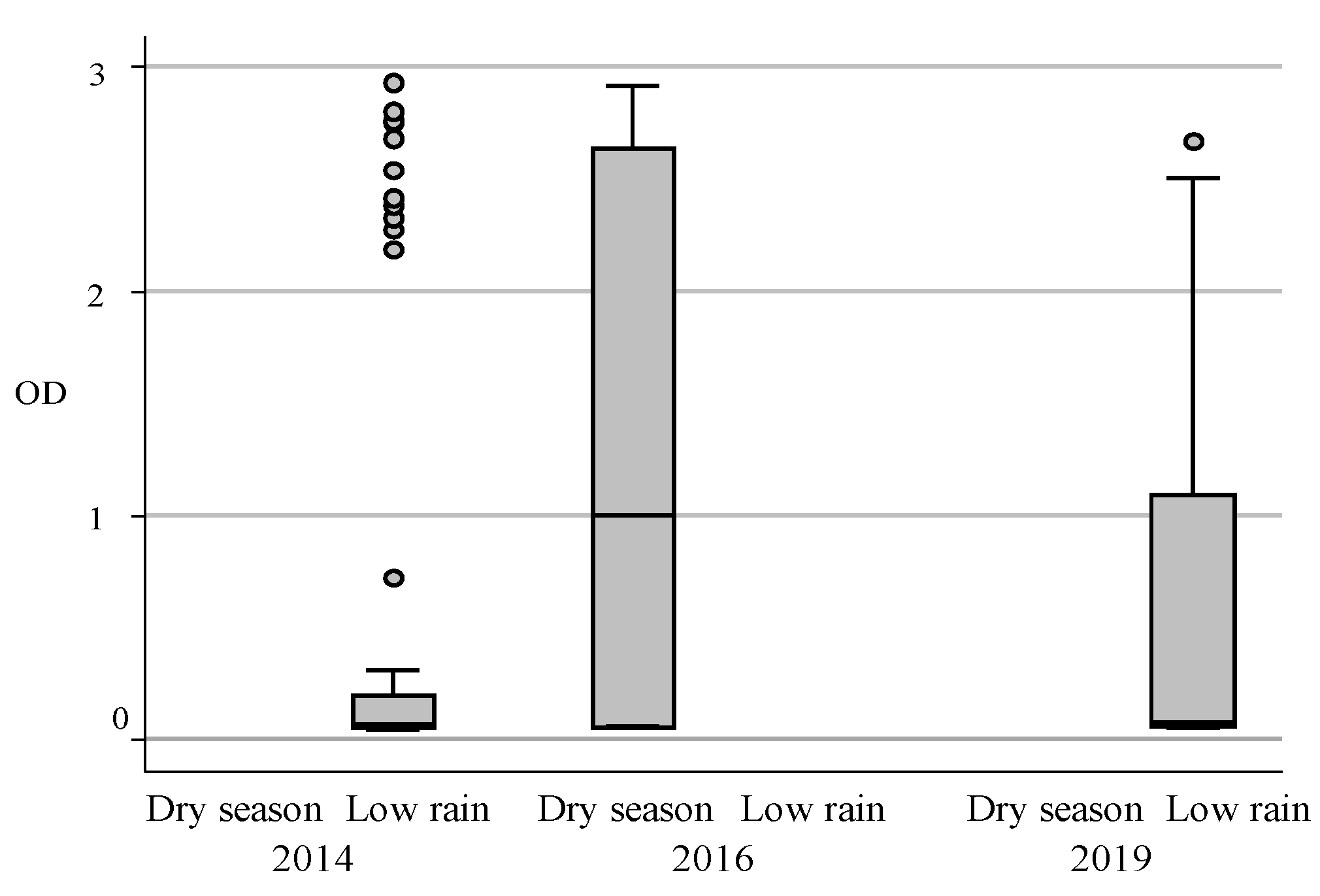
| Sampling year | |
| 2014 (n = 59) | 22.0 |
| 2016 (n = 28) | 100 |
| 2019 (n = 61) | 31.1 |
| Sampling month/season | |
| Low rain (n = 120) | 26.6 |
| Dry (n = 28) | 100 |
| Sites | |
| Emarti (n = 7) | 57.14 |
| Emboseli (n = 4) | 50 |
| Il Poori (n = 4) | 0 |
| Irbaan (n = 8) | 50 |
| Kamok (n = 23) | 47.82 |
| Loigururu (n = 47) | 4.2 |
| Mt. Kenya slopes (n = 9) | 77.7 |
| Olaimutiak (n = 3) | 33.3 |
| Ole Keene (n = 14) | 1.53 |
| Ole Seken (n = 7) | 33.3 |
| Olekusero (n = 1) | 0 |
| Oletontol (n= 2) | 0 |
| Orindo (n = 1) | 0 |
| Orkiu (n = 5) | 0.08 |
| Oseki (n = 2) | 0 |
| Scotts (n = 2) | 50 |
| Sekenani (n = 3) | 33.3 |
| Sirima (n = 6) | 100 |
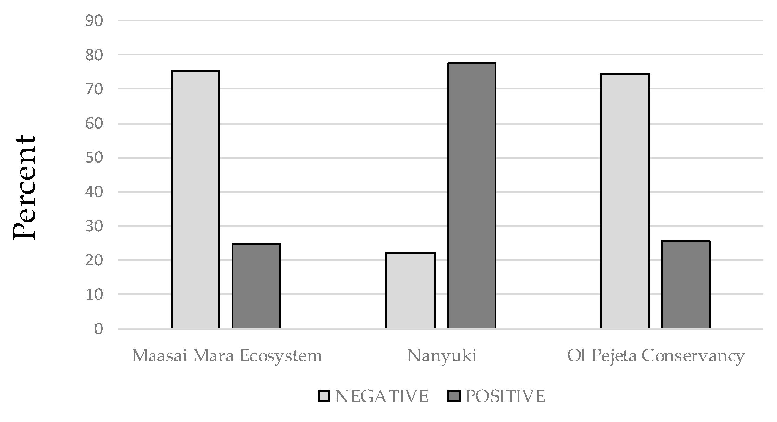
| Ecosystem | |
| Maasai Mara (n = 101) | 24.75 |
| Nanyuki (n = 9) | 77.7 |
| Ol Pejeta (n = 78) | 25.6 |
| ODR | Statistical Variables | ||||
|---|---|---|---|---|---|
| n | Median | IQR | Inter-Herd Variation | Intra-Herd Variation | |
| Ecosystem | |||||
| Maasai Mara | 101 | 0.073 | 0.06; 1.09 | 50.1% | 12.9–172.8% |
| Nanyuki | 9 | 2.668 | 2.509; 2.729 | - | 172% |
| Ol Pejeta | 78 | 0.06 | 0.053; 0.301 | 13.2% | 102.9–133.6% |
| n | Cattle (n = 61) | Sheep (n = 23) | Goat (n = 17) |
|---|---|---|---|
| Seropositive | 19 | 1 | 5 |
| % (95% CI) | 31.0 (0.235–0.386) | 4.34 (−0.046–0.133) | 29.4 (0.052–0.535) |
| Variable | Estimates | OR (95% CI) | p-Value |
|---|---|---|---|
| Age 1 | 0.552 | 1.737 (1.405–2.148) | 0.000 |
| Sex | |||
| Female | Baseline | ||
| Male | −1.028 | 0.357 (0.123–1.038) | 0.058 |
| Season during sampling year | |||
| Dry | Baseline | ||
| Low rain | −1.912 | 0.147 (0.0365–0.596) | 0.007 |
| Ecosystem | |||
| Maasai Mara | Baseline | ||
| Nanyuki | 1.973 | 7.197 (0.813–63.65) | 0.076 |
| Ol Pejeta | 0.525 | 1.690 (0.560–5.102) | 0.351 |
Publisher’s Note: MDPI stays neutral with regard to jurisdictional claims in published maps and institutional affiliations. |
© 2021 by the authors. Licensee MDPI, Basel, Switzerland. This article is an open access article distributed under the terms and conditions of the Creative Commons Attribution (CC BY) license (https://creativecommons.org/licenses/by/4.0/).
Share and Cite
Blanco-Penedo, I.; Obanda, V.; Kingori, E.; Agwanda, B.; Ahlm, C.; Lwande, O.W. Seroepidemiology of Crimean-Congo Hemorrhagic Fever Virus (CCHFV) in Cattle across Three Livestock Pastoral Regions in Kenya. Dairy 2021, 2, 425-434. https://0-doi-org.brum.beds.ac.uk/10.3390/dairy2030034
Blanco-Penedo I, Obanda V, Kingori E, Agwanda B, Ahlm C, Lwande OW. Seroepidemiology of Crimean-Congo Hemorrhagic Fever Virus (CCHFV) in Cattle across Three Livestock Pastoral Regions in Kenya. Dairy. 2021; 2(3):425-434. https://0-doi-org.brum.beds.ac.uk/10.3390/dairy2030034
Chicago/Turabian StyleBlanco-Penedo, Isabel, Vincent Obanda, Edward Kingori, Bernard Agwanda, Clas Ahlm, and Olivia Wesula Lwande. 2021. "Seroepidemiology of Crimean-Congo Hemorrhagic Fever Virus (CCHFV) in Cattle across Three Livestock Pastoral Regions in Kenya" Dairy 2, no. 3: 425-434. https://0-doi-org.brum.beds.ac.uk/10.3390/dairy2030034