
Construction of a Removable Partial Denture (RPD): Comparison between the Analog Procedure and the Selective Laser Melting Procedure


Abstract
:1. Introduction
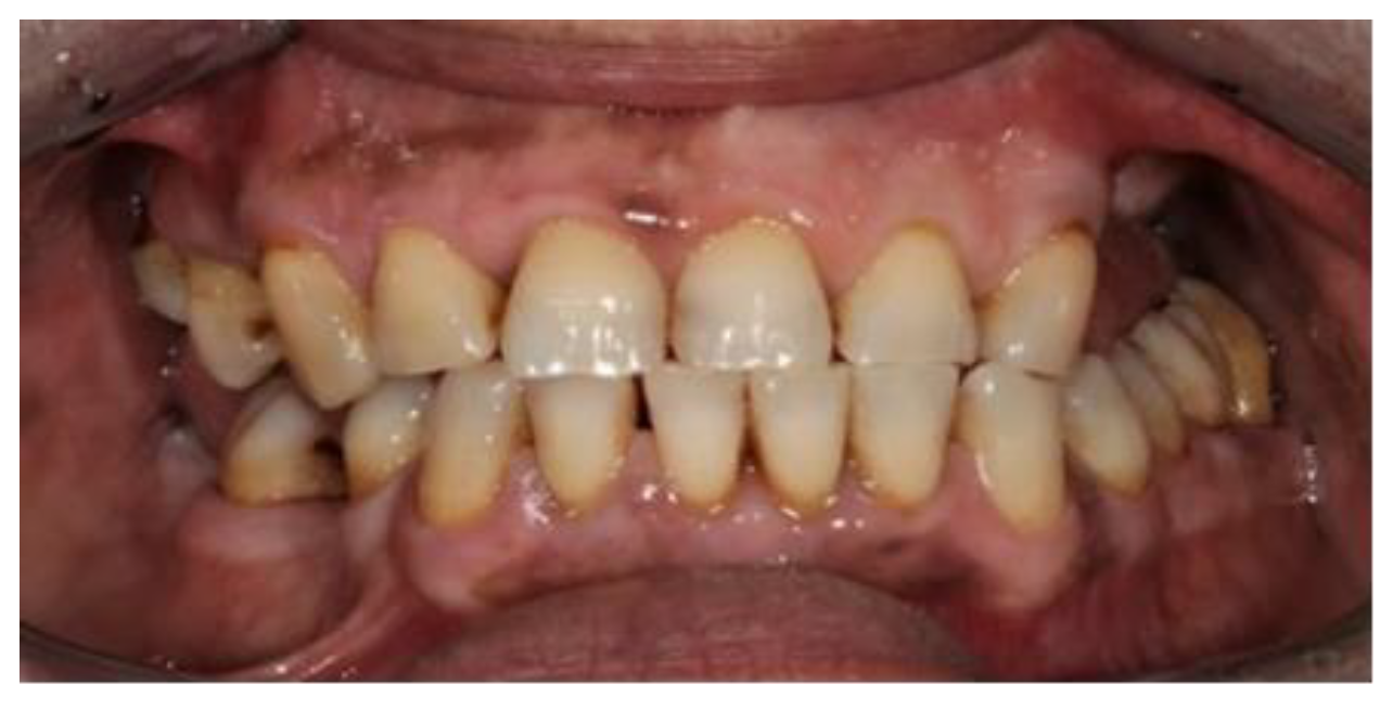
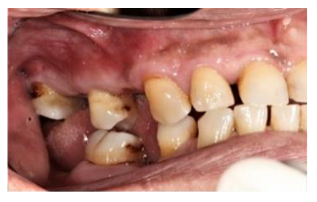
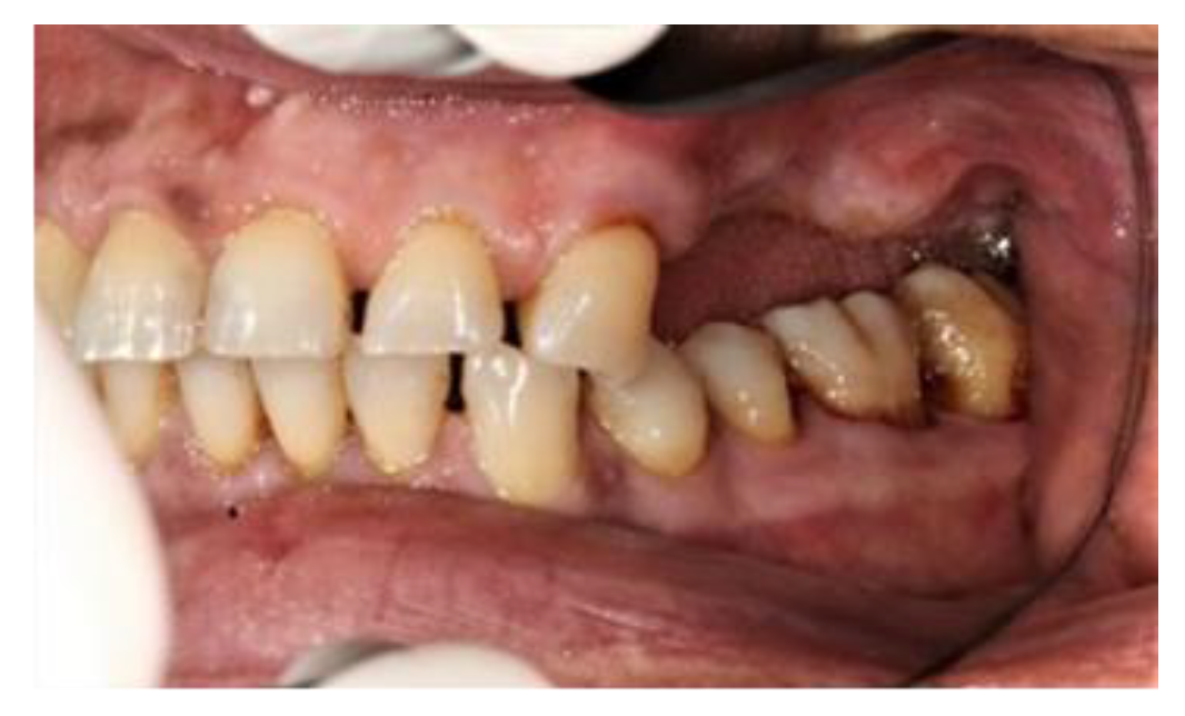
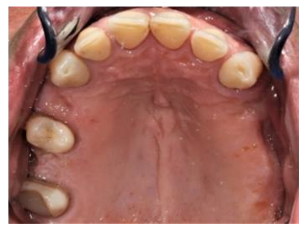
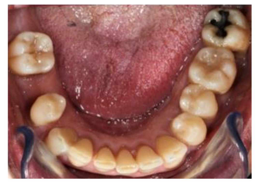
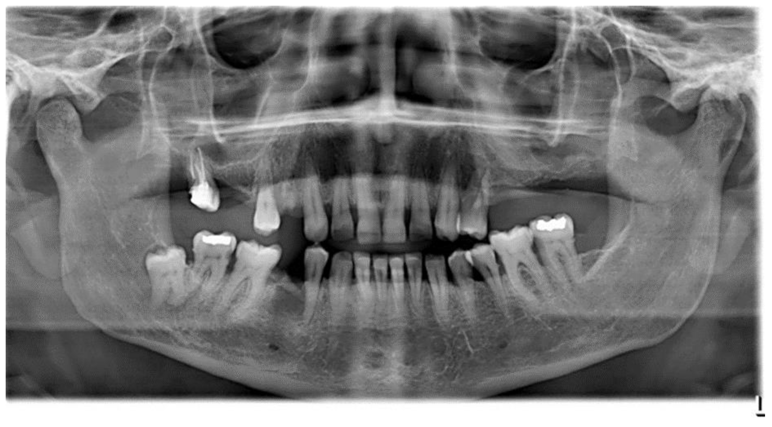
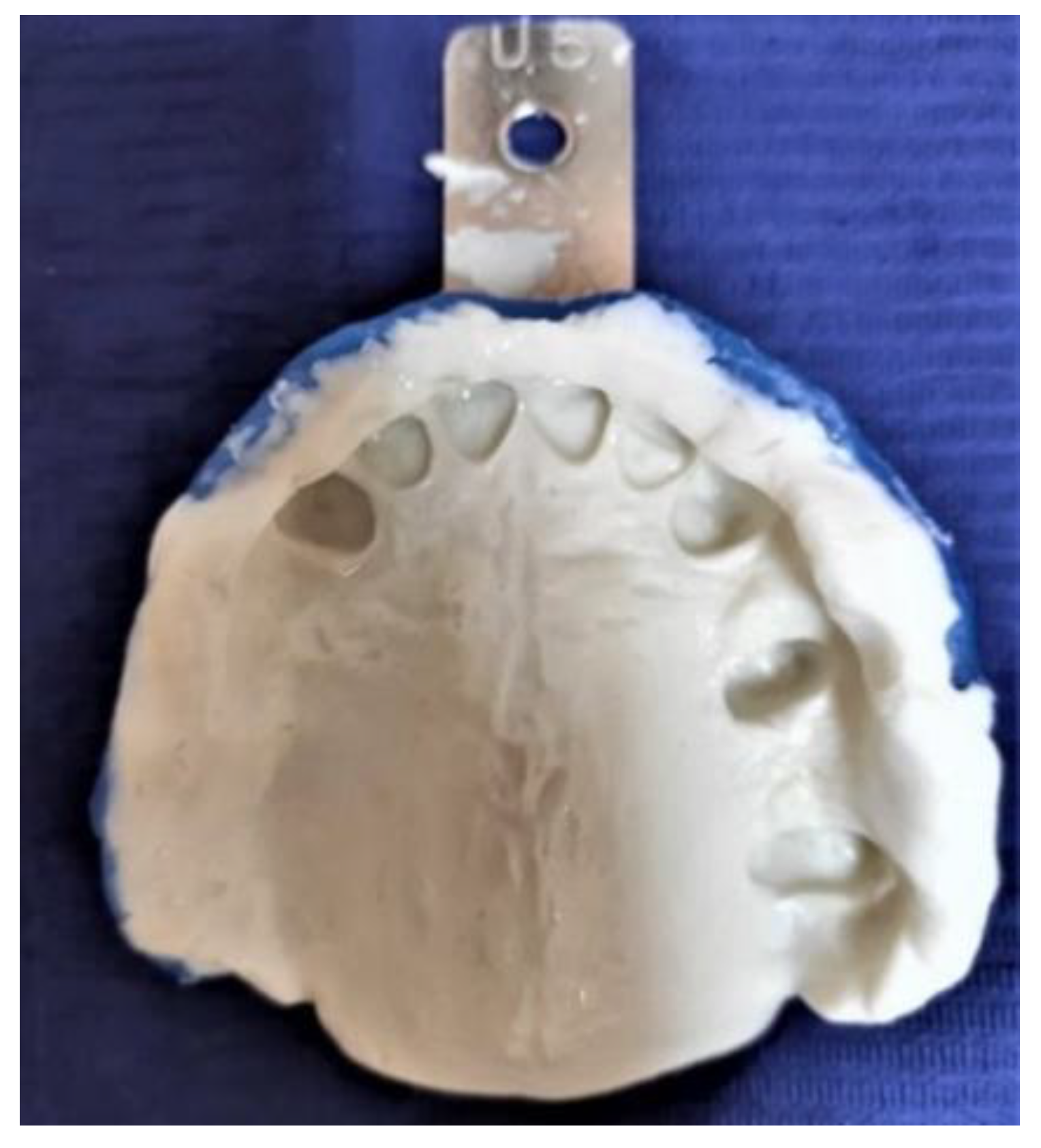
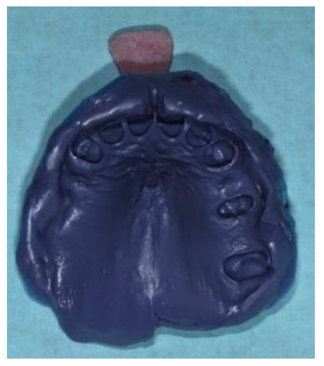
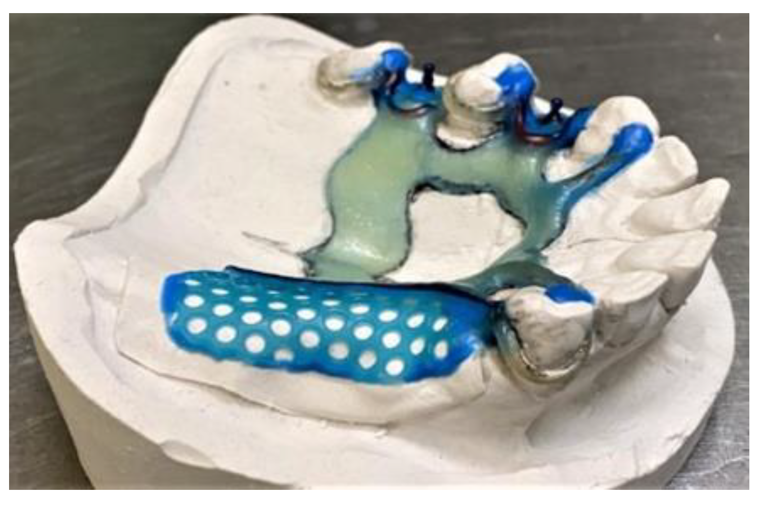
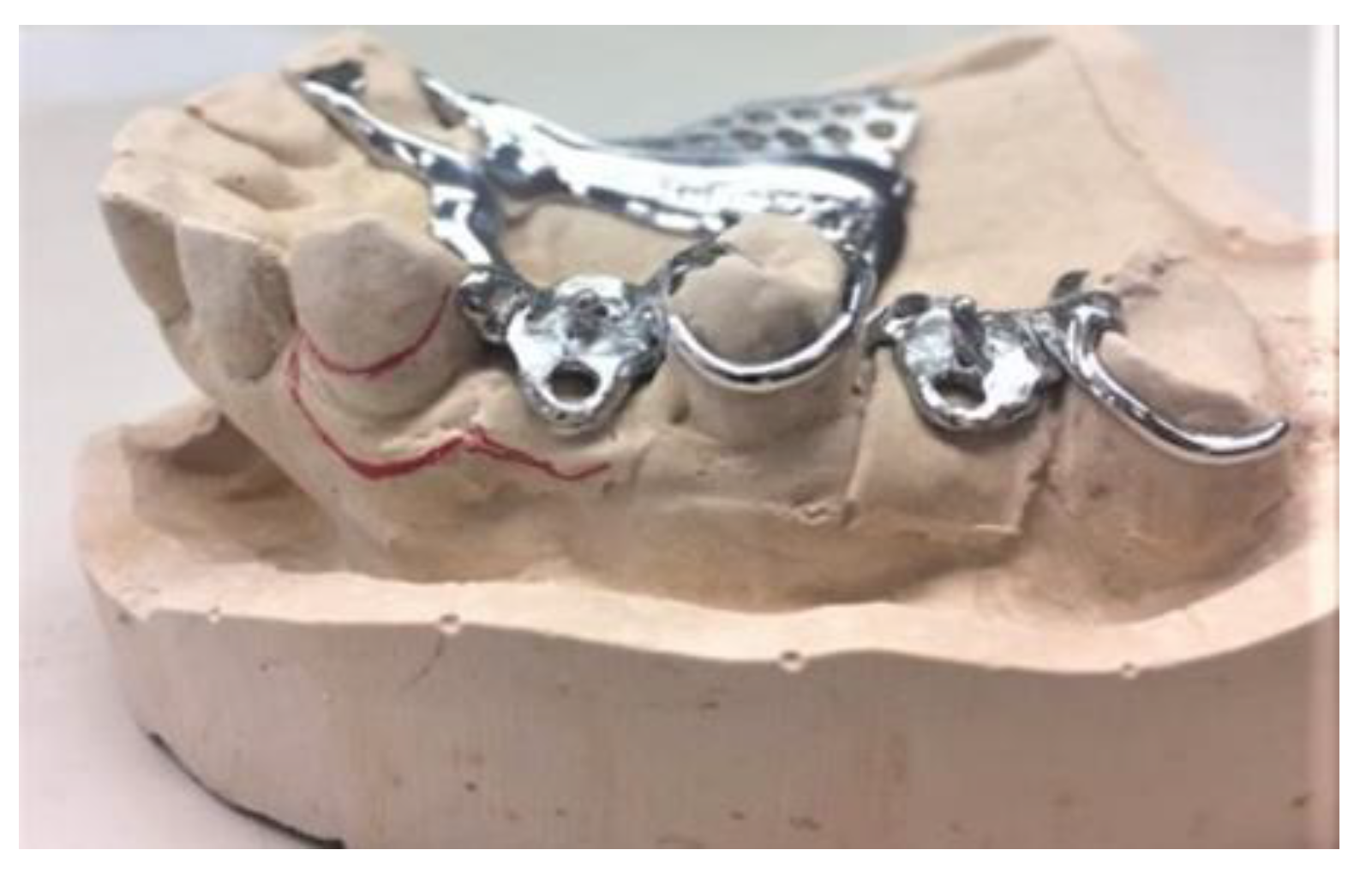
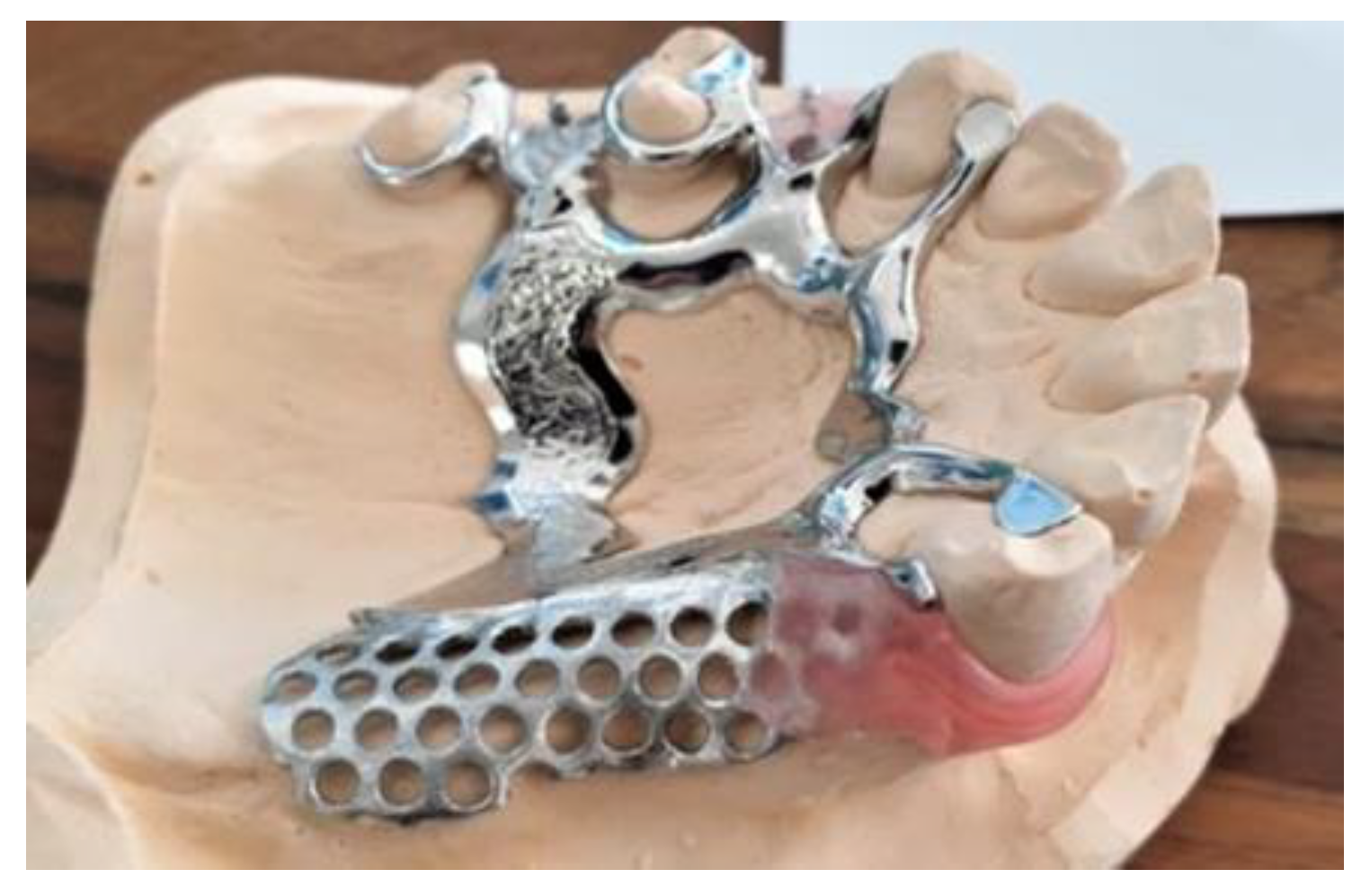
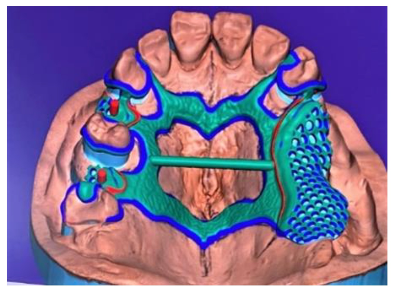
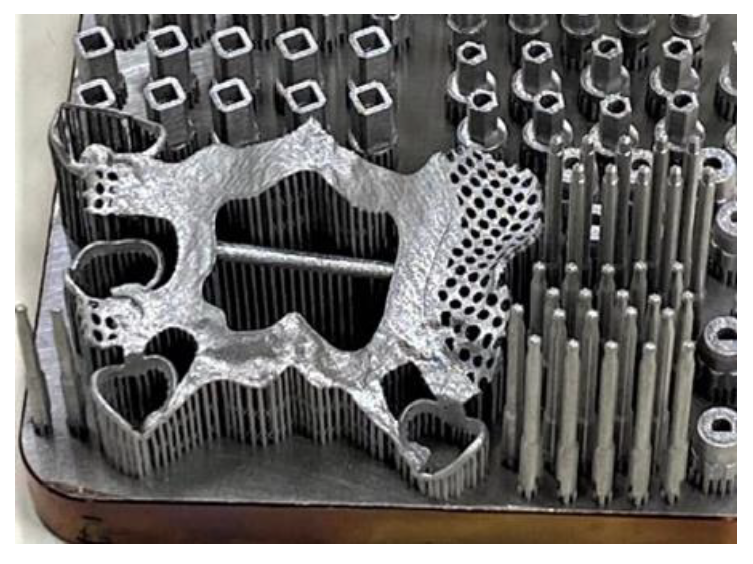
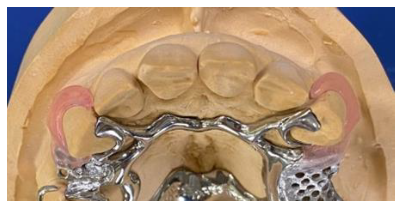
2. Case Report
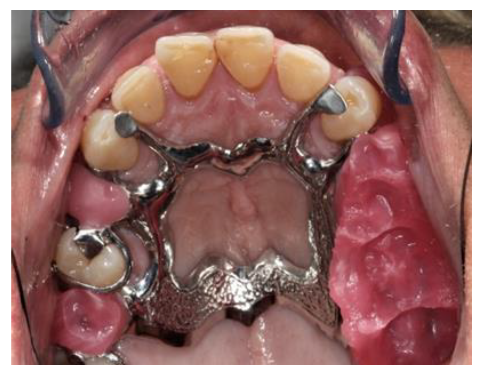
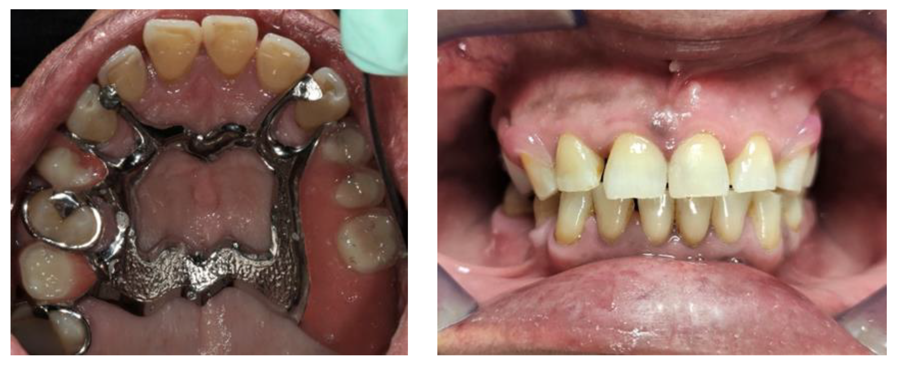
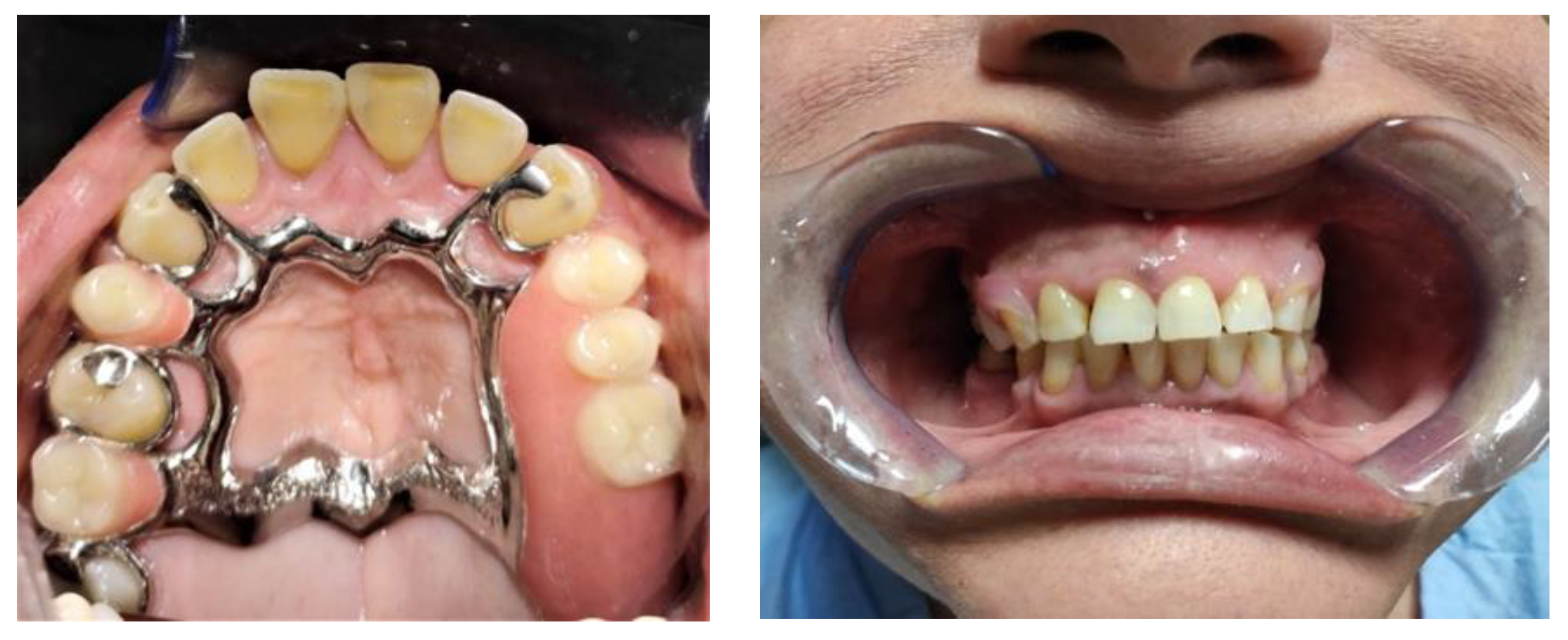
- Evaluation of the prosthesis adaptability to support tissues (teeth, gum and mucosa), with focus on hooks quality and precision;
- Occlusal contacts evaluation through a 40-micron thick articulating paper. This was also used to assess whether by pulling the paper, it was retained in both cases (with and without prosthesis in situ);
- Some pressure was applied with a bolt on several points of the free gap to evaluate whether there was tilting on the opposite side.
3. Discussion
4. Conclusions
Author Contributions
Funding
Institutional Review Board Statement
Informed Consent Statement
Data Availability Statement
Conflicts of Interest
References
- Lamster, I.B. Geriatric periodontology: How the need to care for the aging population can influence the future of the dental profession. Periodontology 2000 2016, 72, 7–12. [Google Scholar] [CrossRef] [PubMed]
- Shao, Z.; Guo, X.; Zhang, Q.; Bronkhorst, E.M.; Zou, D.; Creugers, N.H.J. Masticatory efficiency in patients with partially edentulous dentitions. J. Dent. 2018, 75, 41–47. [Google Scholar] [CrossRef] [PubMed]
- Fenton, A.H. Removable partial prostheses for the elderly. J. Prosthet. Dent. 1994, 72, 532–537. [Google Scholar] [CrossRef]
- Johnson, D.L. Retention for a Removable Partial Denture. J. Prosthodont. 1992, 1, 11–17. [Google Scholar] [CrossRef] [PubMed]
- Laverty, D.P. The Use of 3D Metal Printing (Direct Metal Laser Sintering) in Removable Prosthodontics. Dent. Update 2016, 43, 826–835. [Google Scholar] [CrossRef] [PubMed]
- Xie, W.; Zheng, M.; Wang, J.; Li, X. The effect of build orientation on the microstructure and properties of selective laser melting Ti-6Al-4V for removable partial denture clasps. J. Prosthet. Dent. 2020, 123, 163–172. [Google Scholar] [CrossRef] [PubMed] [Green Version]
- Torii, M.; Nakata, T.; Takahashi, K.; Kawamura, N.; Shimpo, H.; Ohkubo, C. Fitness and retentive force of cobalt-chromium alloy clasps fabricated with repeated laser sintering and milling. J. Prosthodont. Res. 2018, 62, 342–346. [Google Scholar] [CrossRef] [PubMed]
- Dmd, A.S.M.; Evans, Z.P.; Nash, J.; Bs, C.B.; Ms, A.L.; Bacro, T.; Cayouette, M.; Ludlow, M.; Renne, W.G. Evaluation of the trueness and precision of complete arch digital impressions on a human maxilla using seven different intraoral digital impression systems and a laboratory scanner. J. Esthet. Restor. Dent. 2019, 31, 369–377. [Google Scholar] [CrossRef]
- Kattadiyil, M.T.; Mursic, Z.; AlRumaih, H.; Goodacre, C.J. Intraoral scanning of hard and soft tissues for partial removable dental prosthesis fabrication. J. Prosthet. Dent. 2014, 112, 444–448. [Google Scholar] [CrossRef] [PubMed]


| Analog Procedure | Hybrid Procedure | |
|---|---|---|
| 1st appointment: impression taking | Alginate | Digital impression (Omnicam 2.0 Dentsply sirona) |
| 2nd appointment: prosthetic seat preparation and second impression | Polyether (Impregum 3M) | Polyether (Impregum 3M). The model obtained from this impression was transformed into a digital model with a laboratory scanner ((Neway Open Tech 3D) |
| 3rd appointment: measurement of the spatial position of the upper jaw and of the intermaxillary relationship. | Facial arch (Artex) | Facial arch (Artex) |
| 4th appointment: testing of the prosthesis with the acrylic resin teeth mounted in wax, final small adjustments | - | - |
| 5th appointment: delivery of both prostheses and following clinical evaluations were performed | 1. Evaluation of the prosthesis adaptability to support tissues. 2. Occlusal contacts evaluation through a 40-micron thick articulating paper 3. Some pressure was applied with a bolt on several points of the free gap to evaluate whether there was tilting on the opposite side. | - |
Publisher’s Note: MDPI stays neutral with regard to jurisdictional claims in published maps and institutional affiliations. |
© 2021 by the authors. Licensee MDPI, Basel, Switzerland. This article is an open access article distributed under the terms and conditions of the Creative Commons Attribution (CC BY) license (https://creativecommons.org/licenses/by/4.0/).
Share and Cite
Pugliese, A.; Cataneo, E.; Fortunato, L. Construction of a Removable Partial Denture (RPD): Comparison between the Analog Procedure and the Selective Laser Melting Procedure. Prosthesis 2021, 3, 428-436. https://0-doi-org.brum.beds.ac.uk/10.3390/prosthesis3040038
Pugliese A, Cataneo E, Fortunato L. Construction of a Removable Partial Denture (RPD): Comparison between the Analog Procedure and the Selective Laser Melting Procedure. Prosthesis. 2021; 3(4):428-436. https://0-doi-org.brum.beds.ac.uk/10.3390/prosthesis3040038
Chicago/Turabian StylePugliese, Anthony, Enrico Cataneo, and Leonzio Fortunato. 2021. "Construction of a Removable Partial Denture (RPD): Comparison between the Analog Procedure and the Selective Laser Melting Procedure" Prosthesis 3, no. 4: 428-436. https://0-doi-org.brum.beds.ac.uk/10.3390/prosthesis3040038