Review

Corticosteroid Injection Alone or Combined with Surgical Excision of Keloids versus Other Therapies Including Ionising Radiotherapy: A Systematic Review and Meta-Analysis of Randomised Controlled Trials

1
Department of Burn and Plastic Surgery, West China Hospital of Sichuan University, Chengdu 610041, China
2
Department of Dermatology and Copenhagen Wound Healing Center, Bispebjerg Hospital, University of Copenhagen, 2400 Copenhagen, Denmark
3
Burn Injury Research Unit, The University of Western Australia, Perth 6009, Australia
4
Digestive Disease Center, Bispebjerg Hospital, University of Copenhagen, 2400 Copenhagen, Denmark
5
Department of Clinical Medicine, Faculty of Health and Medical Sciences, University of Copenhagen, 2200 Copenhagen, Denmark
6
Plastic Surgery, Royal Perth Hospital, Perth 6009, Australia
7
Department of Nephrology, West China Hospital of Sichuan University, Chengdu 610041, China
8
Paediatric Department, The People’s Hospital of Sichuan Province, Chengdu 610041, China
*
Author to whom correspondence should be addressed.
Submission received: 13 March 2021 / Revised: 29 April 2021 / Accepted: 7 May 2021 / Published: 14 May 2021

Abstract

:
Keloid scars are difficult to manage and remain a therapeutic challenge. Corticosteroid therapy alone or ionising radiation (radiotherapy) alone or combined with surgery are first-line treatments, but the scientific justification for these treatments is unclear. The aim of this systematic review and meta-analysis of randomised controlled trials (RCTs) is to assess the effects of intralesional corticosteroid injection in treating keloids or preventing their recurrence after surgical removal. Searches for RCTs were conducted through the MEDLINE, EMBASE, EBSCO and Cochrane databases from January 1974 to September 2017. Two authors independently reviewed study eligibility, extracted data, analysed the results, and assessed methodological quality. Sixteen RCTs that included more than 814 patients were scrutinised. The quality of evidence for most outcomes was moderate to high. In 10 RCTs, corticosteroid intralesional injections were compared with 5-fluorouracil, etanercept, cryosurgery, botulinum toxin, topical corticosteroid under a silicone dressing, and radiotherapy. Corticosteroid intralesional injections were more effective than radiotherapy (RR 3.3, 95% CI: 1.4–8.1) but equipotent with the other interventions. In conjunction with keloid excision, corticosteroid treatment was compared with radiotherapy, interferon α-2b and verapamil. In two RCTs, there were fewer keloid recurrences (RR 0.43, 95% CI: 0.21–0.89) demonstrated with adjuvant radiotherapy than with corticosteroid injections. More high-quality, large-scale RCTs are required to establish the effectiveness of corticosteroids and other therapies in keloid management.

1. Introduction

Keloid scars are defined as benign outgrowths of fibrous tissue resulting from abnormal wound healing. They have the ability to spread outside the boundaries of an original lesion [1]. As opposed to normal and hypertrophic scars, keloids persist or continue to grow [2,3,4]. Histological examination of keloids has revealed thick collagen fibres and a prominent fascia-like fibrous band in the upper reticular dermis without contraction of the tissue [5]. Epidemiological data indicate that the risk of developing keloids is higher in individuals with darker skin, those younger than 30 years, and in women, but the relevant mechanisms are unknown [6,7].
Cosmetic concerns, pain, erythema, pruritus, paraesthesia, and functional impairment are all symptoms related to keloids [8,9,10]. Thus, keloids present heavy psychological effects and impact an individual’s physical quality of life.
There is no single effective treatment for keloids. Surgical resection of keloids without adjuvant therapy is considered obsolete due to the high recurrence rate [11,12,13].
Intralesional injection with corticosteroids has been used in the treatment of keloids since the middle of the 1960s [14,15,16,17]. Corticosteroids are also used as adjuncts to surgical excision to prevent recurrence. Corticosteroid formulae include hydrocortisone acetate, methylprednisolone, betamethasone, dexamethasone, and triamcinolone acetonide (TAC). TAC is most commonly used and is typically given (10 to 40 mg/mL) at intervals of four to six weeks for several months or until the scar is flattened [18,19,20]. Treatment with corticosteroids can cause local and systemic adverse effects, including delayed wound healing, hypopigmentation, dermal atrophy, telangiectasia, and Cushing’s syndrome [21,22,23,24].
Ionising radiation (radiotherapy) is seldom used as a monotherapy but primarily as an adjuvant to the surgical excision of a keloid substance [25]. The combined intervention was first described in 1906 and is the preferred first-line treatment today for many clinicians [26,27,28,29]. Radiotherapy usually starts one to three days postoperatively in weekly doses of 5 Gray (Gy) up to a total dose of 50 Gy [28]. The radiation sources applied include superficial X-rays or β-rays, brachytherapy (BT) with β-rays and γ-rays, or electron beam [30]. Adverse effects are impaired wound healing [31], hyperpigmentation, malignancy, and recurrence [9]. There is also a concern about carcinogenesis associated with radiotherapy [30], but in a Canadian 10-year retrospective study, none of 96 keloid patients developed cancer following radiotherapy [32]. Reported recurrence rates after surgery with adjuvant radiotherapy vary depending on the radiation regimen and the length of follow-up period [27,33,34].
Other regimens encompass chemotherapeutics (5-fluorouracil (5-FU), mitomycin C, or bleomycin), imiquimod, interferons, verapamil, and mechanical prevention with pressure garments. Topical applications of silicone in various forms, intralesional cryotherapy, and photodynamic therapy have also been advocated [30,35]. The use of stem cells is a promising new option [36].
Therefore, in this systematic review and meta-analysis, we assess the effects of corticosteroids and radiotherapy on keloids and preventing their recurrence after surgical removal in relation to newer therapies. The collected information is valuable for both clinicians and policy makers to improve the clinical decision-making process and implementation of the best treatment practice for keloids.

2. Materials and Methods

We included randomised controlled trials (RCTs) comparing a corticosteroid and/or radiotherapy with no or with other therapies without surgery or in conjunction with surgical removal of the keloid scar. The included studies were approved by an ethics committee and the enrolled patients provided informed written consent.
Patients of any age and skin type with a keloid scar of any aetiology were included. A keloid was defined as abnormal scarring that extends beyond the original wound borders.
The outcomes were (i) changes in the keloid scar at the end of the study as assessed by the Vancouver Scar Scale (VSS) [37], the Patient and Observer Scar Assessment Scale (POSAS) [38], or other clinical or quantitative assessment tools (ruler or volume scans) in studies without surgery and keloid scar recurrence either as present or absent at the original surgical scar removal site in studies with surgery; and (ii) adverse events including dyspigmentation, atrophy, or ulcers (patients within allocated treatment group with adverse effects (yes or no)).

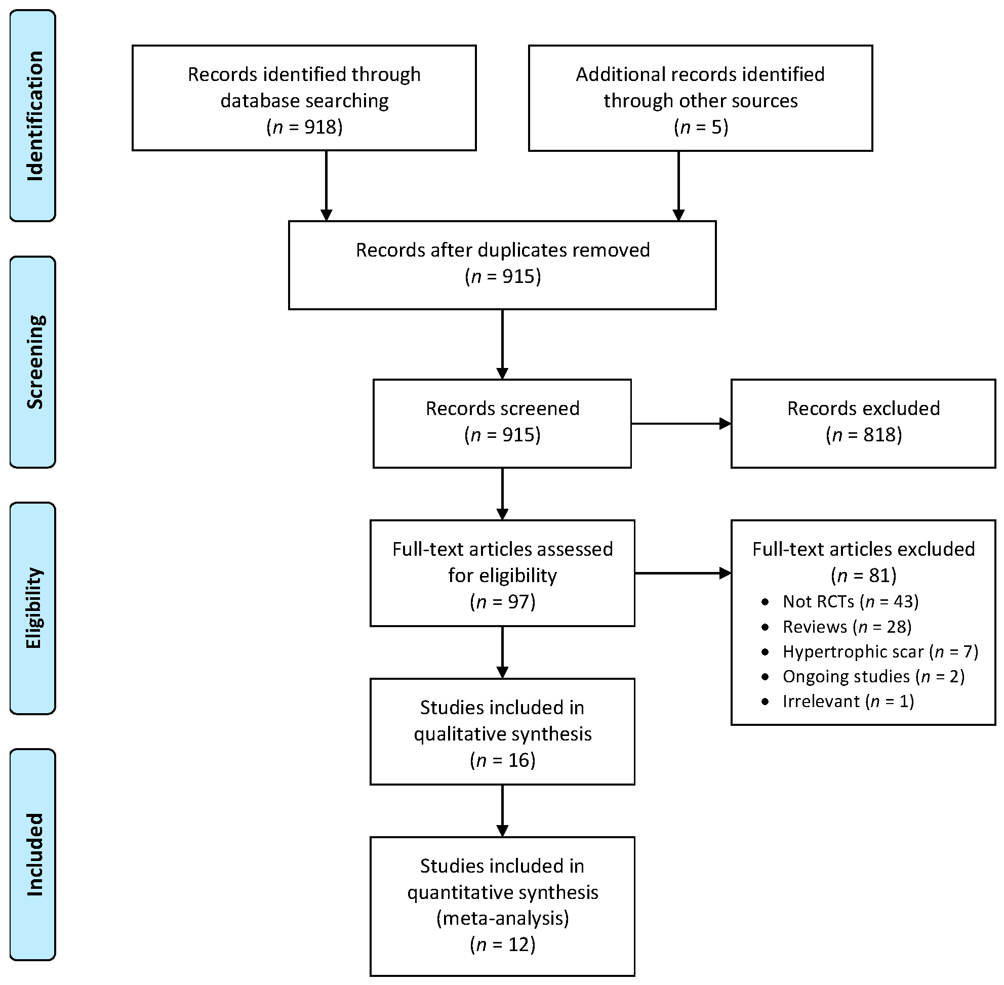
2.1. Search Strategy

Electronic searches were conducted through the MEDLINE, EMBASE, EBSCO, and Cochrane databases from January 1974 to September 2017. The search terms used are given in Table 1. The references of published studies were also reviewed.

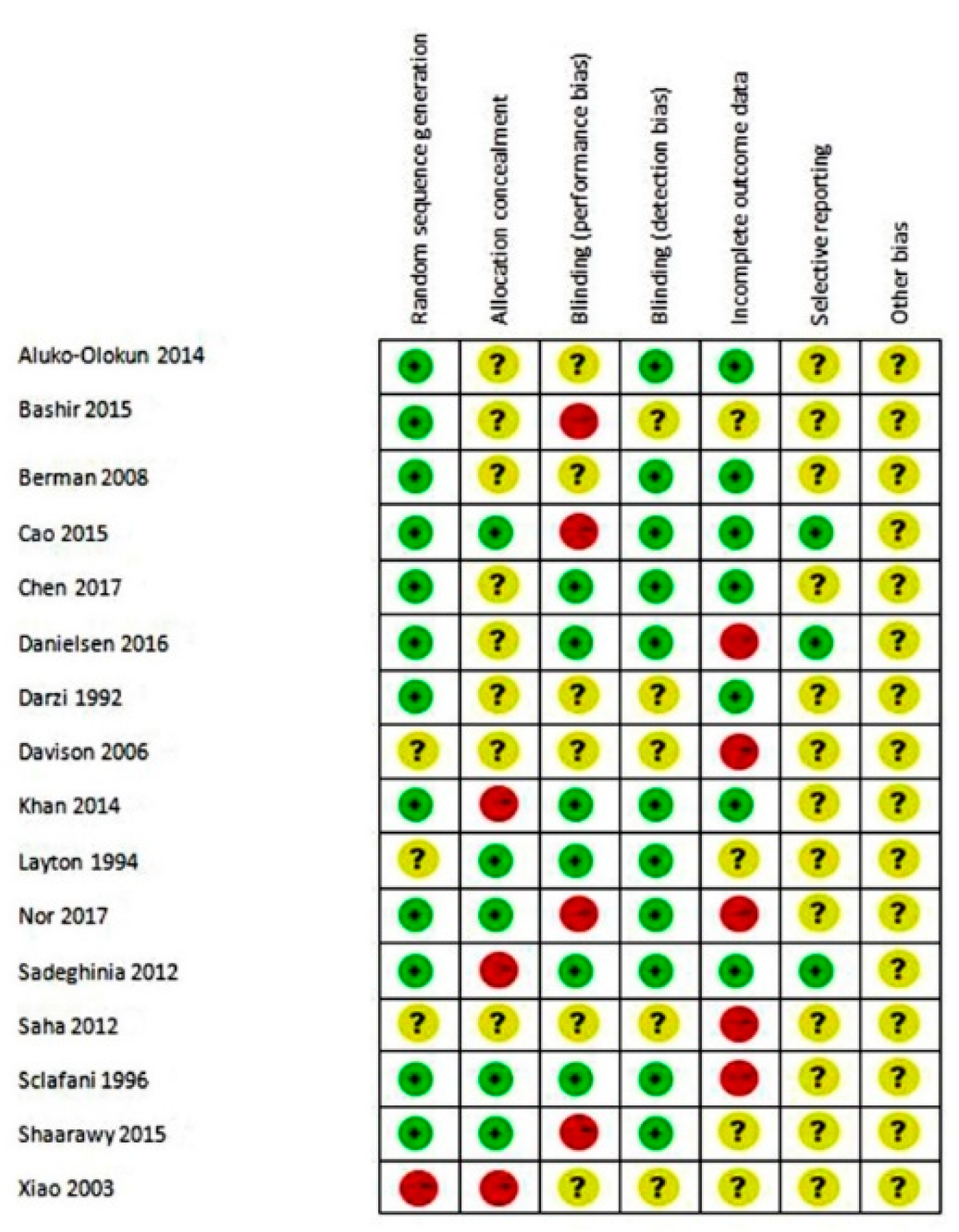
2.2. Data Collection and Analysis

Data collection and analyses were carried out according to the methods outlined in the Cochrane Collaboration. For this review, two review authors (W.R. and P.L.D.) independently assessed the titles and available abstracts of all the studies identified during the initial search and excluded any irrelevant studies. Then, they independently assessed the full-paper copies of the reports of potentially eligible studies using the inclusion criteria. If there were disagreements, these were discussed and resolved between the two authors. Data were extracted by one review author and checked for accuracy by a second review author. Additional unpublished data were requested from primary authors and included when available, but only a fraction of authors responded.
The quality of evidence was assessed by the GRADE (Grading of Recommendations, Assessment, Development and Evaluations) framework as high, moderate, low, or very low quality [39]. The evidence was downgraded from high quality by one level for serious or by two levels for very serious study limitations (risk of bias), indirectness of evidence, serious inconsistency, imprecision of effect estimates or potential publication bias. Two review authors (W.R. and P.L.D.) independently assessed the methodological quality and risk of bias [39].

2.3. Meta-Analysis

We used random effect modelling for both primary (dichotomous) and secondary (continuous) outcomes on the basis that it assumes that treatment effects were not identical in all studies. A fixed effect model assumes that the treatment effect is the same in each study and that differences in the results are only due to chance. We quantified heterogeneity among studies using the I2 inconsistency statistic, which is a measure of interstudy variability. If interstudy variability was low (I2 < 30%), the fixed effect and random effect models yielded very similar results [39]. Statistical analyses were performed using the Review Manager software (Review Manager (RevMan) [Computer program]. Version 5.3. Copenhagen: The Nordic Cochrane Centre, The Cochrane Collaboration, 2014). p < 0.05 was considered statistically significant.

3. Results

The searches yielded a total of 918 articles. Five additional records were identified from the reference lists. After examination of the titles and abstracts independently by the two review authors, 818 studies were excluded due to irrelevant content. After the removal of 8 duplicates, 97 full-text articles remained for further assessment. Finally, the total number of included RCTs was 16 after the omission of 81 full-text articles (Figure 1).
All 16 RCTs compared corticosteroids or radiotherapy with another treatment. The included studies were conducted in 10 countries and involved more than 814 patients aged 10 to 68 years. Characteristics of the included studies are shown in Table 2 and Table 3. The comparator treatments included 5-FU, etanercept, interferon α-2b, cryosurgery, botulinum toxin type A, and radiotherapy/corticosteroids.
The quality of evidence of the individual studies varied markedly. Six of the 16 RCTs met four or more of the seven domain criteria in the risk of bias analysis, as shown in Figure 2 [40,41,44,45,46,47]. The results of our GRADE analyses are shown in Table 2 and Table 3. Three trials were downgraded from high to moderate quality due to attrition bias [44,48,49].
Table 2. RCTs in alphabetical order on the effect of corticosteroid treatment without a keloidectomy.
StudyComparatorPatientsSite of LesionGRADE 1
nAverage age, years
Aluko-Olokun, 2014 [50]Excision with radiotherapy10727Ear, chest, foreheadMQ
Berman, 2008 [42]Etanercept18NS 2NS 2HQ
Chen, 2017 [47]Diprospan + 5-FU,
Diprospan + 5-FU + Nd:YAG laser
69 327Face, neck, trunk, extremitiesHQ
Darzi, 1992 [51]Radiotherapy65 4NS 2NS 2MQ
Khan, 2014 [46]TAC + 5-FU58NS 2NS 2MQ
Layton, 1994 [43]Cryosurgery1120 (males); 28 (females)AcneHQ
Nor, 2017 [52]Topical clobetasol propionate21 529Acne, surgeryMQ
Sadeghinia, 2012 [41] 5-FU4043Face, neck, trunk, limbsHQ
Saha, 2012 [48]5-FU50 633 (TAC); 35 (5-FU)Operations, acne, burnsMQ
Shaarawy, 2015 [53]Botulinum toxin type A2429Posttraumatic, idiopathicHQ
Xiao, 2003 [54]TAC + 5-FU2140.3–68 (range)Chest, shoulder, backMQ
1 GRADE, Grading Recommendation Assessment Development and Evaluation (HQ, high quality; MQ, moderate quality); 2 NS, not stated; 3 62 patients completed the study; 4 65 patients with 58 keloids and 42 hypertrophic scars; 5 17 patients with 34 keloids (17 scars in each arm) completed the study; 6 44 patients completed the study.
Table 3. RCTs in alphabetical order on the effect of corticosteroid treatment in conjunction with a keloidectomy.
StudyComparatorPatientsSite of LesionGRADE 1
nAverage age, years
Bashir, 2015 [55]3 TAC injections7022 (1 injection); 23 (3 injections)EarHQ
Cao, 2015 [45]TAC + 90Sr-90Y6138Chest, shoulder, limb, ear, othersHQ
Danielsen, 2016 [40]Verapamil1432Sternum, shoulder, back, neck, ear, upper arm, forearm, scar area excisedMQ
Darzi, 1992 [51]Radiotherapy65 2NS 3NS 3MQ
Davison, 2006 [49]Interferon α-2b3430Ear, face, abdomen, chest, extremitiesMQ
Sclafani, 1996 [44]Radiotherapy2327 to 29EarMQ
1 GRADE, Grading of Recommendations, Assessment, Development and Evaluations (HQ, high quality; MQ, moderate quality); 2 65 patients with 58 keloids and 42 hypertrophic scars; 3 NS, not stated.

3.1. Studies without Surgery: Primary and Secondary Outcomes

Eleven RCTs listed in Table 2 were retrieved and reviewed in this category [41,42,43,46,47,48,50,51,52,53,54].

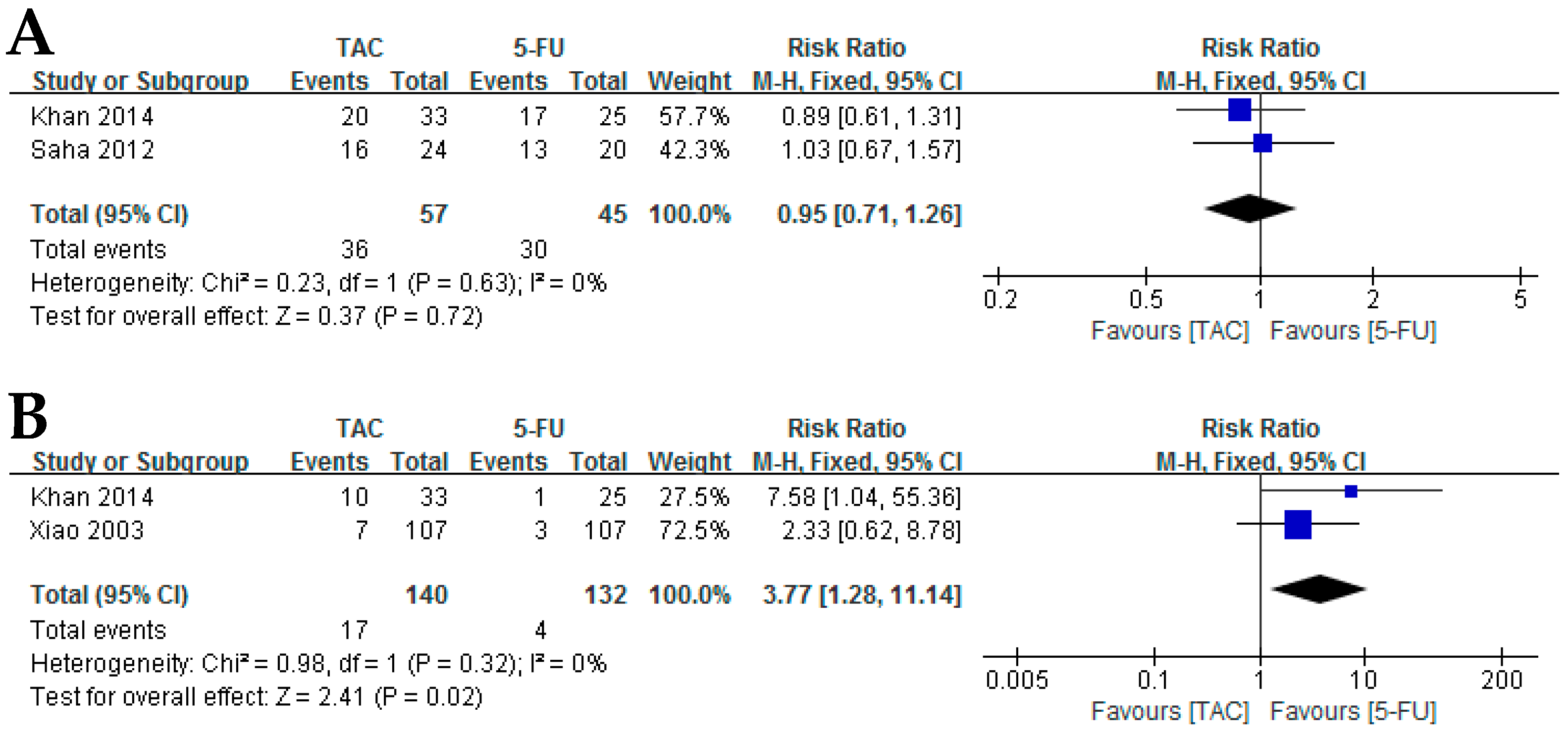
3.1.1. Corticosteroids versus 5-FU

The five RCTs identified involved a total of 431 patients [41,46,47,48,54].
Sadeghinia et al. [41] included 40 patients and compared 5-FU tattooing with an intralesional steroid. At the end of the study (week 44), the height, surface, erythema, induration and pruritus reduction were greater in the 5-FU group than in the TAC group compared with baseline (p < 0.05 for all). No adverse effects were observed.
Khan et al. [46] enrolled 58 patients with keloids (1–5 cm). The patients received intralesional TAC alone (n = 33) or TAC + 5-FU (n = 25). Good to excellent results (>50% improvement) were observed in 20 (61%) patients in the TAC alone group compared to 17 (68%) patients in the TAC + 5-FU group (RR 0.89, 95% CI: 0.61–1.3). Complications (skin atrophy and telangiectasias) were more frequent in the TAC alone group than in the TAC + 5-FU group (RR 7.6, 95% CI: 1.0–55).
Saha et al. [48] included 50 patients with keloids of 8 months to 20 years duration. The major causes of the keloids were operations, acne, and burns. In the 44 patients who completed the RCT, the authors found no difference (p > 0.05) between the TAC (n = 24) and 5-FU (n = 20) groups in the reduction of keloid volume (RR 1.0, 95% CI: 0.67–1.6), pain or itching of the keloids. 5-FU treatment ameliorated hyperpigmentation (p < 0.001) but was associated with more side effects, such as injection pain and superficial ulceration, than with TAC treatment (RR 0.030, 95% CI: 0–0.49, p = 0.014). No haematologic side effects were noted in any of the patients.
The studies by Khan et al. [46] and Saha et al. [48] were pooled (fixed effect, I2 = 0%). The reduction in the volume of keloids (>50%) was not significantly different between the two groups (RR 0.95, 95% CI: 0.71–1.3, p = 0.72), as shown in Figure 3A.
Two hundred and fourteen patients participated in the study by Xiao et al. [54]. The authors compared intralesional TAC with TAC + 5-FU (at volume ratios of 3:1-1:1). At the 6-month follow-up visit, lesion flattening, or softening was more frequent (p < 0.05) in the TAC + 5-FU group than in the TAC alone group. Complications (infection, menstrual disorders and telangiectasia) did not differ significantly between the groups (RR 2.3, 95% CI: 0.62–8.8).
When the data from the studies of Khan et al. [46] and Xiao et al. [54] (fixed effect, I2 = 0%) were pooled, the complications were less frequent in the TAC + 5-FU group (RR 3.8, 95% CI: 1.3–11, p = 0.02) than in the TAC alone group (Figure 3B). The study by Saha et al. [48] was excluded because of high heterogeneity (I2 = 85%).
Chen et al. [47] compared a betamethasone formulation, betamethasone + 5-FU, and betamethasone + 5-FU + Nd:YAG (long-pulsed neodymium-yttrium-aluminium-garnet) in laser treatment in a three-month single-centre RCT for the treatment of 62 keloids. The laser irradiation preceded the injections to inhibit neovascularization. The keloids were >10 mm in length and had not been treated within the last 6 months. Clinical response was assessed by a blinded observer on a 5-grade scale with respect to symptoms, size, height, and induration. The response (good to excellent improvement) was better in the betamethasone + 5-FU group (RR 0.25, 95% CI: 0.14–0.44) and in the betamethasone + 5-FU + Nd:YAG group (RR 0.17, 95% CI: 0.10–0.30) than in the betamethasone group. At the end of the 3-month study period, erythema, toughness, and itching were significantly more reduced in the patients treated with the combination of betamethasone + 5-FU + Nd:YAG than in patients in the other two groups (p < 0.05 for all indices). In the betamethasone group alone, 36% of patients experienced a certain degree of skin atrophy and telangiectasia. Except for transient purpura in the betamethasone + 5-FU + Nd:YAG group, no other adverse effects were observed.

3.1.2. Corticosteroids versus Etanercept

Berman et al. [42] included 18 patients with keloids that lasted for at least 1 year, with lesions less than or equal to 2 cm in size and with no more than 1 episode of recurrence. They found that etanercept (TNF-α inhibitor) improved 5/12 parameters, including significant pruritus reduction, while TAC improved 11/12 parameters at week 8 [42]. These differences were not statistically significant. Although keloids were measured in millimetres and evaluated by the investigator in each subject using a visual analogue scale from 0 to 100, including erythema, pain, tenderness, pigmentation, pruritus and cosmetic assessment, these data were not explained in detail. No adverse effects were reported.

3.1.3. Corticosteroids versus Cryosurgery

One study involving 11 patients with multiple acne keloids was identified. Layton et al. [43] concluded that patients showing minor differences in blood flow between the lesional and perilesional skin also showed no significant difference in response to either treatment modality. Cryosurgery was superior (p < 0.05) to TAC for the more vascularised keloids. No long-term side effects were produced.

3.1.4. Corticosteroids versus Botulinum Toxin Type A

Shaarawy et al. [53] compared the efficacy and safety of intralesional corticosteroid (TAC) treatment with intralesional botulinum toxin in 24 female patients with posttraumatic or idiopathic keloids. There was a significant decrease in the volume of the lesions after treatment (mean difference −3.5, 95% CI: −12–5.0, p < 0.01), with a volume reduction of 83% in the TAC group and 79% in the botulinum toxin group. Softening of lesions compared with the baseline was observed in both groups (p < 0.01), with more (p < 0.01) softening in the TAC group than in the botulinum toxin group. There was a significant decrease in the height of lesions and in the redness score compared with the baseline (p < 0.01) for both groups with no significant difference between the two groups. All patients experienced a reduction (p < 0.01) in their subjective complaints (itching, pain and tenderness). These improvements were more pronounced (p ≤ 0.01) in the botulinum toxin group. Skin atrophy and telangiectasia were observed in three patients in the TAC group (RR 0.14, 95% CI: 0.010–2.5).

3.1.5. Corticosteroid Injection versus Topical Corticosteroid Cream under Silicone Dressing

Nor et al. [52] included 34 keloid scars in 17 patients who allowed a paired design. Two keloids on similar anatomic sites were randomly assigned to receive either daily topical clobetasol propionate 0.05% cream covered with a silicone dressing or monthly intralesional TAC injections. The keloid had shrunk by week 12 with both treatments. The POSAS scores improved with both treatments from baseline to week 12 (p ≤ 0.05), but there was no statistically significant difference in POSAS scores at 4, 8, or 12 weeks between the two treatments. There was a significantly higher rate of adverse effects documented in the intralesional TAC group (RR 2.1, 95% CI: 1.4–3.1). The outcome data were incomplete.

3.1.6. Corticosteroids versus Radiotherapy

One study involving 28 keloids by Darzi et al. [51] was reviewed. Complete flattening was more frequent (p < 0.005) in the TAC group than in the patients receiving radiotherapy (β), whereas mild to no reduction of lesions was more common (p < 0.001) with radiotherapy. Overall, all 17 keloids treated with intralesional TAC showed complete or partial flattening compared with 3 of the 11 keloids treated with radiation alone (RR 3.3, 95% CI: 1.4–8.1). Skin atrophy was observed in one lesion in the TAC group.

3.1.7. Corticosteroids versus Excision Plus Radiotherapy

Aluko-Olokun et al. [50] carried out a study involving 107 patients with facial keloids. The investigators studied the lesion height before and after treatment and found that TAC-treated lesions were flattened to the level of the surrounding skin in 82% of the patients compared with 59% of patients treated with excision plus radiotherapy (p < 0.01). The number of complications did not differ significantly between the two groups (RR 0.95, 95% CI: 0.68–1.3). The authors concluded that intralesional TAC injection was significantly more efficacious than excision plus radiotherapy in the management of facial keloids.

3.2. Studies with Surgery: Primary and Secondary Outcomes

In this category, the six RCTs presented in Table 3 were reviewed [40,44,45,49,51,55].

3.2.1. Single Corticosteroid Injection versus Three Corticosteroid Injections

In the study by Bashir et al. [55], a single intraoperative TAC injection (n = 35) was compared with a single intraoperative and two postoperative TAC injections (n = 35) after the wedge excision of ear keloids. The 70 female patients over 14 years of age presented with post-piercing keloids of the helix not treated previously by any means and amenable to wedge excision without incurring a cosmetic deformity of the ear [55]. The follow-up period ranged from 12 months to 26 months with a mean of 17 ± 3.1 months. The recurrence rate was three (8.5%) in the single intraoperative injection group and two (5.7%) in the intraoperative and postoperative injection group (RR 0.67, 95% CI: 0.12–3.8, p = 0.64). The corresponding complication rates were three (8.5%) and eight (22.8%) (RR 2.7, 95% CI: 0.77–9.2, p = 0.10).

3.2.2. Preoperative and Postoperative Radiotherapy versus Postoperative Radiotherapy

In the study by Darzi et al. [51] on 30 keloids, 20 (67%) did not recur over the follow-up period of 10 years, 9 (30%) recurred, and 1 patient (3.3%) with an ear lobe keloid was lost during follow-up. There was no significant difference in the recurrence of keloids between the two groups (RR 1.2, 95% CI: 0.39–3.5, p > 0.10). Furthermore, the authors compared the effects of early postoperative irradiation (β irradiation) with late postoperative irradiation in both groups; 75% of patients who received early postoperative irradiation showed no recurrence compared to 57% of the patients who received late postoperative irradiation. This effect was not significantly different (RR 0.47, 95% CI: 0.14–1.5, p > 0.10). In the three patients who developed dyspigmentation, the area resumed normal colour within 18 months.

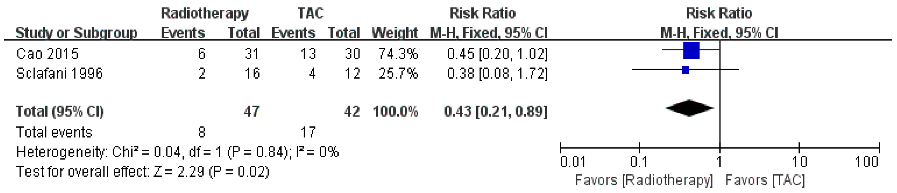
3.2.3. Corticosteroids versus Radiotherapy

There were two studies involving a total of 84 patients with 100 keloids [44,45].
Cao et al. [45] compared corticosteroids (TAC) with TAC plus a 90Sr-90Y applicator (BT) after surgical excision in 61 patients with keloids from 1.9 cm × 1.1 cm to 5.1 cm × 1.3 cm in size. The follow-up periods for efficacy and safety ranged from 12 to 24 months. The cure rate of the TAC group was 43% compared with 71% for the TAC + BT group (RR 1.6, 95% CI: 1.0–2.6, p < 0.05). The recurrence rate in the TAC (43%) group was higher than in the TAC + BT (19%) group (RR 0.45, 95% CI: 0.20–1.0, p < 0.05). The authors reported that complications (weakness, desquamation, blisters, pigmentation, and depigmentation) were more common in the TAC + BT combination group (RR 3.9, 95% CI: 1.2–12).
Intralesional TAC was compared with radiotherapy (X-rays or electron beam) in one RCT conducted by Sclafani et al. [44], including 28 keloids in 23 patients. Four of 12 keloids (33%) recurred after surgery and corticosteroid injection compared with 2 of 16 keloids (13%) after surgery and radiotherapy. This difference was not statistically significant (RR 2.7, 95% CI: 0.58–12). The overall median time to recurrence was 17 months. No adverse side effects were reported in this study.
When pooling the two RCTs (fixed effect, I2 = 0%) [44,45], the recurrence of keloids was less in the radiotherapy group than in the corticosteroid (RR 0.43, 95% CI: 0.21–0.89, p = 0.02) groups (Figure 4).

3.2.4. Corticosteroids versus Interferon α-2b

One RCT was identified that involved 34 patients with 39 keloids [49]. The overall recurrence rate was 28%. Among the 13 keloids that were treated with postoperative intralesional interferon α-2b injections, 7 recurred (54%) compared with 4 of the 26 TAC-treated (15%) keloids, thus presenting a significant difference (RR 0.27, 95% CI: 0.10–0.74). The average time to recurrence was ten months in the interferon group and four months in the TAC group. Notably, 2 of the 11 patients treated with interferon α-2b experienced systemic flu-like symptoms, while no adverse effects were reported in the TAC group. This represents a statistically nonsignificant difference between the two groups (RR 0.10, 95% CI: 0.010–1.9).

3.2.5. Corticosteroids versus Verapamil

One study, involving 14 subjects with 14 keloids compared the calcium channel blocker verapamil with TAC in the prevention of keloid recurrence after surgical excision in a paired design [40]. Survival analysis demonstrated significantly higher keloid recurrence with verapamil compared to TAC 12 months postoperatively (log-rank test, p = 0.01) and a higher overall risk of recurrence with verapamil than with TAC (hazard ratio 8.4, 95% CI: 1.6–44). The study was prematurely terminated according to the stopping criteria (p < 0.05). Four subjects experienced skin hypotrophy when using 2 mg TAC/cm, but none experienced skin hypotrophy when using 1 mg TAC/cm. Verapamil was found to be safe. The study lacked allocation concealment.

4. Discussion

In this systematic review, 16 RCTs with more than 814 included patients were evaluated. The quality of evidence for most outcomes was moderate to high as assessed by the GRADE approach [39]. Information on random sequence generation, allocation concealment, blinding, and/or attrition was sometimes missing. In some studies, keloid and hypertrophic scars were mixed together without subgroup analysis despite their pathological and clinical differences [5,15,56]. There were problems performing the meta-analysis because of a lack of a standardised protocol for keloids, such as age of the scars, dosage, frequency and duration of treatments, follow-up time and assessment scales.
Intralesional corticosteroid injection was compared with the chemotherapeutic 5-FU in 431 patients. Meta-analysis of two RCTs indicated similar efficacy between the two regimens, but 5-FU appeared to cause more adverse effects. Ren et al. [57] advocated the combined use of TAC and 5-FU based on their systematic review. Furthermore, corticosteroid treatment was found to be equipotent to etanercept and cryosurgery. Intralesional botulinum toxin type A was equally effective and better tolerated than intralesional steroid treatment confirming the conclusions of a systematic review from 2019 [58]. Radiotherapy alone was less effective on the keloid mass compared with corticosteroid therapy but eradicated keloid symptoms more effectively than corticosteroids.
The Ca2+ channel blocker verapamil possesses antifibrotic properties. A RCT published in 2018 (not included in our review) compared verapamil with TAC [59]. The investigators found that verapamil had a minimal effect (VSS score reduction of 7%) compared to TAC treatment that reduced the VSS score by 69% after 6 injections over 18 weeks.
Adjuvant radiotherapy following excision is more effective (fewer recurrences) than radiotherapy alone, with BT being the most effective irradiation modality compared to X-ray and electron beam [34]. BT with a high dose rate seems optimal [60]. Furthermore, the recurrence after the excision of a keloid was higher with adjuvant corticosteroid treatment than with radiotherapy [44,45]. The radiation time and the timing of intralesional corticosteroids did not affect the outcomes significantly. Shin et al. [61] compared TAC injections and radiotherapy of surgically excised ear keloids in a systematic review including 25 studies. They concluded that there were no differences in recurrence rate between the TAC (15%) and radiotherapy (14%) groups in their total group of 1105 patients [61]. The discrepancy with our results could be explained by their less strict search criteria. Adverse effects associated with adjuvant radiotherapy are more common than with adjuvant corticosteroid treatment [34]. The recurrence rates with interferon α-2b treatment exceeded those of corticosteroids, despite delaying the time to recurrence [49]. This is in sharp contrast to the findings of Berman et al. [62], who reported lower recurrence rates with interferon α-2b treatment (19%) compared to TAC (58%) and excision alone (51%) in a retrospective study. The recurrence following keloid excision was higher with verapamil than with TAC treatment within one year postoperatively [40].
The effect of the toll-like receptor agonist imiquimod was recently reviewed. Klotz et al. [63] concluded in their systematic review and meta-analysis (7 studies) that imiquimod cream post-excision produces variable results.
There were some potential biases in the review process. First, the follow-up time varied from three months to more than one year. As the characteristics of keloid scars are related to the duration, the results might be potentially biased. Secondly, we did not differentiate between variations in the corticosteroid and radiotherapy treatment protocols. We suggest that these factors are standardised in future RCTs on keloid treatment and prevention.

5. Conclusions

Intralesional corticosteroid treatment remains the first-line monotreatment for keloids, although radiotherapy seems superior as an adjunct in preventing keloid reoccurrence after surgical excision. More high-quality, multicentre, large-scale randomised controlled trials are required to substantiate these conclusions.

Author Contributions

Conceptualization, R.W., P.L.D. and M.S.Å.; methodology, R.W. and P.L.D.; formal analysis, R.W. and P.L.D.; writing—original draft preparation, R.W. and P.L.D.; writing—review and editing, M.S.Å., J.D., X.-X.Z. and Y.M.; supervision, F.W., M.S.Å. and Y.C.; funding acquisition, F.W. and Y.C. All authors have read and agreed to the published version of the manuscript.

Funding

This study was funded by the National Natural Science Foundation of China (81501673).

Institutional Review Board Statement

Studies were approved by an ethics committee.

Informed Consent Statement

The enrolled patients provided informed written consent.

Data Availability Statement

The data presented in this study are available on request from the corresponding author.

Conflicts of Interest

The authors declare no conflict of interest.

Abbreviations

BTBrachytherapy
CIConfidence interval
GRADEGrading of Recommendations, Assessment, Development and Evaluations
FUFluorouracil
POSASPatient and Observer Scar Assessment Scale
RCTRandomised controlled trial
RRRisk ratio
TACTriamcinolone acetonide
VSSVancouver Scar Scale

References

  1. Peacock, E.E., Jr.; Madden, J.W.; Trier, W.C. Biologic basis for the treatment of keloids and hypertrophic scars. South Med. J. 1970, 63, 755–760. [Google Scholar] [CrossRef] [PubMed]
  2. Jackson, I.T.; Bhageshpur, R.; DiNick, V.; Khan, A.; Bhaloo, S. Investigation of recurrence rates among earlobe keloids utilizing various postoperative therapeutic modalities. Eur. J. Plast. Surg. 2001, 24, 88–95. [Google Scholar] [CrossRef]
  3. Seifert, O.; Mrowietz, U. Keloid scarring: Bench and bedside. Arch. Dermatol. Res. 2009, 301, 259–272. [Google Scholar] [CrossRef]
  4. Shirakami, E.; Yamakawa, S.; Hayashida, K. Strategies to prevent hypertrophic scar formation: A review of therapeutic interventions based on molecular evidence. Burns Trauma 2020, 8, tkz003. [Google Scholar] [CrossRef] [PubMed] [Green Version]
  5. Ehrlich, H.P.; Desmouliere, A.; Diegelmann, R.F.; Cohen, I.K.; Compton, C.C.; Garner, W.L.; Kapanci, Y.; Gabbiani, G. Morphological and immunochemical differences between keloid and hypertrophic scar. Am. J. Pathol. 1994, 145, 105–113. [Google Scholar]
  6. Glass, D.A., 2nd. Current understanding of the genetic causes of keloid formation. J. Investig. Dermatol. Symp. Proc. 2017, 18, S50–S53. [Google Scholar] [CrossRef] [Green Version]
  7. Gauglitz, G.G.; Korting, H.C.; Pavicic, T.; Ruzicka, T.; Jeschke, M.G. Hypertrophic scarring and keloids: Pathomechanisms and current and emerging treatment strategies. Mol. Med. 2011, 17, 113–125. [Google Scholar] [CrossRef] [PubMed]
  8. Alhady, S.M.; Sivanantharajah, K. Keloids in various races. A review of 175 cases. Plast. Reconstr. Surg. 1969, 44, 564–566. [Google Scholar] [CrossRef]
  9. Brissett, A.E.; Sherris, D.A. Scar contractures, hypertrophic scars, and keloids. Facial. Plast Surg. 2001, 17, 263–272. [Google Scholar] [CrossRef]
  10. Bock, O.; Schmid-Ott, G.; Malewski, P.; Mrowietz, U. Quality of life of patients with keloid and hypertrophic scarring. Arch. Dermatol. Res. 2006, 297, 433–438. [Google Scholar] [CrossRef]
  11. Pollack, S.V.; Goslen, J.B. The surgical treatment of keloids. J. Dermatol. Surg. Oncol. 1982, 8, 1045–1049. [Google Scholar] [CrossRef] [PubMed]
  12. Cosman, B.; Wolff, M. Correlation of keloid recurrence with completeness of local excision. A negative report. Plast. Reconstr. Surg. 1972, 50, 163–166. [Google Scholar] [CrossRef]
  13. Siotos, C.; Uzosike, A.C.; Hong, H.; Seal, S.M.; Rosson, G.D.; Cooney, C.M.; Cooney, D.S. Keloid Excision and Adjuvant Treatments: A Network Meta-analysis. Ann. Plast. Surg. 2019, 83, 154–162. [Google Scholar] [CrossRef] [PubMed]
  14. Wong, T.S.; Li, J.Z.; Chen, S.; Chan, J.Y.; Gao, W. The efficacy of triamcinolone acetonide in keloid treatment: A systematic review and meta-analysis. Front. Med. 2016, 3, 71. [Google Scholar] [CrossRef] [PubMed] [Green Version]
  15. Chen, M.A.; Davidson, T.M. Scar management: Prevention and treatment strategies. Curr. Opin. Otolaryngol. Head Neck Surg. 2005, 13, 242–247. [Google Scholar] [CrossRef]
  16. Jalali, M.; Bayat, A. Current use of steroids in management of abnormal raised skin scars. Surgeon 2007, 5, 175–180. [Google Scholar] [CrossRef]
  17. Zhang, Z.; Cheng, L.; Wang, R.; Cen, Y.; Li, Z. Effects and safety of triamcinolone acetonide-controlled common therapy in keloid treatment: A Bayesian network meta-analysis. Ther. Clin. Risk Manag. 2018, 14, 973–980. [Google Scholar] [CrossRef] [Green Version]
  18. Chowdri, N.A.; Masarat, M.; Mattoo, A.; Darzi, M.A. Keloids and hypertrophic scars: Results with intraoperative and serial postoperative corticosteroid injection therapy. Aust. N. Z. J. Surg. 1999, 69, 655–659. [Google Scholar] [CrossRef] [Green Version]
  19. Niessen, F.B.; Spauwen, P.H.; Schalkwijk, J.; Kon, M. On the nature of hypertrophic scars and keloids: A review. Plast. Reconstr. Surg. 1999, 104, 1435–1458. [Google Scholar] [CrossRef]
  20. Wang, X.Q.; Liu, Y.K.; Qing, C.; Lu, S.L. A review of the effectiveness of antimitotic drug injections for hypertrophic scars and keloids. Ann. Plast. Surg. 2009, 63, 688–692. [Google Scholar]
  21. Manuskiatti, W.; Fitzpatrick, R.E. Treatment response of keloidal and hypertrophic sternotomy scars: Comparison among intralesional corticosteroid, 5-fluorouracil, and 585-nm flashlamp-pumped pulsed-dye laser treatments. Arch. Dermatol. 2002, 138, 1149–1155. [Google Scholar] [CrossRef] [PubMed]
  22. Alster, T. Laser scar revision: Comparison study of 585-nm pulsed dye laser with and without intralesional corticosteroids. Dermatol. Surg. 2003, 29, 25–29. [Google Scholar] [CrossRef]
  23. Asilian, A.; Darougheh, A.; Shariati, F. New combination of triamcinolone, 5-Fluorouracil, and pulsed-dye laser for treatment of keloid and hypertrophic scars. Dermatol. Surg. 2006, 32, 907–915. [Google Scholar] [PubMed]
  24. Finken, M.J.; Mul, D. Cushing’s syndrome and adrenal insufficiency after intradermal triamcinolone acetonide for keloid scars. Eur. J. Pediatr. 2010, 169, 1147–1149. [Google Scholar] [CrossRef] [PubMed] [Green Version]
  25. van Leeuwen, M.C.; Stokmans, S.C.; Bulstra, A.E.; Meijer, O.W.; Heymans, M.W.; Ket, J.C.; Ritt, M.J.; van Leeuwen, P.A.; Niessen, F.B. Surgical excision with adjuvant irradiation for treatment of keloid scars: A systematic review. Plast. Reconstr. Surg. Glob. Open 2015, 3, e440. [Google Scholar] [CrossRef] [PubMed]
  26. Cheraghi, N.; Cognetta, A., Jr.; Goldberg, D. Radiation therapy for the adjunctive treatment of surgically excised keloids: A review. J. Clin. Aesthet. Dermatol. 2017, 10, 12–15. [Google Scholar]
  27. Yamawaki, S.; Naitoh, M.; Ishiko, T.; Muneuchi, G.; Suzuki, S. Keloids can be forced into remission with surgical excision and radiation, followed by adjuvant therapy. Ann. Plast. Surg. 2011, 67, 402–406. [Google Scholar] [CrossRef]
  28. Recalcati, S.; Caccialanza, M.; Piccinno, R. Postoperative radiotherapy of auricular keloids: A 26-year experience. J. Dermatolog. Treat. 2011, 22, 38–42. [Google Scholar] [CrossRef]
  29. Bennett, K.G.; Kung, T.A.; Hayman, J.A.; Brown, D.L. Treatment of keloids with excision and adjuvant radiation: A single center experience and review of the literature. Ann. Plast. Surg. 2017, 78, 157–161. [Google Scholar] [CrossRef]
  30. Ogawa, R.; Yoshitatsu, S.; Yoshida, K.; Miyashita, T. Is radiation therapy for keloids acceptable? The risk of radiation-induced carcinogenesis. Plast. Reconstr. Surg. 2009, 124, 1196–1201. [Google Scholar] [CrossRef]
  31. Jones, K.; Fuller, C.D.; Luh, J.Y.; Childs, C.C.; Miller, A.R.; Tolcher, A.W.; Herman, T.S.; Thomas, C.R., Jr. Case report and summary of literature: Giant perineal keloids treated with post-excisional radiotherapy. BMC Dermatol. 2006, 6, 7. [Google Scholar] [CrossRef] [PubMed] [Green Version]
  32. Speranza, G.; Sultanem, K.; Muanza, T. Descriptive study of patients receiving excision and radiotherapy for keloids. Int. J. Radiat. Oncol. Biol. Phys. 2008, 71, 1465–1469. [Google Scholar] [CrossRef] [PubMed]
  33. Borok, T.L.; Bray, M.; Sinclair, I.; Plafker, J.; LaBirth, L.; Rollins, C. Role of ionizing irradiation for 393 keloids. Int. J. Radiat. Oncol. Biol. Phys. 1988, 15, 865–870. [Google Scholar] [CrossRef]
  34. Mankowski, P.; Kanevsky, J.; Tomlinson, J.; Dyachenko, A.; Luc, M. Optimizing radiotherapy for keloids: A meta-analysis systematic review comparing recurrence rates between different radiation modalities. Ann. Plast. Surg. 2017, 78, 403–411. [Google Scholar] [CrossRef]
  35. Nie, Z.; Bayat, A.; Behzad, F.; Rhodes, L.E. Positive response of a recurrent keloid scar to topical methyl aminolevulinate-photodynamic therapy. Photodermatol. Photoimmunol. Photomed. 2010, 26, 330–332. [Google Scholar] [CrossRef] [PubMed]
  36. Fong, C.Y.; Biswas, A.; Subramanian, A.; Srinivasan, A.; Choolani, M.; Bongso, A. Human keloid cell characterization and inhibition of growth with human Wharton’s jelly stem cell extracts. J. Cell Biochem. 2014, 115, 826–838. [Google Scholar] [CrossRef]
  37. Baryza, M.J.; Baryza, G.A. The Vancouver Scar Scale: An administration tool and its interrater reliability. J. Burn. Care Rehabil. 1995, 16, 535–538. [Google Scholar] [CrossRef]
  38. Draaijers, L.J.; Tempelman, F.R.; Botman, Y.A.; Tuinebreijer, W.E.; Middelkoop, E.; Kreis, R.W.; van Zuijlen, P.P. The patient and observer scar assessment scale: A reliable and feasible tool for scar evaluation. Plast. Reconstr. Surg. 2004, 113, 1960–1965. [Google Scholar] [CrossRef]
  39. Higgins, J.P.T.; Green, S. Cochrane Handbook for Systematic Reviews of Interventions; The Cochrane Collaboration: Oxford, UK, 2011. [Google Scholar]
  40. Danielsen, P.L.; Rea, S.M.; Wood, F.M.; Fear, M.W.; Viola, H.M.; Hool, L.C.; Gankande, T.U.; Alghamdi, M.; Stevenson, A.W.; Manzur, M.; et al. Verapamil is less effective than triamcinolone for prevention of keloid scar recurrence after excision in a randomized controlled trial. Acta Derm. Venereol. 2016, 96, 774–778. [Google Scholar] [CrossRef] [Green Version]
  41. Sadeghinia, A.; Sadeghinia, S. Comparison of the efficacy of intralesional triamcinolone acetonide and 5-fluorouracil tattooing for the treatment of keloids. Dermatol. Surg. 2012, 38, 104–109. [Google Scholar] [CrossRef]
  42. Berman, B.; Patel, J.K.; Perez, O.A.; Viera, M.H.; Amini, S.; Block, S.; Zell, D.; Tadicherla, S.; Villa, A.; Ramirez, C.; et al. Evaluating the tolerability and efficacy of etanercept compared to triamcinolone acetonide for the intralesional treatment of keloids. J. Drugs Dermatol. 2008, 7, 757–761. [Google Scholar]
  43. Layton, A.M.; Yip, J.; Cunliffe, W.J. A comparison of intralesional triamcinolone and cryosurgery in the treatment of acne keloids. Br. J. Dermatol. 1994, 130, 498–501. [Google Scholar] [CrossRef] [PubMed]
  44. Sclafani, A.P.; Gordon, L.; Chadha, M.; Romo, T., 3rd. Prevention of earlobe keloid recurrence with postoperative corticosteroid injections versus radiation therapy: A randomized, prospective study and review of the literature. Dermatol. Surg. 1996, 22, 569–574. [Google Scholar] [CrossRef] [PubMed]
  45. Cao, C.Y.; Shuai, M.S.; Ding, Z.Q.; Chen, S.T.; Zhao, J.L. Clinical observation of the therapeutic efficacy of surgical excision combined with triamcinolone acetonide injection and 90Sr-90Y applicator brachytherapy for the treatment of keloids. J. Clin. Dermatol. 2015, 44, 813–815. [Google Scholar]
  46. Khan, M.A.; Bashir, M.M.; Khan, F.A. Intralesional triamcinolone alone and in combination with 5-fluorouracil for the treatment of keloid and hypertrophic scars. J. Pak. Med. Assoc. 2014, 64, 1003–1007. [Google Scholar]
  47. Chen, X.E.; Liu, J.; Bin Jameel, A.A.; Valeska, M.; Zhang, J.A.; Xu, Y.; Liu, X.W.; Zhou, H.; Luo, D.; Zhou, B.R. Combined effects of long-pulsed neodymium-yttrium-aluminum-garnet laser, diprospan and 5-fluorouracil in the treatment of keloid scars. Exp. Ther. Med. 2017, 13, 3607–3612. [Google Scholar] [CrossRef] [PubMed] [Green Version]
  48. Saha, A.K.; Mukhopadhyay, M. A comparative clinical study on role of 5-flurouracil versus triamcinolone in the treatment of keloids. Indian J. Surg. 2012, 74, 326–329. [Google Scholar] [CrossRef] [PubMed] [Green Version]
  49. Davison, S.P.; Mess, S.; Kauffman, L.C.; Al-Attar, A. Ineffective treatment of keloids with interferon alpha-2b. Plast Reconstr Surg 2006, 117, 247–252. [Google Scholar] [CrossRef]
  50. Aluko-Olokun, B.; Olaitan, A.A.; Ladeinde, A.L.; Oginni, F.O. The facial keloid: A comparison of treatment outcome between intralesional steroid injection and excision combined with radiotherapy. Eur. J. Plast. Surg. 2014, 37, 361–366. [Google Scholar] [CrossRef]
  51. Darzi, M.A.; Chowdri, N.A.; Kaul, S.K.; Khan, M. Evaluation of various methods of treating keloids and hypertrophic scars: A 10-year follow-up study. Br. J. Plast Surg. 1992, 45, 374–379. [Google Scholar] [CrossRef]
  52. Nor, N.M.; Ismail, R.; Jamil, A.; Shah, S.A.; Imran, F.H. A randomized, single-blind trial of clobetasol propionate 0.05% cream under silicone dressing occlusion versus intra-lesional triamcinolone for treatment of keloid. Clin. Drug Investig. 2017, 37, 295–301. [Google Scholar] [CrossRef] [PubMed]
  53. Shaarawy, E.; Hegazy, R.A.; Abdel Hay, R.M. Intralesional botulinum toxin type A equally effective and better tolerated than intralesional steroid in the treatment of keloids: A randomized controlled trial. J. Cosmet. Dermatol. 2015, 14, 161–166. [Google Scholar] [CrossRef] [PubMed]
  54. Xiao, M.; Shu, Y.; Xie, H.; Li, J.; Liu, Z. The observation on the effect of local injection with triamicnolon acetonide acetate and flurouracil on treating keloid. Chin. J. Derm. 2003, 17, 323–324. [Google Scholar]
  55. Bashir, M.M.; Ahmad, H.; Yousaf, N.; Khan, F.A. Comparison of single intra operative versus an intra operative and two post operative injections of the triamcinolone after wedge excision of keloids of helix. J. Pak. Med. Assoc. 2015, 65, 737–741. [Google Scholar]
  56. Bloemen, M.C.; van der Veer, W.M.; Ulrich, M.M.; van Zuijlen, P.P.; Niessen, F.B.; Middelkoop, E. Prevention and curative management of hypertrophic scar formation. Burns 2009, 35, 463–475. [Google Scholar] [CrossRef]
  57. Ren, Y.; Zhou, X.; Wei, Z.; Lin, W.; Fan, B.; Feng, S. Efficacy and safety of triamcinolone acetonide alone and in combination with 5-fluorouracil for treating hypertrophic scars and keloids: A systematic review and meta-analysis. Int. Wound J. 2017, 14, 480–487. [Google Scholar] [CrossRef]
  58. Bi, M.; Sun, P.; Li, D.; Dong, Z.; Chen, Z. Intralesional injection of botulinum toxin type A compared with intralesional injection of corticosteroid for the treatment of hypertrophic scar and keloid: A systematic review and meta-analysis. Med. Sci. Monit. 2019, 25, 2950–2958. [Google Scholar] [CrossRef] [PubMed]
  59. Abedini, R.; Sasani, P.; Mahmoudi, H.R.; Nasimi, M.; Teymourpour, A.; Shadlou, Z. Comparison of intralesional verapamil versus intralesional corticosteroids in treatment of keloids and hypertrophic scars: A randomized controlled trial. Burns 2018, 44, 1482–1488. [Google Scholar] [CrossRef]
  60. Guix, B.; Henríquez, I.; Andrés, A.; Finestres, F.; Tello, J.I.; Martínez, A. Treatment of keloids by high-dose-rate brachytherapy: A seven-year study. Int. J. Radiat. Oncol. Biol. Phys. 2001, 50, 167–172. [Google Scholar] [CrossRef]
  61. Shin, J.Y.; Lee, J.W.; Roh, S.G.; Lee, N.H.; Yang, K.M. A comparison of the effectiveness of triamcinolone and radiation therapy for ear keloids after surgical excision: A systematic review and meta-analysis. Plast. Reconstr. Surg. 2016, 137, 1718–1725. [Google Scholar] [CrossRef]
  62. Berman, B.; Flores, F. Recurrence rates of excised keloids treated with postoperative triamcinolone acetonide injections or interferon alfa-2b injections. J. Am. Acad. Dermatol. 1997, 37, 755–757. [Google Scholar] [CrossRef]
  63. Klotz, T.; Munn, Z.; Aromataris, E.C.; Greenwood, J.E. Imiquimod to prevent keloid recurrence postexcision: A systematic review and meta-analysis. Wound Repair Regen. 2020, 28, 145–156. [Google Scholar] [CrossRef] [PubMed]
Figure 1. Study flow diagram. Only 12 studies were included in quantitative synthesis (meta-analysis) due to incomplete data presentation in four [40,41,42,43] of the 16 studies.
Ebj 02 00004 g001
Figure 2. Risk of bias summary with review author judgment for the risk of each bias item for the 16 RCTs arranged in alphabetical order.
Ebj 02 00004 g002
Figure 3. Forest plots of corticosteroid (TAC) versus 5-fluorouracil (5-FU) injections. (A) Improvement (reduction of keloid volume >50%). (B) Adverse effects.
Ebj 02 00004 g003
Figure 4. Forest plot of adjuvant corticosteroid injections (TAC) versus adjuvant radiotherapy on keloid recurrence after surgical removal.
Ebj 02 00004 g004
Table 1. Search strategy.
NumberSearch Strategy
1MeSH descriptor: [keloid] explode all trees
2MeSH descriptor: [cicatrix, hypertrophic] explode all trees
3(Keloid* or hypertrophic or hypertrophic or cicatrix): ti, ab, kw
4(“Scar” or “scars” or scarred or scarring): ti, ab, kw
51 OR 2 OR 3 OR 4
6MeSH descriptor: [radiotherapy] explode all trees
7(Radiotherapy* or “radiation therapy”): ti, ab, kw
8MeSH descriptor: [adrenal cortex hormones] explode all trees
9MeSH descriptor: [steroids] explode all trees
10(Corticosteroid* or “hydrocortisone acetate” or methylprednisolone or dexamethasone or triamcinolone or steroid* or betamethasone or glucocort*): ti, ab, kw
116 OR 7 OR 8 OR 9 OR 10
125 AND 11
Publisher’s Note: MDPI stays neutral with regard to jurisdictional claims in published maps and institutional affiliations.

