Article

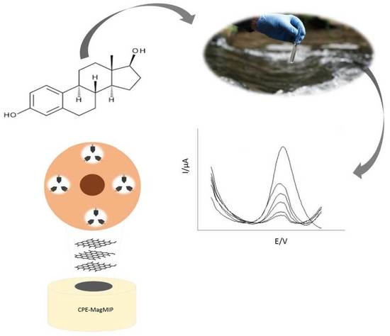
Development of a Chemically Modified Electrode with Magnetic Molecularly Imprinted Polymer (MagMIP) for 17-β-Estradiol Determination in Water Samples

by
Daniela Nunes da Silva
* and
Arnaldo César Pereira
Natural Sciences Department, Federal University of São João del Rei, 74 Praça Dom Helvécio, São João del Rei 36301-160, MG, Brazil
*
Author to whom correspondence should be addressed.
Submission received: 22 September 2022 / Revised: 4 November 2022 / Accepted: 23 November 2022 / Published: 2 December 2022
(This article belongs to the Collection Feature Papers in Electrochemistry)

Abstract

:
The present work consisted of the development of an electrode based on carbon paste modified with magnetic molecularly imprinted polymer (CPE-MagMIP) for 17-β-estradiol (E2) detection. The incorporation of magnetic material (MagMIP) improved sensor performance, an increase of over 317%. The proposed method resulted in a linear response range from 0.5 to 14.0 μM, and the detection limit (LOD) and quantification limit (LOQ) were equal to 0.13 and 0.44 μM, respectively. Under optimized conditions, the developed sensor obtained satisfactory parameters in E2 determination in water samples, demonstrating selectivity, accuracy, and precision, making it a promising method for monitoring E2 in environmental samples.

Graphical Abstract

1. Introduction

17-β-estradiol (E2) is classified as a female sex hormone and plays a relevant role in the regulation and growth of various tissues, such as the reproductive organs, mammary gland, and skeletal [1,2].
E2 is a naturally occurring estrogen, and it is the most potent estrogen in a group of endogenous estrogen steroids that includes estrone and estriol [3]. E2 is one of the main endocrine disruptors present in environmental samples of water [4].
The scientific community’s concern with the presence of estrogens in water has been demonstrated for some years [5]. The presence of estrogens is mainly due to effluents from municipal wastewater treatment. These compounds can cause damage to the endocrine system or other physiological systems, even at low concentration levels (µg or ng L−1) [6,7,8]. Thus, monitoring E2 in water samples is very important and can prevent possible environmental impacts.
Several analytical methods have been developed for the detection of E2, chromatographic [9,10], fluorometric [11,12,13], and electrochemical [14,15,16,17]. Electrochemical sensors have been increasingly used in the most diverse areas, in general, they are simpler devices, allowing in situ analysis with high sensitivity in a short period [18,19].
Another advantage is that these sensors can be modified, promoting better selectivity and accuracy in the analysis [20,21]. Regarding modifiers in sensors, we highlight the molecularly imprinted polymers (MIPs). MIPs are synthetic polymerics, which make it possible to obtain materials with biomimetic recognition for molecules of analytical interest [22,23]. The quality of selectivity of a MIP in the presence of other species and the imprinting effect of MIP for a desirable analyte is evaluated in comparison to an unprinted polymer, called NIP (non-imprinted polymer) [24].
The MIP has been used together with magnetic nanoparticles, generating magnetic molecularly imprinted polymer (MagMIP) [25]. The use of MagMIPs in electrochemical detection improves the access of the target analyte to the selective cavities of the imprinted polymer, provides an effective form of immobilization, and also allows regeneration of the electrodic surface [26].
Khan et al. (2019) developed a biomimetic sensor based on carbon paste with magnetic molecularly imprinted polymer for the detection of methyl green dye. Using square wave adsorptive anodic stripping voltammetry (SWAdASV) the sensor presented a limit of detection of 1.0 × 10⁻⁸ M. The sensor was also applied to river water samples, obtaining excellent precision and accuracy [22].
In another work, Afzali et al. (2022) presented a sensor using a glassy carbon electrode modified by a composite consisting of ionic liquid-based molecularly imprinted polymer and magnetic nanoparticles/graphene oxide for the detection of patulin toxin, using the square wave voltammetry technique. After optimized conditions, the sensor showed a limit of detection of 3.33×10⁻⁴ nM, and it was successfully applied to detect patulin toxin in apple juice samples [27].
Thus, a new electrochemical sensor based on the magnetic molecularly imprinted polymer (CPE-MagMIP) is proposed for the fast, sensitive detection of E2, with high selectivity in water samples.

2. Materials and Methods

2.1. Reagents and Materials

E2 was obtained from the United States Pharmacopeia (USP). Methacrylic acid, ethylene glycol dimethacrylate, iron (II) chloride, estrone, carbendazim, and mineral oil were purchased from Sigma Aldrich® (St. Louis, MO, USA). Monobasic sodium phosphate monohydrate (NaH2PO4.H2O), dibasic sodium phosphate heptahydrate (Na2HPO4.7H2O), hydroquinone, and pure powdered graphite were purchased from Synth® (Diadema, SP, Brazil). 4,4’-Azo-bis-(4-cyanopentaenoic acid) was purchased from Santa Cruz Biotechnology® (Dallas, TX, USA), acetonitrile was purchased from JT Baker® (Mexico City, Mexico), ammonium hydroxide was purchased from Quemis® (Indaiatuba, SP, Brazil), iron(III) chloride was purchased from Vetec® (Duque de Caxias, RJ, Brazil), tetraethyl orthosilicate was purchased from Merck® (Darmstadt, Hessen, Germany), methanol and acetic acid purchased from Dinâmica® (Diadema, SP, Brazil).

2.2. MagMIP Synthesis

The magnetic nanoparticles were obtained by the coprecipitation method, using FeCl3 and FeCl2. These nanoparticles were coated with tetraethylorthosilicate, obtaining Fe3O4@SiO2. MagMIP was synthesized on the surface of the Fe3O4@SiO2 magnetic support, following the molar ratio of 2:10:60 (template: monomer: cross-linker). E2 (template molecule) and methacrylic acid (MAA) (functional monomer) were dissolved in acetonitrile. The magnetic material was added to another container also with acetonitrile. Both systems were kept in an ultrasound bath. After this step, the components of these flasks were mixed to form a single system. Then, ethylene glycol dimethacrylate (EGDMA) (cross-linker) and 4,4′-azo-bis-(4-cyanopentaenoic acid) (ACPA) (radical initiator) were added to this system, which was subjected to ultrasound. Finally, the container was sealed, and the polymerization was carried out in an oven. The MagMIP obtained was washed with methanol: acetic acid solution, to remove E2. MagNIP was prepared following the same steps, exempting the template molecule [28].

2.3. Preparation of CPE-MagMIP

The proposed sensor based on carbon paste consists of 27 mg of graphite powder, 3.0 mg of magnetic molecularly imprinted polymer (synthesized according to the work of our research group) [28], and 8.0 μL of mineral oil (optimized conditions), Figure 1.

2.4. Apparatus

The electrochemical measurements were performed in a Multi Autolab Potentiostat/Galvanostat (PGSTAT101) from Metrohm Autolab (Herisau, Switzerland), interfaced to a microcomputer for data acquisition, and potential control. All operating parameters were controlled by Nova 1.11 software. A conventional cell with three electrodes was used: reference electrode Ag/AgCl/KCl (3.0 M), an auxiliary electrode of platinum wire, and working electrode, CPE-MagMIP, described above. The technique of differential pulse adsorptive stripping voltammetry (DPAdSV) was used in optimized values, accumulation potential 200 mV, and pulse amplitude 100 mV.
The electrochemical performance of an electrode modified with MagMIP was characterized by cyclic voltammetry (CV) and by the electrochemical impedance spectroscopy (EIS) method. Measurements were performed in a solution of KCl (0.1 M) in the presence of K3[Fe (CN)6]/K4[Fe (CN)6] (5.0 mM). For comparison purposes, measurements were also performed using the control polymer, MagNIP.
The materials used in the development of the sensor were characterized by Fourier transform infrared spectroscopy (FT-IR). The spectra were recorded from 4000 to 400 cm−1, employing a Spectrometer Cary 630 FT-IR model.

2.5. Experimental Parameters Optimization

First, the variation of the amount of MagMIP in the carbon paste was performed. The proportion of MagMIP in the paste ranged from 3.3 to 23.3%. With the optimization of the carbon paste components, the influence of pH in the electrolytic medium was evaluated from 5.0 to 8.0, using conditions, phosphate buffer solution, 0.1 M. Finally, the effects of the components of the buffer solutions were studied: PIPES, phosphate, and TRIZMA, all at a concentration of 0.1 M.
The interference of species that can affect river water analysis was investigated. Using the optimized system, we individually evaluated estrone (E1), the same class as E2, hydroquinone (HQ), and carbendazim (Car). The compounds and E2 were maintained at a concentration of 10 M.

2.6. Application in Water Samples

The water samples were analyzed electrochemically by the proposed method. The samples were collected from a river in the city of São João del-Rei-MG (21°11′24″ S, 49°19′42″ W), according to regulations [29]. The samples were filtered with qualitative filter paper, a weight of 80 g/m2. Water samples were prepared by adding a known amount of E2 solution (1.5 and 5.0 M) for standard addition assays in 0.1 M phosphate buffer pH 7.

3. Results

3.1. FT-IR Characterization

Fourier transform infrared spectroscopy was applied to characterize MagMIP and MagNIP. The technique makes it possible to obtain information about the functional groups of these polymers.
Figure 2 shows the MagMIP, MagNIP, and CPE-MagMIP FT-IR spectra. The MagMIP and MagNIP spectra show characteristic peaks of the Si-O-Si group at around 1092 cm−1 and the SiO-H at 943 cm−1, confirming the silica coating on the surface of the magnetic material, Fe3O4@SiO2. The band at 1734 cm−1 (strong) refers to the C=O. The bands at 1270 cm−1 and 2980 cm−1 were attributed to the C-O and C-H, respectively. The absorption peak around 3525 cm−1 was related to the N-H stretching vibration of the amino group [30,31,32]. For CPE-MagMIP the band, characteristic of carbonaceous materials, was detected between 2900 cm−1 [33]. Analyzes made by total reflectance Fourier transform infrared (ATR-FT-IR) for graphite show peaks in the region of 2000 cm−1, characteristic of graphite.
The MagMIP was used in another work, where other characterizations are available, such as scanning electron microscopy (SEM) and transmission electron microscopy (TEM). The images present a uniform surface for the MagMIP, thus showing a better distribution of selective cavities and also indicating the incorporation of the Fe3O4@SiO2 [28].

3.2. Electrochemical Characterization

The electrochemical characterization of the materials obtained was carried out using these materials (MagMIP and MagNIP) in electrodes based on carbon paste, by the CV and EIS methods. CV and EIS allow for obtaining information about the modified surface of the electrode. In the CV (Figure 3A), the CPE showed a higher analytical signal compared to the CPE-MagMIP. Although the cavities in the MgMIP serve as a transport channel for the electrochemical probe, the non-conductive characteristics of the MagMIP contribute to a lower signal [34]. It is important to emphasize that the use of the MagMIP aims to obtain greater selectivity in the determination of E2. For MagNIP, the signal from the electrochemical probe is lower. The decrease in peak current is attributed to the absence of cavities and also to lower porosity and permeability inside the MagNIP [35].
To evaluate the electron transfer that occurs in these different electrodes, EIS analyses were also performed [36]. Figure 3B presents Nyquist diagrams where a characteristic semicircle is observed for all electrode configurations. The charge transfer resistance (Rct) obtained was 1.25 kΩ for the CPE. The Rct increased to 3.50 kΩ with the insertion of the MagMIP, indicating that the electron transfer reaction was lower for the probe. However, a higher analytical signal is expected in the presence of E2, due to the cavities imprinted in the presence of this molecule. The highest Rct value obtained was 4.51 kΩ for the CPE-MagNIP. The EIS results are in agreement with the analysis obtained by CV.

3.3. Modification Influence (MagMIP)

The evaluation of the working electrode configuration shows the influence of the MagMIP on the sensor to determine the E2 (Figure 4a). E2 oxidation occurred at a potential above +0.54 V. The carbon paste electrode (bare electrode) showed a low current intensity. On the other hand, the successful modification with MagMIP is evidenced by the increase in anodic peak current. The electrode modified with MagMIP showed a 317% increase in current intensity relative to the unmodified electrode (Figure 4b). This increase is attributed to the interaction of E2 with MagMIP. The advantage of employing a magnetic MIP is that the impression sites are more available on the surface of the material, which improves the ability to recognize the target molecule [26]. In addition, a control experiment carried out using MagNIP, demonstrates a low affinity of E2 on the electrode surface due to the non-selective characteristics of this material [37].

3.4. Influence of the Amount of MagMIP

Different amounts of MagMIP were used in the construction of the carbon paste electrode to evaluate the influence of the amount of modifier material on the sensor response (Figure 5). The configuration of the working electrode with 10% of MagMIP showed the best response in the oxidation of E2. For percentages of 16.7 and 23.3% of MagMIP, the analytical signal decreased. Higher amounts of MagMIP, while contributing to more imprint sites, may increase resistance to electron transfer.

3.5. Influence of pH

The evaluation of the pH values of the supporting electrolyte showed that the oxidation of E2 is influenced by the pH of the medium (Figure 6). At pH 7 the E2 oxidation showed higher current intensity. The pKa value of E2 is 10.27, so at pH 7 the E2 is in the non-ionized form, which may favor the interaction of the hydroxyl group of E2 with the amino group of MagMIP [38].
Figure 6 also shows a linear relationship between Epa and pH value: Epa (V) = 1.029 − 0.0673 pH (R2 = 0.967). The slope of the equation is 0.0673 V pH−1, an approximate value of the Nernstian value of 0.059 V pH−1. Thus, the number of protons is equal to the number of electrons in the electro-oxidation of E2 on the CPE-MagMIP. Other works report the same conclusion [14,39].

3.6. Influence of Buffer Solution

The influence of different supporting electrolytes on the sensor electrochemical response was also studied. The electrolytes evaluated at pH 7 were phosphate buffer, TRIZMA, and PIPES (all with a concentration of 0.1 M) (Figure 7). The phosphate buffer showed a better voltammetry response because it has multivalent ions with greater mobility in its composition, which contributes to the increase in the ionic strength and consequent increase in the conductivity of the solution [40].

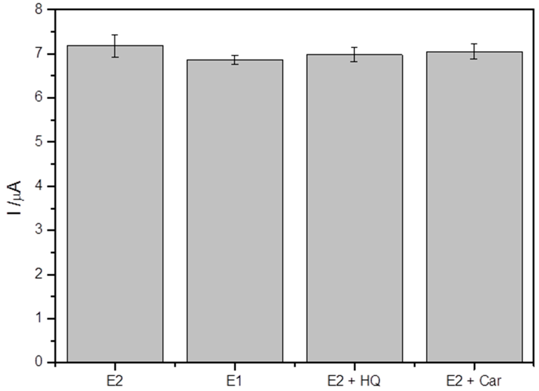
3.7. The Effects of Interferences

The selectivity of the CPE-MagMIP was evaluated in the presence of compounds that can also be found in river water. Figure 8 shows no significant change in relative percentages of E2 response in the presence of hydroquinone (2.78%) and carbendazim (1.81%), thus demonstrating that these compounds do not prevent the determination of E2. Estrone (E1), which is structurally related to the E2 hormone was also evaluated and showed an oxidation peak at the same potential as E2, with a very similar intensity, showing that the sensor also responds to E1 (95.7%) [41].

3.8. Analytical Curve

The CPE-MagMIP was quantitatively analyzed as the E2 concentration was increased from a stock solution of 1 mM. Figure 9A shows the voltammograms obtained for the successive additions. The analytical curve obtained for the proposed sensor is represented in Figure 9B and displayed a linear response range from 0.5 to 14.0 μM, and a sensitivity of 0.79 μA L μmol−1 (R2 = 0.998). Above 14.0 μM, the analytical signal increases non-linearly, which suggests a saturation of the E2 adsorption process on the electrode surface. The detection limit (LOD) and quantification limit (LOQ) were equal to 0.13 and 0.44 μM, respectively, calculated from the parameters of the analytical curve obtained under optimized conditions [42].
Compared to other electroanalytical sensors for E2 (Table 1), the proposed electrode presented relevant characteristics such as ease of handling modification process, low price, and renewable surface. Furthermore, the proposed sensor is less susceptible to interfering species due to modification with the MagMIP.
The reached detection limit is adequate for E2 detection in water samples [43].
Table 1. Comparison of different electroanalytical methods for E2 determination.
Technique/ElectrodeLinear Range (μM)LOD (μM)Ref.
LSV/CDs-PANI/GCE a1.0–1000.043[39]
SWV/GCE/MWCNTs-Pt b0.5–150.18[44]
DPV/BPIDS/GCE c0.1–100.05[45]
DPV/Pt/Pol/HRP/GCE d0.1–2000.105[46]
DPV/CdMoO4/CNS/SPCE e0.05–240.003[47]
DPAdSV/CPE-MagMIP0.5–14.00.13This work
a LSV—linear sweep voltammetry; sensor based on commercially available polyaniline (PANI) and carbon dots (CDs); GCE—glassy carbon electrode. b SVW—square wave voltammetric. GCE/MWCNTs-Pt: glassy carbon electrode-multi-walled carbon nanotubes–platinum. c DPV—differential pulse voltammetry; BPIDS–nanoporous polymeric hydrophobic film. d Conducting polymer and horseradish peroxidase (HRP) modified platinum (Pt) electrode. e Micro stone structured CdMoO4 and carbon nanosphere (CNS); screen-printed carbon electrodes (SPCE).

3.9. Determination of E2 in Water Samples

E2 is commonly found in effluents from municipal wastewater treatment, due to its incomplete removal, and can easily be introduced into aquatic environments [6]. On this basis, river water samples were analyzed by DPAdSV using the standard addition method under optimized conditions. Table 2 shows the results obtained in the application of the proposed method. The high recovery values and low relative standard deviation (RSD) confirm the efficiency of the method and that the present sensor is potentially suitable for this type of analysis.
The detection of E2 in samples, such as water, helps monitor the presence of this compound in the environment, and environmental safety measures can then adopt. A simple method like the one developed can help in this regard.

4. Conclusions

In this work, an electrode based on carbon paste, modified with MagMIP, was developed, which combined the advantages of molecular imprinting technology with the properties of magnetic materials. The sensor showed excellent adsorption and detection capability for E2 in a short analysis time. The developed electrode surface is easily renewed with simple polishing, more than ten times, without decreasing the analytical performance. CPE-MagMIP was used to detect E2 in water samples and showed remarkable performance. It is a promising device for monitoring E2 in this type of sample.

Author Contributions

D.N.d.S.: developed and applied the methodology and wrote the manuscript. A.C.P.: contributed to the development of the entire analytical method and also contributed to the writing of the manuscript. All authors have read and agreed to the published version of the manuscript.

Funding

This research received no external funding.

Institutional Review Board Statement

Not applicable.

Informed Consent Statement

Not applicable.

Data Availability Statement

Not applicable.

Acknowledgments

Fundação de Amparo à Pesquisa do Estado de Minas Gerais (FAPEMIG), Conselho Nacional de Desenvolvimento Científico e Tecnológico (CNPq), and Coordenação de Aperfeiçoamento de Pessoal de Nível Superior (CAPES), INCT-DATREM and to Hanna Leijoto de Oliveira. This work is a collaborative research project of members of the Rede Mineira de Química (RQ-MG) supported by FAPEMIG.

Conflicts of Interest

The authors declare no conflict of interest.

References

  1. Gandhi, A.; Matta, M.K.; Stewart, S.; Chockalingam, A.; Knapton, A.; Rouse, R.; Wu, W.; Patel, V. Quantitative analysis of underivatized 17 β-estradiol using a high-throughput LC–MS/MS assay—Application to support a pharmacokinetic study in ovariectomized guinea pigs. J. Pharm. Biomed. Anal. 2020, 178, 112897. [Google Scholar] [CrossRef] [PubMed]
  2. Smajdor, J.; Piech, R.; Ławrywianiec, M.; Paczosa-Bator, B. Glassy carbon electrode modified with carbon black for sensitive estradiol determination by means of voltammetry and flow injection analysis with amperometric detection. Anal. Biochem. 2018, 544, 7–12. [Google Scholar] [CrossRef] [PubMed]
  3. Yilmaz, B.; Kadioglu, Y. Determination of 17 β-estradiol in pharmaceutical preparation by UV spectrophotometry and high performance liquid chromatography methods. Arab. J. Chem. 2017, 10, S1422–S1428. [Google Scholar] [CrossRef]
  4. Lahcen, A.A.; Baleg, A.A.; Baker, P.; Iwuoha, E.; Amine, A. Synthesis and electrochemical characterization of nanostructured magnetic molecularly imprinted polymers for 17-β-Estradiol determination. Sens. Actuators B Chem. 2017, 241, 698–705. [Google Scholar] [CrossRef]
  5. Arvand, M.; Hemmati, S. Analytical methodology for the electro-catalytic determination of estradiol and progesterone based on graphene quantum dots and poly(sulfosalicylic acid) co-modified electrode. Talanta 2017, 174, 243–255. [Google Scholar] [CrossRef]
  6. Saitoh, T.; Fukushima, K.; Miwa, A. Combined use of surfactant-induced coagulation of poly(allylamine hydrochloride) with peroxidase-mediated degradation for the rapid removal of estrogens and phenolic compounds from water. Sep. Purif. Technol. 2014, 128, 11–17. [Google Scholar] [CrossRef]
  7. Wang, M.; Shi, Q.; Shi, H.; Li, J.; Yang, Y.; Qian, L.; Shao, S.; Gao, S. The promotion effect of phenolic acid compound on the photo-removal of estrogen from water under simulated sunlight irradiation. Chem. Eng. J. 2020, 387, 123999. [Google Scholar] [CrossRef]
  8. Chiu, Y.-W.; Yeh, F.-L.; Shieh, B.-S.; Chen, C.-M.; Lai, H.-T.; Wang, S.-Y.; Huang, D.-J. Development and assays estradiol equivalent concentration from prawn (p-EEQ) in river prawn, Macrobrachium nipponense, in Taiwan. Ecotoxicol. Environ. Saf. 2017, 137, 12–17. [Google Scholar] [CrossRef]
  9. Goeury, K.; Munoz, G.; Duy, S.V.; Prévost, M.; Sauvé, S. Occurrence and seasonal distribution of steroid hormones and bisphenol A in surface waters and suspended sediments of Quebec, Canada. Environ. Adv. 2022, 8, 100199. [Google Scholar] [CrossRef]
  10. D’Orazio, G.; Hernández-Borges, J.; Herrera-Herrera, A.V.; Fanali, S.; Rodríguez-Delgado, M.Á. Determination of estrogenic compounds in milk and yogurt samples by hollow-fibre liquid-phase microextraction-gas chromatography-triple quadrupole mass spectrometry. Anal. Bioanal. Chem. 2016, 408, 7447–7459. [Google Scholar] [CrossRef]
  11. Cheng, Z.; Ling, J.; Zhang, W.; Ding, Y. Rapid detection of 17β-estradiol based on shaddock peel derived fluorescent aptasensor for forensic examination. Forensic Sci. Int. 2022, 331, 111153. [Google Scholar] [CrossRef] [PubMed]
  12. Wei, Q.; Zhang, P.; Pu, H.; Sun, D.-W. A fluorescence aptasensor based on carbon quantum dots and magnetic Fe3O4 nanoparticles for highly sensitive detection of 17β-estradiol. Food Chem. 2022, 373 Pt B, 131591. [Google Scholar] [CrossRef]
  13. Sha, H.; Yan, B. Design of a ratiometric fluorescence sensor based on metal organic frameworks and Ru(bpy)32+-doped silica composites for 17β-Estradiol detection. J. Colloid Interface Sci. 2021, 583, 50–57. [Google Scholar] [CrossRef] [PubMed]
  14. Li, J.; Jiang, J.; Zhao, D.; Xu, Z.; Liu, M.; Deng, P.; Liu, X.; Yang, C.; Qian, D.; Xie, H. Facile synthesis of Pd/N-doped reduced graphene oxide via a moderate wet-chemical route for non-enzymatic electrochemical detection of estradiol. J. Alloys Compd. 2018, 769, 566–575. [Google Scholar] [CrossRef]
  15. Raymundo-Pereira, P.A.; Gomes, N.O.; Machado, S.A.S.; Oliveira, O.N. Simultaneous, ultrasensitive detection of hydroquinone, paracetamol and estradiol for quality control of tap water with a simple electrochemical method. J. Electroanal. Chem. 2019, 848, 113319. [Google Scholar] [CrossRef]
  16. Wen, T.; Wang, M.; Luo, M.; Yu, N.; Xiong, H.; Peng, H. A nanowell-based molecularly imprinted electrochemical sensor for highly sensitive and selective detection of 17β-estradiol in food samples. Food Chem. 2019, 297, 124968. [Google Scholar] [CrossRef] [PubMed]
  17. Wang, A.; Ding, Y.; Li, L.; Duan, D.; Mei, Q.; Zhuang, Q.; Cui, S.; He, X. A novel electrochemical enzyme biosensor for detection of 17β-estradiol by mediated electron-transfer system. Talanta 2018, 192, 478–485. [Google Scholar] [CrossRef]
  18. Li, R.; Li, F.; Zhe, T.; Li, M.; Liu, Y.; Wang, L. Three-dimensional (3D) hierarchical structure engineering of AuNPs/Co(OH)2 nanocomposite on carbon cloth: An advanced and efficient electrode for highly sensitive and specific determination of nitrite. Sens. Actuators B Chem. 2021, 342, 130061. [Google Scholar] [CrossRef]
  19. Wu, T.; Xia, D.; Xu, J.; Ye, C.; Zhang, D.; Deng, D.; Zhang, J.; Huang, G. Sequential injection-square wave voltammetric sensor for phosphate detection in freshwater using silanized multi-walled carbon nanotubes and gold nanoparticles. Microchem. J. 2021, 167, 106311. [Google Scholar] [CrossRef]
  20. Cárdenas, A.; Frontana, C. Evaluation of a Carbon Ink Chemically Modified Electrode Incorporating a Copper-Neocuproine Complex for the Quantification of Antioxidants. Sens. Actuators B Chem. 2020, 313, 128070. [Google Scholar] [CrossRef]
  21. Lima, G.V.; Mello, L.S.; Dockal, E.R.; de Oliveira, M.F. Electrochemical determination of ethanol on carbon paste electrode chemically modified with [N,N′-cis-1,2-cyclohexylene bis (salicylideneaminate)] nickel(II) Schiff base complex. Microchem. J. 2022, 175, 107209. [Google Scholar] [CrossRef]
  22. Khan, S.; Wong, A.; Zanoni, M.V.B.; Sotomayor, M.D.P.T. Electrochemical sensors based on biomimetic magnetic molecularly imprinted polymer for selective quantification of methyl green in environmental samples. Mater. Sci. Eng. C 2019, 103, 109825. [Google Scholar] [CrossRef] [PubMed]
  23. Cui, B.; Liu, P.; Liu, X.; Liu, S.; Zhang, Z. Molecularly imprinted polymers for electrochemical detection and analysis: Progress and perspectives. J. Mater. Res. Technol. 2020, 9, 12568–12584. [Google Scholar] [CrossRef]
  24. Tarley, C.R.T.; Sotomayor, M.D.P.T.; Kubota, L.T. Polímeros biomiméticos em química analítica. Parte 1: Preparo e aplicações de MIP (“Molecularly Imprinted Polymers”) em técnicas de extração e separação. Quím. Nova 2005, 28, 1076–1086. [Google Scholar] [CrossRef] [Green Version]
  25. Jiang, H.; Jiang, D.; Shao, J.; Sun, X. Magnetic molecularly imprinted polymer nanoparticles based electrochemical sensor for the measurement of Gram-negative bacterial quorum signaling molecules (N-acyl-homoserine-lactones). Biosens. Bioelectron. 2016, 75, 411–419. [Google Scholar] [CrossRef]
  26. Yáñez-Sedeño, P.; Campuzano, S.; Pingarrón, J.M. Electrochemical sensors based on magnetic molecularly imprinted polymers: A review. Anal. Chim. Acta 2017, 960, 1–17. [Google Scholar] [CrossRef]
  27. Afzali, Z.; Mohadesi, A.; Karimi, M.A.; Fathirad, F. A highly selective and sensitive electrochemical sensor based on graphene oxide and molecularly imprinted polymer magnetic nanocomposite for patulin determination. Microchem. J. 2022, 177, 107215. [Google Scholar] [CrossRef]
  28. Da Silva, D.N.; de Oliveira, H.L.; Borges, K.B.; Pereira, A.C. Sensitive determination of 17β-estradiol using a Magneto Sensor Based on Magnetic Molecularly Imprinted Polymer. Electroanalysis 2021, 33, 506–514. [Google Scholar] [CrossRef]
  29. Brandão, C.J.; Botelho, M.J.C.; Sato, M.I.Z. (Eds.) Guia Nacional de Coleta e Preservação de Amostras: Água, Sedimento, Comunidades Aquáticas e Efluentes Líquidos; Companhia Ambiental do Estado de São Paulo: São Paulo, Brazil; Agência Nacional de Águas: Brasília, Brazil, 2011. [Google Scholar]
  30. Santos, A.C.F.; de Araújo, O.R.P.; Moura, F.A.; Khan, S.; Tanaka, A.A.; Santana, A.E.G.; Pividori, M.I.; Sotomayor, M.P.T.; Goulart, M.O.F. Development of magnetic nanoparticles modified with new molecularly imprinted polymer (MIPs) for selective analysis of glutathione. Sens. Actuators B Chem. 2021, 344, 130171. [Google Scholar] [CrossRef]
  31. Nadali, A.; Leili, M.; Afkhami, A.; Bahrami, A.; Karami, M. Synthesize and application of magnetic molecularly imprinted polymers (mag-MIPs) to extract 1-Aminopyrene from the human urine sample. J. Environ. Chem. Eng. 2021, 9, 106253. [Google Scholar] [CrossRef]
  32. De Oliveira, H.L.; Pires, B.C.; Teixeira, L.S.; Dinali, L.A.F.; Simões, N.S.; de Souza, W.B.; Borges, K.B. Novel restricted access material combined to molecularly imprinted polymer for selective magnetic solid-phase extraction of estrogens from human urine. Microchem. J. 2019, 149, 104043. [Google Scholar] [CrossRef]
  33. Marco, J.P.; Borges, K.B.; Tarley, C.R.T.; Ribeiro, E.S.; Pereira, A.C. Development and application of an electrochemical biosensor based on carbon paste and silica modified with niobium oxide, alumina and DNA (SiO2/Al2O3/Nb2O5/DNA) for amitriptyline determination. J. Electroanal. Chem 2013, 704, 159–168. [Google Scholar] [CrossRef]
  34. Li, Y.; Zhao, X.; Li, P.; Huang, Y.; Wang, J.; Zhang, J. Highly sensitive Fe3O4 nanobeads/graphene-based molecularly imprinted electrochemical sensor for 17β-estradiol in water. Anal. Chim. Acta 2015, 884, 106–113. [Google Scholar] [CrossRef] [PubMed]
  35. El-Sharif, H.F.; Patel, S.; Ndunda, E.N.; Reddy, S.M. Electrochemical detection of dioctyl phthalate using molecularly imprinted polymer modified screen-printed electrodes. Anal. Chim. Acta 2022, 1196, 339547. [Google Scholar] [CrossRef]
  36. Wong, A.; Santos, A.M.; Fava, E.L.; Fatibello-Filho, O.; Sotomayor, M.D.P.T. Voltammetric determination of 17β-estradiol in different matrices using a screen-printed sensor modified with CuPc, Printex 6L carbon and Nafion film. Microchem. J. 2019, 147, 365–373. [Google Scholar] [CrossRef]
  37. Braga, G.B.; Oliveira, A.E.F.; Pereira, A.C. Total Determination of Estrogenic Phenolic Compounds in River Water Using a Sensor Based on Reduced Graphene Oxide and Molecularly Imprinted Polymer. Electroanalysis 2018, 30, 2176–2184. [Google Scholar] [CrossRef]
  38. Liu, W.; Li, H.; Yu, S.; Zhang, J.; Zheng, W.; Niu, L.; Li, G. Poly(3,6-diamino-9-ethylcarbazole) based molecularly imprinted polymer sensor for ultra-sensitive and selective detection of 17-β-estradiol in biological fluids. Biosens. Bioelectron. 2018, 104, 79–86. [Google Scholar] [CrossRef]
  39. Supchocksoonthorn, P.; Alvior Sinoy, M.C.; de Luna, M.D.G.; Paoprasert, P. Facile fabrication of 17β-estradiol electrochemical sensor using polyaniline/carbon dot-coated glassy carbon electrode with synergistically enhanced electrochemical stability. Talanta 2021, 235, 122782. [Google Scholar] [CrossRef]
  40. Zoski, C.G. Handbook of Electrochemistry, 1st ed.; Elsevier: Amsterdam, The Netherlands, 2007. [Google Scholar]
  41. Özcan, A.; Topçuoğulları, D. Voltammetric determination of 17-β-estradiol by cysteamine self-assembled gold nanoparticle modified fumed silica decorated graphene nanoribbon nanocomposite. Sens. Actuators B Chem. 2017, 250, 85–90. [Google Scholar] [CrossRef]
  42. ICH Expert Working Group. ICH-harmonised Tripartite Guideline, validation of analytical procedures: Methodology, technical requirements for the registration of pharmaceuticals for human use. In Proceedings of the International Conference on Harmonization, Geneva, Switzerland, 30 March 1995. [Google Scholar]
  43. Torres, N.H.; de Oliveira, G.S.S.; Ferreira, L.F.R.; Américo-Pinheiro, J.H.P.; Eguiluz, K.I.B.; Salazar-Banda, G.R. Environmental aspects of hormones estriol, 17β-estradiol and 17α-ethinylestradiol: Electrochemical processes as next-generation technologies for their removal in water matrices. Chemosphere 2021, 267, 128888. [Google Scholar] [CrossRef]
  44. Lin, X.; Li, Y. A sensitive determination of estrogens with a Pt nano-clusters/multi-walled carbon nanotubes modified glassy carbon electrode. Biosens. Bioelectron. 2006, 22, 253–259. [Google Scholar] [CrossRef] [PubMed]
  45. Wang, Z.; Wang, P.; Tu, X.; Wu, Y.; Zhan, G.; Li, C. A novel electrochemical sensor for estradiol based on nanoporous polymeric film bearing poly{1-butyl-3-[3-(N-pyrrole)propyl]imidazole dodecyl sulfonate} moiety. Sens. Actuators B Chem. 2014, 193, 190–197. [Google Scholar] [CrossRef]
  46. Spychalska, K.; Zając, D.; Cabaj, J. Electrochemical biosensor for detection of 17β-estradiol using semi-conducting polymer and horseradish peroxidase. RSC Adv. 2020, 10, 9079–9087. [Google Scholar] [CrossRef] [PubMed] [Green Version]
  47. Shanmugam, R.; Alagumalai, K.; Chen, S.-M.; Ganesan, T. Electrochemical evaluation of organic pollutant estradiol in industrial effluents. J. Environ. Chem. Eng. 2021, 9, 105723. [Google Scholar] [CrossRef]
Figure 1. Schematic representation of the working electrode.
Electrochem 03 00053 g001
Figure 2. FT-IR spectrum of the MagNIP, MagMIP, and CPE-MagMIP.
Electrochem 03 00053 g002
Figure 3. Electrochemical characterization in KCl 0.1 M containing 5 mM K3[Fe (CN)6]/K4[Fe (CN)6]: (A) CV voltammograms and (B) Nyquist plots; electrodes: CPE; CPE-MagMIP, and CPE-MagNIP.
Electrochem 03 00053 g003
Figure 4. (A) DPAdSV voltammograms obtained for different working electrode configurations. (B) Relation between the peak current and different working electrodes. Conditions: 0.1 M phosphate buffer solution, pH 7.0 in the presence of 10.0 μM of E2.
Electrochem 03 00053 g004
Figure 5. Influence of the amount of MagMIP on the anodic peak current. Conditions: 0.1 M phosphate buffer solution, pH 7.0 in the presence of 10.0 μM of E2.
Electrochem 03 00053 g005
Figure 6. Influence of the pH on sensor response. Conditions: 0.1 M phosphate buffer solution in the presence of 10.0 μM of E2.
Electrochem 03 00053 g006
Figure 7. Influence of buffer solution on sensor response. Conditions: 0.1 M buffer solution in the presence of 10.0 μM of E2.
Electrochem 03 00053 g007
Figure 8. Effects of interferences on the response of the proposed sensor for E2. Conditions: 0.1 M phosphate buffer solution, pH 7.0.
Electrochem 03 00053 g008
Figure 9. (A) DPAdSV voltammograms obtained with successive additions of E2; (B) analytical curve obtained from the 9A voltammograms. Conditions: 0.1 M phosphate buffer solution, pH 7.0.
Electrochem 03 00053 g009
Table 2. Determination of E2 in river water samples.
Added Concentration (μM)Detected Concentration (μM) ± RSD aRecovery (%)
0ND b-
1.51.47 ± 0.0898.3
5.04.84 ± 0.2896.8
a Triplicate analysis. b ND = not detected.
Publisher’s Note: MDPI stays neutral with regard to jurisdictional claims in published maps and institutional affiliations.

