Article

AmnioClip-Plus as Sutureless Alternative to Amniotic Membrane Transplantation to Improve Healing of Ocular Surface Disorders

1
German Society for Tissue Transplantation (DGFG) gGmbH, 30625 Hannover, Germany
2
Department of Ophthalmology, Klinikum Chemnitz gGmbH, 09116 Chemnitz, Germany
*
Author to whom correspondence should be addressed.
Submission received: 30 July 2021 / Revised: 1 October 2021 / Accepted: 14 October 2021 / Published: 18 October 2021

Abstract

:
The medicinal benefits of amniotic membrane transplantation for ocular surface disorders are well accepted worldwide. Even in high-risk keratoplasties, the concomitant use of amniotic membrane has demonstrated its value in improving graft survival. However, its seam-associated application can lead to an additional trauma. The AmnioClip ring system, into which the amniotic membrane is clamped (AmnioClip-plus, AC+), was developed to avoid this surgical intervention. The AC+ is placed on the cornea, similar to a contact lens, under local anesthesia and can therefore be applied repeatedly. Clinical practice demonstrates the easy handling, good compatibility, and efficacy of this minimally invasive method.

1. Introduction

Ocular surface disorders or defects of the cornea are often difficult to treat and can lead to clouding of the cornea with consecutive visual impairment or blindness. In addition, disrupted ocular surfaces, for example, due to limbal stem cell deficiency (LSCD), jeopardize the healing and prognosis for survival of a corneal graft after keratoplasty. Furthermore, if keratoplasty is performed as an emergency procedure on an inflamed eye, immunologic graft rejection can also lead to a worse outcome [1].
In as early as 1940, De Röth [2] described amniotic membrane transplantation (AMT) as a suitable therapy for conjunctival reconstruction. Besides pharmacological therapy [3,4,5], the suturing of the human amniotic membrane (hAM) is now firmly established in clinical practice today, especially for the treatment of persistent epithelial defects, including neurotrophic corneal ulcers [6,7], as well as for reconstruction in conjunctival injuries and pterygium surgery [8,9,10]. In addition, AMT has also been demonstrated to be effective as a simultaneous treatment to perforating keratoplasty in eyes with a history of limbal stem cell deficiencies [11] or eyes with a risk of epithelial healing problems [12].
Moreover, different groups [13,14] showed that AMT ensured a rapid decrease in the inflammatory reaction and the fast re-epithelialization could help to avoid an emergency keratoplasty and improve the prognosis of graft survival in elective keratoplasty. The successful and safe use of hAM in ophthalmological therapies is confirmed by the results of Paolin et al. [15], with more than 5300 amniotic membrane treatments from 2003 to 2014. Data from the German Society for Tissue Transplantation (DGFG) affirm the safe production and application, as no infection after AMT has been reported to date for almost 13,000 amniotic membranes that have been provided for transplantation, among others, for application in high-risk keratoplasty since 2008.

1.1. Anti-Inflammatory and Anti-Angiogenic Properties of the Amniotic Membrane Support Wound Healing and Reconstruction of the Ocular Surface

The amniotic membrane is the inner membrane of the amniotic sac facing the fetus. HAM consists of an epithelium, a relatively thick basal membrane, and a vascular stroma (Figure 1), and combines typical characteristics and properties that make it ideal for use in many medical fields. It is a non-immunogenic tissue [16,17] with antibacterial [18], anti-inflammatory, and anti-angiogenic properties [19,20,21,22], as well as anti-fibroblastic activity [9,12]. In addition, multiple factors in the amniotic membrane stimulate cell migration and cell proliferation, which are very important for wound healing. [23].

1.2. Disadvantages of Suture-Associated Amniotic Membrane Transplantation

A major disadvantage of conventional AMT is that the amniotic membrane must usually be fixed to the ocular surface with sutures. Suturing means an additional trauma for the patient, as the eye surface is already affected by the underlying disease. The basic inflammatory condition, together with the suturing, often leads to heavy bleeding in the conjunctiva. Sewing directly into the cornea can also be unfavorable in cases of severe pathologies. The current work presents a system to avoid this trauma and summarizes user experiences after receiving marketing authorization.

2. Materials and Methods

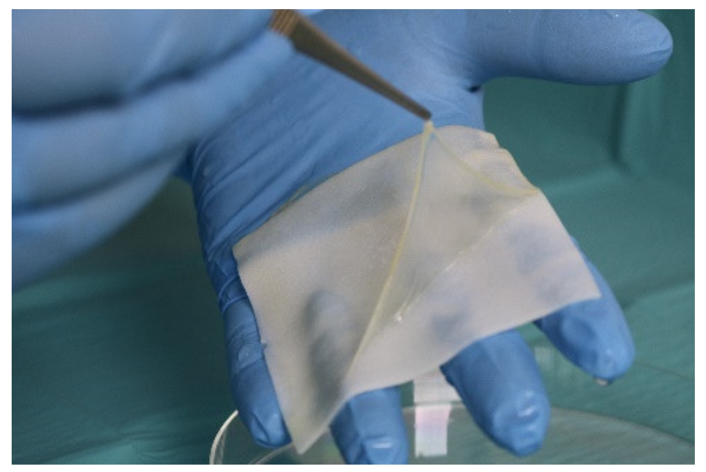
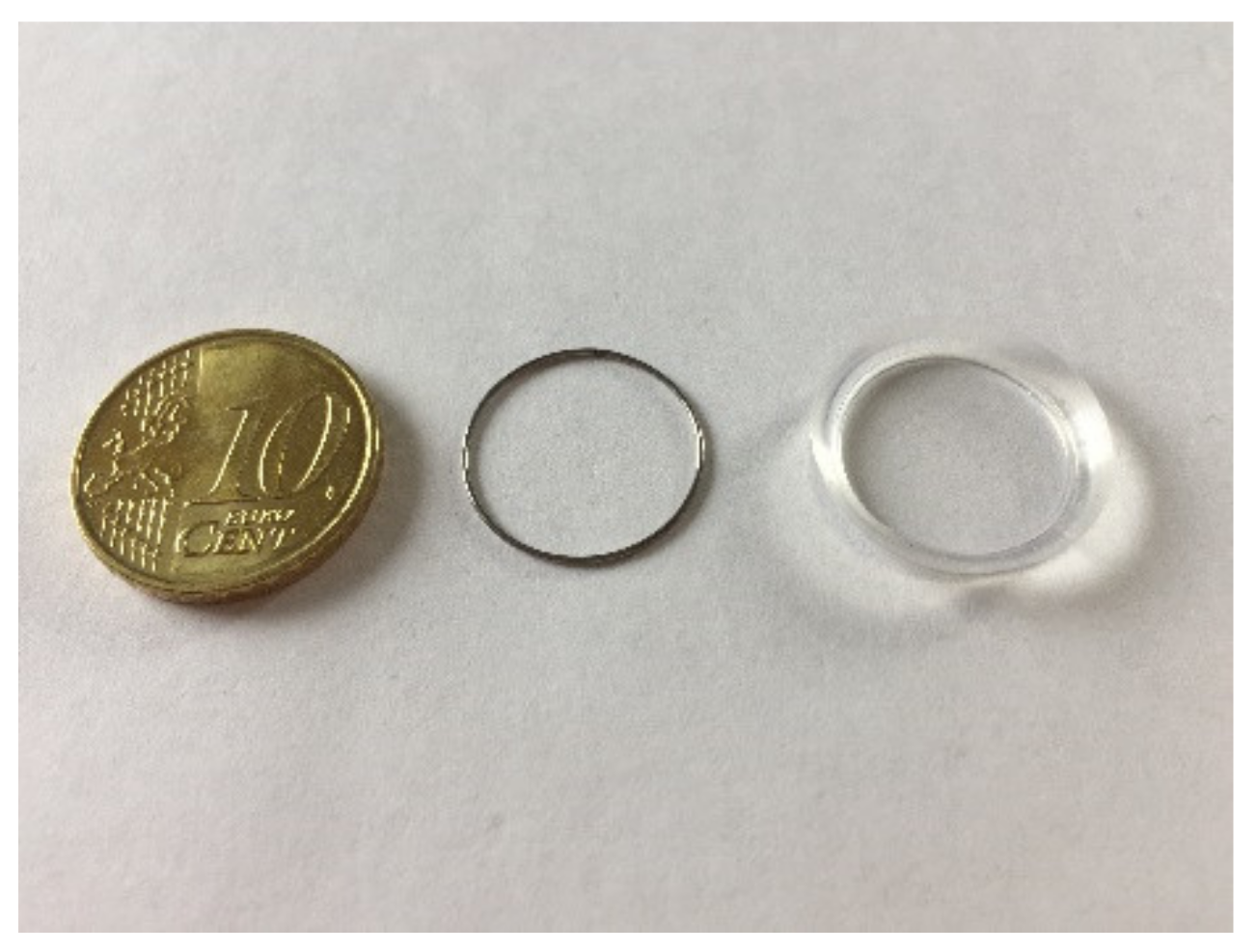
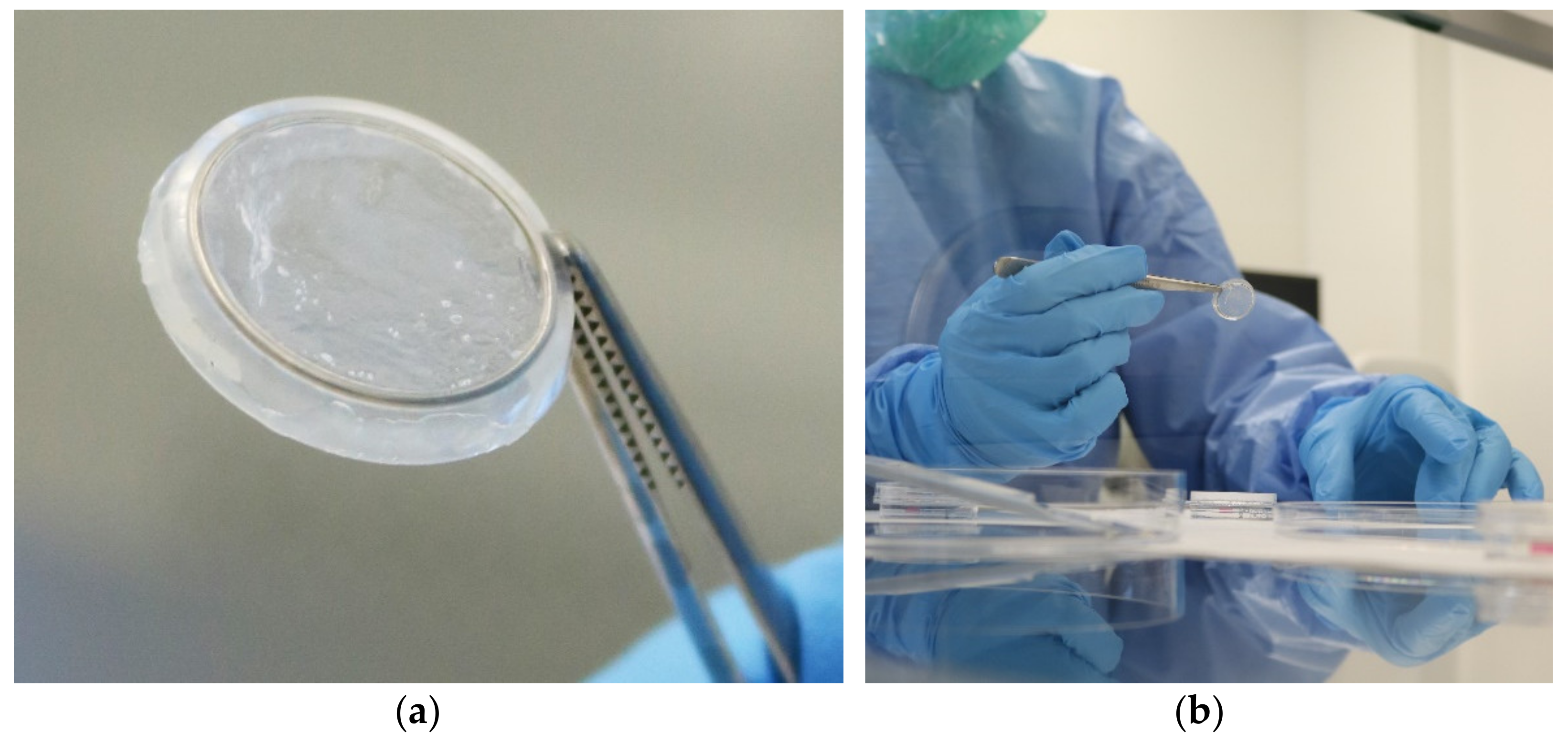
2.1. Development of the AmnioClip-Plus Ring System for Minimally Invasive Application without Sutures

An interdisciplinary team has developed a ring system, so-called AmnioClip (AC), into which the amniotic membrane is clamped. The resulting AmnioClip-plus (AC+) can be placed on the ocular surface without any suture technique [24,25]. The patient can wear the AC+ as comfortably as they would a contact lens, and additional surgical trauma is avoided.
The AC consists of two rings: an inner ring made of stainless-steel wire, which is completely enclosed by the amniotic membrane, and an outer ring made of silicone (Figure 2). The outer ring has a groove on the inside in which the inner ring with the amniotic membrane fits. The ring tapers outwards, so that the eyelid can slide tangentially over the AC. The assembled AC+ has a total diameter of 19 mm.
The amniotic membrane used for the AC, as with the hAM for conventional AMT, is obtained in a planned caesarean section birth with the donor’s consent. Prior to this, a comprehensive donor evaluation and infection diagnostic analysis in accordance with the criteria of good practice is performed [26]. After careful preparation, the hAM is pre-cut to an approximate size of 30 mm × 30 mm by means of a scalpel and clamped into the AC using a specially developed device. The orientation of the hAM can be chosen so that the stromal or epithelial side rests on the eye surface during application. The production of this tissue preparation AC+ (Figure 3a) takes place under standardized conditions in a clean room of a tissue bank (Figure 3b). The AC+ can be deep-frozen stored without impairing its effectiveness [27].

2.2. The AmnioClip-Plus Is an Authority-Approved Tissue Preparation

In Germany, tissue preparations are regulated by the Medicinal Products Act (AMG), so that an authorization according to §21a AMG from the higher federal authority (Paul-Ehrlich-Institute, PEI) is essential for the delivery of AC+ for clinical use. The approval procedure for the AC+ required extensive validation and testing.
In order to be able to obtain this approval, the AC+ was first tested in clinical practice in a monocentric pilot study (ID no. NCT02168790), with 7 patients described in detail elsewhere, followed by an expanded access study to prove safety with 5 patients [24]. These 12 patients included in the two prospective interventional trials suffered from keratitis with epithelial defect (n = 2), corneal ulcer (n = 6), or corneal dystrophies with epithelial defects (n = 4). In a total of 20 applications (7 + 5 = 12 patients, 13 eyes: 1 patient received treatment on both eyes, 7 patients had consecutively 2 AC+) the AC+ has shown itself to be well tolerated and effective. The patients’ feedback was recorded by means of a specially developed questionnaire and compared with the clinical data.
After the detailed evaluation, the AC+ was approved by the Paul-Ehrlich-Institute as a tissue preparation in January 2019 (PEI.G.11968.01.1).
In accordance with a condition for this approval, the physician’s professional assessment and the patient’s opinions were recorded for the first AC+ that was provided for clinical usage. The specially developed questionnaires, which were completed and returned independently by the attending physician and the patient, were evaluated after the application period. In the present paper, 45 patients that received one of the first AC+ were included in this retrospective evaluation. For the treatment of these 45 patients, one questionnaire from the treating physician was available each, as well as a total of 81 patient feedback forms, as some patients were interviewed at several time points after the treatment (up to 4 weeks follow-up).

3. Results

3.1. Clinical Experience Demonstrates the Easy Handling and Good Tolerability of the AmnioClip-Plus

After the evaluation of the total 45 questionnaires completed by the treating physicians, it became apparent that, to date, the AC+ system has mainly been used for the treatment of ulcers and inflammations of different geneses (~61%, Table 1), but was also used for persistent erosion or chemical burn of the cornea and keratoconjunctivitis sicca in graft versus host disease (GvHD). In some cases, the disease had been present for more than 12 months and more than 60% of patients had already undergone conventional suture-associated AMT [28].
In contrast to conventional AMT, where retrobulbar or general anesthesia might be necessary, the AC+ can be applied to the cornea on an outpatient basis in sterile conditions under drip anesthesia. Consequently, the majority of surgeons (98%) rate the insertion procedure as simple. Most patients provided the same feedback and additionally described the process as painless. Only a special anatomy of the eye can lead to somewhat more difficult and uncomfortable procedures in individual cases (Table 2). These results were obtained from the evaluation of the 81 questionnaires filled out by the 45 patients.
The AC+ is approved for application for up to 14 days. So far, most users took advantage of this period. Some application times were shorter (1 to 7 days), but most of these applications showed an improvement in the condition.
No complications emerged in the majority of applications while wearing the AC+. In a few cases, membrane degradation seemed to occur during the treatment period. However, such degradation is not due to clamping in the ring system but can also occur in AMT with sutures because of the different nature of the amniotic membrane as a biological material. This degradation apparently had no influence on the effect of the AC+ treatment, as the disease nevertheless improved.

3.2. The AC+ Improves the Clinical Picture and Well-Being of the Patient

In more than 70% of the patients, both the general condition and the disease improved after using the AC+. This effect could not be linked to the length of time the clip was worn or the duration of the disease. Unexpectedly, only 50% of the patients treated with the AC+ reported a reduction in pain.

4. Discussion and Conclusions

The medical benefit of amniotic membrane treatment is accepted worldwide and has been documented in numerous publications [29]. At the same time, the limitation of suture-associated application is known [24,25]. To avoid this further stressing surgical intervention, the AC ring system was developed in which the amniotic membrane is clamped (AmnioClip-plus, AC+) and applied to the surface of the eye without any sutures. Therefore, the AC+ treatment can be carried out several times without risk in an out-patient manner, also because complex anesthetic measures can be avoided.
The questionnaires evaluated so far have shown that the use of the AC+ is rated positively overall by expert medical assessment and patient feedback. As already shown in the hospital exemption study, the ring was easy to insert and only occasionally caused persistent foreign body sensation or impairment. However, discussion came up as to whether clamping in the ring system might interfere with the effect of the amniotic membrane and whether conventional suture fixation would better support healing. The responses indicate that the effect of the amniotic membrane in the ring system is comparable or even superior to that after suture-associated AMT. AC+ also improved diseases that had not been cured after conventional AMT. Although conventional AMT may allow the hAM to remain on the eye surface longer, the gentle application method of the AC+ without sutures appears to promote healing just as much or even faster. This is also the conclusion of a study in the USA, where a similar product is already being used clinically without particular complications having been described so far. [30,31]. In contrast to the more adaptable silicone of the AC+, the other product’s ring is made of hard plastic, which may have led to lower wearing comfort for the patients. In general, sutureless alternatives are gaining more and more interest due to the disadvantages of conventional AMT described above. Besides its application, similar to a contact lens, fixation of hAM by means of bioadhesives, e.g., fibrin glue, is also used. This method also achieves good healing results, but graft dehiscence may be a problem in this procedure, and focal inflammation of host conjunctiva and pyogenic granuloma were reported after using fibrin glue to fix the hAM [32,33]. Besides this, commercially available safe fibrin glue is deemed a rather expensive substrate.
The exact mode of hAM’s action in healing support is still unclear and the influence of the orientation of the hAM (stromal or epithelial) facing the eye has not yet been systematically investigated. There are two ways to apply the amniotic membrane: as a patch or onlay or as a graft or inlay. The application of AmnioClip-plus is like a patch and its greatest effectiveness might be found in those refractory corneal ulcers that, with this treatment, spontaneous healing is equal to or less than two weeks. Here, the single-layer membrane in the AC+ seems to be sufficient for the success of the therapy, as it could be shown that multi-layer AMT did not achieve better results [7]. Generally, similar interactions on a cellular or molecular level regarding the release of AM factors independent from the way of application are suspected; this is a topic of ongoing research. Without considering those questions, the AC+ gains important advantages through its easy attachment and removal, which allows for a faster or repeated intervention in the process.
The AC+ system has proven itself to be stable. Only 1 patient out of 65 applications (45 patients in retrospective study together with 20 applications from previous pilot studies) reported loss of the AC+, which was caused by a deviating anatomy of the treated eye. Based on user experience to date, it would be beneficial to be able to better respond to different patient eye anatomies. For this reason, other sizes will be developed in future in addition to the current ring diameter of the AC+.
The AC+ technique, being easier to perform, reduces anesthesia complications, operating room time, and reduces the risk of corneal astigmatism due to sutures and thus also opens new treatment options for diseases such as “dry eye syndrome”. These advantages compensate for the higher initial material costs.

Summary of Clinical Advantages of the AmnioClip-Plus

Below is a summary of the advantages of the AmnioClip-plus:
-
Avoidance of surgical trauma for the patient;
-
No costly anesthetic procedures;
-
Minimizing an accompanying surgical risk;
-
Extensive absence of pain;
-
Easy handling of the amniotic membrane;
-
Consumer protection by providing the quality-tested combination product;
-
Safe production in the clean room of a tissue bank;
-
Short-term repetition of application possible;
-
Cost saving because of ambulant application;
-
Enabling extended therapy for prolonged/chronic defects such as “dry-eye syndrome”.
The evaluation of the questionnaires showed that the AC+ has not yet been used as an adjunctive measure in high-risk keratoplasties, although it is known from the literature that AMT has a positive effect on the corneal graft survival in these cases [11,12,13,14]. In future, systematic studies should be conducted to examine whether the use of AC+ could improve corneal graft survival after high-risk keratoplasty since it is a simple, gentle, and non-immunogenic treatment method.

Author Contributions

Conceptualization, N.H., M.B. and K.E.; Methodology, N.H., K.E. and N.D.; Investigation, N.D., K.E., A.-K.S. and N.M.; Resources, M.B.; Writing—Original Draft Preparation, N.H., A.-K.S. and K.E.; Writing—Review and Editing, all authors; Visualization, K.K.; Supervision, M.B. and K.E.; Project Administration, N.H. and N.M. All authors have read and agreed to the published version of the manuscript.

Funding

This work received no external funding.

Institutional Review Board Statement

All procedures performed in studies involving human participants were in accordance with the ethical standards of the institutional and national research committee and with the 1964 Helsinki declaration and its later amendments of comparable ethical standards. Approval for the study was obtained from the local ethics committee and registered at www.clinicaltrials.gov (ID no. NCT02168790).

Informed Consent Statement

Informed consent was obtained from all subjects involved in the study.

Data Availability Statement

Data presented in this study are partially available on request from the corresponding author. The data are not publicly available due to national data protection laws.

Conflicts of Interest

N.D. and K.E. declare no conflict of interest. N.H., A.-K.S., K.K., N.M. and M.B. are employees of the German Society for Tissue Transplantation (DGFG).

References

  1. Zazzo, A.D.; Kheirkhah, A.; Abud, T.B.; Goyal, S.; Dana, R. Management of high-risk corneal transplantation. Surv. Ophthalmol. 2017, 62, 816–827. [Google Scholar] [CrossRef] [PubMed]
  2. De Röth, A. Plastic repair of conjunctival defects with fetal membranes. Arch. Ophthalmol. 1946, 23, 522–525. [Google Scholar] [CrossRef]
  3. Lee, S.H.; Tseng, S.C. Amniotic membrane transplantation for persistent epithelial defects with ulceration. Am. J. Ophthalmol. 1997, 123, 303–312. [Google Scholar] [CrossRef]
  4. Kruse, F.E.; Rohrschneider, K.; Völcker, H.E. Multilayer amniotic membrane transplantation for reconstruction of deep corneal ulcers. Ophthalmology 1999, 106, 1504–1510. [Google Scholar] [CrossRef]
  5. Seitz, B. Amnionmembrantransplantation. Unverzichtbare Therapieoption bei persistierenden cornealen Epitheldefekten. Z. Prakt. Augenheilkund. 2003, 24, 445–450. [Google Scholar]
  6. Schuerch, K.; Baeriswyl, A.; Frueh, B.E.; Christoph, T. Efficacy of Amniotic Membrane Transplantation for the Treatment of Corneal Ulcers. Cornea 2020, 39, 479–483. [Google Scholar] [CrossRef]
  7. Lacorzana, J.; Campos, A.; Brocal-Sánchez, M.; Marín-Nieto, J.; Durán-Carrasco, O.; Fernández-Núñez, E.C.; López-Jiménez, A.; González-Gutiérrez, J.L.; Petsoglou, C.; García Serrano, J.L. Visual Acuity and Number of Amniotic Membrane Layers as Indicators of Ef-ficacy in Amniotic Membrane Transplantation for Corneal Ulcers: A Multicenter Study. J. Clin. Med. 2021, 10, 3234. [Google Scholar] [CrossRef]
  8. Dua, H.S.; Azuara-Blanco, A. Amniotic membrane transplantation. Br. J. Ophthalmol. 1999, 83, 748–752. [Google Scholar] [CrossRef] [Green Version]
  9. Kruse, F.S.; Meller, D. Die Amnionmembrantransplantation zur Rekonstruktion der Augenoberfläche. Ophthalmologe 2001, 98, 801–810. [Google Scholar] [CrossRef]
  10. Meller, D.; Pauklin, M.; Thomasen, H.; Westekemper, H.; Steuhl, K.-P. Amniotic Membrane Transplantation in the Human Eye. Dtsch. Arztebl. Int. 2011, 108, 243–248. [Google Scholar] [CrossRef]
  11. Borderie, V.M.; Levy, O.; Georgeon, C.; Bouheraoua, N. Simultaneous penetrating keratoplasty and amniotic membrane transplantation in eyes with a history of limbal stem cell deficiency. J. Fr. Ophtalmol. 2018, 41, 583–591. [Google Scholar] [CrossRef]
  12. Seitz, B.; Das, S.; Sauer, R.; Hofmann-Rummelt, C.; Beckmann, M.W.; Kruse, F.E. Simultaneous amniotic membrane patch in high-risk keratoplasty. Cornea 2011, 30, 269–272. [Google Scholar] [CrossRef]
  13. Dietrich, T.; Sauer, R.; Hofmann-Rummelt, C.; Langenbucher, A.; Seitz, B. Simultaneous amniotic membrane transplantation in emergency penetrating keratoplasty: A therapeutic option for severe corneal ulcerations and melting disorders. Br. J. Ophthalmol. 2011, 95, 1034–1035. [Google Scholar] [CrossRef] [Green Version]
  14. Hoffmann, S.; Szentmáry, N.; Seitz, B. Amniotic Membrane Transplantation for the Treatment of Infectious Ulcerative Keratitis Before Elective Penetrating Keratoplasty. Cornea 2013, 32, 1321–1325. [Google Scholar] [CrossRef] [PubMed]
  15. Paolin, A.; Cogliati, E.; Trojan, D.; Griffoni, C.; Grassetto, A.; Elbadawy, H.M.; Ponzin, D. Amniotic membranes in ophthalmology: Long term data on transplantation outcomes. Cell Tissue Bank. 2015, 17, 51–58. [Google Scholar] [CrossRef] [PubMed] [Green Version]
  16. Adinolfi, M.; Akle, C.A.; McColl, I.; Fensom, A.H.; Tansley, L.; Connolly, P.; Hsi, B.-L.; Faulk, W.P.; Travers, P.; Bodmer, W.F. Expression of HLA antigens, beta 2-microglobulin and enzymes by human amniotic epithelial cells. Nature 1982, 295, 325–327. [Google Scholar] [CrossRef]
  17. Akle, C.A.; Welsh, K.J.; Adinolfi, M.; Leibowitz, S.; Mccoll, I. Immunogenicity of human amniotic epithelial cells after transplantation into volunteers. Lancet 1981, 318, 1003–1005. [Google Scholar] [CrossRef]
  18. Robson, M.C.; Krizek, T.J. The effect of human amniotic membranes on the bacteria population of infected rat burns. Ann. Surg. 1973, 177, 144–149. [Google Scholar] [CrossRef]
  19. Hao, Y.; Ma, D.H.-K.; Hwang, D.G.; Kim, W.-S.; Zhang, F. Identification of antiangiogenic and antiinflammatory proteins in human amniotic membrane. Cornea 2000, 19, 348–352. [Google Scholar] [CrossRef]
  20. Solomon, A.; Rosenblatt, M.; Monroy, D.; Ji, Z.; Pflugfelder, S.; Tseng, S. Suppression of Interleukin-1α and Interleukin-1β in the human corneal epithelial cells cultured on the amniotic membrane matrix. Br. J. Ophthalmol. 2001, 85, 444–449. [Google Scholar] [CrossRef] [PubMed] [Green Version]
  21. Shimmura, S.; Shimazaki, J.; Ohashi, Y.; Tsubota, K. Antiinflammatory effects of amniotic membrane transplantation in ocular surface disorders. Cornea 2001, 20, 408–413. [Google Scholar] [CrossRef]
  22. Shao, C.; Sima, J.; Zhang, S.X.; Jin, J.; Reinach, P.; Wang, Z.; Ma, J.-X. Suppression of corneal neovascularization by PEDF release from human amniotic membrans. Investig. Ophthalmol. Vis. Sci. 2004, 45, 1758–1762. [Google Scholar] [CrossRef] [Green Version]
  23. Koizumi, N.; Inatomi, T.; Sotozono, C.; Fullwood, H.J.; Quantock, A.J.; Kinoshita, S. Growth factor mRNA and protein in preserved human amniotic membrane. Curr. Eye Res. 2000, 20, 173–177. [Google Scholar] [CrossRef]
  24. Engelmann, K.; Kotomin, I.; Knipper, A.; Werner, C. Suture-free amniotic membrane transplantation. Ophthalmologe 2013, 110, 675–680. [Google Scholar] [CrossRef]
  25. Kotomin, I.; Valtink, M.; Hofmann, K.; Frenzel, A.; Morawietz, H.; Werner, C.; Funk, R.H.W.; Engelmann, K. Sutureless fixation of amniotic membrane for therapy of ocular surface disorders. PLoS ONE 2015, 10, e0125035. [Google Scholar] [CrossRef] [PubMed]
  26. Thomasen, H.; Schroeter, J.; Reinhard, T.; Seitz, B.; Steuhl, K.-P.; Meller, D. Good practice procedures for acquisition and preparation of cryopreserved human amniotic membranes from donor placentas. Ophthalmologe 2018, 115, 855–867. [Google Scholar] [CrossRef] [PubMed]
  27. Thomasen, H.; Pauklin, M.; Noelle, B.; Geerling, G.; Vetter, J.; Steven, P.; Steuhl, K.-P.; Meller, D. The effect of long-term storage on the biological and histological properties of cryopreserved amniotic membranes. Curr. Eye Res. 2011, 36, 247–255. [Google Scholar] [CrossRef]
  28. Hofmann, N.; Salz, A.-K.; Becke, K.; Möhle, N.; Börgel, M.; Diedenhofen, N.; Engelmann, K. AmnioClip-plus—Die nahtlose Amnionmembranbehandlung. Ophthalmo-Chir. 2020, 32, 179–183. [Google Scholar]
  29. Costa, E.; Murta, J.N. Amniotic Membrane in Ophthalmology. In Amniotic Membrane, 1st ed.; Mamede, A.C., Botelho, M.F., Eds.; Springer: Dordrecht, The Netherlands, 2015; pp. 105–122. [Google Scholar]
  30. Pachigolla, G.; Prasher, P.; Pascuale, M.; McCulley, J.; McHenry, J.G.; Mootha, V.V. Evaluation of the role of ProKera in the management of ocular surface and orbital disorders. Eye Contact Lens 2009, 35, 172–175. [Google Scholar] [CrossRef]
  31. Brocks, D.; Mead, O.G.; Tighe, S.; Tseng, S.C.G. Self-Retained Cryopreserved Amniotic Membrane for the Management of Corneal Ulcers. Clin. Ophthalmol. 2020, 14, 1437–1443. [Google Scholar] [CrossRef]
  32. Ozcan, A.; Esen, E.; Ciloglu, E. Sutureless amniotic membrane transplantation following excision of ocular surface neoplasia. Int. J. Ophthalmol. 2015, 8, 637–640. [Google Scholar] [PubMed]
  33. Kheirkhah, A.; Casas, V.; Blanco, G.; Li, W.; Hayashida, Y.; Chen, Y.-T.; Tseng, S.C.G. Amniotic membrane transplantation with fibrin glue for conjunctivochalasis. Am. J. Ophthalmol. 2007, 144, 311–313. [Google Scholar] [CrossRef] [PubMed]
Figure 1. Human amniotic membrane (hAM) on a carrier sheet during preparation.
Transplantology 02 00040 g001
Figure 2. AmnioClip (AC) ring system.
Transplantology 02 00040 g002
Figure 3. (a) AmnioClip-plus (AC+): hAM is clamped between the two rings; (b) the AC+ is prepared in a clean room of a tissue bank under sterile conditions.
Transplantology 02 00040 g003
Table 1. Indications for AC+ treatment. In some cases, several indications apply to one patient, so that the number of indications mentioned exceeds the number of patients treated.
IndicationMention for X [Number] Patients
Ulcer21
Inflammation/keratitis16
Keratoconjunctivitis sicca9
Corneal erosion4
Corneal chemical burn5
Corneal scars *2
Fuch’s endothelial dystrophy1
Bullous cornea decompensation3
Total of mentions61
* Information on the use of AC+ from the treating physician.
Table 2. Patients’ feedback from 81 questionnaires (in some cases, patients were interviewed at several points during and after treatment, so that the total number of questionnaires analyzed exceeded the number of 45 patients included).
Surveyed CategoriesSummarized Results
Insertion procedure >73% easy and painless
occasionally experienced as unpleasant
only complicated with uncommon anatomy of the eye
Foreign body sensation52% none
if yes, usually until 1–3 days and only moderately
Pain due to AC+>85% none
Stability of AC+loss and dislocation of the AC+ is only a rare event and if so, mostly due to the deviating anatomy (1 patient from 45)in individual cases hAM may dissolve before the end of the intended wearing time
Personal evaluation of the benefitfrom ~68% regarded as helpful
only a few cases of discomfort
Publisher’s Note: MDPI stays neutral with regard to jurisdictional claims in published maps and institutional affiliations.

