Review

Innate Immunity Response to BK Virus Infection in Polyomavirus-Associated Nephropathy in Kidney Transplant Recipients

by
Wiwat Chancharoenthana
1,2,* and
Asada Leelahavanichkul
3,4
1
Tropical Nephrology Research Unit, Department of Clinical Tropical Medicine, Faculty of Tropical Medicine, Mahidol University, Bangkok 10400, Thailand
2
Tropical Immunology and Translational Research Unit, Department of Clinical Tropical Medicine, Faculty of Tropical Medicine, Mahidol University, Bangkok 10400, Thailand
3
Immunology Unit, Department of Microbiology, Faculty of Medicine, Chulalongkorn University, Bangkok 10330, Thailand
4
Translational Research in Inflammation and Immunology Research Unit (TRIRU), Department of Microbiology, Chulalongkorn University, Bangkok 10330, Thailand
*
Author to whom correspondence should be addressed.
Submission received: 27 November 2021 / Revised: 27 December 2021 / Accepted: 4 January 2022 / Published: 6 January 2022
(This article belongs to the Special Issue Advances in Transplant Infectious Diseases)

Abstract

:
BK polyomavirus (BKV) mainly causes infection in uroepithelial and renal tubular epithelial cells of either immunocompetent or immunocompromised hosts. Despite asymptomatic or mild clinical features in immunocompetent hosts with BK infection, serious complications are frequently found in immunocompromised patients, especially patients with kidney transplantation. Accordingly, BKV-associated nephropathy (BKVN) demonstrates a wide range of clinical manifestations, including ureteric stenosis and hemorrhagic cystitis. In addition, BKV re-infection in post-kidney transplantation is also a main cause of kidney allograft dysfunction and graft loss. Since the direct anti-BKV is unavailable, immune response against BKV infection is the main mechanism for organism control and might be a novel strategy to treat or suppress BKV. As such, the innate immunity, consisting of immune cells and soluble molecules, does not only suppress BKV but also enhances the subsequent adaptive immunity to eradicate the virus. Furthermore, the re-activation of BKV in BKVN of kidney-transplanted recipients seems to be related to the status of innate immunity. Therefore, this review aims to collate the most recent knowledge of innate immune response against BKV and the association between the innate immunity status of kidney-transplanted recipients and BKV re-activation.

1. Introduction

BK polyomavirus (BKV) is a double-stranded DNA virus in Polyomaviridae family that is originally described in 1971 from a patient with kidney transplantation [1]. As a primary infection, BKV causes a common viral infection in children without the significant residual complications, but becomes a dormant form in healthy individuals [2]. In kidney-transplanted recipients, BKV is associated with a wide range of clinical presentations, including viruria, viremia, ureteric stenosis, and hemorrhagic cystitis [3]. Although the understanding of the pathogenesis of BKV-associated nephropathy (BKVN) has changed clinical practice in kidney transplantation, BKVN is still common in post-transplant recipients. Accordingly, 10–30% of post-transplant recipients have reported BK viremia and 10% of the patients have reported BKVN, along with the progression to allograft loss in more than 50–60% of the cases [4]. The high rate of BKVN-induced allograft loss is probably due to the uncertainty in disease pathogenesis. In contrast to kidney-transplanted recipients, the most common manifestation of BKV-associated complications in hematopoietic stem cell transplant (HSCT) recipients is hemorrhagic cystitis [5].
In addition, the transmission mode and the causes of primary infection of BK virus in childhood are still unclear, possibly transmitted through the respiratory route, as supported by the frequent respiratory tract involvement and the detectable polyomavirus-specific DNA sequences in tonsils of children [6]. Moreover, BKV also enters the blood stream via the shedding of virus from tonsils and disseminates to other organs, including the kidneys. In the resolution phase of BKV infection, BKV remains lifelong in the uroepithelium, with occasional shedding (as asymptomatic viruria) or symptomatic reactivation (such as hemorrhagic cystitis) depending on host immunity [5,7]. Interestingly, BK virus can be found in various tissues, such as brain and secondary lymphoid tissues [8,9]. During BKV infection, the innate immune response is an important immunity for the suppression and the eradication of virus. The major innate immune cells are monocytes (macrophages), natural killer (NK) cells, dendritic cells (DCs), and neutrophils. In parallel, the soluble molecules are a complement, ficolins and lectins [10], transferrin and lactoferrin [11]. It seems that innate immunity may be a key parameter against BKV and BKVN pathogenesis. Hence, this review aims to describe recent knowledge of the cellular and molecular immunobiology that is involved in the host immune system against BKV, particularly the innate immune response and the innate immunity in kidney-transplanted recipients with BKVN.

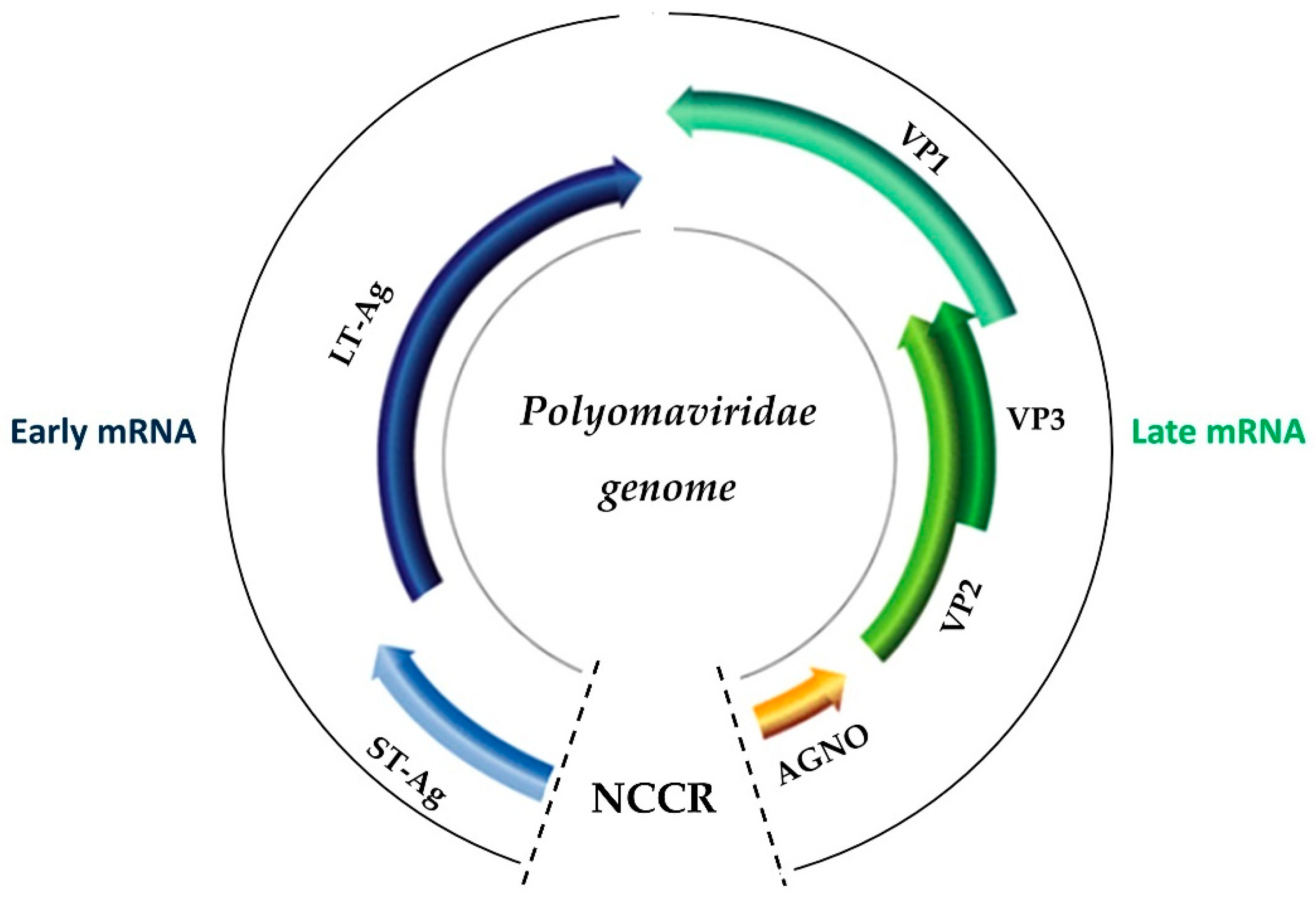
2. The Biology of BK Virus

All members of the Polyomaviridae family share a common virion structure, including capsid, the genomic part, and the high levels of genetic homology. Of note, BKV is a small non-enveloped virus with an icosahedral capsid of 40–44 mm in diameter. Viral capsid protein is comprised of three main components. While VP1, VP2, and VP3, contain DNA-binding domains [12], VP1 is the only viral protein that is exposed to the outer part of the virion. Thus, VP1 is the most important part in terms of host–pathogen (virus) communication. On the other hand, VP2 and VP3 are minor proteins that work closer with the histone and genomic parts of DNA. These new minor capsid proteins have been postulated as the interconnection molecule between VP1 and the structures of chromatin chains [13]. Unlike the rest of the polyomaviruses, BKV enters the host cells via caveolae and endocytic pathways before carrying into the endoplasmic reticulum [14]. Then, VP2 and VP3 mediate the BKV entry into the nucleus via importin after the uncoating of VP1. In the nucleus, the BKV genome is still episomal in human cells without integrating into the host DNA [15].
The BK genome (Figure 1) consists of approximately 5300 bp, contains gene encoding for the viral replication proteins (large T antigen (LTA), and agnoprotein); a non-coding control region (NCCR); and VP1, VP2, and VP3. As such, NCCR is a hypervariable region containing various binding sites for host cellular regulatory factors, referred to as hypervariable regulatory regions (HVRR) [16]. Two different forms of BKV, archetype and rearranged variants, are defined based on the DNA sequences of NCCR. While the former variant is able to separate from the healthy volunteer, the latter can most often be isolated only from patient serum [17]. The viral replication and virulence of BK are enhanced by specific NCCR break-point, leading to gene rearrangement and fatal outcomes [18]. In addition, BK virus can be categorized into four genotypes (rather than variants) according to the difference in NCCR and VP1. The genotype I is most common in humans, as subgroup Ib2 is common in the American and European, whereas subgroup IVc2 is common in the Asian [16,19]. In contrast, the genotypes II and III are only in the minor population. Although the pathogenesis of permanently dormant BKV in the host is still unclear, BKV might remain in the latent stage or maintains in a low level of gene expression. Clinically, the persistence of BKV in kidney-transplanted recipients is associated an increased risk of the allograft loss [20]. The most recent data suggest that the immune evasion from BKV miRNAs override the negative autoregulatory feedback in kidney transplant patients with unclear mechanism [21].

3. Innate Immunity in the BK Virus

3.1. An Overview of the Innate Immune Response in the BK Virus

Macrophages are major cells for viral control through phagocytosis and opsonization, whereas NK cells are mainly involved in antibody-dependent cellular cytotoxicity (ADCC) [22]. Macrophages coupled with dendritic cells (DCs) also stimulate the innate immunity response through major histocompatibility complexes (MHCs). The deficiency of DCs is associated with the development of BK viremia in kidney-transplanted recipients [23,24]. The connection between MHC and cell recognition of viral pathogens then works with pathogen-associated molecular patterns (PAMPs) through both intracellular and extracellular receptors. Regarding those receptors, toll-like receptors (TLRs), formyl-methionine receptors, scavenger receptors, and inflammasomes are the main receptors involved in the innate immune response against BKV infection.
Innate immune mediators also cause allograft dysfunction in BKVN of kidney-transplanted recipients through the increase in most inflammatory cytokines in allograft tissue, i.e., interleukin (IL)-6, interferon gamma-induced protein 10 (IP-10)/C-X-C motif chemokine ligand 10 (CXCL10), IL-8/CXCL8, and monocyte chemoattractant protein-1 (MCP-1)/C-C motif chemokine ligand 2 (CCL2) [25]. In addition, several mediators that associated with renal fibrosis including transforming growth factor (TGF)-β, matrix metalloproteinase (MMP)-2, and MMP-9 also increase BKV expression in patient allografts [26]. Moreover, toll-like receptors (TLRs), on the cellular or endosomal membrane, recognize pathogen-associated molecular patterns (PAMPs) and damaged-associated molecular patterns (DAMPs) during BKV infection and trigger several transcription factors, such as nuclear factor kappa B (NF-KB) through myeloid differentiation primary response 88 (MyD88) [27] or TIR-domain-containing adapter-inducing interferon-β (TRIF) signaling pathways [28]. The inflammatory responses against BKV are important for organism control and organ damage during infection.

3.2. Immunity Cells against the BK Virus

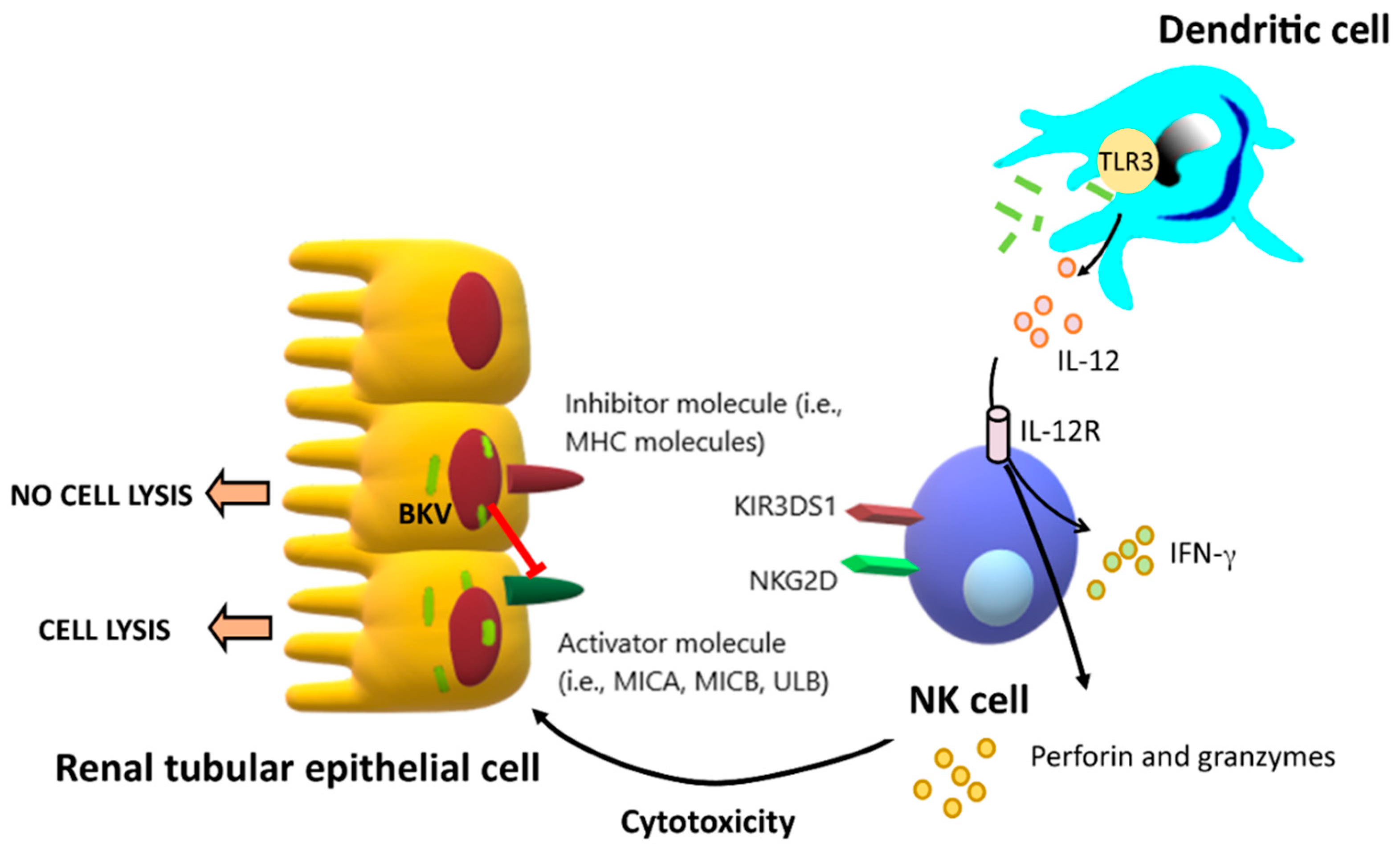
3.2.1. Natural Killer Cells (NK Cells)

NK cells are the most important immunity against BKV-infected uroepithelial cells through the recognition of the MHC class I downregulation, as well as the upregulation of MHC class I polypeptide-related sequence A (MICA), MHC class I polypeptide-related sequence B (MICB), and UL16-binding proteins (ULBP) that induce apoptosis in these cells by antibody-dependent cellular cytotoxicity (ADCC) [29,30] (Figure 2). Accordingly, Trydzenskaya et al. [22] evaluated the role of killer-cell immunoglobulin-like receptors (KIRs) on NK cells in the pathogenesis of BKV infection in kidney-transplanted recipients through the interaction of KIR bindings with both donor and recipient human leukocyte antigen (HLA) ligands by performing genotyping analysis of kidney-transplanted recipients (BKVN group (n = 48) versus control group (n = 110). They demonstrated a specific telomeric gene motif to be associated with severe BKVN, even during the attenuation of immunosuppression, and severe BKVN is associated with lower numbers of activating receptors. This reveals the role of low NK activating receptor (KIR3DS1) and the crucial roles of the immune recognition of NK cells in BKV infection.
In addition, the natural killer group 2D (NKG2D) receptor, an activating receptor that is mostly expressed in NK cells, NKT cells (natural killer T cells), and subsets of γδ T cells [31] is also important in BKV infection. Although NKG2D abundance is normally very low, NKG2D receptors could be rapidly induced during the activation by viruses and other cell stress conditions. Engagement of NKG2D stimulates the production of cytokines and cytotoxic molecules that mediate both inhibitory and activating signals, which depend on the intensity and duration of ligand engagement. In humans, the NKG2D ligands are MICA, MICB, and six members of the ULBP family [31]. Bauman et al. [32] demonstrated that BKV downregulates ULBP3, the stress-induced ligand recognized by the killer receptor NKG2D, through viral microRNA (miRNA) that reduces NK cells killing activities. As such, NK cells more efficiently eradicate the infected cells with the inhibition of viral 3p miRNA. Because NKG2D is also expressed by various T cell subsets [31], BKV might also downregulate ULBP3 of the adaptive immunity through miRNA. In addition, sustained engagement of the NKG2D receptor not only impairs NKG2D expression, but also reduces activated signaling of other receptors of NK cells. Koch et al. [33] demonstrated profound downregulation and dysfunction of Ly49D, an inhibitory NK cells signaling, in NK cells with chronic NKG2D engagement in an animal study results in renal allograft dysfunction. In parallel, NK cells lacking inhibitory Ly49 receptors for donor MHC class I molecules are sufficient to promote cardiac allograft rejection [34]. Furthermore, some degree of KIRs-ligand mismatching between donors and recipients is estimated to occur in more than 50% of HLA non-identical renal transplantation, leading to variable impacts in the outcomes. KIR-ligand mismatches have been reported as factors associated with reduced long-term allograft survival in HLA-incompatible kidney transplantation [35]. Altogether, increased activities with the inhibition of viral killing activity of NK cells through KIR-ligand mismatches are associated with kidney allograft rejection or a long-standing stage of viremia in kidney-transplanted recipients with BKVN [36].

3.2.2. Dendritic Cells

Macrophages and dendritic cells (DCs) are the front lines of defense against viruses and DAMPs through pattern recognition receptors (PRRs), specialized endocytic mechanisms, cytokines, and chemokines. The roles of aryl hydrocarbon receptor (AhR), one of the receptors against nucleic acid in specific T cell subpopulations and B cells in adaptive immunity are well known, and AhR is associated with macrophage polarization [37]. Bouatou et al. [38] demonstrated high AhR expression in kidney allografts of patients with BKVN and a higher proportion of nuclear AhR expression in CD68+ and CD45+ cells that infiltrates in kidney allografts in comparison with the allografts of T cell mediated rejection.
In parallel, dendritic cells (DCs) are a type of innate immune cells for the adaptive immune activation due to the ability of antigen presentation and naïve T cell stimulation. Many viruses efficiently reduce DCs functions, resulting in the impairment of viral elimination processes. DCs can be classified into different subtypes, and each of them expresses a wide range of corresponding molecules and receptors that help the clearance of viruses. DCs are one of the most important immune cells that produce interferon (IFN), a cytokine with viral protection properties. Of note, type I IFN (α and β) is strongly associated with antiviral responses [39]. Regarding BKV, Womer and colleagues [40] were the first group that demonstrated the reduced number of DCs in the peripheral blood of kidney-transplanted recipients with biopsy-proven BKVN. Therefore, it seems to raise the hypothesis of the reduction in DCs predisposed to BKV reactivation. Peripheral blood DC monitoring may be an early BKVN diagnostic tool for vulnerable kidney-transplanted recipients [23]. While immature DCs engulf viral antigens via endocytic pathways, mature DCs with the antigens migrate to draining lymph nodes, where they present viral peptides to CD8+ and CD4+ T lymphocytes through MHC class I and class II [41]. In fact, the maturation process of DCs is enhanced by trafficking to the nearest lymphoid organs to process antigen presentation and initiate adaptive immune responses [42]. With proper activation by DCs, virus-specific T cells are clonally expanded and differentiate into virus-specific effector T cells. Subsequently, CD8+ T cells further differentiate into cytotoxic T lymphocytes (CTL). Several studies have demonstrated the potential relative roles between DCs and virus-specific T cells. Dorn et al. [43] demonstrated hamster polyomavirus (HaPyV) major capsid protein VP1-derived virus-like particles (VLPs) as a potential carrier for a human tumor-associated CTL epitope, indicating the potential of chimeric HaPyV VP1-derived VLPs as a delivery vehicle for immunotherapeutic targets. Additionally, BK virus-specific CD8+ T cells require the mature monocyte-derived DCs for the highest immune response activity [44]. Hence, DCs in the presence of viral specific T cells could be potential vehicles for novel approaches for BKV vaccination and adoptive T cell therapies against BKVN.

3.2.3. Neutrophils and Eosinophils

Neutrophils, the first immune cells that are recruited into infection sites in bacterial and fungal infection [45], might be important in BKV infection as BKVN reactivation is more common in kidney-transplanted recipients with anti-neutrophil cytoplasmic antibody-associated vasculitis [46,47]. Indeed, defensins, small (18–45 amino acids) cationic peptides that form three disulfide bonds, are also associated with viral control. Human defensins (HDs) are divided into two families, α- and β-defensins. As such, α-defensins are composed of human neutrophil peptides (HNP) 1–4, which are expressed in HD5–6 immune cells that are found in the small intestine and HD5 cells that are found in the urogenital tract [48]. In parallel, β-defensins are produced by many different epithelial tissues and some immune cells [48]. In BKV infection, α-defensin human neutrophil protein 1 (HNP1) and human α-defensin 5 (HD5) inhibit BKV infection by targeting an early event in the viral lifecycle. Moreover, HD5 has anti-polyomavirus activity and neutralizes polyomaviruses in a serum-independent manner [49]. On the other hand, neutrophil extracellular traps (NETs) [46] mediate antiviral defense by trapping and inactivating viruses with the extruded meshwork of chromatin fibers that are decorated with granule-derived antimicrobial peptides and enzymes, such as neutrophil elastase, cathepsin G, and myeloperoxidase (MPO). Moreover, other neutrophil mechanisms, including phagocytosis, reactive oxygen species (ROS) generation, and the release of microbicidal molecules from granules (also called degranulation) might also be important in BKV control. For eosinophils, there is only one report that described the association between the main pathological features of BKVN with tubulitis and the numerous eosinophils in the collecting ducts and distal tubules [50]. However, the association between eosinophil and BKV needs more studies for the conclusion.

3.2.4. Gamma-Delta (γδ) T Cells

Since gamma-delta (γδ) T cells are an important part of unconventional T lymphocytes, they have been counted as innate immunity as a result of the ability to recognize various antigens without the presence of the MHC molecules. Indeed, γδ T cells attack the target cells through their cytotoxic activity as well as the activation of neighboring cellular immunity. Although γδ T cells have been described as a potential effector cell population against many pathogens, including bacteria, virus, fungi, and parasites, there are limited data on BKV infection. Different clones of γδ T cells (Vδ2 and Vγ45 γδ T cells) were demonstrated to be reactive against CMV-infected cells in allogeneic stem cell transplantation with CMV disease [51]. In addition, CD16 expression by γδ T cells could induce IFN-γ responses by opsonized CMV virions independently of T cell receptor (TCR) engagement, along with providing intrinsic antibody-dependent cell-mediated cytotoxicity (ADCC) potential [52]. In an in vitro study, the culture generated up to 66% of γδ TCRs, which were found to be active against BK infected cells [53]. Accordingly, the role of γδ T cells in BKV infection does not only serve as sentinels in the innate immune response toward BK infection but also as a potential bridge between the innate and cellular immunity.

3.3. Complement System of the Innate Immune Response against the BK Virus

Activation of the complement system typically occurs via three main pathways—the classical, alternative, and lectin-dependent pathways—that induce antiviral effects through direct viral neutralization, inflammatory responses, chemotaxis promotion, and the enhancement of the adaptive immunity. Although the complement system has multifaceted roles in both innate and adaptive immunity against all stages of viral infection, data on complement of BKV infection is still lacking. There is increased complement-related rejection after BKVN [54], but the true causal effects of the complement system towards BKV eradication are still unclear. In kidney transplantation, high mannose-binding lectin (MBL), a complement activation molecule, is associated with increased tissue damage following the ischemia-reperfusion injury, but MBL deficiency was not associated with an increased risk of BK viremia [55]. Perhaps MBL is activated by damaged tissue from BKVN, but can not control BKV or the role of the gene encoding for the mannose-binding lectin, MBL2 [10]. However, the impact of classical and alternative pathways in BKV infection is still unclear.

3.4. Toll-like Receptors in the Innate Immunity Response against the BK Virus

Toll-like receptors (TLRs) are fundamental sensor molecules of the innate immune response against any virus, and they detect conserved molecular signatures of a wide range of microbial pathogens and initiate innate immune responses via distinct signaling pathways. Many TLRs are implicated in the early interplay of host cells with invading viruses, which regulate either viral replication or host responses—ultimately impacting viral pathogenesis. Signaling pathways triggered by the BK virus involve TLRs, melanoma differentiation-associated protein 5 (MDA5), retinoic acid inducible gene-I (RIG-I), and possibly the stimulator of interferon genes (STING).
Regarding innate immunity sensors, the toll-like receptors against BKV are the most well-known molecules in terms of immune functions. A study by Heutinck et al. [56] reveals that the intracellular viral sensors, including TLR3, MDA5, and RIG-I, are regulated in cytomegalovirus, Epstein–Barr virus, and BKV-infected tubular epithelial cells. In contrast, Ribeiro et al. [25] demonstrated that activation of the viral DNA and the double-stranded RNA sensors TLR3 provided strong signals in the epithelial cells of distal cortical tubules and the collecting ducts of nephron, while stimulation of RIG-I had no effect on BKVN. However, in an in vitro study, they found that collecting duct cells significantly expressed TLR3 intracellularly, and activation of TLR3 and RIG-I enhanced expression of cytokines, chemokines, and IFN-β mRNA. Thus, the inflammation could be specifically blocked by siRNA to TLR3 [25]. Hence, there is an efficient antiviral immune response with TLR3 and RIG-I upregulation.
Nevertheless, the most recent study on the NACHT, LRR, and PYD domains containing the protein 3 (NLRP3) inflammasome—an oligomeric complex comprised of the NOD-like receptor NLRP3, the adaptor ASC, and caspase-1—demonstrates that the NLRP3 complex is crucial to the host’s defense against various viruses because it promotes IL-1β and IL-18 secretion and induces pyroptosis [57]. Furthermore, NLRP3 recognizes a variety of pathogen-associated molecular patterns (PAMPs) and danger-associated molecular patterns (DAMPs) generated during viral replication that trigger the NLRP3 inflammasome-dependent antiviral immune responses and facilitate viral eradication [58,59]. Meanwhile, as mentioned earlier, the connections between MHC and the cell recognition of the BKV collaborate with pathogen-associated molecular patterns (PAMPs) through TLRs and inflammasomes against BKV infection. Thus, the inflammasome might also involve the innate immunity against BKV infection. Further studies are warranted.

3.5. Cytokines in the Innate Immunity Response to the BK Virus

As mentioned above, the host’s response to the BK virus necessitates the integrated action of the innate immunity, largely NK cells, macrophages, DCs, and granulocytes. An effective immune response requires crosstalk among the various immune cells as an intercellular network, which is partly mediated by cytokines and chemokines. Cytokines are low molecular weight soluble proteins that modulate various immune functions upon induction by pathogens. Cytokines also control the magnitude and kinetics of immune responses by inducing or inhibiting the activation, proliferation, and/or differentiation of various target cells, and they also regulate the production of antibodies and other cytokines and chemokines [60]. In addition, interferon (IFN)-γ and the tumor necrosis factor (TNF)-α are important anti-viral cytokines produced by NK cells [61], and interleukin (IL)-12 is an important cytokine from activated macrophages and DCs to induce anti-viral adaptive immunity [60]. Indeed, BKVN demonstrated high IL-12 [53], possibly due to the increased DC functions. So far, the evidence of cytokine roles in terms of defense against viruses is still controversial. Although TNF-α and IFN-β have been demonstrated as major cytokines for controlling the spread of the BKV in kidney-transplanted recipients with BKVN [62], the papillomavirus-like particles are also able to induce expression of IL-6, TNF-α, and IFN-α by DCs [63,64]. However, Liu et al. [65] demonstrated that the expression of immune-related genes (antigen presentation, cytotoxicity, and inflammation) is significantly higher in kidney-transplanted recipients with BKVN than in those with stable allografts. The authors also concluded that immune cell-, cytokine-, chemokine-, and inflammation-related pathways are significantly activated in BKVN, while metabolism- and renal development-related pathways were significantly downregulated in BKVN [65]. Accordingly, the proinflammatory cytokines are not only an important innate immune constituent for the inhibition of BKVN; they also participate in the pathogenesis of BKVN and may provide a theoretical basis for preventing the occurrence of BKVN and finding novel treatments.

3.6. Notch Signaling and Nuclear Factors

Notch signaling pathway regulates cell proliferation, cell differentiation, and cell death (apoptosis) during the development and normal homeostasis. In human, there are four Notch receptors (Notch 1–4) that are involved in transducing specific extracellular signals [66]. One of the most established functions of Notch signaling in the immune system is the differentiation of lymphoid B and T cell lineages [67]. Notch functions also enhance regulatory T (Treg) cell function, activate the T cell network, as well as stimulate T helper cell differentiation [68,69,70]. Of note, Treg cells act as a gate-keeper of multiple immune reactions, including overwhelming immune suppression and transplant rejection [71,72]. Nuclear factor-kappa B (NF-кB) transcription factors critically integrate the balancing of immune response. NF-кB is always triggered by multiple immune signals, which serves as a cell-intrinsic key factor for Treg cells [73]. In addition to NF-кB, nuclear factor of activated T cells (NFAT) is also a crucial transcription factor during HaPyV infection through binding the viral promoter and regulating viral transcription and infection. The NFAT binding sites have a very sophisticated function among subunits of NK-кB, leading to control either positively or negatively regulatory networks and viral infection [74].
On the other hand, during respiratory syncytial viral infection, Notch signaling has prominent functions, for example, through increased levels of DLL4 ligand expression mediated by the MyD88 intracellular signaling pathway that further enhance production of CD8+ and CD4+ T cells, Th2 cytokines, and decreased IFN-γ [75], increased methylation of H3K4 nearby the Foxp3 locus [76], as well as decreased the expression of domain of SET and MYDN containing protein 3 (SMYD3) and expression of FOXP3 in induced Treg cells, leading to an increase in IL-17A [76]. Although little is known about the role of Notch on HaPyV, it has been postulated that the role of BKV infection is based on the induction of telomerase activity, deregulation of multiple important signaling pathways for proliferation (phosphoinositide-3 kinase–Akt/protein kinase B, Wnt, and Ras/Raf/mitogen-activated kinase, STAT3, and Notch signaling pathway) [77,78,79]. As such, the participation of Notch signaling during BKV infection could be an essential tool to control viral replication [80]. More studies are warranted.

4. BKVAN and Allograft Rejection

During BKV infection, CD8+ T cells play a major role for surveillance and anti-viral cells against HaPyV, leading to the development of BKV-specific T cells in circulation and allograft tissue. In addition to CD8+ T cells, as a result of VP1 and LT-Ag stimulation, plenty of immune cells and cytokines are stimulated as the continuum cascades, including IFN-γ, TNF-α, and granzymes [81]. This process provides synergistic effects to DCs and NK cells (Figure 2). Accordingly, IFN-γ has been suggested as a marker of BK viremia in transplant recipients [82]. In a study by Comoli and colleagues [83], kidney transplant recipients with BKV infection—active or non-active—had a mean BKV-specific lymphocyte frequency 2 log lower than healthy recipients assessed by enzyme-linked immunospot assay (ELISpot), suggesting an impact of immunosuppression on BKV immunosurveillance. However, a later study by Chakera et al. showed no correlation between BKV-specific T cells and BKV activity [84]. Perhaps recipients recovering from BK viremia had an ultimately improved T cell response [85]. On the other hand, over-activation of T cell following BKV infection leads to T cell exhaustion and loss of polyfunctional responses [86]. As such, lymphoid organs or allograft tissue may play a central restoration role in immune competence.
T cells also recognize peptide antigens presented by HLA molecules; therefore, the number of HLA matching could affect the T cell response toward BKV infection [87]. Recipients with high HLA mismatch are more aggressively suppressed leading to attenuating anti-viral response. Interestingly, male recipients, HLA-A2, B44, and DR15, but not DR7, were reported as protective against BK viremia infection [88].
Little is known about the true causal link between allograft rejection and BKV infection. Although there is severe inflammation found in allograft tissue, assembling both histology [89,90] and genetics [36,91] to acute allograft rejection, decreased immunosuppression drugs may increase rejection risk. Perhaps the most important question remains, does this process trigger de novo donor-human leukocyte antigen (HLA) specific antibodies (dnDSA)? In the study by Patel and colleagues [92], kidney transplant recipients with a higher number of HLA mismatched and viral clearance may increase the risk of dnDSA after BKV infection. Of note, BKVAN also triggers dnDSA within 14 months of BKV diagnosis, which a risk factor for subsequent antibody-mediated rejection (hazard ratios, HR 4.75) and allograft loss (HR 2.63) [93]. Therefore, monitoring for dnDSA in recipients being managed for BKV infection may be warranted.

5. Conclusions

The interaction between the innate immune response and BKV is an important framework. To date, there is little information regarding how innate immunity mediators work in controlling BKV infection. Most studies have focused on the post-transplanted BKV infection setting, termed BKV-associated nephropathy (BKVN). Among the innate immune system, natural killer (NK) cells are the most important innate immunity cells that interact with the BK virus. Killer cell immunoglobulin-like receptors (KIR) as a central molecule of the others for defense against the BKV as the activating KIR, particularly those of the KIR3DS1 genotype, play a crucial role in the recognition of BKV replication and are related to clinical severity. Defensins are another innate mediator that inhibit BKV activity by blocking the BKV from binding to host cells. In addition, innate immunity mediators also have a role in kidney allograft loss in kidney-transplanted recipients with BKVN. BKVN is associated with intrinsic renal inflammation through increased expression of several interleukins and other cytokines. Furthermore, the microenvironment, due to the BKV infection, also promotes the inflammatory stages in allograft that finally induce kidney allograft fibrosis. As such, the harness on innate immunity against BKV infection might be important in organ transplantation. However, prior to reaching that point, the prevention of BKVN remains the priority for kidney-transplanted recipients who seek to avoid widespread BKV in the kidney allograft, combined with the challenging problems of allo-sensitization.

Author Contributions

Conceptualization, W.C. and A.L.; writing—original draft preparation, W.C.; writing—review and editing, W.C. and A.L.; funding acquisition, W.C. All authors have read and agreed to the published version of the manuscript.

Funding

This work was funded by Mahidol University—Basic Research Fund: fiscal year 2021 (FRB640032) and Basic Research Fund: fiscal year 2022 (FRB650007/0185), and Health System Research Institute (HSRI)-Flagship Project Fund: fiscal year 2020 (W.C).

Conflicts of Interest

The authors declare no conflict of interest. The funders had no role in the writing of the manuscript, or in the decision to publish the results.

References

  1. Gardner, S.D.; Field, A.M.; Coleman, D.V.; Hulme, B. New human papovavirus (B.K.) isolated from urine after renal transplantation. Lancet 1971, 1, 1253–1257. [Google Scholar] [CrossRef]
  2. Hussain, I.; Tasneem, F.; Umer, M.; Pervaiz, A.; Raza, M.; Arshad, M.I.; Shahzad, N. Specific and quantitative detection of Human polyomaviruses BKPyV and JCPyV in the healthy Pakistani population. Virol. J. 2017, 14, 86. [Google Scholar] [CrossRef] [PubMed] [Green Version]
  3. Ramos, E.; Drachenberg, C.B.; Wali, R.; Hirsch, H.H. The decade of polyomavirus BK-associated nephropathy: State of affairs. Transplantation 2009, 87, 621–630. [Google Scholar] [CrossRef] [PubMed]
  4. Hirsch, H.H.; Randhawa, P.; AST Infectious Diseases Community of Practice. BK polyomavirus in solid organ transplantation. Am. J. Transplant. 2013, 13 (Suppl. 4), 179–188. [Google Scholar] [CrossRef] [PubMed]
  5. Laskin, B.L.; Denburg, M.R.; Furth, S.L.; Moatz, T.; Altrich, M.; Kleiboeker, S.; Lutzko, C.; Zhu, X.; Blackard, J.T.; Jodele, S.; et al. The natural history of BK polyomavirus and the host immune response after stem cell transplantation. Clin. Infect. Dis. 2019, 71, 3044–3054. [Google Scholar] [CrossRef]
  6. Patel, N.C.; Vilchez, R.A.; Killen, D.E.; Zanwar, P.; Sroller, V.; Eldin, K.W.; López-Terrada, D.; Butel, J.S. Detection of polyomavirus SV40 in tonsils from immunocompetent children. J. Clin. Virol. 2008, 43, 66–72. [Google Scholar] [CrossRef] [Green Version]
  7. Babel, N.; Fendt, J.; Karaivanov, S.; Bold, G.; Arnold, S.; Sefrin, A.; Lieske, E.; Hoffzimmer, M.; Dziubianau, M.; Bethke, N.; et al. Sustained BK viruria as an early marker for the development of BKV-associated nephropathy: Analysis of 4128 urine and serum samples. Transplantation 2009, 88, 89–95. [Google Scholar] [CrossRef]
  8. Takahashi, K.; Sato, Y.; Sekizuka, T.; Kuroda, M.; Suzuki, T.; Hasegawa, H.; Katano, H. High expression of JC polyomavirus-encoded microRNAs in progressive multifocal leukoencephalopathy tissues and its repressive role in virus replication. PLoS Pathog. 2020, 16, e1008523. [Google Scholar] [CrossRef]
  9. Papa, N.; Zanotta, N.; Knowles, A.; Orzan, E.; Comar, M. Detection of Malawi polyomavirus sequences in secondary lymphoid tissues from Italian healthy children: A transient site of infection. Virol. J. 2016, 13, 97. [Google Scholar] [CrossRef] [Green Version]
  10. Comar, M.; Segat, L.; Crovella, S.; Bovenzi, M.; Cortini, E.; Tognon, M. The significance of mannose-binding lectin gene polymorphisms on the risk of BK virus coinfection in women with human papillomavirus–positive cervical lesions. Hum. Immunol. 2011, 72, 663–666. [Google Scholar] [CrossRef]
  11. Longhi, G.; Pietropaolo, V.; Mischitelli, M.; Longhi, C.; Conte, M.P.; Marchetti, M.; Tinari, A.; Valenti, P.; Degener, A.M.; Seganti, L.; et al. Lactoferrin inhibits early steps of human BK polyomavirus infection. Antivir. Res. 2006, 72, 145–152. [Google Scholar] [CrossRef] [PubMed]
  12. Hurdiss, D.L.; Frank, M.; Snowden, J.S.; Macdonald, A.; Ranson, N.A. The Structure of an Infectious Human Polyomavirus and Its Interactions with Cellular Receptors. Structure 2018, 26, 839–847.e3. [Google Scholar] [CrossRef] [Green Version]
  13. Hurdiss, D.L.; Morgan, E.L.; Thompson, R.F.; Prescott, E.L.; Panou, M.M.; Macdonald, A.; Ranson, N.A. New Structural Insights into the Genome and Minor Capsid Proteins of BK Polyomavirus using Cryo-Electron Microscopy. Structure 2016, 24, 528–536. [Google Scholar] [CrossRef] [Green Version]
  14. Moriyama, T.; Sorokin, A. Intracellular trafficking pathway of BK Virus in human renal proximal tubular epithelial cells. Virology 2008, 371, 336–349. [Google Scholar] [CrossRef] [Green Version]
  15. Cubitt, C.L. Molecular genetics of the BK virus. Adv. Exp. Med. Biol. 2006, 577, 85–95. [Google Scholar]
  16. Hussain, I.; Tasneem, F.; Gilani, U.S.; Arshad, M.I.; Farhan Ul Haque, M.; Abbas, Z.; Umer, M.; Shahzad, N. Human BK and JC polyomaviruses: Molecular insights and prevalence in Asia. Virus Res. 2020, 278, 197860. [Google Scholar] [CrossRef] [PubMed]
  17. Olsen, G.H.; Hirsch, H.H.; Rinaldo, C.H. Functional analysis of polyomavirus BK non-coding control region quasispecies from kidney-transplanted recipients. J. Med. Virol. 2009, 81, 1959–1967. [Google Scholar] [CrossRef] [PubMed]
  18. Roy, S.; Mieczkowski, P.A.; Weida, C.; Huo, J.; Roehrs, P.; Singh, H.K.; Nickeleit, V. BK polyomavirus nephropathy with systemic viral spread: Whole genome sequencing data from a fatal case of BKPyV infection. Transplant. Infect. Dis. 2020, 22, e13269. [Google Scholar] [CrossRef] [PubMed]
  19. Zhong, S.; Jeong, B.H.; Ikegaya, H.; Kim, Y.S.; Xu, Y.; Zhu, M.; Chao, Y.; Suzuki, M.; Kitamura, T.; Homma, Y.; et al. Comparison of the distribution patterns of BK polyomavirus lineages among China, Korea and Japan: Implications for human migrations in northeast Asia. Microbiol. Immunol. 2009, 53, 266–276. [Google Scholar] [CrossRef]
  20. Nickeleit, V.; Singh, H.K.; Dadhania, D.; Cornea, V.; El-Husseini, A.; Castellanos, A.; Davis, V.G.; Waid, T.; Seshan, S.V. The 2018 Banff Working Group classification of definitive polyomavirus nephropathy: A multicenter validation study in the modern era. Am. J. Transplant. 2020, 21, 669–680. [Google Scholar] [CrossRef]
  21. Huang, Y.; Zeng, G.; Randhawa, P.S. Detection of BKV encoded mature MicroRNAs in kidney transplant patients: Clinical and biologic insights. J. Clin. Virol. 2019, 119, 6–10. [Google Scholar] [CrossRef]
  22. Trydzenskaya, H.; Juerchott, K.; Lachmann, N.; Kotsch, K.; Kunert, K.; Weist, B.; Schonemann, C.; Schindler, R.; Nickel, P.; Melzig, M.F.; et al. The genetic predisposition of natural killer cell to BK virus-associated nephropathy in renal transplant patients. Kidney Int. 2013, 84, 359–365. [Google Scholar] [CrossRef] [Green Version]
  23. Womer, K.L.; Huang, Y.; Herren, H.; Dibadj, K.; Peng, R.; Murawski, M.; Shraybman, R.; Patton, P.; Clare-Salzler, M.J.; Kaplan, B. Dendritic cell deficiency associated with development of BK viremia and nephropathy in renal transplant recipients. Transplantation 2010, 89, 115–123. [Google Scholar] [CrossRef]
  24. Yapici, U.; Kers, J.; Slavujevic-Letic, I.; Stokman, G.; Roelofs, J.J.; van Aalderen, M.C.; Groothoff, J.W.; de Boer, O.J.; van der Pant, K.A.; Claessen, N.; et al. Intragraft Blood Dendritic Cell Antigen-1-Positive Myeloid Dendritic Cells Increase during BK Polyomavirus-Associated Nephropathy. J. Am. Soc. Nephrol. 2016, 27, 2502–2510. [Google Scholar] [CrossRef] [PubMed] [Green Version]
  25. Ribeiro, A.; Wörnle, M.; Motamedi, N.; Anders, H.J.; Gröne, E.F.; Nitschko, H.; Kurktschiev, P.; Debiec, H.; Kretzler, M.; Cohen, C.D.; et al. Activation of innate immune defense mechanisms contributes to polyomavirus BK-associated nephropathy. Kidney Int. 2012, 81, 100–111. [Google Scholar] [CrossRef] [PubMed] [Green Version]
  26. Mannon, R.B.; Hoffmann, S.C.; Kampen, R.L.; Cheng, O.C.; Kleiner, D.E.; Ryschkewitsch, C.; Curfman, B.; Major, E.; Hale, D.A.; Kirk, A.D. Molecular evaluation of BK polyomavirus nephropathy. Am. J. Transplant. 2005, 5, 2883–2893. [Google Scholar] [CrossRef] [PubMed]
  27. Federico, S.; Pozzetti, L.; Papa, A.; Carullo, G.; Gemma, S.; Butini, S.; Campiani, G.; Relitti, N. Modulation of the Innate Immune Response by Targeting Toll-like Receptors: A Perspective on their Agonists and Antagonists. J. Med. Chem. 2020, 63, 13466–13513. [Google Scholar] [CrossRef]
  28. Kariminik, A.; Yaghobi, R.; Dabiri, S. Innate Immunity and BK Virus: Prospective Strategies. Viral. Immunol. 2016, 29, 74–82. [Google Scholar] [CrossRef]
  29. Nainu, F.; Shiratsuchi, A.; Nakanishi, Y. Induction of Apoptosis and Subsequent Phagocytosis of Virus-Infected Cells as an Antiviral Mechanism. Front. Immunol. 2017, 8, 1220. [Google Scholar] [CrossRef]
  30. Warren, H.S.; Smyth, M.J. NK cells and apoptosis. Immunol. Cell Biol. 1999, 77, 64–75. [Google Scholar] [CrossRef]
  31. Raulet, D.H. Roles of the NKG2D immunoreceptor and its ligands. Nat. Rev. Immunol. 2003, 3, 781–790. [Google Scholar] [CrossRef]
  32. Bauman, Y.; Nachmani, D.; Vitenshtein, A.; Tsukerman, P.; Drayman, N.; Stern-Ginossar, N.; Lankry, D.; Gruda, R.; Mandelboim, O. An identical miRNA of the human JC and BK polyoma viruses targets the stress-induced ligand ULBP3 to escape immune elimination. Cell Host Microbe 2011, 9, 93–102. [Google Scholar] [CrossRef] [Green Version]
  33. Koch, C.; Kim, Y.; Zöller, T.; Born, C.; Steinle, A. Chronic NKG2D Engagement In Vivo Differentially Impacts NK Cell Responsiveness by Activating NK Receptors. Front. Immunol. 2017, 8, 1466. [Google Scholar] [CrossRef] [Green Version]
  34. McNerney, M.E.; Lee, K.-M.; Zhou, P.; Molinero, L.; Mashayekhi, M.; Guzior, D.; Sattar, H.; Kuppireddi, S.; Wang, C.-R.; Kumar, V.; et al. Role of Natural Killer Cell Subsets in Cardiac Allograft Rejection. Am. J. Transplant. 2006, 6, 505–513. [Google Scholar] [CrossRef]
  35. van Bergen, J.; Thompson, A.; Haasnoot, G.W.; Roodnat, J.I.; de Fijter, J.W.; Claas, F.H.; Koning, F.; Doxiadis, I.I. KIR-ligand mismatches are associated with reduced long-term graft survival in HLA-compatible kidney transplantation. Am. J. Transplant. 2011, 11, 1959–1964. [Google Scholar] [CrossRef] [PubMed]
  36. Lubetzky, M.; Bao, Y.; Broin, P.O.; Marfo, K.; Ajaimy, M.; Aljanabi, A.; de Boccardo, G.; Golden, A.; Akalin, E. Genomics of BK viremia in kidney-transplanted recipients. Transplantation 2014, 97, 451–456. [Google Scholar] [CrossRef] [PubMed]
  37. Zhu, J.; Luo, L.; Tian, L.; Yin, S.; Ma, X.; Cheng, S.; Tang, W.; Yu, J.; Ma, W.; Zhou, X.; et al. Aryl Hydrocarbon Receptor Promotes IL-10 Expression in Inflammatory Macrophages Through Src-STAT3 Signaling Pathway. Front. Immunol. 2018, 9, 2033. [Google Scholar] [CrossRef] [PubMed] [Green Version]
  38. Bouatou, Y.; Stokman, G.; Claessen, N.; Roelofs, J.J.T.H.; Bemelman, F.; Kers, J.; Florquin, S. Aryl hydrocarbon receptor expression by macrophages and lymphocytes within infiltrates in BK polyomavirus associated nephropathy. Transplant. Immunol. 2018, 47, 18–21. [Google Scholar] [CrossRef]
  39. Soto, J.A.; Gálvez, N.M.S.; Andrade, C.A.; Pacheco, G.A.; Bohmwald, K.; Berrios, R.V.; Bueno, S.M.; Kalergis, A.M. The Role of Dendritic Cells During Infections Caused by Highly Prevalent Viruses. Front. Immunol. 2020, 11, 1513. [Google Scholar] [CrossRef]
  40. Womer, K.L.; Peng, R.; Patton, P.R.; Murawski, M.R.; Bucci, M.; Kaleem, A.; Schold, J.; Efron, P.A.; Hemming, A.W.; Srinivas, T.R.; et al. The effects of renal transplantation on peripheral blood dendritic cells. Clin. Transplant. 2005, 19, 659–667. [Google Scholar] [CrossRef] [PubMed]
  41. Audiger, C.; Rahman, M.J.; Yun, T.J.; Tarbell, K.V.; Lesage, S. The Importance of Dendritic Cells in Maintaining Immune Tolerance. J. Immunol. 2017, 198, 2223–2231. [Google Scholar] [CrossRef] [Green Version]
  42. de Winde, C.M.; Munday, C.; Acton, S.E. Molecular mechanisms of dendritic cell migration in immunity and cancer. Med. Microbiol. Immunol. 2020, 209, 515–529. [Google Scholar] [CrossRef]
  43. Dorn, D.C.; Lawatscheck, R.; Zvirbliene, A.; Aleksaite, E.; Pecher, G.; Sasnauskas, K.; Ozel, M.; Raftery, M.; Schönrich, G.; Ulrich, R.G.; et al. Cellular and humoral immunogenicity of hamster polyomavirus-derived virus-like particles harboring a mucin 1 cytotoxic T-cell epitope. Viral. Immunol. 2008, 21, 12–27. [Google Scholar] [CrossRef]
  44. Wilhelm, M.; Kaur, A.; Wernli, M.; Hirsch, H.H. BK Polyomavirus (BKPyV)-specific CD8 T-cell expansion in vitro using 27mer peptide antigens for developing adoptive T-cell transfer and vaccination. J. Infect. Dis. 2020, 223, 1410–1422. [Google Scholar] [CrossRef]
  45. Naumenko, V.; Turk, M.; Jenne, C.N.; Kim, S.-J. Neutrophils in viral infection. Cell Tissue Res. 2018, 371, 505–516. [Google Scholar] [CrossRef] [PubMed]
  46. Brinkmann, V.; Reichard, U.; Goosmann, C.; Fauler, B.; Uhlemann, Y.; Weiss, D.S.; Weinrauch, Y.; Zychlinsky, A. Neutrophil extracellular traps kill bacteria. Science 2004, 303, 1532–1535. [Google Scholar] [CrossRef] [PubMed]
  47. Rau, S.; Schönermarck, U.; Jäger, G.; Stangl, M.; Guba, M.; Meiser, B.; Fischereder, M.; Habicht, A. BK virus-associated nephropathy: Neutrophil gelatinase-associated lipocalin as a new diagnostic tool? Clin. Transplant. 2013, 27, E184–E191. [Google Scholar] [CrossRef] [PubMed]
  48. Klotman, M.E.; Chang, T.L. Defensins in innate antiviral immunity. Nat. Rev. Immunol. 2006, 6, 447–456. [Google Scholar] [CrossRef] [PubMed]
  49. Dugan, A.S.; Maginnis, M.S.; Jordan, J.A.; Gasparovic, M.L.; Manley, K.; Page, R.; Williams, G.; Porter, E.; O’Hara, B.A.; Atwood, W.J. Human alpha-defensins inhibit BK virus infection by aggregating virions and blocking binding to host cells. J. Biol. Chem. 2008, 283, 31125–31132. [Google Scholar] [CrossRef] [Green Version]
  50. Yamanaka, K.; Oka, K.; Nakazawa, S.; Hirai, T.; Kishikawa, H.; Nishimura, K.; Kyo, M.; Ichikawa, Y. Immunohistochemical features of BK virus nephropathy in renal transplant recipients. Clin. Transplant. 2012, 26 (Suppl. 24), 20–24. [Google Scholar] [CrossRef]
  51. Knight, A.; Madrigal, A.J.; Grace, S.; Sivakumaran, J.; Kottaridis, P.; Mackinnon, S.; Travers, P.J.; Lowdell, M.W. The role of Vδ2-negative γδ T cells during cytomegalovirus reactivation in recipients of allogeneic stem cell transplantation. Blood 2010, 116, 2164–2172. [Google Scholar] [CrossRef] [Green Version]
  52. Couzi, L.; Pitard, V.; Sicard, X.; Garrigue, I.; Hawchar, O.; Merville, P.; Moreau, J.F.; Déchanet-Merville, J. Antibody-dependent anti-cytomegalovirus activity of human γδ T cells expressing CD16 (FcγRIIIa). Blood 2012, 119, 1418–1427. [Google Scholar] [CrossRef]
  53. Comoli, P.; Basso, S.; Azzi, A.; Moretta, A.; De Santis, R.; Del Galdo, F.; De Palma, R.; Valente, U.; Nocera, A.; Perfumo, F.; et al. Dendritic cells pulsed with polyomavirus BK antigen induce ex vivo polyoma BK virus-specific cytotoxic T-cell lines in seropositive healthy individuals and renal transplant recipients. J. Am. Soc. Nephrol. 2003, 14, 3197–3204. [Google Scholar] [CrossRef] [Green Version]
  54. Honsová, E.; Lodererová, A.; Viklický, O.; Boucek, P. BK-virus nephropathy and simultaneous C4d positive staining in renal allografts. Cesk. Patol. 2005, 41, 163–166. [Google Scholar] [PubMed]
  55. Liman, P.; Babel, N.; Schachtner, T.; Unterwalder, N.; König, J.; Hofmann, J.; Reinke, P.; Nickel, P. Mannose-binding lectin deficiency is not associated with increased risk for polyomavirus nephropathy. Transpl. Immunol. 2012, 26, 123–127. [Google Scholar] [CrossRef] [PubMed]
  56. Heutinck, K.M.; Kassies, J.; Florquin, S.; ten Berge, I.J.; Hamann, J.; Rowshani, A.T. SerpinB9 expression in human renal tubular epithelial cells is induced by triggering of the viral dsRNA sensors TLR3, MDA5 and RIG-I. Nephrol. Dial. Transplant. 2012, 27, 2746–2754. [Google Scholar] [CrossRef] [PubMed] [Green Version]
  57. Mitoma, H.; Hanabuchi, S.; Kim, T.; Bao, M.; Zhang, Z.; Sugimoto, N.; Liu, Y.J. The DHX33 RNA helicase senses cytosolic RNA and activates the NLRP3 inflammasome. Immunity 2013, 39, 123–135. [Google Scholar] [CrossRef] [PubMed] [Green Version]
  58. Swanson, K.V.; Deng, M.; Ting, J.P.Y. The NLRP3 inflammasome: Molecular activation and regulation to therapeutics. Nat. Rev. Immunol. 2019, 19, 477–489. [Google Scholar] [CrossRef] [PubMed]
  59. Zhao, C.; Zhao, W. NLRP3 Inflammasome—A Key Player in Antiviral Responses. Front. Immunol. 2020, 11, 211. [Google Scholar] [CrossRef] [PubMed] [Green Version]
  60. Mogensen, T.H.; Paludan, S.R. Molecular pathways in virus-induced cytokine production. Microbiol. Mol. Biol. Rev. 2001, 65, 131–150. [Google Scholar] [CrossRef] [Green Version]
  61. Ferrari, L.; Martelli, P.; Saleri, R.; De Angelis, E.; Ferrarini, G.; Cavalli, V.; Passeri, B.; Bazzoli, G.; Ogno, G.; Magliani, W.; et al. An engineered anti-idiotypic antibody-derived killer peptide (KP) early activates swine inflammatory monocytes, CD3(+)CD16(+) natural killer T cells and CD4(+)CD8α(+) double positive CD8β(+) cytotoxic T lymphocytes associated with TNF-α and IFN-γ secretion. Comp. Immunol. Microbiol. Infect. Dis. 2020, 72, 101523. [Google Scholar] [CrossRef]
  62. Heutinck, K.M.; Rowshani, A.T.; Kassies, J.; Claessen, N.; van Donselaar-van der Pant, K.A.; Bemelman, F.J.; Eldering, E.; van Lier, R.A.; Florquin, S.; Ten Berge, I.J.; et al. Viral double-stranded RNA sensors induce antiviral, pro-inflammatory, and pro-apoptotic responses in human renal tubular epithelial cells. Kidney Int. 2012, 82, 664–675. [Google Scholar] [CrossRef] [PubMed] [Green Version]
  63. Lenz, P.; Day, P.M.; Pang, Y.Y.; Frye, S.A.; Jensen, P.N.; Lowy, D.R.; Schiller, J.T. Papillomavirus-like particles induce acute activation of dendritic cells. J. Immunol. 2001, 166, 5346–5355. [Google Scholar] [CrossRef] [Green Version]
  64. Yang, R.; Murillo, F.M.; Cui, H.; Blosser, R.; Uematsu, S.; Takeda, K.; Akira, S.; Viscidi, R.P.; Roden, R.B. Papillomavirus-like particles stimulate murine bone marrow-derived dendritic cells to produce alpha interferon and Th1 immune responses via MyD88. J. Virol. 2004, 78, 11152–11160. [Google Scholar] [CrossRef] [PubMed] [Green Version]
  65. Liu, Y.; Zhou, S.; Hu, J.; Xu, W.; Liu, D.; Liao, J.; Liao, G.; Guo, Z.; Li, Y.; Yang, S.; et al. Characterization of aberrant pathways activation and immune microenviroment of BK virus associated nephropathy. Aging 2020, 12, 14434–14451. [Google Scholar] [CrossRef]
  66. Radtke, F.; Fasnacht, N.; Macdonald, H.R. Notch signaling in the immune system. Immunity 2010, 32, 14–27. [Google Scholar] [CrossRef] [Green Version]
  67. Tanigaki, K.; Honjo, T. Regulation of lymphocyte development by Notch signaling. Nat. Immunol. 2007, 8, 451–456. [Google Scholar] [CrossRef]
  68. Ostroukhova, M.; Qi, Z.; Oriss, T.B.; Dixon-McCarthy, B.; Ray, P.; Ray, A. Treg-mediated immunosuppression involves activation of the Notch-HES1 axis by membrane-bound TGF-beta. J. Clin. Investig. 2006, 116, 996–1004. [Google Scholar] [CrossRef] [Green Version]
  69. Eagar, T.N.; Tang, Q.; Wolfe, M.; He, Y.; Pear, W.S.; Bluestone, J.A. Notch 1 signaling regulates peripheral T cell activation. Immunity 2004, 20, 407–415. [Google Scholar] [CrossRef] [Green Version]
  70. Amsen, D.; Antov, A.; Jankovic, D.; Sher, A.; Radtke, F.; Souabni, A.; Busslinger, M.; McCright, B.; Gridley, T.; Flavell, R.A. Direct regulation of Gata3 expression determines the T helper differentiation potential of Notch. Immunity 2007, 27, 89–99. [Google Scholar] [CrossRef] [PubMed] [Green Version]
  71. Sakaguchi, S.; Ono, M.; Setoguchi, R.; Yagi, H.; Hori, S.; Fehervari, Z.; Shimizu, J.; Takahashi, T.; Nomura, T. Foxp3+ CD25+ CD4+ natural regulatory T cells in dominant self-tolerance and autoimmune disease. Immunol. Rev. 2006, 212, 8–27. [Google Scholar] [CrossRef]
  72. Josefowicz, S.Z.; Lu, L.F.; Rudensky, A.Y. Regulatory T cells: Mechanisms of differentiation and function. Ann. Rev. Immunol. 2012, 30, 531–564. [Google Scholar] [CrossRef] [PubMed]
  73. Oeckinghaus, A.; Hayden, M.S.; Ghosh, S. Crosstalk in NF-κB signaling pathways. Nat. Immunol. 2011, 12, 695–708. [Google Scholar] [CrossRef] [PubMed]
  74. Manley, K.; O’Hara, B.A.; Atwood, W.J. Nuclear factor of activated T-cells (NFAT) plays a role in SV40 infection. Virology 2008, 372, 48–55. [Google Scholar] [CrossRef] [Green Version]
  75. Schaller, M.A.; Neupane, R.; Rudd, B.D.; Kunkel, S.L.; Kallal, L.E.; Lincoln, P.; Lowe, J.B.; Man, Y.; Lukacs, N.W. Notch ligand Delta-like 4 regulates disease pathogenesis during respiratory viral infections by modulating Th2 cytokines. J. Exp. Med. 2007, 204, 2925–2934. [Google Scholar] [CrossRef] [Green Version]
  76. Ting, H.A.; de Almeida Nagata, D.; Rasky, A.J.; Malinczak, C.A.; Maillard, I.P.; Schaller, M.A.; Lukacs, N.W. Notch ligand Delta-like 4 induces epigenetic regulation of Treg cell differentiation and function in viral infection. Mucosal Immunol. 2018, 11, 1524–1536. [Google Scholar] [CrossRef] [PubMed] [Green Version]
  77. Sontag, E.; Fedorov, S.; Kamibayashi, C.; Robbins, D.; Cobb, M.; Mumby, M. The interaction of SV40 small tumor antigen with protein phosphatase 2A stimulates the map kinase pathway and induces cell proliferation. Cell 1993, 75, 887–897. [Google Scholar] [CrossRef]
  78. Papadimitriou, J.C.; Randhawa, P.; Rinaldo, C.H.; Drachenberg, C.B.; Alexiev, B.; Hirsch, H.H. BK Polyomavirus Infection and Renourinary Tumorigenesis. Am. J. Transplant. 2016, 16, 398–406. [Google Scholar] [CrossRef]
  79. Moens, U.; Van Ghelue, M.; Johannessen, M. Oncogenic potentials of the human polyomavirus regulatory proteins. Cell. Mol. Life Sci. 2007, 64, 1656–1678. [Google Scholar] [CrossRef]
  80. Castro, R.C.; Gonçales, R.A.; Zambuzi, F.A.; Frantz, F.G. Notch signaling pathway in infectious diseases: Role in the regulation of immune response. Inflamm. Res. 2021, 70, 261–274. [Google Scholar] [CrossRef]
  81. Zhou, W.; Sharma, M.; Martinez, J.; Srivastava, T.; Diamond, D.J.; Knowles, W.; Lacey, S.F. Functional characterization of BK virus-specific CD4+ T cells with cytotoxic potential in seropositive adults. Viral Immunol. 2007, 20, 379–388. [Google Scholar] [CrossRef]
  82. Hammer, M.H.; Brestrich, G.; Andree, H.; Engelmann, E.; Rosenberger, C.; Tillmann, H.; Zwinger, S.; Babel, N.; Nickel, P.; Volk, H.D.; et al. HLA type-independent method to monitor polyoma BK virus-specific CD4 and CD8 T-cell immunity. Am. J. Transplant. 2006, 6, 625–631. [Google Scholar] [CrossRef]
  83. Comoli, P.; Azzi, A.; Maccario, R.; Basso, S.; Botti, G.; Basile, G.; Fontana, I.; Labirio, M.; Cometa, A.; Poli, F.; et al. Polyomavirus BK-specific immunity after kidney transplantation. Transplantation 2004, 78, 1229–1232. [Google Scholar] [CrossRef] [Green Version]
  84. Chakera, A.; Bennett, S.; Lawrence, S.; Morteau, O.; Mason, P.D.; O’Callaghan, C.A.; Cornall, R.J. Antigen-specific T cell responses to BK polyomavirus antigens identify functional anti-viral immunity and may help to guide immunosuppression following renal transplantation. Clin. Exp. Immunol. 2011, 165, 401–409. [Google Scholar] [CrossRef]
  85. Schachtner, T.; Müller, K.; Stein, M.; Diezemann, C.; Sefrin, A.; Babel, N.; Reinke, P. BK virus-specific immunity kinetics: A predictor of recovery from polyomavirus BK-associated nephropathy. Am. J. Transplant. 2011, 11, 2443–2452. [Google Scholar] [CrossRef]
  86. Schmidt, T.; Adam, C.; Hirsch, H.H.; Janssen, M.W.; Wolf, M.; Dirks, J.; Kardas, P.; Ahlenstiel-Grunow, T.; Pape, L.; Rohrer, T.; et al. BK polyomavirus-specific cellular immune responses are age-dependent and strongly correlate with phases of virus replication. Am. J. Transplant. 2014, 14, 1334–1345. [Google Scholar] [CrossRef] [PubMed] [Green Version]
  87. Awadalla, Y.; Randhawa, P.; Ruppert, K.; Zeevi, A.; Duquesnoy, R.J. HLA mismatching increases the risk of BK virus nephropathy in renal transplant recipients. Am. J. Transplant. 2004, 4, 1691–1696. [Google Scholar] [CrossRef] [PubMed]
  88. Masutani, K.; Ninomiya, T.; Randhawa, P. HLA-A2, HLA-B44 and HLA-DR15 are associated with lower risk of BK viremia. Nephrol. Dial. Transplant. 2013, 28, 3119–3126. [Google Scholar] [CrossRef]
  89. Menter, T.; Mayr, M.; Schaub, S.; Mihatsch, M.J.; Hirsch, H.H.; Hopfer, H. Pathology of resolving polyomavirus-associated nephropathy. Am. J. Transplant. 2013, 13, 1474–1483. [Google Scholar] [CrossRef] [PubMed]
  90. Kojc, N.; Aleš Rigler, A.; Mlinšek, G.; Kovač, D.; Ferluga, D.; Arnol, M. Outcome of polyomavirus nephropathy in renal transplant patients: A single-center experience. Clin. Nephrol. 2017, 88, 109–114. [Google Scholar] [CrossRef]
  91. Burek Kamenaric, M.; Ivkovic, V.; Kovacevic Vojtusek, I.; Zunec, R. The Role of HLA and KIR Immunogenetics in BK Virus Infection after Kidney Transplantation. Viruses 2020, 12, 1417. [Google Scholar] [CrossRef] [PubMed]
  92. Patel, S.J.; Kuten, S.A.; Knight, R.J.; Graviss, E.A.; Nguyen, D.; Gaber, A.O. Incidence and Factors Associated with De Novo DSA After BK Viremia in Renal Transplant Recipients. Clin. Transplant. 2016, 32, 103–109. [Google Scholar]
  93. Cheungpasitporn, W.; Kremers, W.K.; Lorenz, E.; Amer, H.; Cosio, F.G.; Stegall, M.D.; Gandhi, M.J.; Schinstock, C.A. De novo donor-specific antibody following BK nephropathy: The incidence and association with antibody-mediated rejection. Clin. Transplant. 2018, 32, e13194. [Google Scholar] [CrossRef] [PubMed]
Figure 1. Schematic map of Polyomaviridae genome. The BK virus (BKV) genome is a closed circular, double-stranded DNA molecule. The transcription of early mRNA and late mRNA coding regions is a counter-direction from the origin of replication within the non-coding control region (NCCR). The early coding region encodes LT-Ag and ST-Ag, whereas the late coding region encodes AGNO protein and capsid proteins VP1, VP2, and VP3.
Transplantology 03 00003 g001
Figure 2. The interaction between dendritic cell and NK cell during BK virus infection. BK virus (BKV) escapes the innate immunity through the inhibition of several activator molecules and the induction of transforming growth factor (TGF)-β in the infected renal tubular epithelial cells to suppress NK cell functions. Meanwhile, NK cell eradicates BKV through antibody-dependent cellular cytotoxicity (ADCC) mechanism. NK, natural killer cell; IFN, interferon; IL, interleukin; TLR, toll-like receptor.
Transplantology 03 00003 g002
Publisher’s Note: MDPI stays neutral with regard to jurisdictional claims in published maps and institutional affiliations.

