Proceeding Paper

The Structural Flexibility of Cold- and Warm-Adapted Enzymes (Endonucleases I) by Molecular Dynamics Simulation †

by
Djamila Benrezkallah
1,*,
Hayat Sediki
2,3 and
Abdelghani Mohammed Krallafa
3
1
Departement of Basic Teaching in Science and Technology, Faculty of Technology, Djillali Liabes University, 22000 Sidi Bel Abbes, Algeria
2
Department of Materials Technology, Faculty of Physics, University of Science and Technology Mohamed Boudiaf, Oran 31000, Algeria
3
LCPM, Chemistry Department, Faculty of Sciences, University of Oran 1, Ahmed Benbella, Es-Senia, Oran 31000, Algeria
*
Author to whom correspondence should be addressed.
Presented at the 24th International Electronic Conference on Synthetic Organic Chemistry, 15 November–15 December 2020; Available online: https://ecsoc-24.sciforum.net/.
Published: 14 November 2020

Abstract

:
The constraints exerted on the molecular edifices by different environmental parameters are not the same, which is translated by different adaptive strategies. Thus, for the extracellular and periplasmic enzymes of marine organisms which are directly exposed to environments in which large variations in temperature and salinity can occur, it is not an easy task to separate the adaptation of the enzyme to one of the two environmental parameters without the involvement of the other. In such a scenario, a comparative study of a marine psychrophilic and an estuarine mesophilic endonuclease I wasundertaken. The different salt optima of the enzymes were taken into consideration when the temperature-dependent enzymatic properties were characterized, but the results did not show an adaptive strategy at the molecular edifice. For that purpose, we employed multiple all-atom explicit solvent and ions molecular dynamics simulations, in conjunction with different temperatures at the nanosecond time scale to analyzethe structural flexibility of the cold adapted enzyme, endonuclease I psychrophile marine (VsEndA), and its mesophile homologous enzyme, endonuclease I mesophile estuarine (VcEndA). The Root Mean Square Fluctuation (RMSF) profiles of the two enzymes are almost similar with the most flexible residues located at the loop regions for both enzymes. We underlined a different trend against temperature for the two enzymes. The cold adapted enzyme was dominated by the lowest temperatures of T = 300 and T = 318 K, compared to its warm adapted homologous counterpart for which the highest temperature studiedwas T = 326 K as the dominantone. The lifetimes of the hydrogen bonding network of the most flexible residues of both enzymes correlate well with the RMSF of the considered enzymes.

1. Introduction

Temperature is among the most important environmental factors of any living specie. The identification of the mechanisms of adaptation to different temperature conditions are of crucial importance, both in fundamental research [1,2,3,4] and for industrial applications which are aimed at developing new biocatalysts that are active at various ranges of temperatures [5,6].Temperature affects the kinetic energy of molecules, including that of biomolecules such as proteins and their rates of collision and reaction, the strength of the molecular interactions, and other physico-chemical properties. The relationship between the temperature of growth of the organisms and the biophysical properties of their proteins were extensively explored [7]. The most dominant effect of the temperature is on the stability of proteins, such as proteins unfolding (i.e., the loss of their 3D distinct structures) beyond a certain temperature (the melting point, Tm).
The environmental adaptation to extreme temperatures haveended in enzymes which possess the appropriate melting points (for example, high global configurational stability). However, the adaptation of the temperature also creates different rate-temperature profiles so that enzymes present a maximal rate to the temperature of growth of the organisms.Thermophiles enzymes arethe most studied example. They are strongly thermostable and expose maximal rates at temperatures of ≥60 °C [8].
In agreement with their high configurational stability, thermophiles enzymes expose a higher level of structural packaging [9,10,11] and a lower configurational flexibility when compared to mesophiles enzymes and enzymes of organisms living in moderate temperatures (20 °C–45 °C) [8,9,10]. Psychrophiles enzymes, isolated from organisms adapted to cold environments, include the other extreme. They are considered very unstable in terms of configurational stability and are flexible enough near 0 °C [7,12,13,14].
In their respective operating temperatures, the catalytic efficiencies (values of kcat/km) of thermophiles, mesophiles, and psychrophiles enzymes seem similar [15]. Consequently, from an evolutionary point of view, each of the three classes of enzymes is just as competent on the metabolic plan in its own operational physiological temperature. The functional characterization of extremophiles proteins that is concentrated on, up to now, is what is obvious that is, the effects of temperature on thermophiles proteins and psychrophiles, and the salinity on halophiles proteins. However, several environments in which extremophiles prosper are extreme toward more than a single parameter. For example, in the field of psychrophiles research, the majority of enzymes studied to date were extracellular and of marine origin [16]; consequently, a problem arises in the conclusions which must be drawn from the mechanisms of cold adaptation. Are the observed adjustments a result of the real adaptation to the low temperature, or the combination of an adaptation to salt and to cold?
The choice of the non-marine psychrophiles (of fresh water) as an objective of study is proposed as a solution [16]. The interaction between both types of adaptation is interesting in itself; however, it is possible to conceive experiences in a way which facilitates the separation of both effects. The first steps toward this approach were taken in a comparative study of the endonuclease I psychrophile marine and mesophile estuarine (VsEndA and VcEndA, respectively) [17]. The different optimum for salt of both enzymes was considered when the enzymatic properties dependent on the temperature were characterized. In the discussion, the authors underlined the importance of the measures made in the buffer solutions which were as physiological as possible.
Molecular dynamics simulation is the best suited tool available to estimate the base comparative of the thermostability of homologous proteins; psychrophiles and mesophiles. Consequently, several comparative molecular dynamics studies aimed to estimate the dependence of the dynamics of psychrophiles and mesophiles proteins towards temperature [18,19,20,21,22]. Recently, a comparative study of the two homologues enzymes VsEndA and VcEndA using molecular dynamics simulation at T = 296 K and 150 mM of NaCl for both enzymes was reported [21]. The two enzymes did not show a different trend in terms of structural flexibility when the root mean square fluctuation (RMSF) of both enzymes was compared. Except for the C-terminal region, which is more rigid in VcEndA which is caused by more electrostatic interactions and hydrogen bonds. With the aim to clarify the role of the salt effect when handling the temperature study of homologous enzymes, we report here the analysis of molecular dynamics simulation trajectories of the psychrophile enzyme Vibrio SalmonicidaVsEndA and the mesophile enzyme Vibrio cholera VcEndA. The optimum values of salt concentration for each enzyme were used [17] when the temperature effect was considered:175 mM for VcEndA, and 425 mM for VsEndA, respectively.

2. Materials and Methods

All molecular dynamics simulations presented in this study are realized using the CHARMm27 force field [23,24] implemented in the GROMACS 4.5.3 [25,26] software, using periodic conditions. The starting crystal structures of both enzymes, the psychrophile (VsEndA) and the mesophile (VcEndA), were downloaded from the protein data bank, with the PDBentries 2PU3 and 2g7f [17], respectively. Missing amino acid residues in VsEndA (19–22; ALA-PRO-PRO-SER) and in VcEndA (20–22; ALA-PRO-ILE) were added using a simple homology modelling procedure in the modeler [27]. The full-length structures were obtained by treating the original structures from the PDB as a template and building a comparative model using the full sequence. Finally, a loop refinement was done on the model generated with all the residues. Hydrogen atoms were added with the GROMACS program. The native structures were centeredin a dodecahedral box and immersed with TIP3P water molecules [28]: 8611 and 8660, for VsEndA and VcEndA, respectively. The minimal distance between the protein atoms and the edge of the box was 1 nm. So, we obtained a box size of 8.326 × 8.326 × 5.887 nm3, and 7.474 × 7.474 × 5.285 nm3, for VsEndA and VcEndA, respectively. To ensure the electro neutrality of the system, 16 and 5 water molecules were replaced by Cl- ions, in the simulation box of VsEndA and VcEndA, respectively. Additional Cl- and Na+ ions were added (36 Cl/31 Na+ and 120 Cl/104 Na+) to create the desired salt concentrations of 175 mM for VcEndA and 425 mM for VsEndA, respectively.
In order to ensure equilibrium of the MD sample, several steps were followed as described earlier [22]. Productive MD simulations were performed for 10 ns in the NPT ensemble using the Berendsen coupling algorithm ensemble [29] at different temperatures (T = 276 K, T = 300 K, T = 318 K and T = 326 K) with a pressure P = 1 bar and 2 fs time-step with the LINCS [30] algorithm. The Van der Waals and Coulomb interactions were truncated at 1.2 nm and the electrostatic interactions were calculated using the Particle Mesh Ewald (PME) method [31,32]. The non-bonded pair list was updated every 5 steps and conformations were stored every 2 ps.
The root mean square deviation (RMSD) was calculated with g_rms for the backbone atoms of each protein and by taking the starting structure of the simulation as the reference structure. The radius of gyration and the secondary structures were calculated using the g_gyrate and do_dssp programs, respectively. The root mean square fluctuation (RMSF) per residue wascalculated with r_rmsf for the Cα atoms of each protein and by taking as the reference structure the average structure of the simulation. Hydrogen bonding lifetimes were calculated using g_hbond and the -ac autocorrelation analysis option using the method developed by Luzar and Chandler [33,34].

3. Results and Discussion

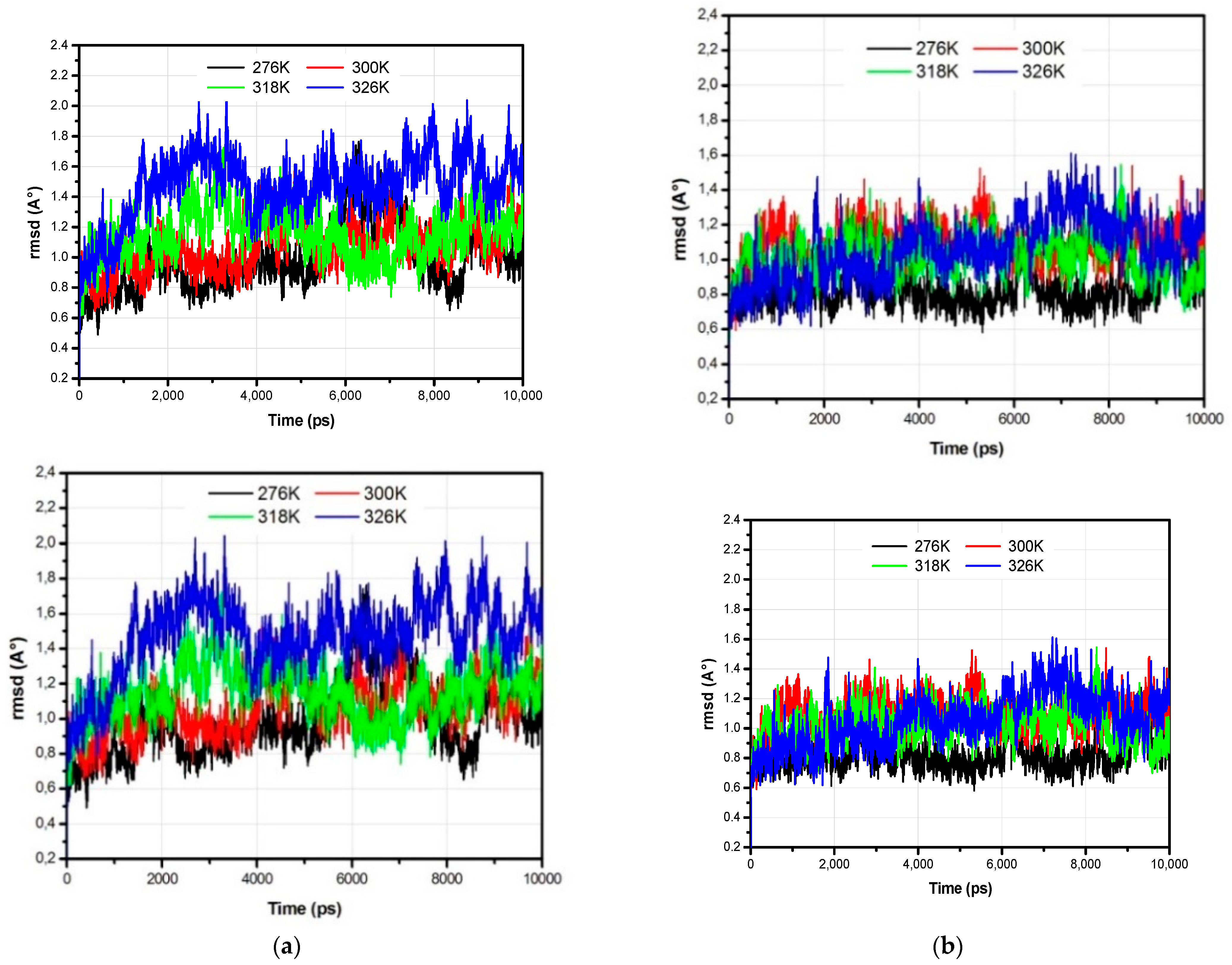
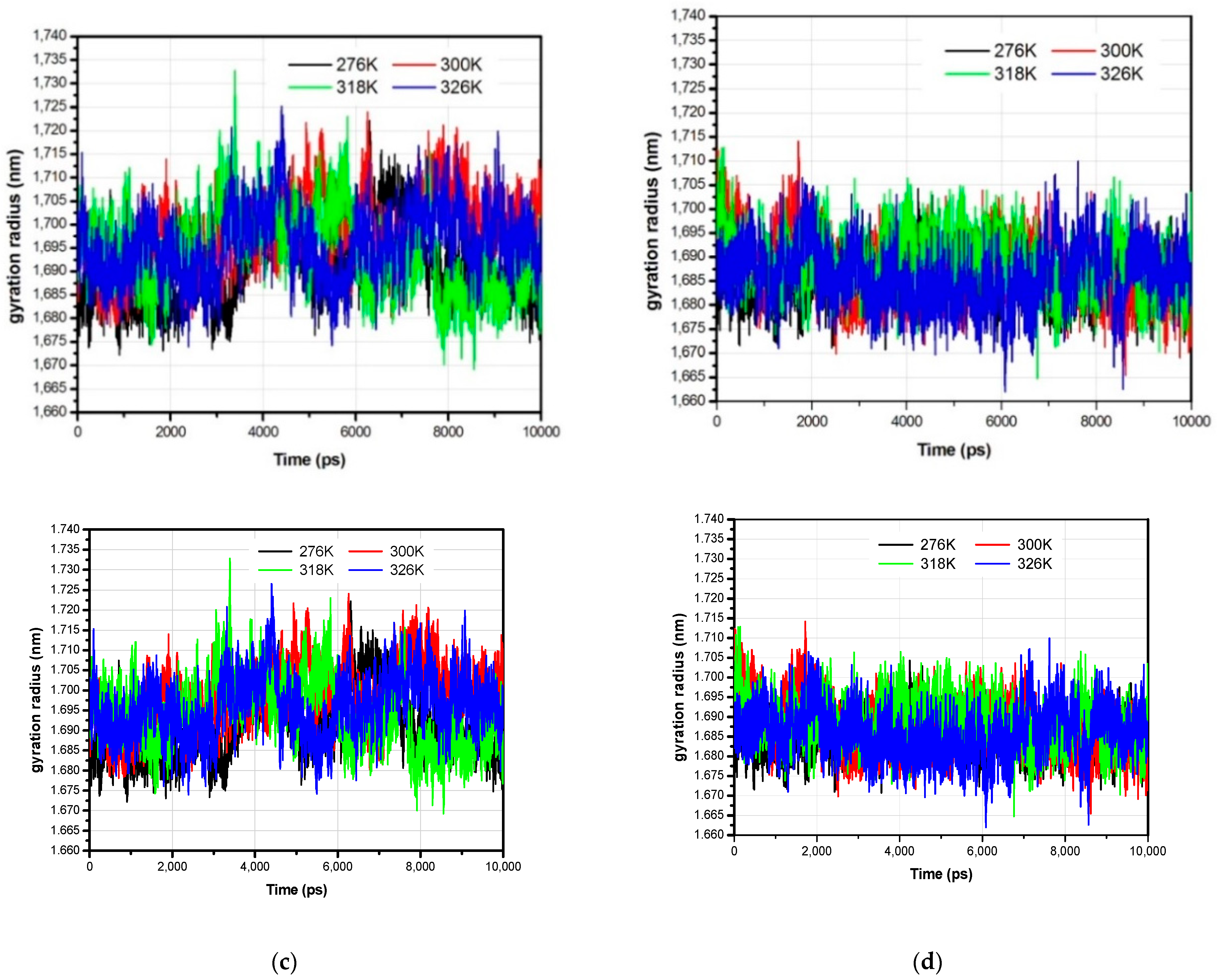
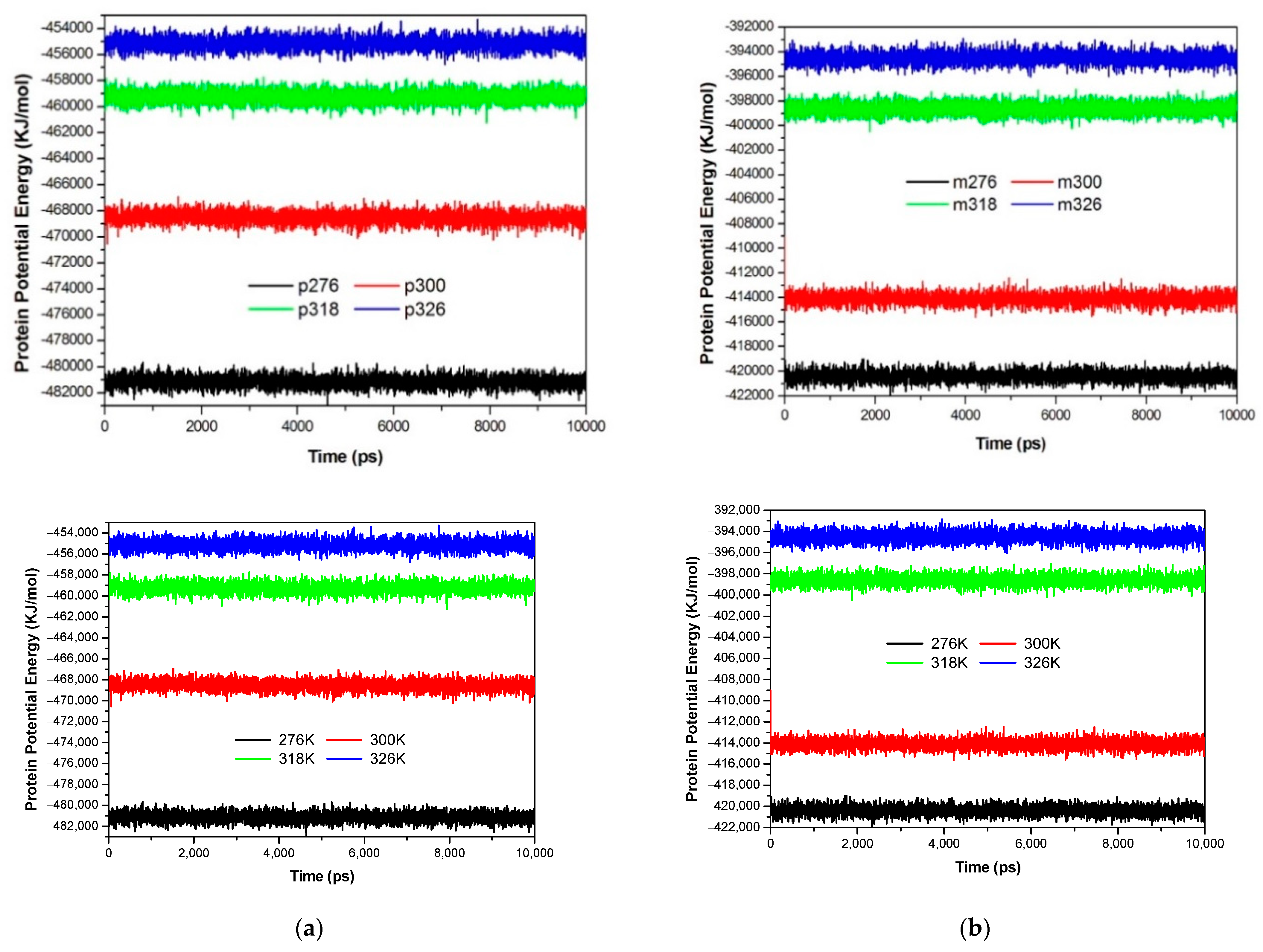
3.1. The Global Structural Stability

The root mean square deviation (RMSD) calculated for the backbone of the psychrophile enzyme VsEndA and its homologous counterpart, the mesophile enzyme VcEndA, are reported in Figure 1a,b. In general, we notice that the values of the RMSD for both proteins do not exceed 2.5 Å, reflecting rather stable trajectories. The increase in the temperature does not really influence the RMSD of the mesophile enzyme (VcEndA) as shown in Figure 1b, especially for temperatures ranging from 300 K to 326 K, with rather close average values of the RMSD of 1.09 Å, 1.02 Å and 1.06 Å at temperatures of 300 K, 318 K and 326 K, respectively. However, the lowest curve of the RMSD recorded corresponds to the temperature of 276 K, fluctuating around an average value of 0.82 Å. The RMSD of the VcEndA enzyme reaches the equilibrium more quickly than that of the VsEndA enzyme, occurring at times reaching 4 ns.
The VsEndA enzyme presents values of the RMSD that are more important than those of its homologous counterpart, the VcEndA, particularly at temperatures of 276 K, 318 K and 326 K, and where the corresponding average values of 0.99 Å, 1.15 Å and 1.47 Å were observed. However, for the temperature of 300 K, the average value of the RMSD for the VsEndA is lower than that observed for the VcEndA (1.06 Å). In comparison with the mesophile enzyme, the RMSD plots of the psychrophile enzyme are more distinct to each other, and the RMSD increases with the temperature to reach a maximal average value of 1.47 Å for the calculated trajectory at 326 K. An additional parameter reflecting the structural changes is the gyration radius (Rg) of the protein, a measure of the compactness of the structure of the protein (Figure 1c,d) illustrating the gyration radius along the trajectory of both enzymes VsEndA and VcEndA, respectively.
The potential energy of the system as the sum of bond, angle, torsion, and non-bonded terms can also be used as a simple measure of system stability. Thus, we present the evolution of the potential energy of both enzymes during the molecular dynamics simulations, for the different values of temperature (Figure 2). The potential energy plots show that all the molecular systems were well equilibrated and remained stable through the 10 ns of molecular dynamics simulations. For both enzymes, we observe the same trend concerning the temperature; the potential energy increases with temperature.
As the temperature of the system is increased, the internal energy of the molecule will increase too, which is consistent with the structural flexibility. The internal energy may include the translational energy, vibrational and rotational energy, as well as the energy involved in chemical bonding and that involved in non-bonding interactions. So with the increase in temperature, the system has to overcome more energy barriers to stabilize; thus, the potential energy values for both enzymes are higher than at low temperatures. Finally, the potential energies of the psychrophile enzyme and the mesophile enzyme stabilize to different values for the same temperature due to different topological terms.

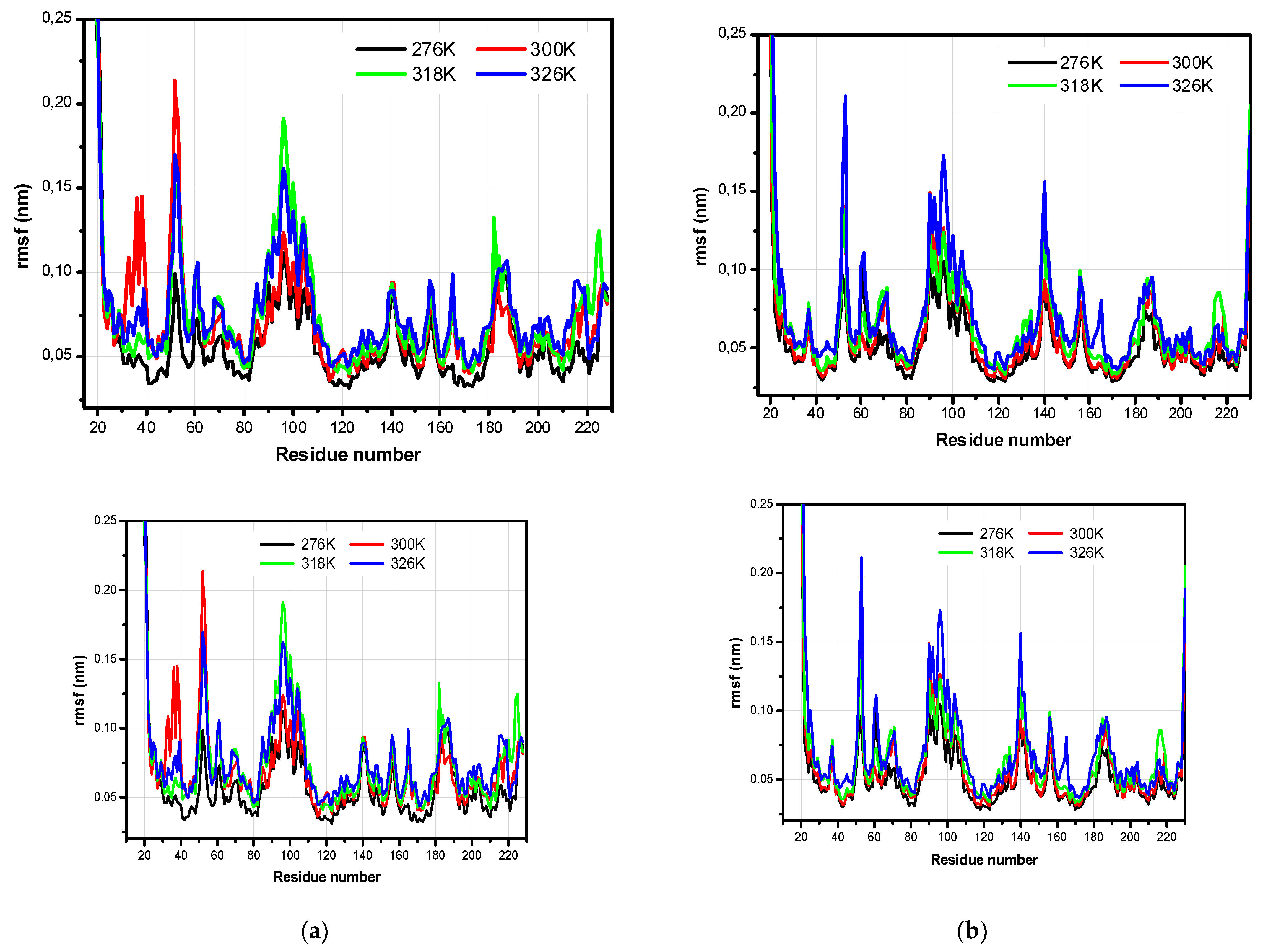
3.2. The Structural Flexibility

A more detailed picture of the difference in the mobility of residues, within a simulation and between two homologous enzymes, can be obtained from the RMSF (root mean square fluctuation) plot along the trajectory. The RMSF wascalculated from Cα atoms of the average structure of the simulation and for different temperatures.
Figure 3 showsan illustration of the RMSF of Cα atoms within a residue of both enzymes, the psychrophile (VsEndA) and the mesophile (VcEndA). The protein regions characterized by large values of the RMSF are in most cases situated in corresponding positions in VsEndA and VcEndA, although the intensity of the fluctuations is often different. The analysis of the RMSF values clearly shows the important structural flexibility of the loop region (residues 51–53), with regard to the remaining structure of both proteins (Figure 3a,b). When we compare the maximal value of the structural flexibility of this loop region between the psychrophile and mesophile enzymes, we find almost similar values of the RMSF: 0.2133 nm recorded for the residue ASN52 at the temperature T = 300 K for VsEndA (Figure 3a), and 0.2112 nm recorded for the residue LYS53 at the temperature T = 326 K for its homologous counterpart, VcEndA (Figure 3b).This region of the protein is of crucial importance as it is close to the binding site of DNA in the structure of Vvn(Vibrio vulnificus nuclease) [17] and thus, can potentially have an effect upon the catalytic activity of both enzymes.
Another important region (residues 90–96) presents a very high RMSF for both homologousenzymes. Belonging to the α5 motif in the secondary structure of both proteins (Figure 3, Figure 4, Figure 5 and Figure 6), and located near the active site ARG99, the LYS96 has a value of the RMSF equal to 0.191 nm at T = 318 K (Figure 3a), this residue is replaced by the polar residue GLN96 in the structure of the VcEndA, with the highest value of the RMSF (0.1729 nm) registered at T = 326 K, for this residue (Figure 3b).
Two more regions of the VsEndA enzyme (residues 182–184 and 224–225) wereobserved with a high flexibility at T = 318 K. Located in a loop region, between two motifsα (α8 and α9), the LYS182 reaches the maximal value of the RMSF equal to 0.1324 nm. Furthermore, the RMSF of the polar residue TYR225 located in the motif α11, reaches the value of 0.1248 nm atthe same temperature T = 318 K (Figure 3a).The loop region of the VsEndA (36–38) is also characterized by an important flexibility recorded at T = 300 K, with the highest value of the RMSF observed for the polar residue TYR38 (0.1451 nm). On the other hand, this same loop region of the mesophile VcEndA enzyme, is rather stable under the influence of the change in temperature and does not show a different structural mobility.
The RMSF profile of the VcEndA enzyme is also marked by an increase inthe flexibility of the hydrophobic residue GLY140 at the temperature T = 326 K (0.1564 nm). Located in the loop region between two ß sheets (ß5 and ß6), this residue is preserved in the primary structure of the VsEndA but has no influence on the flexibility of the psychrophile enzyme.
At the opposite of the mesophile enzyme where the structural flexibility is dominated by a single temperature (with the highest studied at T = 326 K), the psychrophile enzyme shows a different behavior with respect to the temperature effect. The dominant temperatures on the structural flexibility of this enzyme are lower than its mesophile homologous counterpart; in particular, at T = 300 K and T = 318 K. Furthermore, itaffects two main regions: ASN52 and LYS96, with greater RMSF intensities than its mesophile homologous counterpart.
Comparable structural flexibilities were observed between the VsEndA and VcEndA enzymes for the loop N-terminal region, for all temperatures studied here (Figure 3). However, the C-terminal region of the VcEndA enzyme (Figure 3b) shows a more pronounced flexibility than its homologous counterpart, the VsEndA (Figure 3a), for the simple reason that the primary structure of the latter ends with the CYS228 residue (Figure 4) involved in a disulfure bridge with the CYS207 residue, which impedes its flexibility, whereas the primary structure of the VcEndA ends with the ASN230 residue (Figure 4), which is free at the end of the polypeptide chain of the enzyme. The superposition of the mean structures from the trajectories of molecular dynamics simulation, for the two enzymes at each considered temperature reveal the same more flexible regions than the rmsf profile (Figure 7).

3.3. Hydrogen Bond Lifetime

3.3.1. The Psychrophile Enzyme VsEndA

Intermolecular and intramolecular hydrogen bond lifetimes, calculated for the most flexible residues of the psychrophile enzyme VsEndA, TYR38, ASN52, LYS96, LYS182 and TYR225, according to temperature, are included in Table 1.
The most flexible residues (TYR38 and ASN52) of the VsEndA enzyme at T = 300 K (Figure 3a), compared to the other temperatures, present the lowest values of the hydrogen bond lifetimes between the surrounding water molecules and the backbone part (BB) of the residues in question, atthe same temperature (4.664 ps for the TYR38 and 2.636 ps for the ASN52 (Table 1)). This is in good agreement with the flexibility profiles of the enzyme (Figure 3a).
The smallest values of the hydrogen bond lifetimes formed with the backbone part (BB) of the LYS96 and the water molecules are observed for the highest temperatures, 318 K and 326 K, with 1.046 ps and 0.918 ps, respectively. This result of the lifetime values joins perfectly with the RMSF profile according to the temperatures (Figure 3a), where the RMSF values are the highest at 0.191 nm and 0.161 nm for the temperature 318 K and 326 K, respectively. Although the difference between the two values of the hydrogen bonding lifetimes at T = 318 and T = 326 K is relatively small, the value recorded at T = 326 K, 0.918 ps is due to the diffusion of the water molecules surrounding the backbone of the lysine, which increases with the temperature.
The role of the dynamics of the solvent in the structural relaxation of globular proteins was already studied at the microscopic level. The molecular dynamics results of the globular protein Ribonuclease A (RNase), establish firmly a correlation between the mobility of water molecules on the surface of the protein and the diffusive movements of the latter [37]. The complete structural relaxation requires a relaxation of the network of the hydrogen bonding through the translational movement of water molecules; this is as important with the rise of the temperature, where we noted shorter hydrogen bonding lifetimes.
A similar explanation is given with regard to the side chain (SC) of LYS96 which forms shorter hydrogen bonds with water at T = 326 K. The hydrogen bonding lifetimes between the protein and the side chain of LYS96 are relatively low at318 K when comparing to the other temperatures;thisled us to believe that, as well as the intermolecular hydrogen bonds formed with water molecules, the intramolecular hydrogen bonds can also influence the dynamics of the protein.
Both residues, LYS182 and TYR225 are characterized by shorter intermolecular and intramolecular hydrogen bond lifetimes at T = 326 K, compared with the other temperatures. However, we can clearly see that lifetimes of hydrogen bonding recorded at T = 318 K between the side chain of LYS182 and water (1.743 ps) and between the backbone of TYR225 and water (3.549 ps) are relatively close to those recorded at T = 326 K, which supports the conclusion that the hydrogen bond lifetimes calculated for the most flexible regions of the VsEndA enzyme are in correlation with the RMSF profile of the same enzyme.

3.3.2. The Mesophile Enzyme VcEndA

In Table 2 we report the intramolecular and intermolecular hydrogen bonding lifetimes formed with the most flexible residues of the mesophile enzyme VcEndA: LYS53, GLN96 and GLY140, for the various values of the temperature.We can clearly see, according to Table 2, that the hydrogen bonding lifetimes formed between the considered residues and water molecules are the shortest at T = 326 K, compared with the other values of the temperature. Such results areattributed to the important diffusion of water molecules onto the surface of the mesophile enzyme at the highest temperature studied (326 K).

4. Conclusions

Temperature is one of the most important factors that govern the enzyme dynamics and activity. Therefore, it is necessary to understand the molecular mechanisms of the adaptation to the temperature of the contemporary enzymes.
In a first step toward such an objective, we undertook, in this paper, the effect of temperature on the pair of homologous enzymes: the cold adapted enzyme, the Vibrio SalmonicidaVsEndA and the mesophile enzyme Vibrio cholera VcEndA. The molecular dynamics simulations with various temperatures are carried out withthe optimal conditions in salt (NaCl) for every enzyme, such as the experimental study already made on the pair of enzymes [17]. The structural analysis, in terms of RMSD, gyration radius and secondary structure of these two enzymes reveals stable trajectories at the considered temperatures, 276 K, 300 K, 318 K and 326 K. The RMSFprofiles of both enzymes are similar with the most flexible residues of both homologousenzymesappearing at the loop regions and are identical in the primary structure of both enzymes (residues 52–53 and 96). However, the temperature effect is not the same for these considered regions. The cold adapted enzyme presents a structural flexibility more important at low temperatures (300 K and 318 K), compared with the mesophile enzyme, for which the temperature which presents major flexibility is the highest studied (326 K). The lifetimes of hydrogen bonds of almost all of the most flexible residues of both enzymes correlate well with the RMSF of the considered enzyme.

Institutional Review Board Statement

Not applicable.

Informed Consent Statement

Not applicable.

Data Availability Statement

All data are available in the manuscript.

Acknowledgments

The Computations were performed on the Al-Farabi Cluster computer of the Ecole Nationale Polytechnique Oran; Maurice Audin.

References

  1. D’Amico, S.; Collins, T.; Marx, J.C.; Feller, G.; Gerday, C. Psychrophilic microorganisms: Challenges for life. EMBO Rep. 2006, 7, 385–389. [Google Scholar] [CrossRef]
  2. Muga, A.; Moro, F. Thermal adaptation of heat shock proteins. Curr. Protein Pept. Sci. 2008, 9, 552–566. [Google Scholar] [CrossRef]
  3. Somero, G.N. Adaptation of enzymes to temperature: Searching for basic strategies. Comp. Biochem. Physiol. B 2004, 139, 321–333. [Google Scholar] [CrossRef]
  4. Trivedi, S.; Gehlot, H.S.; Rao, S.R. Protein thermostability in Archaea and Eubacteria. Genet. Mol. Res. 2006, 5, 816–827. [Google Scholar]
  5. Daniel, R.M.; Danson, M.J.; Eisenthal, R.; Lee, C.K.; Peterson, M.E. The effect of temperature on enzyme activity: New insights and their implications. Extremophiles 2008, 12, 51–59. [Google Scholar] [CrossRef]
  6. Schiraldi, C.; De Rosa, M. The production of biocatalysts and biomolecules from extremophiles. Trends Biotechnol. 2002, 20, 515–521. [Google Scholar] [CrossRef]
  7. Chiuri, R.; Maiorano, G.; Rizzello, A.; del Mercato, L.L.; Cingolani, R.; Maffia, M.; Pompa, P.P. Exploring local flexibility/rigidity in psychrophilic and mesophilic carbonicanhydrases. Biophys. J. 2009, 96, 1586–1596. [Google Scholar] [CrossRef]
  8. Li, W.F.; Zhou, X.X.; Lu, P. Structural features of thermozymes. Biotechnol. Adv. 2005, 23, 271–281. [Google Scholar] [CrossRef]
  9. Vieille, C.; Zeikus, G.J. Hyperthermophilic enzymes: Sources, uses, and molecular mechanisms for thermostability. Microbiol. Mol. Biol. Rev. 2001, 65, 1–43. [Google Scholar] [CrossRef]
  10. Sterner, R.; Liebl, W. Thermophilic adaptation of proteins. Crit. Rev. Biochem. Mol. Biol. 2001, 36, 39–106. [Google Scholar] [CrossRef]
  11. Del Vecchio, P.; Elias, M.; Merone, L.; Graziano, G.; Dupuy, J.; Mandrich, L.; Manco, G. Structural determinants of the high thermal stability of SsoPox from the hyperthermophilic archaeon Sulfolobus Solfataricus. Extremophiles 2009, 13, 461–470. [Google Scholar] [CrossRef]
  12. Feller, G. Protein stability and enzyme activity at extreme biological temperatures. J. Phys. Condens. Matter 2010, 22, 323101. [Google Scholar] [CrossRef]
  13. Feller, G.; Gerday, C. Psychrophilic enzymes: Hot topics in cold adaptation. Nat. Rev. Microbiol. 2003, 1, 200–208. [Google Scholar] [CrossRef]
  14. Liang, Z.X.; Tsigos, I.; Lee, T.; Bouriotis, V.; Resing, K.A.; Ahn, N.G.; Klinman, J.P. Evidence for increased local flexibility in psychrophilic alcohol dehydrogenase relative to its thermophilic homologue. Biochemistry 2004, 43, 14676–14683. [Google Scholar] [CrossRef]
  15. Georlette, D.; Damien, B.; Blaise, V.; Depiereux, E.; Uversky, V.N.; Gerday, C.; Feller, G. Structural and functional adaptations to extreme temperatures inpsychrophilic, mesophilic, and thermophilic DNA ligases. J. Biol. Chem. 2003, 278, 37015–37023. [Google Scholar] [CrossRef]
  16. Siddiqui, K.S.; Cavicchioli, R. Cold-adapted enzymes. Annu. Rev. Biochem. 2006, 75, 403–433. [Google Scholar] [CrossRef]
  17. Altermark, B.; Niiranen, L.; Willassen, N.P.; Smalas, A.O.; Moe, E. Comparative studies of endonuclease I from cold-adapted Vibrio salmonicida and mesophilic Vibrio cholerae. FEBS J. 2007, 274, 252–263. [Google Scholar] [CrossRef] [PubMed]
  18. Papaleo, E.; Riccardi, L.; Villa, C.; Fantucci, P.; De Gioia, L. Flexibility and enzymatic cold adaptation: A comparative molecular dynamics investigation of the elastase family. Biochim. Biophys. Acta 2006, 1764, 1397–1406. [Google Scholar] [CrossRef]
  19. Spiwok, V.; Lipovova, P.; Skalova, T.; Duskova, J.; Dohnalek, J.; Hasek, J.; Kralova, B. Cold-active enzymes studied by comparative molecular dynamics simulation. J. Mol. Model. 2007, 13, 485–497. [Google Scholar] [CrossRef]
  20. Kundu, S.; Roy, D. Comparative structural studies of psychrophilic and mesophilic proteinhomologues by molecular dynamics simulation. J. Mol. Graph. Model. 2009, 27, 871–880. [Google Scholar] [CrossRef]
  21. Michetti, D.; Brandsdal, B.O.; Bon, D.; Isaksen, G.V.; Tiberti, M.; Papaleo, E. A comparative study of cold- and warm-adapted Endonucleases A using sequence analyses and molecular dynamics simulations. PLoS ONE 2017, 12, e0169586. [Google Scholar] [CrossRef]
  22. Benrezkallah, D.; Dauchez, M.; Krallafa, A.M. Molecular dynamics of the salt dependence of a cold-adapted enzyme: Endonuclease I. J. Biomol. Struct. Dyn. 2015, 33, 2511–2521. [Google Scholar] [CrossRef]
  23. MacKerell, A.D., Jr.; Bashford, D.; Bellott, M.; Dunbrack, R.L.; Evanseck, J.D.; Field, M.J.; Karplus, M. All-Atom Empirical Potential for Molecular Modeling and Dynamics Studies of Proteins. J. Phys. Chem. B 1998, 102, 3586–3616. [Google Scholar] [CrossRef]
  24. MacKerell, J.A.D.; Feig, M.; Brooks, C.L., III. Extending the treatment of backbone energetics in protein force fields: Limitations of gas-phase quantum mechanics in reproducing protein conformational distributions in molecular dynamics simulations. J. Comput. Chem. 2004, 25, 1400–1415. [Google Scholar] [CrossRef]
  25. Hess, B.; Kutzner, C.; Van Der Spoel, D.; Lindahl, E. GROMACS 4: Algorithms for highly efficient, load-balanced, and scalable molecular simulation. J. Chem. Theory Comput. 2008, 4, 435–447. [Google Scholar] [CrossRef]
  26. Van Der Spoel, D.; Lindahl, E.; Hess, B.; Groenhof, G.; Mark, A.E.; Berendsen, H.J.C. GROMACS: Fast, flexible, and free. J. Comput. Chem. 2005, 26, 1701–1718. [Google Scholar] [CrossRef]
  27. Šali, A.; Blundel, T.L. Comparative protein modelling by satisfaction of spatial restraints. J. Mol. Biol. 1993, 234, 779–815. [Google Scholar] [CrossRef]
  28. Jorgensen, W.L.; Chandrasekhar, J.; Madura, J.D.; Impey, R.W.; Klein, M.L. Comparison of simple potential functions for simulating liquid water. J. Chem. Phys. 1983, 79, 926–935. [Google Scholar] [CrossRef]
  29. Berendsen, H.J.C.; Postma, J.P.M.; DiNola, A.; Haak, J.R. Molecular dynamics with coupling to an external bath. J. Chem. Phys. 1984, 81, 3684–3690. [Google Scholar] [CrossRef]
  30. Hess, B.; Bekker, H.; Berendsen, H.J.C.; Fraaije, J.G.E.M. LINCS: A Linear Constraint Solver for Molecular Simulations. J. Comput. Chem. 1997, 18, 1463–1472. [Google Scholar] [CrossRef]
  31. Darden, T.; York, D.; Pedersen, L. Particle mesh Ewald: An N log (N) method for Ewald sums in large systems. J. Chem. Phys. 1993, 98, 10089–10092. [Google Scholar] [CrossRef]
  32. Essmann, U.; Perera, L.; Berkowitz, M.L.; Darden, T.; Lee, H.; Pedersen, L.G. A smooth particle mesh ewald potential. J. Chem. Phys. 1995, 103, 8577–8592. [Google Scholar] [CrossRef]
  33. Luzar, A.; Chandler, D. Hydrogen-bond kinetics in liquid water. Nature 1996, 397, 55–57. [Google Scholar] [CrossRef]
  34. Luzar, A. Resolving the hydrogen bond dynamics conundrum. J. Chem. Phys. 2000, 113, 10663–10675. [Google Scholar] [CrossRef]
  35. Robert, X.; Gouet, P. Deciphering key features in protein structures with the new ENDscript server. Nucleic Acids Res. 2014, 42, 320–324. [Google Scholar] [CrossRef]
  36. Humphrey, W.; Dalke, A.; Schulten, K. VMD: Visual molecular dynamics. J. Mol. Graph. 1996, 14, 33–38. [Google Scholar] [CrossRef]
  37. Tarek, M.; Tobias, D.J. Role of Protein-Water Hydrogen Bond Dynamics in the Protein Dynamical Transition. Phys. Rev. Lett. 2002, 88, 138101. [Google Scholar] [CrossRef]
Figure 1. Root meansquare deviation (a,b), and the gyration radius as a function of temperature (c,d) of the endonuclease I psychrophile marine (VsEndA) (at left) and endonuclease I mesophile estuarine (VcEndA) (at right).
Chemproc 03 00128 g001aChemproc 03 00128 g001b
Figure 2. Potentialenergy of VsEndA (a), and VcEndA (b).
Chemproc 03 00128 g002
Figure 3. The root meansquare fluctuation (RMSF) as a function of temperature, (a) VsEndA and (b) VcEndA.
Chemproc 03 00128 g003
Figure 4. Structure alignment of VsEndA and VcEndA enzymes, using the ESPript 3.0 program [35]. The numbers indicate cysteines involved in disulfure bridges.
Chemproc 03 00128 g004
Figure 5. Secondary structure of VsEndA as a function of temperature; (a) 276 K, (b) 300 K, (c) 318 K, (d) 326 K.
Chemproc 03 00128 g005
Figure 6. Secondary structure of VcEndA as a function of temperature; (a) T = 276 K, (b) T = 300 K, (c) T = 318 K and (d) T = 326 K.
Chemproc 03 00128 g006
Figure 7. Superposition of the mean structures of the VsEndA (a), and the VcEndA (b), at different temperatures; black, red, green, blue, corresponding to T = 276 K, T = 300 K, T = 318 K and T = 326 K respectively. The structure alignments were made with VMD 1.9.2 [36]. Residues in CPK representation illustrate the most flexible parts of the enzymes.
Chemproc 03 00128 g007
Table 1. Intermolecular and intramolecular hydrogen bond lifetimes (ps) for VsEndA enzyme as a function of temperature.
Temperature276 K300 K318 K326 K
Tyr38_BB_Wat24.5044.664−66.2915.167
Tyr38_SC_Wat5.1364.4802.3122.503
Tyr38_BB_P87.630357.496382.37642.699
Tyr38_SC_Pnone38.040nonenone
Asn52_BB_Wat3.9382.6368.4532.644
Asn52_SC_Wat2.5681.8321.3000.989
Asn52_BB_Pnone97.19nonenone
Asn52_SC_Pnone130.6619.132−93.1
Lys96_BB_Wat1.6751.4881.0460.918
Lys96_SC_Wat5.8493.0922.0691.665
Lys96_BB_P50.97959.33790.84919.532
Lys96_SC_P31.26219.12617.47721.534
Lys182_BB_Wat13.875121.496−16.2772.157
Lys182_SC_Wat4.3172.9031.7431.677
Lys182_BB_P199.917226.47279.35437.415
Lys182_SC_P−11.89415.507797.64676.648
Tyr225_BB_Wat20.7377.0273.5493.436
Tyr225_SC_Wat2.5901.8511.5421.368
Tyr225_BB_P854.846401.057775.720159.523
Tyr225_SC_P198.984103.911510.5682.001
Table 2. Intermolecular and intramolecular hydrogen bonds lifetimes (ps) for VcEndA enzyme as a function of temperature.
Temperature276 K300 K318 K326 K
Lys53_BB_Wat7.78616.2417.6915.408
Lys53_SC_Wat4.3623.4711.9061.878
Lys53_BB_Pnone−2748.66537.42825.194
Lys53_SC_P79.268101.08780.11796.125
Gln96_BB_Wat2.0431.6370.9360.937
Gln96_SC_Wat1.8461.2930.8210.559
Gln96_BB_P144.256504.24730.88096.679
Gln96_SC_P143.45771.14737.07222.270
Gly140_Wat8.6309.9203.7711.400
Gly140_PnoneNone−93.4160.001
Publisher’s Note: MDPI stays neutral with regard to jurisdictional claims in published maps and institutional affiliations.

