Proceeding Paper

pMyc and pMax Peptides Nanosystems and the Potential Treatment of Prostate Cancer, In Vitro Assays †

by
Samuel Longoria-García
1,
Celia N. Sánchez-Domínguez
1,
Margarita Sánchez-Domínguez
2,
Jesús R. Delgado-Balderas
3 and
Hugo L. Gallardo-Blanco
4,*
1
Departamento de Bioquímica y Medicina Molecular, Facultad de Medicina, Universidad Autónoma de Nuevo León, Av. Dr. José Eleuterio González 235, Mitras Centro, Monterrey 64460, Mexico
2
Grupo de Química Coloidal e Interfacial Aplicada a Nanociencia y Formulaciones, Centro de Investigación en Materiales Avanzados, Unidad Monterrey, Alianza Norte 202, Apodaca 66000, Mexico
3
Facultad de Ciencias Químicas, Universidad Autónoma de Nuevo León, Avenida Universidad s/n, Cd. Universitaria, San Nicolás de los Garza 66455, Mexico
4
Departamento de Genética, Hospital Universitario “José Eleuterio González”, Universidad Autónoma de Nuevo León, Av. Dr. José Eleuterio González 235, Mitras Centro, Monterrey 64460, Mexico
*
Author to whom correspondence should be addressed.
Presented at the 4th International Online Conference on Nanomaterials, 5–19 May 2023; Available online: https://iocn2023.sciforum.net.
Published: 5 May 2023
(This article belongs to the Proceedings of The 4th International Online Conference on Nanomaterials)

Abstract

:
The Myc transcription factor and its associated Max protein have essential roles in the development of several types of cancers, including prostate cancer. They dimerize into a Myc–Max heterodimer and bind to DNA sequences known as enhancer boxes (E-box). Therefore, disrupting the binding of these E-boxes to derange transcription is a promising strategy for treating cancer. Using computational biology tools, we designed pMyc and pMax peptides from Myc and Max reference sequences and evaluated their ability to bind to E-boxes through an electrophoretic mobility shift assay (EMSA). We then coupled them to AuNPs and evaluated their hemocompatibility and cytotoxic effects in three different prostate adenocarcinoma cell lines and a non-cancerous cell line The EMSA results suggested that the pMyc–pMax dimers bound to CMEs. The hemolysis test showed little hemolytic activity for the nanosystems (NS) at the three concentrations evaluated. The cell viability assays showed mixed results, depending on which cell line was being evaluated. Overall, the results suggest that NS with pMyc and pMax peptides might be suitable for further research regarding Myc-driven prostate adenocarcinomas.

1. Introduction

The Myc transcription factor is the protein encoded by the oncogene with the same name. It regulates several essential cell processes that can lead to a cancerous phenotype if the Myc protein is unregulated. In its sequence, Myc contains a basic helix-loop-helix domain and a leucine zipper domain, allowing for many dimerization interactions with different proteins and resulting in different gene expression patterns [1].
The Myc-associated factor X, also known as Max, is Myc’s primary binding partner; the Myc:Max dimer is responsible for the transcription initiation of its target genes. The Myc:Max dimer binds to a DNA sequence known as the enhancer box (E-box); this sequence is located in the promoters of the target genes and has the following sequence: CACGTG (CME). The dimer’s affinity is higher for this sequence than other degenerated sequences known as non-canonical E-boxes [2].
The MYC oncogene is overexpressed in prostate cancer (PCa), which has been determined to be the second most common cancer in men worldwide [3,4]. With this in mind, is has been suggested that disrupting the Myc:Max dimer at the oncogenic level, or its binding into E-boxes, could be a promising strategy for treating cancer [5]. Designing peptides through computational biology tools to target Myc could lead to new therapeutic approaches for PCa and other Myc-related cancer types. However, the primary limitation of this approach is the difficulty in delivering these peptides into the cell nuclei. Several strategies can be used to achieve this, and one of them is nanotechnology. This work aimed at constructing pMyc:pMax:AuNPs nanosystems (NS) using Myc- and Max-derived peptides and gold nanoparticles, and at evaluating their effect in PCa cell lines.

2. Materials and Methods

2.1. Peptide Design and Synthesis

We used the protein reference sequences for Myc (NP_002458.2) and Max (NP_660087.1) obtained from Genbank available at the NCBI; we also used the Swiss-Model for protein homology modeling [6], Cn3D for visualizing the macromolecular structure [7], and ScooP for predicting the thermal stability [8]. The pMyc and pMax peptides were chemically synthesized using Accura’s Custom Peptide Synthesis service (Accura, Monterrey, Mexico).

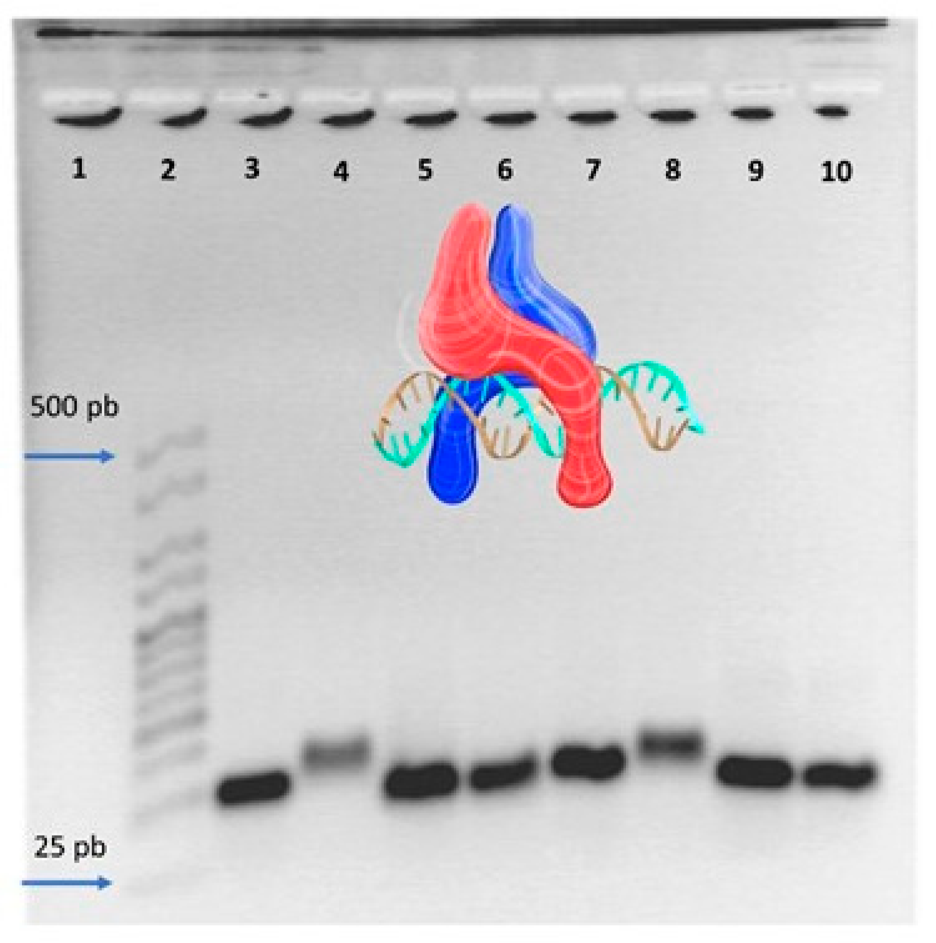
2.2. Electrophoretic Mobility Shift Assay

To avoid undesired oligonucleotides mismatches in the electrophoretic mobility shift assay (EMSA), we designed ssDNA oligonucleotides with similar melting point temperatures using the OligoAnalyzer tool Version. 3.1, Integrated DNA Technologies (Coralville, IA, USA). These oligonucleotides were synthesized using Integrated DNA Technologies’ Custom DNA oligos service (Integrated DNA Technologies, Coralville, IA, USA) (Table 1).

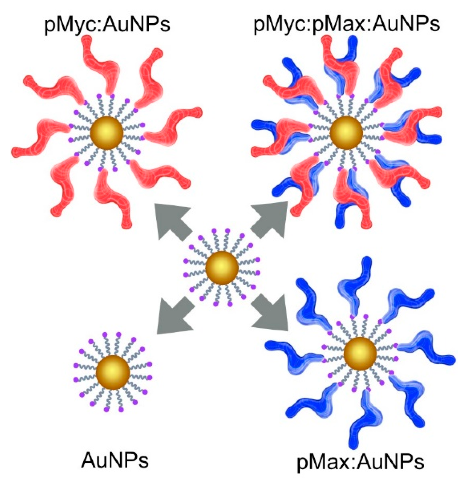
2.3. Nanosystem Construction
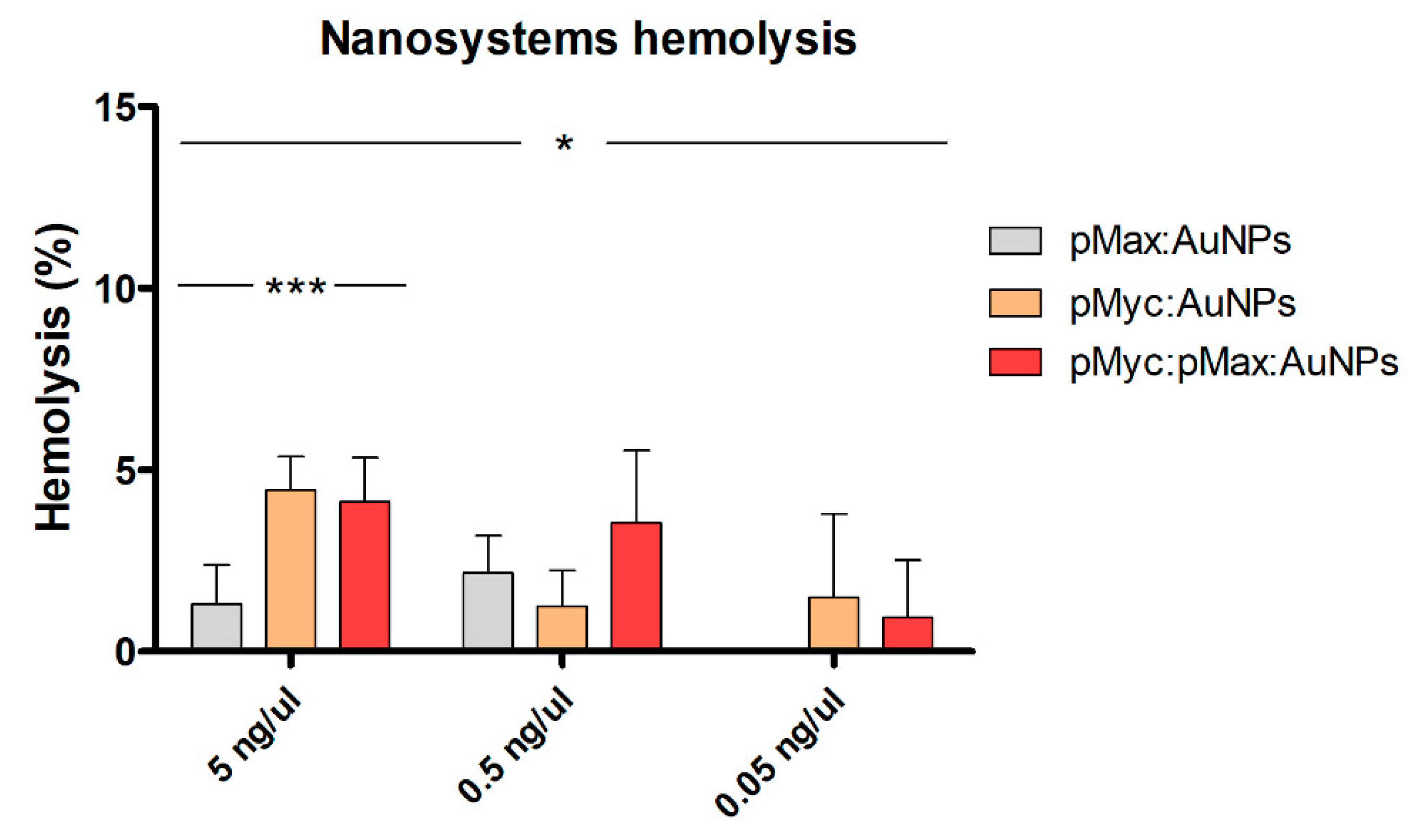
Three different NSs were constructed, as depicted in Figure 1: pMyc:AuNPs with only the pMyc peptide, pMax:AuNPs with only the pMax peptide, and pMyc:pMax:AuNPs with both peptides. We used Maleimide 5 nm Gold Nanoparticle Conjugation kits (cat. No 900458-1EA Cytodiagnostics, Burlington, ON, Canada) for constructing the NSs. All the AuNPs had polyethylene glycol (PEG) and maleimide for peptide conjugation.

2.4. Hemolysis Test

We extracted 2 mL of blood from a volunteer; this sample was centrifuged and the pellet was washed to obtain an erythrocyte solution. Finally, we read the absorbance spectra with an ND-1000 Nanodrop at 415 nm to obtain the hemolysis percentage.

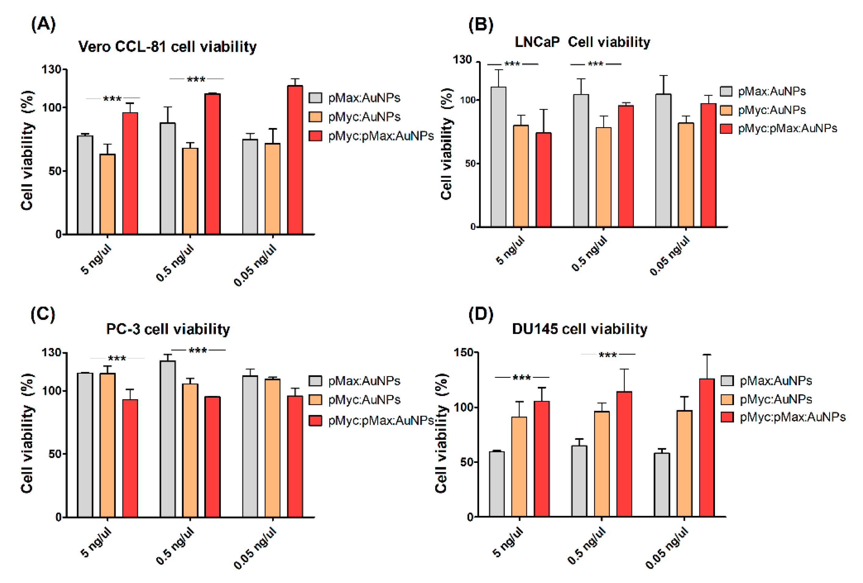
2.5. Cell Viability Assays

All the cells were obtained from the American Type Culture Collection (ATCC, Manassas, VA, USA). PC-3 and Vero CCL-81 cells were cultured in DMEM (Thermofisher, Waltham, MA, USA, Cat. No. 11965092), and LNCaP and DU145 were cultured in RPMI 1640 (Gibco, Miami, FL, USA, Cat. No. 72400047). The cell viability was determined following the Cell Proliferation Kit I (MTT) (Roche, Branchburg, NJ, USA, Cat. No. 11465007001) protocol. The formazan crystals were solubilized with isopropanol (pH = 3) and read in a BioTek Cytation3 Imaging reader at 570 nm and 651 nm.

2.6. Statistical Analyzes

A two-way ANOVA was used along with the Bonferroni correction for this work’s statistical analysis. GraphPad Prism 5 was used for the statistical analysis and figures.

3. Results and Discussion

The Cn3D rendering showed that the structure predicted for the pMyc:pMax heterodimer conserved the recognition and binding site to the CACGTG canonical E-box sequence. Furthermore, the ScooP modeling showed a high thermal stability in the heterodimer of up to 76.6 °C. The ScooP results can be seen in Table 2.
Figure 2 shows a shift on the electrophoretic mobility when the designed oligonucleotides containing the CACGTG sequence were incubated with the pMyc:pMax heterodimer (lanes 4 and 8). These results suggest the binding of the peptides to the E-box. On the other hand, this shift was not observed with the oligonucleotides carrying the non-E-box element.
According to the ASTM-F756-17 standard practice for assessing hemolysis in materials, all the NSs were materials with no hemolysis at the lowest concentration (Figure 3). pMyc:AuNPs at 5 ng/μL have low hemolytic properties under the ASTM-F756-17 norm. The other peptide conjugates that have been reported also showed a hemolysis of <10% [9], whereas the other peptide-AuNPs NSs reported a <1% hemolysis [10].
Figure 4 shows the results of the cell viability assays. We obtained mixed results depending on the NS used and the cell line evaluated. In Vero CCL-81, pMyc:pMax.AuNPs showed little to no cytotoxic effect. In LNCaP, pMyc:AuNPs had the highest cytotoxic effect. With different pMyc:pMax disruption compounds, Carabet and colleagues obtained a reduced cell viability of <20% for the LNCaPs. Holmes and colleagues used a small-molecule Myc inhibitor (MYCi975) and obtained a cell viability reduction of 28.4%; however, their assays involved treatments in the order of micromolar concentrations [11,12]. These results show that different compounds can affect cell viability in different proportions. In PC-3 cells, little cytotoxicity can be seen; only pMyc:pMax:AuNPs had a cytotoxic effect in these cells. Finally, in DU145 cells, the most significant cytotoxic effect was shown by the pMax:AuNPs, whereas pMyc:AuNPs had a mild effect.

4. Conclusions

The pMyc and pMax peptides had a predicted thermal stability well over 37 °C and were shown to recognize and bind to canonical E-boxes in an EMSA assay. Their hemolytic properties allowed us to determine the concentrations for a cell viability assay evaluation. All the NSs that we tested were considered to have low hemolytic properties at the three different concentrations evaluated. The cell viability was affected at different levels, depending on the cell line evaluated. Our results suggest that pMyc:pMAx:AuNPs could potentially have a cytotoxic effect by binding to the E-boxes in cell nuclei, reducing the cell viability.

Author Contributions

Conceptualization, H.L.G.-B., C.N.S.-D. and S.L.-G.; methodology, S.L.-G.; validation, J.R.D.-B., S.L.-G. and H.L.G.-B.; formal analysis, H.L.G.-B., S.L.-G., C.N.S.-D. and M.S.-D.; investigation, S.L.-G. and M.S.-D.; resources, H.L.G.-B.; data curation, H.L.G.-B., C.N.S.-D. and M.S.-D.; writing—original draft preparation, S.L.-G.; writing—review and editing, S.L.-G., H.L.G.-B., C.N.S.-D., M.S.-D., J.R.D.-B.; visualization, S.L.-G. and H.L.G.-B.; supervision, H.L.G.-B. and C.N.S.-D.; project administration, C.N.S.-D. and H.L.G.-B.; funding acquisition, H.L.G.-B. All authors have read and agreed to the published version of the manuscript.

Funding

This study was funded by Consejo Nacional de Humanidades, Ciencia y Tecnología (CONAHCYT), Call for Basic Scientific Research 2017–2018 with the grant number A1-S-9859.

Institutional Review Board Statement

The study was conducted according to the guidelines of the Declaration of Helsinki and approved by the Committee of the School of Medicine of the Universidad Autónoma de Nuevo León (protocol code BI22-00002 with an approval date of 11 April 2022).

Informed Consent Statement

Patient consent was waived since the blood sample was obtained from a healthy donor after the donation consent form was signed.

Data Availability Statement

Not applicable.

Conflicts of Interest

The authors declare no conflict of interest.

References

  1. Conacci-Sorrell, M.; McFerrin, L.; Eisenman, R.N. An Overview of MYC and Its Interactome. Cold Spring Harb. Perspect. Med. 2014, 4, a014357. [Google Scholar] [CrossRef] [PubMed]
  2. Allevato, M.; Bolotin, E.; Grossman, M.; Mane-Padros, D.; Sladek, F.M.; Martinez, E. Sequence-Specific DNA Binding by MYC/MAX to Low-Affinity Non-E-Box Motifs. PLoS ONE 2017, 12, e0180147. [Google Scholar] [CrossRef] [PubMed]
  3. Barsouk, A.; Padala, S.A.; Vakiti, A.; Mohammed, A.; Saginala, K.; Thandra, K.C.; Rawla, P.; Barsouk, A. Epidemiology, Staging and Management of Prostate Cancer. Med. Sci. 2020, 8, 28. [Google Scholar] [CrossRef] [PubMed]
  4. Gandaglia, G.; Leni, R.; Bray, F.; Fleshner, N.; Freedland, S.J.; Kibel, A.; Stattin, P.; Van Poppel, H.; La Vecchia, C. Epidemiology and Prevention of Prostate Cancer. Eur. Urol. Oncol. 2021, 4, 877–892. [Google Scholar] [CrossRef] [PubMed]
  5. Chen, H.; Liu, H.; Qing, G. Targeting Oncogenic Myc as a Strategy for Cancer Treatment. Signal Transduct. Target. Ther. 2018, 3, 5. [Google Scholar] [CrossRef]
  6. Waterhouse, A.; Bertoni, M.; Bienert, S.; Studer, G.; Tauriello, G.; Gumienny, R.; Heer, F.T.; De Beer, T.A.P.; Rempfer, C.; Bordoli, L.; et al. SWISS-MODEL: Homology Modelling of Protein Structures and Complexes. Nucleic Acids Res. 2018, 46, W296–W303. [Google Scholar] [CrossRef]
  7. Wang, Y.; Geer, L.Y.; Chappey, C.; Kans, J.A.; Bryant, S.H. Cn3D: Sequence and Structure Views for Entrez. Trends Biochem. Sci. 2000, 25, 300–302. [Google Scholar] [CrossRef] [PubMed]
  8. Pucci, F.; Kwasigroch, J.M.; Rooman, M. SCooP: An Accurate and Fast Predictor of Protein Stability Curves as a Function of Temperature. Bioinformatics 2017, 33, 3415–3422. [Google Scholar] [CrossRef] [PubMed]
  9. Jabir, M.S.; Taha, A.A.; Sahib, U.I.; Taqi, Z.J.; Al-Shammari, A.M.; Salman, A.S. Novel of Nano Delivery System for Linalool Loaded on Gold Nanoparticles Conjugated with CALNN Peptide for Application in Drug Uptake and Induction of Cell Death on Breast Cancer Cell Line. Mater. Sci. Eng. C 2019, 94, 949–964. [Google Scholar] [CrossRef] [PubMed]
  10. Verimli, N.; Demiral, A.; Yılmaz, H.; Çulha, M.; Erdem, S.S. Design of Dense Brush Conformation Bearing Gold Nanoparticles as Theranostic Agent for Cancer. Appl. Biochem. Biotechnol. 2019, 189, 709–728. [Google Scholar] [CrossRef] [PubMed]
  11. Carabet, L.A.; Lallous, N.; Leblanc, E.; Ban, F.; Morin, H.; Lawn, S.; Ghaidi, F.; Lee, J.; Mills, I.G.; Gleave, M.E.; et al. Computer-Aided Drug Discovery of Myc-Max Inhibitors as Potential Therapeutics for Prostate Cancer. Eur. J. Med. Chem. 2018, 160, 108–119. [Google Scholar] [CrossRef] [PubMed]
  12. Holmes, A.G.; Parker, J.B.; Sagar, V.; Truica, M.I.; Soni, P.N.; Han, H.; Schiltz, G.E.; Abdulkadir, S.A.; Chakravarti, D. A MYC Inhibitor Selectively Alters the MYC and MAX Cistromes and Modulates the Epigenomic Landscape to Regulate Target Gene Expression. Sci. Adv. 2022, 8, eabh3635. [Google Scholar] [CrossRef] [PubMed]
Figure 1. Schematic depiction of the constructed nanosystems with either individual pMyc and pMax homodimers or pMyc:pMax heterodimer.
Materproc 14 00046 g001
Figure 2. Resolved EMSAs with pMyc:pMax. Lanes were filled and resolved as the following: 1, empty; 2, Bioline Hypperladder 25 base pairs; 3, CME; 4, CME and peptides; 5, Ctrl; 6, Ctrl and peptides; 7, CME-Allevato; 8, CME-Allevato and peptides; 9, NE; and 10, NE and peptides.
Materproc 14 00046 g002
Figure 3. Hemolytic properties of the different nanosystems at three different concentrations. * p < 0.05; *** p < 0.001.
Materproc 14 00046 g003
Figure 4. Cell viability assays for the different nanosystems in the four different cell lines. *** p < 0.001. (A) Cell viability of Vero CCL-81 cells with the three NSs. (B) Cell viability of LNCaP cells with three NSs. (C) Cell viability of PC-3 cells with three NSs. (D) Cell viability of DU145 cells with three NSs.
Materproc 14 00046 g004
Table 1. Designed oligonucleotides for the EMSA carrying E-box and non E-box sequences.
ICSequence
CME Allevato F5′ CCG GCC ACG TGC ACG TGT TAA TAG CTC AGA CTA CTG TGT CGA CG 3′
CME Allevato R5′ CGT CGA CAC AGT AGT CTG AGC TAT TAA CAC GTG CAC GTG GCC GG 3′
CME F5′ AGA TCT CGA GCT GCA TGC TGT ACA CGT GAT GTC GTA CGT CGA GCT CTA GT 3′
CME R5′ ACT AGA GCT CGA CGT ACG ACA TCA CGT GTA CAG CAT GCA GCT CGA GAT CT 3′
NE F5′ AGA TCT CGA GCT GCA TGC TGT AAA CGT TAT GTC GTA CGT CGA GCT CTA GT 3′
NE R5′ ACT AGA GCT CGA CGT ACG ACA TAA CGT TTA CAG CAT GCA GCT CGA GAT CT 3′
CTRL F5′ AGA TCT CGA GCT GCA TGC TGT ATT AGC AAT GTC GTT ATC AGA GCT CTA GT 3′
CTRL R5′ ACT AGA GCT CTG ATA ACG ACA TTG CTA ATA CAG CAT GCA GCT CGA GAT CT 3′
IC, oligonucleotide identification code; CME, canonical E-box; NE, non-E-box element; CTRL, control. Bold, target E-box sequence; Underline, non-CME sequence; F, forward sequence; and R, reverse sequence.
Table 2. Thermal stability prediction for the designed peptides.
PeptideTm (°C)ΔG (kcal/mol)
pMyc80.1−8.4
pMax81.4−6
pMyc:pMax dimer76.6−4.2
Disclaimer/Publisher’s Note: The statements, opinions and data contained in all publications are solely those of the individual author(s) and contributor(s) and not of MDPI and/or the editor(s). MDPI and/or the editor(s) disclaim responsibility for any injury to people or property resulting from any ideas, methods, instructions or products referred to in the content.

