Article

Rare TERT Promoter Mutations Present in Benign and Malignant Cutaneous Vascular Tumors

1
Department of Dermatology, University Hospital Essen, University Duisburg-Essen and German Cancer Consortium (DKTK), 45147 Essen, Germany
2
Dermatohistologie am Stachus, 80331 München, Germany
3
Dermatopathologie Duisburg, 47166 Duisburg, Germany
4
MVZ Dermatopathologie Friedrichshafen, Bodensee, 88048 Friedrichshafen, Germany
5
Dermatopathologie bei Mainz, 55268 Nieder-Olm, Germany
*
Authors to whom correspondence should be addressed.
Submission received: 10 May 2021 / Revised: 6 June 2021 / Accepted: 21 June 2021 / Published: 25 June 2021

Abstract

:
Mutations in the promoter of the telomerase reverse transcriptase (TERT) gene have been described as the most common hot-spot mutations in different solid tumors. High frequencies of TERT promoter mutations have been reported to occur in tumors arising in tissues with low rates of self-renewal. For cutaneous vascular tumors, the prevalence of TERT promoter mutations has not yet been investigated in larger mixed cohorts. With targeted next-generation sequencing (NGS), we screened for different known recurrent TERT promoter mutations in various cutaneous vascular proliferations. In our cohort of 104 representative cutaneous vascular proliferations, we identified 7 TERT promoter mutations. We could show that 4 of 64 (6.3%) hemangiomas and vascular malformations harbored TERT promoter mutations (1 Chr.5:1295228 C > T mutations, 1 Chr.5:1295228_9 CC > TT mutation, and 2 Chr.5:1295250 C > T mutations), 1 of 19 (5.3%) angiosarcomas harbored a Chr.5:1295250 C > T TERT promoter mutation, and 2 of 21 (9.5%) Kaposi’s sarcomas harbored TERT promoter mutations (2 Chr.5:1295250 C > T mutations). To our knowledge, this is the first general description of the distribution of TERT promoter mutations in a mixed cohort of cutaneous vascular tumors, revealing that TERT promoter mutations seem to occur with low prevalence in both benign and malignant cutaneous vascular proliferations.

Graphical Abstract

1. Introduction

Mutations in the promoter of the telomerase reverse transcriptase (TERT) gene have been described as the most common hot-spot mutations in various solid tumors [1]. These mutations induce TERT promoter activity and subsequent TERT gene transcription [2,3]. The recurrent hotspot point mutations, primarily affecting two sites, were first identified in melanomas [2,3] and consequently described in more than 50 distinct cancer types [1]. Based on their hg 19 genomic coordinates, the mutations are referred to as Chr.5:1295228 C > T (228 C > T) and Chr.5:1295250 C > T (250 C > T), being located 124 and 146 bp upstream of the translation start. For different solid tumors, both the sole occurrence of TERT promoter mutations and the frequent occurrence with other activating oncogenes, e.g., BRAF in thyroid cancer [4,5] or FGFR3 in bladder cancer [6], have been described to promote aggressiveness [7,8,9] and to be associated with poorer prognosis [10,11]. TERT promoter mutation status has been described as an independent prognostic marker in melanoma [12,13] and head and neck cancer [14]. For many entities, it remains unclear whether TERT promoter mutations are mandatory at an early stage of tumorigenesis or for sustained tumor growth [1,15,16]. High frequencies of TERT promoter mutations have been reported to occur in tumors arising in tissues with low rates of self-renewal [17].
Comparative genetic analysis of TERT promoter mutation status may aid to understand the role of these mutations in tumorigenesis. In melanocytic tumors, TERT promoter mutations are rarely found in benign common nevi; however, they are more frequent in dysplastic or atypical nevi and most frequently present in melanomas [18]. TERT promoter mutations with a UV mutation signature are also frequently present in the most common cutaneous neoplasm (basal cell carcinomas and cutaneous squamous cell carcinomas) [19,20].
We hypothesized that TERT promoter mutations may also be present in cutaneous vascular neoplasms. To determine the presence and frequency of TERT promoter mutations in cutaneous vascular entities and observe if the mutation frequency is enhanced in malignant tumor entities, we analyzed a larger cohort of hemangiomas, vascular malformations, Kaposi‘s sarcomas, and angiosarcomas by next-generation sequencing (NGS).

2. Materials and Methods

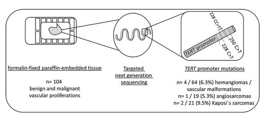
2.1. Sample Selection

Vascular tumor samples were obtained from the Department of Dermatology University Hospital Essen (n = 38), Dermatopathologie bei Mainz (n = 43), Dermatopathologie Duisburg (n = 8), and Dermatopathologie Friedrichshafen (n = 15), Germany. All tumor samples included in the study were primary cutaneous proliferations (no metastases). All cases were screened by at least one experienced board-certified dermatopathologist (KGG, EH, JS, TM, HM). The study was performed in accordance with the approval of the ethics committee of the University of Duisburg-Essen (IRB-number 20–9688-BO). The cohort has been described previously [21,22]. Hemangiomas with characteristic morphology were sub-classified as cherry hemangioma, a term not applied in the current ISSVA classification [23].

2.2. DNA Isolation

DNA was isolated from 10-µm-thick sections, cut from formalin-fixed, paraffin-embedded tumor tissues. The sections were de-paraffinized and the whole tissue was manually macrodissected. DNA isolation was performed with the QIAamp DNA Mini Kit (Qiagen, Hilden, Germany) according to the manufacturer’s instructions.

2.3. Targeted Sequencing

PCR amplification of the TERT promoter region was performed using primers: hTERT_F ACGAACGTGGCCAGCGGCAG and hTERT_R CTGGCGTCCCTGCACCCTGG (474 bp product), or primers hTERT_short_F CAGCGCTGCCTGAAACTC and hTERT_short_R GTCCTGCCCCTTCACCTT (163 bp product) as previously described [2]. PCR products were used as templates for sequencing after purification with the QIAquick PCR Purification Kit (Qiagen (Hilden, Germany)). Adapter ligation and barcoding of individual samples were done by applying the NEBNext Ultra DNA Library Prep Mastermix Set and NEBNext Multiplex Oligos for Illumina from New England Biolabs. Up to 100 samples were sequenced in parallel on an Illumina MiSeq next-generation sequencer. Sequencing analysis was performed applying the CLC Cancer Research Workbench from QIAGEN®. In brief, the following steps were applied: The workflow in CLC included adapter trimming and read pair merging before mapping to the human reference genome (hg19). Insertions and deletions as well as single nucleotide variant detection, local realignment, and primer trimming followed. Additional information was then obtained regarding potential mutation type, known single nucleotide polymorphisms, and conservation scores by cross-referencing varying databases (COSMIC, ClinVar, dbSNP, 1000 Genomes Project, HAPMAP, and PhastCons-Conservation_scores_hg19). The resulting csv files were analyzed manually and in all cases, the location of the recurrent TERT promoter mutations were assessed (Chr.5:1295–228, −242, and −250). Protein coding gene mutations were identified as previously described [2]. The mean coverage achieved of the TERT promoter region in all samples was 865 reads. Mutations were reported if the overall coverage of the mutation site was ≥30 reads, ≥5 reads reported the mutated variant, and the frequency of mutated reads was ≥2%.

3. Results

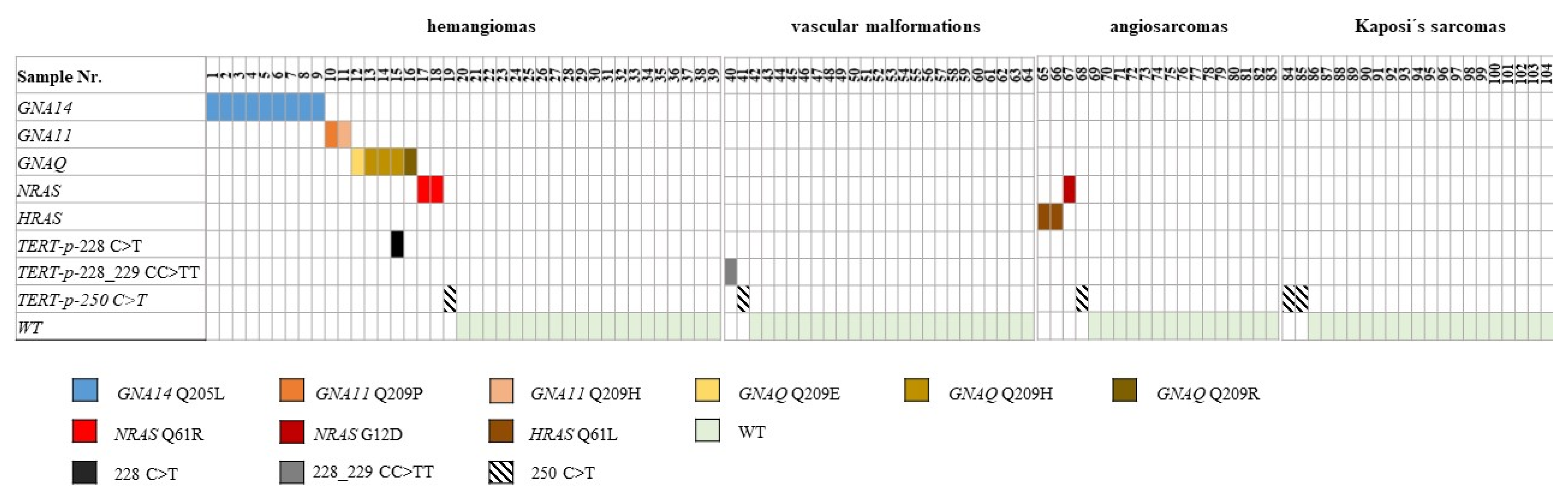
3.1. Mutation Analysis for TERT Promoter Mutations

In our cohort of 104 vascular tumors from 102 patients, the TERT promoter region showed wild-type reads in 97 tumors (93.3%) and harbored one mutation in 7 cases (6.7%) (Table 1). Mutations identified were located at the previously described hotspots: Chr.5:1295228 C > T, Chr.5:1295228_1295229 CC > TT, or Chr.5:1295250 C > T. All annotations are reported according to human genome assembly 19 (hg19). For simplicity, the mutations will further be referred to using solely the last three digits of the chromosome location nomenclature representing the first nucleotide altered as 228 C > T, 228 CC > TT, and 250 C > T, respectively.
In our cohort of benign hemangiomas and vascular malformations, 4 TERT promoter mutations could be detected in 64 samples (6.3%) (Table 1). Targeted amplicon next-generation sequencing identified 1228 C > T, 1228 CC > TT, and 2250 C > T mutations (Figure 1). One hemangioma harbored a 228 C > T TERT promoter and a GNAQ c.627 A > C, Q209H mutation. No hemangiomas or vascular malformations harbored more than one TERT promoter mutation (Table 1 and Table 2).
In the cohort of 19 angiosarcomas, one 250 C > T TERT promoter mutation was identified (5.3%). In those angiosarcomas with a known activating oncogene mutation (2 HRAS c.182 A > T, Q61L and 1 NRAS c.35 G > A, G12D), no TERT promoter mutations were identified (Table 1 and Figure 1).
In the cohort of Kaposi‘s sarcomas, two TERT promoter mutations were identified in two tumors, respectively (9.5%) (Table 1 and Figure 1).
The resulting amino acid changes are color-coded according to the scheme underneath the illustration. WT = wild-type for GNA14, GNA11, GNAQ, HRAS, NRAS, and TERT-promoter-mutation

3.2. Associations of Clinical and Pathological Parameters with TERT Promoter Status

An analysis with available clinicopathological data was performed. In the three different cohorts, statistically significant associations could not be detected between TERT promoter status, oncogene mutation status and patient age, sex, and sites of involvement, respectively (Table 1). We could not identify differences in histomorphological patterns between vascular proliferations harboring a TERT promoter mutation, an oncogene mutation, or no mutation (“wild-type”) [2] (Figure 2). Occurrence of TERT promoter mutations was not statistically significantly associated with prior radiotherapy (angiosarcomas), identification of human herpes virus 8 (HHV8), or HIV infection (Kaposi’s sarcomas).

4. Discussion

We analyzed the presence of TERT promoter mutations in a previously characterized cohort of vascular tumors comprising benign hemangiomas, vascular malformations, angiosarcomas, and Kaposi‘s sarcomas by targeted next-generation sequencing [2]. All proliferations showed a low frequency of TERT promoter mutations.
In other neoplasms, TERT promoter mutations can be present in both benign and malignant tumor entities with the same origin but are often elevated in the malignant entity [24,25,26,27]. In our cohort, we could show that both benign and malignant cutaneous vascular proliferations harbor a low frequency of different TERT promoter mutations (6.3% in hemangiomas and vascular malformations, 5.3% in angiosarcomas, and 9.5% in Kaposi‘s sarcomas).
In the literature, the frequency and types of TERT promoter mutation vary greatly depending on the type of tumor [4,5,6,7,8,9]. The recurrent mutations we found in the TERT promoter were at previously reported hotspots [2,3] and had a UV signature represented by [28] C > T or CC > TT changes [29,30]. C > T mutations can be identified in tumors developing independent of UV exposure, e.g., bladder cancer [6]. However, UV light is still considered to play a major role in the induction of both C > T and CC > TT in cutaneous tumors [13,29,30]. CC >> TT alterations are considered pathognomonic for UV induction [29,30] and could be identified in one benign venous malformation localized on the head/neck of a 70-year-old man. As a UV-mutation profile has been described for angiosarcomas arising in sun-exposed regions [31], we analyzed the association of TERT promoter mutation occurrence and localization. We identified a 250 C > T TERT promoter mutation in 1 of 15 angiosarcomas on the ventral trunk after prior radiotherapy due to breast cancer. In the other six angiosarcomas known to have arisen following radiotherapy due to breast cancer, no TERT promoter mutation could be identified.
Of the seven cutaneous vascular proliferations harboring one TERT promoter mutation, two were located in the head/neck region, two on the ventral trunk, and two on the lower extremity (one with unknown localization). Although proliferations with TERT promoter mutations were identified in areas favoring sun exposure, a statistically significant association between the occurrence of a TERT promoter mutation and localization could not be calculated in any of our cohorts.
The prevalence of TERT promoter mutations detected (hemangiomas/vascular malformations (6.3%), angiosarcomas (5.3%), and Kaposi‘s sarcomas (9.5%)) was low in both benign and malignant cutaneous vascular proliferations. Rare TERT promoter mutations in angiosarcomas fit previous studies [28,32]. GNA14, GNA11, and GNAQ mutations are frequent in congenital and cherry hemangiomas [33,34,35] (cherry or senile hemangiomas is still a widely used term, although not recognized as an individual entity according to the ISSVA classification 2018. This classification would group these lesions as a form of pyogenic granuloma or lobular capillary hemangioma). A single hemangioma harbored a 228 C > T TERT promotor and a GNAQ c.627 A > C, Q209H mutation. Otherwise, no co-occurrence with known GNA or RAS mutations was observed. In melanoma, TERT promoter mutations frequently co-occur with activating BRAF mutations [36,37]. In our study, a similar co-occurrence of mutations could not be observed.
The role for the TERT (telomerase reverse transcriptase) protein in cutaneous vascular tumors remains intriguing. Our finding suggests that it promotes tumor proliferation in a few cutaneous vascular proliferations. However, based on the frequency of mutations, a mutation of the promoter to increase protein activity appears to be non-mandatory in most tumors. Potentially, gains of the region do occur in vascular tumors lacking TERT promoter mutations. Additionally, alternative lengthening of telomeres (ALT), e.g., mutations in ATRX or DAXX, may contribute to the progression of cutaneous vascular proliferations. These mechanisms, both generally occurring much rarer than TERT promoter mutations [38], could not be assessed in the assay we applied and will need to be examined in future studies.
The overall frequency (<10%) and its variation between tumor groups was low. This suggests determining TERT promoter mutation status is of no diagnostic aid in terms of classifying tumor entities or predicting prognosis.
A limitation of our study is the lack of detailed clinical data, including therapy and follow-up information. Strengths of our study are the considerable number of vascular tumors included in the analysis (n = 104), making it the largest cohort of vascular tumors analyzed for the presence of TERT promoter mutations.

5. Conclusions

In conclusion, we could show that TERT promoter mutations are present in both benign and malignant cutaneous vascular tumors and malformations. The prevalence was 6.3% in hemangiomas and vascular malformations, 5.3% in angiosarcomas, and 9.5% in Kaposi‘s sarcomas. In our comparative analysis, we could not confirm the tendency observed in other cutaneous tumors of TERT promoter mutations being more prevalent in malignant entities. While TERT promoter mutations appear to be relevant in a small percentage of tumors, the majority of these tumors arise independent of these mutations.

Author Contributions

Conceptualization, methodology and software, K.G.G. and P.J.; validation, P.J., D.S., E.H., K.G.G.; histological analysis, H.M., J.S., T.M., E.H., K.G.G.; investigation all authors.; data curation, P.J. and K.G.G.; writing—original draft preparation, P.J., H.M., T.M., E.H., K.G.G.; writing—review and editing, all authors; visualization and supervision, P.J. and K.G.G.; project administration, P.J. and K.G.G. All authors have read and agreed to the published version of the manuscript.

Funding

This research received no external funding.

Institutional Review Board Statement

The study was conducted according to the guidelines of the Declaration of Helsinki, and approved by the Ethics Committee of the University of Duisburg-Essen (IRB-number 20–9688-BO, date of approval: 26 November 2020).

Informed Consent Statement

Informed consent was obtained from all subjects involved in the study.

Data Availability Statement

The datasets presented in this study can be found in online repositories. The names of the repository/repositories and accession number(s) can be found below: https://0-www-ncbi-nlm-nih-gov.brum.beds.ac.uk/genbank/, PRJNA717731 (accessed on 27 March 2021).

Conflicts of Interest

The authors declare no conflict of interest.

References

  1. Bell, R.J.; Rube, H.T.; Xavier-Magalhaes, A.; Costa, B.M.; Mancini, A.; Song, J.S.; Costello, J.F. Understanding TERT Promoter Mutations: A Common Path to Immortality. Mol. Cancer Res. 2016, 14, 315–323. [Google Scholar] [CrossRef] [Green Version]
  2. Horn, S.; Figl, A.; Rachakonda, P.S.; Fischer, C.; Sucker, A.; Gast, A.; Kadel, S.; Moll, I.; Nagore, E.; Hemminki, K.; et al. TERT promoter mutations in familial and sporadic melanoma. Science 2013, 339, 959–961. [Google Scholar] [CrossRef] [Green Version]
  3. Huang, F.W.; Hodis, E.; Xu, M.J.; Kryukov, G.V.; Chin, L.; Garraway, L.A. Highly recurrent TERT promoter mutations in human melanoma. Science 2013, 339, 957–959. [Google Scholar] [CrossRef] [PubMed] [Green Version]
  4. Liu, X.; Qu, S.; Liu, R.; Sheng, C.; Shi, X.; Zhu, G.; Murugan, A.K.; Guan, H.; Yu, H.; Wang, Y.; et al. TERT promoter mutations and their association with BRAF V600E mutation and aggressive clinicopathological characteristics of thyroid cancer. J. Clin. Endocrinol. Metab. 2014, 99, E1130–E1136. [Google Scholar] [CrossRef] [Green Version]
  5. Rusinek, D.; Pfeifer, A.; Krajewska, J.; Oczko-Wojciechowska, M.; Handkiewicz-Junak, D.; Pawlaczek, A.; Zebracka-Gala, J.; Kowalska, M.; Cyplinska, R.; Zembala-Nozynska, E.; et al. Coexistence of TERT Promoter Mutations and the BRAF V600E Alteration and Its Impact on Histopathological Features of Papillary Thyroid Carcinoma in a Selected Series of Polish Patients. Int. J. Mol. Sci. 2018, 19, 2647. [Google Scholar] [CrossRef] [Green Version]
  6. Hosen, I.; Rachakonda, P.S.; Heidenreich, B.; de Verdier, P.J.; Ryk, C.; Steineck, G.; Hemminki, K.; Kumar, R. Mutations in TERT promoter and FGFR3 and telomere length in bladder cancer. Int. J. Cancer 2015, 137, 1621–1629. [Google Scholar] [CrossRef] [PubMed]
  7. Spiegl-Kreinecker, S.; Lotsch, D.; Neumayer, K.; Kastler, L.; Gojo, J.; Pirker, C.; Pichler, J.; Weis, S.; Kumar, R.; Webersinke, G.; et al. TERT promoter mutations are associated with poor prognosis and cell immortalization in meningioma. Neuro. Oncol. 2018, 20, 1584–1593. [Google Scholar] [CrossRef] [PubMed] [Green Version]
  8. Ren, H.; Shen, Y.; Hu, D.; He, W.; Zhou, J.; Cao, Y.; Mao, Y.; Dou, Y.; Xiong, W.; Xiao, Q.; et al. Co-existence of BRAF(V600E) and TERT promoter mutations in papillary thyroid carcinoma is associated with tumor aggressiveness, but not with lymph node metastasis. Cancer Manag. Res. 2018, 10, 1005–1013. [Google Scholar] [CrossRef] [Green Version]
  9. Jeong, D.E.; Woo, S.R.; Nam, H.; Nam, D.H.; Lee, J.H.; Joo, K.M. Preclinical and clinical implications of TERT promoter mutation in glioblastoma multiforme. Oncol. Lett. 2017, 14, 8213–8219. [Google Scholar] [CrossRef]
  10. Campos, M.A.; Macedo, S.; Fernandes, M.; Pestana, A.; Pardal, J.; Batista, R.; Vinagre, J.; Sanches, A.; Baptista, A.; Lopes, J.M.; et al. TERT promoter mutations are associated with poor prognosis in cutaneous squamous cell carcinoma. J. Am. Acad. Dermatol. 2019, 80, 660–669 e666. [Google Scholar] [CrossRef] [PubMed]
  11. Malkki, H. Neuro-oncology: TERT promoter mutations could indicate poor prognosis in glioblastoma. Nat. Rev. Neurol. 2014, 10, 546. [Google Scholar] [CrossRef] [PubMed]
  12. Blateau, P.; Coyaud, E.; Laurent, E.; Beganton, B.; Ducros, V.; Chauchard, G.; Vendrell, J.A.; Solassol, J. TERT Promoter Mutation as an Independent Prognostic Marker for Poor Prognosis MAPK Inhibitors-Treated Melanoma. Cancers 2020, 12, 2224. [Google Scholar] [CrossRef] [PubMed]
  13. Griewank, K.G.; Murali, R.; Puig-Butille, J.A.; Schilling, B.; Livingstone, E.; Potrony, M.; Carrera, C.; Schimming, T.; Moller, I.; Schwamborn, M.; et al. TERT promoter mutation status as an independent prognostic factor in cutaneous melanoma. J. Natl. Cancer Inst. 2014, 106. [Google Scholar] [CrossRef] [PubMed]
  14. Arantes, L.; Cruvinel-Carloni, A.; de Carvalho, A.C.; Sorroche, B.P.; Carvalho, A.L.; Scapulatempo-Neto, C.; Reis, R.M. TERT Promoter Mutation C228T Increases Risk for Tumor Recurrence and Death in Head and Neck Cancer Patients. Front. Oncol. 2020, 10, 1275. [Google Scholar] [CrossRef] [PubMed]
  15. Borah, S.; Xi, L.; Zaug, A.J.; Powell, N.M.; Dancik, G.M.; Cohen, S.B.; Costello, J.C.; Theodorescu, D.; Cech, T.R. Cancer. TERT promoter mutations and telomerase reactivation in urothelial cancer. Science 2015, 347, 1006–1010. [Google Scholar] [CrossRef] [PubMed] [Green Version]
  16. Chiba, K.; Lorbeer, F.K.; Shain, A.H.; McSwiggen, D.T.; Schruf, E.; Oh, A.; Ryu, J.; Darzacq, X.; Bastian, B.C.; Hockemeyer, D. Mutations in the promoter of the telomerase gene TERT contribute to tumorigenesis by a two-step mechanism. Science 2017, 357, 1416–1420. [Google Scholar] [CrossRef] [Green Version]
  17. Killela, P.J.; Reitman, Z.J.; Jiao, Y.; Bettegowda, C.; Agrawal, N.; Diaz, L.A., Jr.; Friedman, A.H.; Friedman, H.; Gallia, G.L.; Giovanella, B.C.; et al. TERT promoter mutations occur frequently in gliomas and a subset of tumors derived from cells with low rates of self-renewal. Proc. Natl. Acad. Sci. USA 2013, 110, 6021–6026. [Google Scholar] [CrossRef] [Green Version]
  18. Shain, A.H.; Yeh, I.; Kovalyshyn, I.; Sriharan, A.; Talevich, E.; Gagnon, A.; Dummer, R.; North, J.; Pincus, L.; Ruben, B.; et al. The Genetic Evolution of Melanoma from Precursor Lesions. N. Engl. J. Med. 2015, 373, 1926–1936. [Google Scholar] [CrossRef]
  19. Griewank, K.G.; Murali, R.; Schilling, B.; Schimming, T.; Moller, I.; Moll, I.; Schwamborn, M.; Sucker, A.; Zimmer, L.; Schadendorf, D.; et al. TERT promoter mutations are frequent in cutaneous basal cell carcinoma and squamous cell carcinoma. PLoS ONE 2013, 8, e80354. [Google Scholar] [CrossRef]
  20. Scott, G.A.; Laughlin, T.S.; Rothberg, P.G. Mutations of the TERT promoter are common in basal cell carcinoma and squamous cell carcinoma. Mod. Pathol. 2014, 27, 516–523. [Google Scholar] [CrossRef] [PubMed] [Green Version]
  21. Murali, R.; Chandramohan, R.; Moller, I.; Scholz, S.L.; Berger, M.; Huberman, K.; Viale, A.; Pirun, M.; Socci, N.D.; Bouvier, N.; et al. Targeted massively parallel sequencing of angiosarcomas reveals frequent activation of the mitogen activated protein kinase pathway. Oncotarget 2015, 6, 36041–36052. [Google Scholar] [CrossRef] [Green Version]
  22. Jansen, P.; Müller, H.; Lodde, G.C.; Zaremba, A.; Möller, I.; Sucker, A.; Paschen, A.; Esser, S.; Schaller, J.; Gunzer, M.; et al. GNA14, GNA11, and GNAQ Mutations Are Frequent in Benign but Not Malignant Cutaneous Vascular Tumors. Front. Genet. 2021, 12, 656. [Google Scholar] [CrossRef] [PubMed]
  23. ISSVA Classification for Vascular Anomalies. 2018. Available online: https://www.issva.org/UserFiles/file/ISSVA-Classification-2018.pdf (accessed on 23 June 2021).
  24. Koopmans, A.E.; Ober, K.; Dubbink, H.J.; Paridaens, D.; Naus, N.C.; Belunek, S.; Krist, B.; Post, E.; Zwarthoff, E.C.; de Klein, A.; et al. Prevalence and implications of TERT promoter mutation in uveal and conjunctival melanoma and in benign and premalignant conjunctival melanocytic lesions. Investig. Ophthalmol. Visual Sci. 2014, 55, 6024–6030. [Google Scholar] [CrossRef] [PubMed] [Green Version]
  25. Munoz-Jimenez, M.T.; Blanco, L.; Ruano, Y.; Carrillo, R.; Santos-Briz, A.; Riveiro-Falkenbach, E.; Requena, L.; Kutzner, H.; Garrido, M.C.; Rodriguez-Peralto, J.L. TERT promoter mutation in sebaceous neoplasms. Virchows Arch. 2021. [Google Scholar] [CrossRef]
  26. Jansen, P.; Cosgarea, I.; Murali, R.; Moller, I.; Sucker, A.; Franklin, C.; Paschen, A.; Zaremba, A.; Brinker, T.J.; Stoffels, I.; et al. Frequent Occurrence of NRAS and BRAF Mutations in Human Acral Naevi. Cancers 2019, 11, 546. [Google Scholar] [CrossRef] [Green Version]
  27. Zaremba, A.; Murali, R.; Jansen, P.; Moller, I.; Sucker, A.; Paschen, A.; Zimmer, L.; Livingstone, E.; Brinker, T.J.; Hadaschik, E.; et al. Clinical and genetic analysis of melanomas arising in acral sites. Eur. J. Cancer 2019, 119, 66–76. [Google Scholar] [CrossRef] [PubMed]
  28. Painter, C.A.; Jain, E.; Tomson, B.N.; Dunphy, M.; Stoddard, R.E.; Thomas, B.S.; Damon, A.L.; Shah, S.; Kim, D.; Gomez Tejeda Zanudo, J.; et al. The Angiosarcoma Project: Enabling genomic and clinical discoveries in a rare cancer through patient-partnered research. Nat. Med. 2020, 26, 181–187. [Google Scholar] [CrossRef]
  29. Pleasance, E.D.; Cheetham, R.K.; Stephens, P.J.; McBride, D.J.; Humphray, S.J.; Greenman, C.D.; Varela, I.; Lin, M.L.; Ordonez, G.R.; Bignell, G.R.; et al. A comprehensive catalogue of somatic mutations from a human cancer genome. Nature 2010, 463, 191–196. [Google Scholar] [CrossRef] [PubMed]
  30. Brash, D.E.; Rudolph, J.A.; Simon, J.A.; Lin, A.; McKenna, G.J.; Baden, H.P.; Halperin, A.J.; Ponten, J. A role for sunlight in skin cancer: UV-induced p53 mutations in squamous cell carcinoma. Proc. Natl. Acad. Sci. USA 1991, 88, 10124–10128. [Google Scholar] [CrossRef] [Green Version]
  31. Chan, J.Y.; Lim, J.Q.; Yeong, J.; Ravi, V.; Guan, P.; Boot, A.; Tay, T.K.Y.; Selvarajan, S.; Md Nasir, N.D.; Loh, J.H.; et al. Multiomic analysis and immunoprofiling reveal distinct subtypes of human angiosarcoma. J. Clin. Investig. 2020, 130, 5833–5846. [Google Scholar] [CrossRef]
  32. Koelsche, C.; Renner, M.; Hartmann, W.; Brandt, R.; Lehner, B.; Waldburger, N.; Alldinger, I.; Schmitt, T.; Egerer, G.; Penzel, R.; et al. TERT promoter hotspot mutations are recurrent in myxoid liposarcomas but rare in other soft tissue sarcoma entities. J. Exp. Clin. Cancer Res. 2014, 33, 33. [Google Scholar] [CrossRef] [PubMed] [Green Version]
  33. Liau, J.Y.; Lee, J.C.; Tsai, J.H.; Chen, C.C.; Chung, Y.C.; Wang, Y.H. High frequency of GNA14, GNAQ, and GNA11 mutations in cherry hemangioma: A histopathological and molecular study of 85 cases indicating GNA14 as the most commonly mutated gene in vascular neoplasms. Mod. Pathol. 2019, 32, 1657–1665. [Google Scholar] [CrossRef] [PubMed]
  34. Liau, J.Y.; Lee, J.C.; Tsai, J.H.; Chen, C.C.; Wang, Y.H.; Chung, Y.C. Thrombotic Hemangioma With Organizing/Anastomosing Features: Expanding the Spectrum of GNA-mutated Hemangiomas With a Predilection for the Skin of the Lower Abdominal Regions. Am. J. Surg. Pathol. 2020, 44, 255–262. [Google Scholar] [CrossRef]
  35. Ayturk, U.M.; Couto, J.A.; Hann, S.; Mulliken, J.B.; Williams, K.L.; Huang, A.Y.; Fishman, S.J.; Boyd, T.K.; Kozakewich, H.P.; Bischoff, J.; et al. Somatic Activating Mutations in GNAQ and GNA11 Are Associated with Congenital Hemangioma. Am. J. Hum. Genet. 2016, 98, 789–795. [Google Scholar] [CrossRef] [PubMed] [Green Version]
  36. Zehir, A.; Benayed, R.; Shah, R.H.; Syed, A.; Middha, S.; Kim, H.R.; Srinivasan, P.; Gao, J.; Chakravarty, D.; Devlin, S.M.; et al. Mutational landscape of metastatic cancer revealed from prospective clinical sequencing of 10,000 patients. Nat. Med. 2017, 23, 703–713. [Google Scholar] [CrossRef] [PubMed]
  37. Macerola, E.; Loggini, B.; Giannini, R.; Garavello, G.; Giordano, M.; Proietti, A.; Niccoli, C.; Basolo, F.; Fontanini, G. Coexistence of TERT promoter and BRAF mutations in cutaneous melanoma is associated with more clinicopathological features of aggressiveness. Virchows Arch. 2015, 467, 177–184. [Google Scholar] [CrossRef]
  38. Shay, J.W.; Wright, W.E. Telomeres and telomerase: Three decades of progress. Nat. Rev. Genet. 2019, 20, 299–309. [Google Scholar] [CrossRef]
Figure 1. Distribution of TERT promoter mutations and activating mutations identified in vascular tumors.
Dermato 01 00003 g001
Figure 2. Representative histologic images of cherry hemangiomas * and two angiosarcomas, with and without a TERT promoter mutation (The bars represent a distance of 200 μm. * cherry hemangiomas are grouped as lobular capillary hemangiomas according to the current ISSVA classification [23]).
Dermato 01 00003 g002
Table 1. Clinical variables of vascular proliferations with oncogene and TERT promoter mutations.
Hemangioma/Vascular MalformationAllWTGNA MutantRAS MutantTERT-P-Mutantp-Value *
n = 64n = 43n = 16 $n = 2n = 4 $
Mean age (years)53544860570.4
SexFemale28205 $03 $0.36
Male35 #2210 #21
Sites of involvementhead/neck21163 $12 $0.06
ventral trunk168710
dorsal trunk137600
upper extremity54001
lower extremity77000
LND21001
AngiosarcomaAllWTGNA MutantRAS MutantTERT-P-Mutantp-Value *
n = 19n = 15n = 0n = 3n = 1
Mean age (years)6765-75740.4
SexFemale1511-311.0
Male4400
Sites of involvementhead/neck86-201.0
ventral trunk75-11
dorsal trunk00-00
upper extremity00-00
lower extremity11-00
LND33-00
Kaposi‘s sarcomaAllWTGNA MutantRAS MutantTERT-P-Mutantp-Value *
n = 21n = 19n = 0n = 0n = 2
Mean age (years)5658--340.1
SexFemale44--01.0
Male16 !14 !2
Sites of involvementhead/neck22--00.13
ventral trunk21--1
dorsal trunk00--0
upper extremity21--1
lower extremity1313--0
LND22--0
LND—localization not determined, TERT-P-mutant = TERT-promoter mutation (228 C > T, 228 CC > TT or 250 C > T), WT = wild-type for GNA14, GNA11, GNAQ, HRAS, NRAS and TERT-promoter-mutation, # one male had two hemangiomas both showing a GNA14 mutation, $ one female patient showed a GNAQ and a TERT-promoter (228 C > T)-mutation, ! one male had two Kaposi‘s sarcomas, * Age—Kruskal–Wallis test; all others—Fisher exact test.
Table 2. Oncogene and TERT promoter mutations in histological subtypes of hemangiomas and vascular malformations.
Hemangioma/Vascular MalformationAllWTGNA MutantRAS MutantTERT-P-Mutant
n = 64n = 43n = 16 $n = 2n = 4 $
lobular capillary/pyogenic granuloma73310
microvenular66000
cherry/senile #251013 $12 $
tufted11000
Angiokeratoma +33000
Arteriovenous +44000
superficial hemosiderotic lymphovascular +x22000
venous/cavernous +1614002
# not listed as an independent entity according to ISSVA (23), would alternatively be seen as a form of lobular capillary hemangioma (or pyogenic granuloma); + classified as malformations according to ISSVA; x previously referred to as targetoid hemosiderotic; $ one tumor harbored a GNAQ and TERT-promoter (228 C > T)-mutations.
Publisher’s Note: MDPI stays neutral with regard to jurisdictional claims in published maps and institutional affiliations.

