Article

Dermacentor variabilis Does Not Transstadially Transmit the U.S. Isolate of Theileria orientalis Ikeda: A Controlled Acquisition and Transmission Study

1
Department of Veterinary Microbiology and Pathology, Washington State University, Pullman, WA 99164, USA
2
Department of Animal Medicine, The Faculty of Veterinary Medicine, The University of Alexandria, Alexandria 21944, Egypt
3
Animal Disease Research Unit, United States Department of Agriculture-Agricultural Research Service, Pullman, WA 99164, USA
4
Invasive Insect Biocontrol and Behavior Laboratory, United States Department of Agriculture-Agricultural Research Service, Beltsville, MD 20705, USA
*
Author to whom correspondence should be addressed.
Submission received: 12 July 2023 / Revised: 5 September 2023 / Accepted: 7 September 2023 / Published: 14 September 2023

Abstract

:
Theileria orientalis Ikeda, an emerging U.S. bovine hemoparasite, causes anemia, abortion, ill-thrift, and occasionally death. While Haemaphysalis longicornis is the primary vector, it is possible that other U.S. ticks are capable of parasite transmission and may contribute to disease spread. Dermacentor variabilis is highly prevalent in the U.S., exhibits a similar geographical distribution to T. orientalis, and is a competent vector of the related parasite, Theileria equi. Herein, we conducted controlled acquisition and transmission studies using splenectomized calves to assess whether D. variabilis can transstadially transmit T. orientalis. D. variabilis nymphs were applied to an infected, splenectomized calf for parasite acquisition and subsequently incubated to molt into adults. Freshly molted adults were applied to two splenectomized T. orientalis-naïve calves to investigate parasite transmission. Calves were monitored for 59 days, and no evidence of parasite transmission was detected using PCR for the T. orientalis Ikeda major piroplasm surface protein gene, blood smear cytology, complete blood counts, or physical examination. Salivary glands from a subset of D. variabilis adults were assessed for T. orientalis using PCR, and the parasite was not detected. These findings support the conclusion that D. variabilis is not capable of transstadial transmission of the U.S. T. orientalis Ikeda isolate.

1. Introduction

Bovine oriental theileriosis is caused by the tick-borne protozoan parasite Theileria orientalis [1,2]. Like other non-transforming Theileria species [1,3], the symptomatic phase of T. orientalis infection is characterized by infection of erythrocytes and resultant erythrolysis [1,4].
T. orientalis is classified into eleven genotypes based on targeted amplification and sequencing of the major piroplasm surface protein (MPSP) and small subunit (SSU) rRNA genes [2,3,5,6]. These genotypes include Chitose (type 1), Ikeda (type 2), buffeli (type 3), types 4 to 8, and types N1, N2, and N3 [2,5,6,7,8,9]. Most T. orientalis genotypes cause a relatively minor disease known as benign oriental bovine theileriosis [9,10]. The Ikeda and Chitose genotypes, in contrast, are more virulent, causing more severe clinical disease. These genotypes have been associated with relatively recent outbreaks in the U.S., Asia, Australia, and New Zealand [4,6,11,12,13,14,15,16].
Infection with virulent T. orientalis genotypes can cause severe hemolytic anemia, lethargy, anorexia, fever, icterus, abortion, stillbirth, inappetence, tachypnea, tachycardia, and death in some cases [12,16,17,18]. The Ikeda genotype is associated with mortality rates of up to 6% in infected cattle [14,15,19]. Transplacental transmission of the parasite has been reported in up to 10% of infected cows, and abortions are sometimes a frequent occurrence during these outbreaks [20,21]. In this regard, T. orientalis Ikeda is associated with significant economic losses. For instance, the Australian beef industry suffers indirect costs of approximately AUD 19.6 million annually, and New Zealand suffers losses of more than NZD 400 per cow due to reduced meat and milk yields [22,23,24]. The lack of efficient diagnostic tests, efficacious treatment strategies, and vaccines for the control of the parasite further exacerbates these challenges.
The first T. orientalis Ikeda outbreak in the U.S. was reported in 2017 [4] and coincided with the detection of Haemaphysalis longicornis ticks in the same region [25,26,27,28,29], which were later shown to be competent vectors of the U.S. T. orientalis Ikeda isolate [26]. The source of H. longicornis introduction to the U.S. remains unknown, but, at the time of publication, it had spread to at least 19 U.S. states [30]. Control of H. longicornis is challenging because the tick species can withstand diverse climatic conditions [31], infests a wide range of host species, and reproduces via parthenogenesis, in which females do not require males to produce offspring [29,30,32]. It is therefore not surprising that since the tick’s emergence in Virginia, T. orientalis Ikeda has spread within the U.S. and has also been reported in Tennessee, West Virginia, North Carolina, Pennsylvania, Arkansas, New York, Tennessee, and Kentucky [4,27,29,30,32,33].
Assessment of the ability of other tick species, especially those native to the U.S. [34], to transmit T. orientalis Ikeda is key to comprehensively understanding the threat that the parasite poses to the American cattle population. H. longicornis belongs to the family Ixodidae [32], and evaluation of the competence of other U.S. ixodid ticks for T. orientalis Ikeda is key in the determination of its potential spread. Our group recently showed that the cattle fever tick (also known as the southern cattle tick), Rhipicephalus microplus, is likely not a competent vector of U.S. isolates of T. orientalis Ikeda [35]. In the present study, we assessed the competence of the American dog tick, Dermacentor variabilis, for the U.S. isolate of T. orientalis Ikeda using controlled acquisition and transmission studies in calves. D. variabilis was chosen for testing because: (1) It belongs to the Ixodidae family [32]; (2) it is the most widely distributed native north American tick species [36] and is well-established within the geographic range in which T. orientalis Ikeda is spreading [34,37]; and (3) it has been shown to transmit a related parasite, Theileria equi, to horses [38,39]. The findings obtained from this study are summarized herein.

2. Results

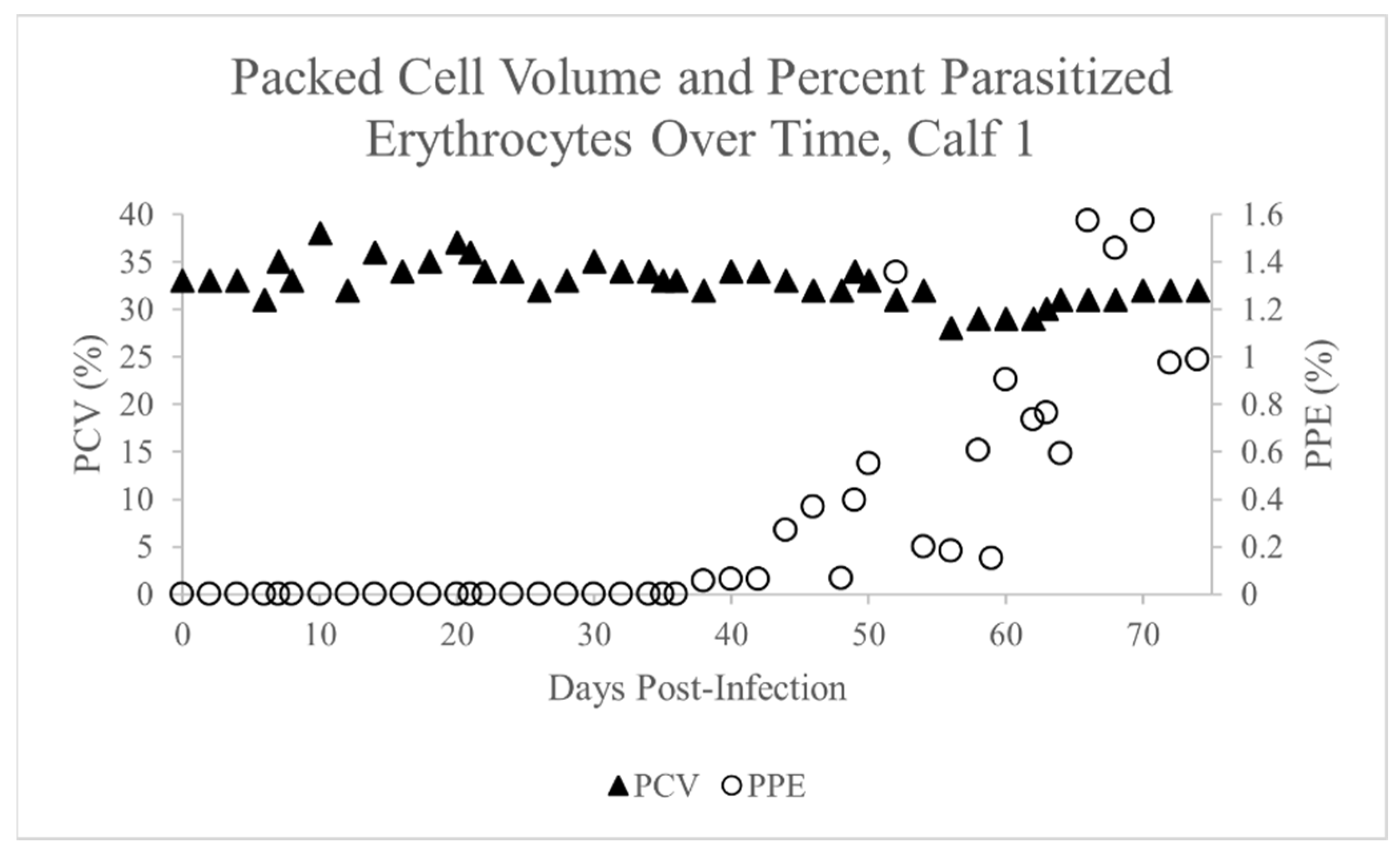
2.1. Theileria Orientalis Infection of Calf 1

Peripheral blood from Calf 1 became PCR-positive for T. orientalis Ikeda on day 16 post-infection and remained PCR-positive for the remainder of the experiment (Table 1). Merozoites were first detected within erythrocytes via blood smear cytology on day 38 post-infection. As noted previously [26,35], intraerythrocytic merozoites were pear-shaped and approximately 1–2.5 µm × 0.5 µm. The percent parasitized erythrocytes (PPE) varied from 0.06% to 1.57% for the remainder of the experiment (Figure 1). The packed cell volume (PCV) declined only mildly to a nadir of 28% on day 56 but returned to a pre-infection level of 32% by day 74 (Figure 1). Calf 1 did not develop any clinical signs of severe T. orientalis, and no other significant abnormalities were detected on the complete blood count (CBC) or serum chemistry panel.

2.2. Dermacentor variabilis Acquisition Feed on Calf 1

D. variabilis nymphs were applied to Calf 1 in two batches of 676 nymphs each. The first batch was applied on day 46 post-infection, and 467 replete nymphs from this batch were collected from 5 to 12 days post-application. The PPE during this time ranged from 0.07 to 1.36% (Figure 1). The second batch was applied on day 59 post-infection, and 213 replete nymphs from this batch were collected from 5 to 10 days post-application. The PPE during this time ranged from 0.59 to 1.57% (Figure 1). Replete nymphs were placed in an incubator and allowed to molt to the adult stage.

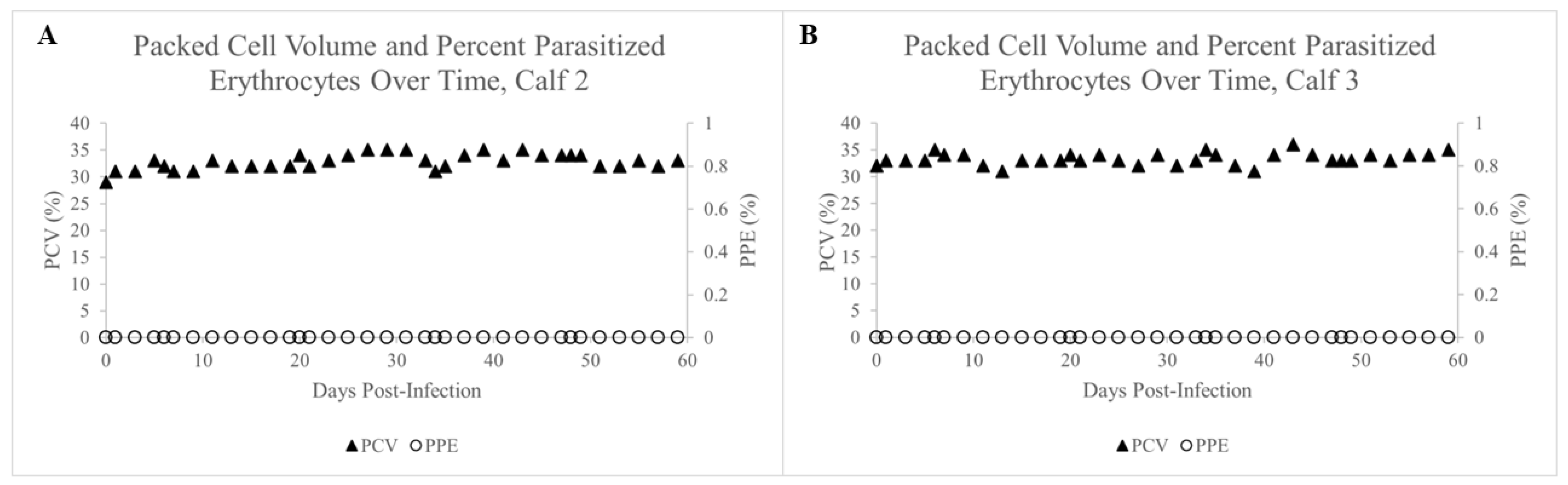
2.3. Failure of Adult D. variabilis Ticks to Acquire and Transmit T. orientalis to Calves 2 and 3

Amounts of 285–286 adult ticks from batches 1 and 2 were combined and applied to calves 2 and 3 as described above. After four days of feeding, five male and five female ticks were removed from each calf, dissected, and their salivary glands tested for T. orientalis via PCR. T. orientalis was not detected in the salivary glands of any of the assayed ticks (Table 2), suggesting that D. variabilis ticks did not acquire T. orientalis while feeding on Calf 1.
Of the 286 ticks applied to Calf 2, 197 (69%) were fed to repletion. Of the 285 ticks applied to Calf 3, 148 (52%) were fed to repletion. Calves 2 and 3 were monitored for evidence of T. orientalis infection for 59 days. Peripheral blood was assessed weekly for T. orientalis Ikeda using T. orientalis MPSP PCR, but T. orientalis was never detected in either calf. Furthermore, no organisms were visualized via blood smear cytology for the duration of the experiment (Figure 2A,B). Neither calf exhibited clinical signs consistent with T. orientalis (e.g., fever, icterus, inappetence, weakness), a decline in PCV (Figure 2A,B), or significant abnormalities in CBC or serum chemistry panel parameters during this time period. Since these calves were splenectomized and thus incredibly susceptible to hemoparasitic infections like T. orientalis, the lack of parasite detection via PCR or cytology, coupled with the complete absence of clinical signs of T. orientalis, strongly supports the conclusion that adult D. variabilis ticks that had been fed as nymphs on a T. orientalis-infected calf failed to transmit T. orientalis in this experiment. This confirms that transstadial transmission from the nymphal tick stage to the adult tick stage failed to occur in this experiment. This conclusion is further strengthened by the fact that each splenectomized calf was infested by a large number of exposed adult ticks, increasing the likelihood of detection of even rare transmission events within the exposed tick population.

3. Discussion

This study assessed whether D. variabilis, a broadly distributed North American tick, could transstadially transmit the U.S. T. orientalis Ikeda isolate. We found that D. variabilis could neither acquire T. orientalis Ikeda while feeding as nymphs nor maintain infection through molting to the adult stage. Our findings suggest D. variabilis is unlikely to be a competent vector of T. orientalis Ikeda.
The assessment of intrastadial and transovarial transmission of T. orientalis by D. variabilis was beyond the scope of the current work. D. variabilis was shown to be competent to transmit Theileria equi intrastadially between horses [38,39,40]. Intrastadial transmission requires movement of adult ticks between infected and susceptible hosts; this can occur when partially fed female ticks are groomed off and subsequently reattach [41] or when male ticks move between hosts seeking females for mating [42,43,44]. Anaplasma marginale is routinely transmitted between infected and susceptible cattle [45,46] in this way. The ability of D. variabilis to intrastadially transmit T. orientalis should be assessed in future studies. Transovarial transmission was not assessed in the current study; however, as the lack of transovarial transmission is a defining characteristic of the Theileria sp. genus [47], it is incredibly unlikely that T. orientalis is transovarially transmitted.
Dermacentor variabilis ticks are known to be efficient vectors of both Rickettsia rickettsii, the causative agent of Rocky Mountain Spotted Fever, and Francisella tularensis, which causes tularemia [48]. Rickettsia rickettsii is a coccobacillary, obligate, intracellular organism that is transmitted transstadially between tick life stages and transovarially from adult females to their offspring [49]. Francisella tularensis is a gram-negative coccobacillus that is transstadially transmitted between tick life stages [50]. Interestingly, for both R. rickettsii and F. tularensis, most of the pathogen lifecycle occurs in the mammalian host as opposed to the tick vector [51]. In contrast, for Theileria species, the tick is the definitive host of the parasite since sexual stage development occurs within the tick, potentially highlighting the importance of tick-specific physiological processes for maintaining viable infection [1]. Given the marked differences between the tick-stage life cycle of T. orientalis Ikeda and that of most pathogens for which D. variabilis is a known competent and efficient vector, it is possible that T. orientalis Ikeda is incapable of completing its life cycle within the tick.
One notable exception to consider is the ability of D. variabilis to intrastadially transmit the related parasite, T. equi [38,39]. Although experimental studies have revealed this form of transmission is possible in T. equi, the epidemiological significance of intrastadial T. equi transmission by D. variabilis has yet to be shown. The overall efficiency, or capacity, of a given tick vector is influenced by several factors, including its competence to transmit the pathogen, host preferences, tick and host behavior, tick prevalence, and the life cycle characteristics of each pathogen [47]. Thus, although D. variabilis is a competent intrastadial vector of T. equi, it likely has a low overall vector capacity for that parasite. As stated above, further studies to assess whether D. variabilis is capable of intrastadial transmission of T. orientalis are warranted.
Furthermore, host use per life stage of D. variabilis may limit its ability to successfully transmit T. orientalis Ikeda to cattle. Dermacentor variabilis is a three-host tick, requiring three different hosts for blood meals to complete its life cycle [52]. Larval D. variabilis are known to feed on small mammals such as mice, voles, chipmunks, shrews, and rabbits, while nymphs and adults are likely to feed on medium and large mammals such as raccoons, domestic pets (dogs and cats), coyotes, humans, and cattle [37,53,54,55,56,57]. Given that the larval stages of D. variabilis are not known to feed on cattle or hosts that naturally carry T. orientalis Ikeda, it is unlikely that transstadial transmission between the larval and nymphal stages of D. variabilis will occur in nature.
Because D. variabilis is a multi-host tick and transstadial transmission of T. orientalis Ikeda between nymphs and adult stages does not occur, this tick is unlikely to contribute more than minimally, if at all, to transmission of T. orientalis Ikeda in the U.S. Despite this, several caveats should be considered. Our study used ticks obtained from a long-term captive-bred colony of D. variabilis. Despite occasional introductions of wild-type ticks into these colonies, there are likely minimal genetic differences among colonies of ticks and wild-type D. variabilis, which, although unlikely, might impact vector competence. The strain of T. orientalis Ikeda used for this study was obtained from a recent case of T. orientalis Ikeda [4,26] and is likely representative of field strains. Although our study was conducted in vivo and as close to natural conditions as possible, field conditions may vary, and factors such as co-feeding events next to infected ticks, local adaptation among tick populations, and variable pathogen strains might impact the ability of D. variabilis to transmit T. orientalis Ikeda. Finally, it is possible that D. variaibilis can transmit T. orientalis intrastadially, as this form of transmission was not assessed in the current study. Although D. variabilis is unlikely to influence transmission patterns of T. orientalis Ikeda, future work should address the capacity of other native tick species to carry and transmit T. orientalis Ikeda in the U.S.

4. Materials and Methods

4.1. Cattle

All aspects of this study involving animals were approved by the University of Idaho Institutional Animal Care and Use Committee, protocol number 2021-37. Three 2–4-month-old Holstein-cross steers were utilized for this study. Steers were obtained from a local dairy were quarantined for two weeks to ensure adequate health prior to inclusion in the study. All three steers underwent splenectomy at the Washington State University Veterinary Teaching Hospital and were allowed to recover for 4–8 weeks following surgery. Splenectomy was elected to increase the likelihood of infection transmission and detection, even if D. variabilis is a less efficient vector. Once recovered, health status was verified via complete blood count (CBC) and serum chemistry panel, and calves were found to be negative for T. orientalis Ikeda via PCR for the T. orientalis Ikeda MPSP gene (referenced below).

4.2. Infection of Calf 1 and Tick Acquisition Feeding

Calf 1 was infected with the U.S. T. orientalis Ikeda isolate as described previously [26,35]. Briefly, 12 mL of cryopreserved T. orientalis Ikeda-infected erythrocyte stabilate was administered intravenously on day 0 (8.4 mL from stabilate batch 1804/3-2-22, parasitemia 2.5%; 2.4 mL from stabilate batch 1697/5-12-20, parasitemia 0.7%; and 1.2 mL from stabilate batch 1726/7-2-20, parasitemia 0.48%). Beginning one week after inoculation, temperature, pulse, respiratory rate, attitude, and appetite were assessed daily, and CBC, chemistry panel, and T. orientalis PCR were performed weekly. Blood was collected via jugular venipuncture. PCR for the T. orientalis major piroplasm surface protein (MPSP) gene was performed as previously described [35]. Packed cell volume (PCV) and percent parasitized erythrocytes (PPE) were assessed via blood smear cytology every 48 h using this equation: ((Total parasites in 5 fields)/(Erythrocyte count in ¼ of a field × 20)) × 100, as previously described [35]. To maximize the chance of T. orientalis acquisition by the ticks, D. variabilis nymphs were applied to the calf in two batches: the first batch of 676 nymphs was applied eight days after initial detection of merozoites via blood smear cytology (30 days after initial PCR-positive result), and the second batch of 676 nymphs was applied 21 days after initial detection of merozoites via blood smear cytology (43 days after initial PCR-positive result). Nymphs were applied to the back of the calf under separate fabric patches, as previously described [35], and allowed to feed to repletion. Replete nymphs were collected and placed in an incubator to molt to the adult stage. Following the collection of fed nymphs, Calf 1 was humanely euthanized via intravenous injection of sodium pentobarbital (Fatal Plus®, Vortech Pharmaceuticals, Dearborn, MI, USA).

4.3. Tick Transmission Feeding on Calves 2 and 3

Four to eight weeks after molting, T. orientalis-exposed D. variabilis adults from both batches of nymphs fed on Calf 1 were combined, applied under cloth patches to the backs of calves 2 and 3 (172 females and 114 males on calf 2 and 171 females and 114 males on calf 3), and allowed to feed to repletion. Four days after application, five male and five female ticks were forcibly removed from each calf, dissected, and their intact salivary glands removed, macerated, and tested via T. orientalis MPSP PCR for the presence of T. orientalis as described in [35]. Calves 2 and 3 were monitored for T. orientalis infection using the same protocol described above for Calf 1. Calves were monitored for 59 days and then humanely euthanized via intravenous injection of sodium pentobarbital. A monitoring period of 59 days was selected because it is approximately three times as long as the pre-patent period of the U.S. isolate of T. orientalis Ikeda when transmitted by H. longicornis ticks [26], thereby increasing the likelihood of parasite detection in the event that D. variabilis proved to be less efficient at parasite transmission.

Author Contributions

Conceptualization, L.M.F., C.K.O., D.R.H., K.C.P., K.O. and G.A.S.; methodology, L.M.F., C.K.O., D.R.H. and G.A.S.; formal analysis, L.M.F., C.K.O., A.H. and D.R.H.; investigation, L.M.F., C.K.O., D.R.H. and A.H., resources, L.M.F.; data curation, L.M.F.; writing—original draft preparation, C.K.O., A.H., K.O., K.C.P. and L.M.F.; writing—review and editing, L.M.F., D.R.H., C.K.O., A.H., K.O., K.C.P. and G.A.S.; visualization, L.M.F.; supervision, L.M.F., D.R.H. and C.K.O.; project administration, L.M.F.; funding acquisition, L.M.F. All authors have read and agreed to the published version of the manuscript.

Funding

The research was funded by USDA-ARS CRIS# 2090-32000-044-000-D.

Institutional Review Board Statement

These animal experiments were approved by the University of Idaho Institutional Animal Care and Use Committees, Protocol number 2021-37.

Informed Consent Statement

Not applicable.

Data Availability Statement

All data are provided in the manuscript.

Acknowledgments

The authors wish to acknowledge the technical expertise of Shelby Beckner and Nic Durfee. We also wish to acknowledge Kevin Lahmers for sharing T. orientalis Ikeda stabilate. Finally, we wish to acknowledge the animal handling expertise of Megan Jacks and the ADRU Hemoparasite Barn crew, and the tick rearing and handling expertise of Gavin Scoles.

Conflicts of Interest

The authors declare no conflict of interest.

References

  1. Watts, J.G.; Playford, M.C.; Hickey, K.L. Theileria orientalis: A review. N. Z. Vet. J. 2016, 64, 3–9. [Google Scholar] [CrossRef] [PubMed]
  2. Sivakumar, T.; Hayashida, K.; Sugimoto, C.; Yokoyama, N. Evolution and genetic diversity of Theileria. Infect. Genet. Evol. 2014, 27, 250–263. [Google Scholar] [CrossRef] [PubMed]
  3. Hayashida, K.; Hara, Y.; Abe, T.; Yamasaki, C.; Toyoda, A.; Kosuge, T.; Suzuki, Y.; Sato, Y.; Kawashima, S.; Katayama, T.; et al. Comparative genome analysis of three eukaryotic parasites with differing abilities to transform leukocytes reveals key mediators of Theileria-induced leukocyte transformation. mBio 2012, 3, e00204-12. [Google Scholar] [CrossRef]
  4. Oakes, V.J.; Yabsley, M.J.; Schwartz, D.; LeRoith, T.; Bissett, C.; Broaddus, C.; Schlater, J.L.; Todd, S.M.; Boes, K.M.; Brookhart, M.; et al. Theileria orientalis Ikeda Genotype in Cattle, Virginia, USA. Emerg. Infect. Dis. 2019, 25, 1653–1659. [Google Scholar] [CrossRef]
  5. Kakuda, T.; Shiki, M.; Kubota, S.; Sugimoto, C.; Brown, W.C.; Kosum, C.; Nopporn, S.; Onuma, M. Phylogeny of benign Theileria species from cattle in Thailand, China and the U.S.A. based on the major piroplasm surface protein and small subunit ribosomal RNA genes. Int. J. Parasitol. 1998, 28, 1261–1267. [Google Scholar] [CrossRef] [PubMed]
  6. Kamau, J.; de Vos, A.J.; Playford, M.; Salim, B.; Kinyanjui, P.; Sugimoto, C. Emergence of new types of Theileria orientalis in Australian cattle and possible cause of theileriosis outbreaks. Parasit. Vectors 2011, 4, 22. [Google Scholar] [CrossRef]
  7. Gubbels, M.J.; Hong, Y.; van der Weide, M.; Qi, B.; Nijman, I.J.; Guangyuan, L.; Jongejan, F. Molecular characterisation of the Theileria buffeli/orientalis group. Int. J. Parasitol. 2000, 30, 943–952. [Google Scholar] [CrossRef]
  8. Khukhuu, A.; Lan, D.T.B.; Long, P.T.; Ueno, A.; Li, Y.; Luo, Y.; de Macedo, A.C.C.; Matsumoto, K.; Inokuma, H.; Kawazu, S.-I.; et al. Molecular epidemiological survey of Theileria orientalis in Thua Thien Hue Province, Vietnam. J. Vet. Med. Sci. 2011, 73, 701–705. [Google Scholar] [CrossRef]
  9. Jeong, W.; Yoon, S.H.; An, D.J.; Cho, S.-H.; Lee, K.-K.; Kim, J.-Y. A molecular phylogeny of the benign Theileria parasites based on major piroplasm surface protein (MPSP) gene sequences. Parasitology 2010, 137, 241–249. [Google Scholar] [CrossRef]
  10. Fujisaki, K.; Kawazu, S.; Kamio, T. The taxonomy of the bovine Theileria spp. Parasitol. Today 1994, 10, 31–33. [Google Scholar] [CrossRef]
  11. Ota, N.; Mizuno, D.; Kuboki, N.; Igarashi, I.; Nakamura, Y.; Yamashina, H.; Hanzaike, T.; Fujii, K.; Onoe, S.; Hata, H.; et al. Epidemiological survey of Theileria orientalis infection in grazing cattle in the eastern part of Hokkaido, Japan. J. Vet. Med. Sci. 2009, 71, 937–944. [Google Scholar] [CrossRef] [PubMed]
  12. McFadden, A.M.; Rawdon, T.G.; Meyer, J.; Makin, J.; Morley, C.M.; Clough, R.R.; Tham, K.; Mullner, P.; Geysen, D. An outbreak of haemolytic anaemia associated with infection of Theileria orientalis in naive cattle. N. Z. Vet. J. 2011, 59, 79–85. [Google Scholar] [CrossRef]
  13. McFadden, A.M.; Vink, D.; Pulford, D.; Lawrence, K.; Gias, E.; Heath, A.; McFadden, C.; Bingham, P. Monitoring an epidemic of Theileria-associated bovine anaemia (Ikeda) in cattle herds in New Zealand. Prev. Vet. Med. 2016, 125, 31–37. [Google Scholar] [CrossRef] [PubMed]
  14. Eamens, G.J.; Bailey, G.; Jenkins, C.; Gonsalves, J.R. Significance of Theileria orientalis types in individual affected beef herds in New South Wales based on clinical, smear and PCR findings. Vet. Parasitol. 2013, 196, 96–105. [Google Scholar] [CrossRef] [PubMed]
  15. Eamens, G.J.; Gonsalves, J.R.; Jenkins, C.; Collins, D.; Bailey, G. Theileria orientalis MPSP types in Australian cattle herds associated with outbreaks of clinical disease and their association with clinical pathology findings. Vet. Parasitol. 2013, 191, 209–217. [Google Scholar] [CrossRef] [PubMed]
  16. Islam, M.K.; Jabbar, A.; Campbell, B.E.; Cantacessi, C.; Gasser, R.B. Bovine theileriosis—An emerging problem in south-eastern Australia? Infect. Genet. Evol. 2011, 11, 2095–2097. [Google Scholar] [CrossRef]
  17. Lawrence, K.E.; Forsyth, S.; Vaatstra, B.; McFadden, A.; Pulford, D.; Govindaraju, K.; Pomroy, W. Clinical haematology and biochemistry profiles of cattle naturally infected with Theileria orientalis Ikeda type in New Zealand. N. Z. Vet. J. 2018, 66, 21–29. [Google Scholar] [CrossRef]
  18. McFadden, A.; Hart, M.; Bueno, I.; Ha, H.; Heath, A.; Pulford, D. Monitoring Theileria orientalis (Ikeda)-associated bovine anaemia in affected cattle over time. Vet. Parasitol. 2017, 245, 29–33. [Google Scholar] [CrossRef]
  19. Vink, W.D.; Lawrence, K.; McFadden, A.; Bingham, P. An assessment of the herd-level impact of the Theileria orientalis (Ikeda) epidemic of cattle in New Zealand, 2012–2013: A mixed methods approach. N. Z. Vet. J. 2016, 64, 48–54. [Google Scholar] [CrossRef]
  20. Swilks, E.; Fell, S.A.; Hammer, J.F.; Sales, N.; Krebs, G.L.; Jenkins, C. Transplacental transmission of Theileria orientalis occurs at a low rate in field-affected cattle: Infection in utero does not appear to be a major cause of abortion. Parasit. Vectors 2017, 10, 227. [Google Scholar] [CrossRef]
  21. Mekata, H.; Minamino, T.; Mikurino, Y.; Yamamoto, M.; Yoshida, A.; Nonaka, N.; Horii, Y. Evaluation of the natural vertical transmission of Theileria orientalis. Vet. Parasitol. 2018, 263, 1–4. [Google Scholar] [CrossRef] [PubMed]
  22. Perera, P.K.; Gasser, R.B.; Anderson, G.A.; Jeffers, M.; Bell, C.M.; Jabbar, A. Epidemiological survey following oriental theileriosis outbreaks in Victoria, Australia, on selected cattle farms. Vet. Parasitol. 2013, 197, 509–521. [Google Scholar] [CrossRef] [PubMed]
  23. Perera, P.K.; Gasser, R.B.; Firestone, S.M.; Anderson, G.A.; Malmo, J.; Davis, G.; Beggs, D.S.; Jabbar, A. Oriental theileriosis in dairy cows causes a significant milk production loss. Parasit. Vectors 2014, 7, 73. [Google Scholar] [CrossRef]
  24. Lawrence, K.E.; Gedye, K.; Pomroy, W.E. A longitudinal study of the effect of Theileria orientalis Ikeda type infection on three New Zealand dairy farms naturally infected at pasture. Vet. Parasitol. 2019, 276, 108977. [Google Scholar] [CrossRef] [PubMed]
  25. Beard, C.B.; Occi, J.; Bonilla, D.L.; Egizi, A.M.; Fonseca, D.M.; Mertins, J.W.; Backenson, B.P.; Bajwa, W.I.; Barbarin, A.M.; Bertone, M.A.; et al. Multistate Infestation with the Exotic Disease-Vector Tick Haemaphysalis longicornis—United States, August 2017-September 2018. MMWR Morb. Mortal. Wkly. Rep. 2018, 67, 1310–1313. [Google Scholar] [CrossRef]
  26. Dinkel, K.D.; Herndon, D.R.; Noh, S.M.; Lahmers, K.K.; Todd, S.M.; Ueti, M.W.; Scoles, G.A.; Mason, K.L.; Fry, L.M. A U.S. isolate of Theileria orientalis, Ikeda genotype, is transmitted to cattle by the invasive Asian longhorned tick, Haemaphysalis longicornis. Parasit. Vectors 2021, 14, 157. [Google Scholar] [CrossRef]
  27. Rainey, T.; Occi, J.L.; Robbins, R.G.; Egizi, A. Discovery of Haemaphysalis longicornis (Ixodida: Ixodidae) Parasitizing a Sheep in New Jersey, United States. J. Med. Entomol. 2018, 55, 757–759. [Google Scholar] [CrossRef]
  28. Thompson, A.T.; White, S.; Shaw, D.; Egizi, A.; Lahmers, K.; Ruder, M.G.; Yabsley, M.J. Theileria orientalis Ikeda in host-seeking Haemaphysalis longicornis in Virginia, U. S.A. Ticks Tick Borne Dis. 2020, 11, 101450. [Google Scholar] [CrossRef]
  29. Tufts, D.M.; VanAcker, M.C.; Fernandez, M.P.; DeNicola, A.; Egizi, A.; Diuk-Wasser, M.A. Distribution, Host-Seeking Phenology, and Host and Habitat Associations of Haemaphysalis longicornis Ticks, Staten Island, New York, USA. Emerg. Infect. Dis. 2019, 25, 792–796. [Google Scholar] [CrossRef]
  30. Bonilla, D.L. National Haemaphysalis longicornis (Asian longhorned tick) Situation Report; United State Department of Argriculture, USDA: Washington, DC, USA, 2023.
  31. Heath, A. Biology, ecology and distribution of the tick, Haemaphysalis longicornis Neumann (Acari: Ixodidae) in New Zealand. N. Z. Vet. J. 2016, 64, 10–20. [Google Scholar] [CrossRef]
  32. Yabsley, M.J.; Thompson, A.T. Haemaphysalis longicornis (Asian longhorned tick). Trends Parasitol. 2023, 39, 305–306. [Google Scholar] [CrossRef] [PubMed]
  33. Marendy, D.; Baker, K.; Emery, D.; Rolls, P.; Stutchbury, R. Haemaphysalis longicornis: The life-cycle on dogs and cattle, with confirmation of its vector status for Theileria orientalis in Australia. Vet. Parasitol. 2020, 277S, 100022. [Google Scholar] [CrossRef]
  34. Alkishe, A.; Raghavan, R.K.; Peterson, A.T. Likely Geographic Distributional Shifts among Medically Important Tick Species and Tick-Associated Diseases under Climate Change in North America: A Review. Insects 2021, 12, 225. [Google Scholar] [CrossRef] [PubMed]
  35. Onzere, C.K.; Herndon, D.R.; Hassan, A.; Oyen, K.; Poh, K.C.; Scoles, G.A.; Fry, L.M. A U.S. Isolate of Theileria orientalis Ikeda Is Not Transstadially Transmitted to Cattle by Rhipicephalus microplus. Pathogens 2023, 12, 559. [Google Scholar]
  36. Kaufman, E.L.; Stone, N.E.; Scoles, G.A.; Hepp, C.M.; Busch, J.D.; Wagner, D.M. Range-wide genetic analysis of Dermacentor variabilis and its Francisella-like endosymbionts demonstrates phylogeographic concordance between both taxa. Parasit. Vectors 2018, 11, 306. [Google Scholar] [CrossRef] [PubMed]
  37. Sonenshine, D.E.; Stout, I.J. Ticks infesting medium-sized wild mammals in two forest localities in Virginia (Acarina: Ixodidae). J. Med. Entomol. 1971, 8, 217–227. [Google Scholar] [CrossRef]
  38. Scoles, G.A.; Hutcheson, H.J.; Schlater, J.L.; Hennager, S.G.; Pelzel, A.M.; Knowles, D.P. Equine piroplasmosis associated with Amblyomma cajennense Ticks, Texas, USA. Emerg. Infect. Dis. 2011, 17, 1903–1905. [Google Scholar] [CrossRef]
  39. Scoles, G.A.; Ueti, M.W. Amblyomma cajennense is an intrastadial biological vector of Theileria equi. Parasit. Vectors 2013, 6, 306. [Google Scholar] [CrossRef]
  40. Stiller, D.; Goff, W.L.; Johnson, L.W.; Knowles, D.P. Dermacentor variabilis and boophilus microplus (Acari: Ixodidae): Experimental vectors of Babesia equi to equids. J. Med. Entomol. 2002, 39, 667–670. [Google Scholar] [CrossRef]
  41. Wang, H.; Henbest, P.J.; Nuttall, P.A. Successful interrupted feeding of adult Rhipicephalus appendiculatus (Ixodidae) is accompanied by reprogramming of salivary gland protein expression. Parasitology 1999, 119 Pt 2, 143–149. [Google Scholar] [CrossRef]
  42. Lysyk, T.J. Movement of male Dermacentor andersoni (Acari: Ixodidae) among cattle. J. Med. Entomol. 2013, 50, 977–985. [Google Scholar] [CrossRef] [PubMed]
  43. Little, S.E.; Hostetler, J.; Kocan, K.M. Movement of Rhipicephalus sanguineus adults between co-housed dogs during active feeding. Vet. Parasitol. 2007, 150, 139–145. [Google Scholar] [CrossRef] [PubMed]
  44. Mazzucco Panizza, M.N.; Rossner, M.V.; Signorini, M.L.; Nava, S. Migration of Rhipicephalus microplus ticks among cattle. Med. Vet. Entomol. 2023, 37, 418–421. [Google Scholar] [CrossRef] [PubMed]
  45. Scoles, G.A.; Broce, A.B.; Lysyk, T.J.; Palmer, G.H. Relative efficiency of biological transmission of Anaplasma marginale (Rickettsiales: Anaplasmataceae) by Dermacentor andersoni (Acari: Ixodidae) compared with mechanical transmission by Stomoxys calcitrans (Diptera: Muscidae). J. Med. Entomol. 2005, 42, 668–675. [Google Scholar] [CrossRef] [PubMed]
  46. Stiller, D.; Coan, M.E. Recent developments in elucidating tick vector relationships for anaplasmosis and equine piroplasmosis. Vet. Parasitol. 1995, 57, 97–108. [Google Scholar] [CrossRef] [PubMed]
  47. Scoles, G.A.; Ueti, M.W. Vector ecology of equine piroplasmosis. Annu. Rev. Entomol. 2015, 60, 561–580. [Google Scholar] [CrossRef]
  48. Jongejan, F.; Uilenberg, G. The global importance of ticks. Parasitology 2004, 129, S3–S14. [Google Scholar] [CrossRef]
  49. McDade, J.E.; Newhouse, V.F. Natural history of Rickettsia rickettsii. Annu. Rev. Microbiol. 1986, 40, 287–309. [Google Scholar] [CrossRef]
  50. Hopla, C.E. The ecology of tularemia. Adv. Vet. Sci. Comp. Med. 1974, 18, 25–53. [Google Scholar]
  51. Heinzen, R.A.; Grieshaber, S.S.; Van Kirk, L.S.; Devin, C.J. Dynamics of actin-based movement by Rickettsia rickettsii in vero cells. Infect. Immun. 1999, 67, 4201–4207. [Google Scholar] [CrossRef]
  52. Mullen, G.R.; Durden, L.A. (Eds.) Medical and Veterinary Entomology; Academic Press: London, UK, 2019; pp. 603–672. [Google Scholar]
  53. Saleh, M.N.; Allen, K.E.; Lineberry, M.W.; Little, S.E.; Reichard, M.V. Ticks infesting dogs and cats in North America: Biology, geographic distribution, and pathogen transmission. Vet. Parasitol. 2021, 294, 109392. [Google Scholar] [CrossRef] [PubMed]
  54. Saleh, M.N.; Sundstrom, K.D.; Duncan, K.T.; Ientile, M.M.; Jordy, J.; Ghosh, P.; Little, S.E. Show us your ticks: A survey of ticks infesting dogs and cats across the USA. Parasit. Vectors 2019, 12, 595. [Google Scholar] [CrossRef] [PubMed]
  55. Little, S.E.; Barrett, A.W.; Nagamori, Y.; Herrin, B.H.; Normile, D.; Heaney, K.; Armstrong, R. Ticks from cats in the United States: Patterns of infestation and infection with pathogens. Vet. Parasitol. 2018, 257, 15–20. [Google Scholar] [CrossRef] [PubMed]
  56. Kollars, T.M., Jr.; Oliver, J.H., Jr.; Masters, E.J.; Kollars, P.G.; Durden, L.A. Host utilization and seasonal occurrence of Dermacentor species (Acari:Ixodidae) in Missouri, USA. Exp. Appl. Acarol. 2000, 24, 631–643. [Google Scholar] [CrossRef] [PubMed]
  57. Dergousoff, S.J.; Galloway, T.D.; Lindsay, L.R.; Curry, P.S.; Chilton, N.B. Range expansion of Dermacentor variabilis and Dermacentor andersoni (Acari: Ixodidae) near their northern distributional limits. J. Med. Entomol. 2013, 50, 510–520. [Google Scholar] [CrossRef]
Figure 1. Packed cell volume (PCV) and percent parasitized erythrocytes (PPE) over time, Calf 1. Intraerythrocytic merozoites were first detected on day 38 post-inoculation, and from that point to the end of the experiment, PPE ranged from 0.06% to 1.57%. The PCV declined mildly to a nadir of 28% on day 56 but returned to pre-infection levels (32% by day 74).
Parasitologia 03 00029 g001
Figure 2. Packed cell volume (PCV) and percent parasitized erythrocytes (PPE) over time, Calves 2 (A) and 3 (B). In both calves, no parasites were detected, and the PCV remained normal during the monitoring period following tick application.
Parasitologia 03 00029 g002
Table 1. Weekly Theileria orientalis peripheral blood PCR results for Calves 1–3.
1234567891011
Calf 1+++++++++
Calf 2N/AN/A
Calf 3N/AN/A
Table 2. Results of Theileria orientalis PCR performed on adult Dermacentor variabilis salivary glands.
Tick BatchFemales TestedMales TestedNumber Positive
Group 1 Adults550
Group 2 Adults550
Disclaimer/Publisher’s Note: The statements, opinions and data contained in all publications are solely those of the individual author(s) and contributor(s) and not of MDPI and/or the editor(s). MDPI and/or the editor(s) disclaim responsibility for any injury to people or property resulting from any ideas, methods, instructions or products referred to in the content.

