Article

Induction of the CD24 Surface Antigen in Primary Undifferentiated Human Adipose Progenitor Cells by the Hedgehog Signaling Pathway

1
Foundation for Cardiological Research and Education (FCRE), Cardiocentro Ticino, 6807 Taverne, Switzerland
2
Department of Plastic, Reconstructive, and Aesthetic Surgery, EOC, 6900 Lugano, Switzerland
3
Faculty of Biomedical Sciences, Università della Svizzera Italiana, 6900 Lugano, Switzerland
*
Author to whom correspondence should be addressed.
Submission received: 28 May 2021 / Revised: 28 June 2021 / Accepted: 5 July 2021 / Published: 21 July 2021

Abstract

:
In the murine model system of adipogenesis, the CD24 cell surface protein represents a valuable marker to label undifferentiated adipose progenitor cells. Indeed, when injected into the residual fat pads of lipodystrophic mice, these CD24 positive cells reconstitute a normal white adipose tissue (WAT) depot. Unluckily, similar studies in humans are rare and incomplete. This is because it is impossible to obtain large numbers of primary CD24 positive human adipose stem cells (hASCs). This study shows that primary hASCs start to express the glycosylphosphatidylinositol (GPI)-anchored CD24 protein when cultured with a chemically defined medium supplemented with molecules that activate the Hedgehog (Hh) signaling pathway. Therefore, this in vitro system may help understand the biology and role in adipogenesis of the CD24-positive hASCs. The induced cells’ phenotype was studied by flow cytometry, Real-Time Quantitative Polymerase Chain Reaction (RT-qPCR) techniques, and their secretion profile. The results show that CD24 positive cells are early undifferentiated progenitors expressing molecules related to the angiogenic pathway.

1. Introduction

In 2002, Zuk et al. published a study describing a novel adult stem cell population isolated from adipose tissue [1,2]. Since then, adipose stem cells (ASCs) have become one of the most popular adult cell populations and have already found their way into the clinic for cell therapeutic applications [3,4,5] Furthermore, the endocrine functions of these cells have made them very useful and interesting for regenerative medicine [6,7,8]. At the beginning of the new millennium, almost simultaneously with the first description of ASCs, several research groups reported the existence of other adult stem cells of mesodermal origin found in other organs or tissues. This led the International Society of Cellular Therapy (ISCT) to publish a position paper to give standard guidelines to researchers studying this type of stem cell [9]. However, shortly after, the researchers realized that these adult stem cells of mesodermal provenance are different and have peculiarities depending on the tissue of origin. This new vision, which also concerns the ASCs, was expressed in a position article in a joint statement of the International Federation for Adipose Therapeutics and Science (IFATS) and the ISCT [10]. Indeed, recently, cutting-edge technologies have confirmed a substantial difference between ASCs and bone marrow-derived mesenchymal stem cells (BM-MSCs), the two most studied types of adult stem cells of mesodermal origin [11,12].
In recent years, developmental biology has made incredible advances, and this has helped understand the reasons for these differences at the molecular level. In fact, during ontogenesis, the mesoderm is specified and diversified very early, and at the end of the gastrulation, there are five different mesodermal subtypes [13]. This phenomenon can also be reproduced in vitro, starting from embryonic stem cells. When these cells were cultured with the appropriate induction media, mesodermal subtypes were obtained, differing in their gene expression profile and their ability to give rise to specific organs or tissue [14,15]. Thus, the subcutaneous adipose tissue and its stem cells derive from the somatic lateral plate mesoderm, while the BM-MSCs derive from the paraxial mesoderm [16,17]. Different ontogenesis explains the differences in surface and gene expression markers between ASCs and BM-MSCs [11,12].
A clear milestone in the study of adipose tissue biology was the characterization of murine adipose tissue stem cells (ASCs) in 2008 [18]. They were described as a rare subpopulation of early adipocyte progenitors defined as CD24+ CD29+ CD34+ Sca-1 (Ly6A)+ in mouse white adipose tissue (WAT) [19,20]. Importantly, Rodeheffer et al. showed that the CD24 positive cells in the mouse adipose stroma could form a normal-size fat depot in vivo and complement the diabetic phenotype of lipodystrophic mice. These studies demonstrated that the adipose tissue stromal fraction’s CD24 positive cells could proliferate and differentiate into functional, mature adipocytes to reconstitute a fully functional adipose depot in vivo [18]. Thus, they have the attributes to be termed “stem cells” of murine adipose tissue. These findings were confirmed and extended in 2013 with a subsequent study [19,20].
The human CD24 gene encodes a primary short (80-amino acid) protein. However, its mature form contains only 27 amino acids with multiple N-or O-glycosylation sites. It is primarily localized at the plasma membrane as a glycosylphosphatidylinositol (GPI)-anchored molecule with variable molecular weight ranging from 27–70 kD [21,22,23]. Surprisingly, immunohistochemical stainings revealed the CD24 polypeptide in the cytoplasm [24,25] and even in the nucleus [26]. Thus, many studies suggested a particular role of the CD24 protein for tumor progression [27,28]. These observations raise the question of whether CD24 can act as a signaling molecule. Strikingly, although CD24 lacks an intracellular domain, antibody-mediated cross-linking experiments showed that CD24 itself has a signaling capacity [29]. Thus, D Craig Ayre and Sherri L. Christian postulated that CD24 might act in a cell-specific manner dependent on its specific glycosylation pattern, as a membrane-organizing factor regulating the association and localization of cell surface receptors with their canonical ligands to promote or inhibit receptor activation [30]. While CD24 is considered a marker for the high stem-like capacity of murine adipose stem cells [18,19,20], the role of CD24 in human ASCs (hASCs) has yet to be completely elucidated. Indeed, the presence of CD24 positive cells in human adipose tissue is poorly documented in the literature. In two early publications, it was reported that the percentage of cells resulting as positive to a single-parameter flow cytometric analysis was about 5% [31,32]. However, a study focusing on the role of CD24 in human subcutaneous adipose tissue was recently published. Florian M. Hatzmann et al. reported that approximately 3% of human SVF ASCs were positive for CD24. Furthermore, their data suggested that the stemness of human CD24 positive ASCs was higher than that of CD24 negative ASCs and that this protein was necessary for adequate ASC proliferation and adipogenesis [33]. Although CD24 is not yet part of the panel suggested by IFATS and ISCT to characterize ASC, all these data cannot be ignored and elect CD24 as a very useful and exciting marker for studying the biology of subcutaneous adipose tissue stem cell in mice and man. Furthermore, what is known about the functional tasks of CD24 in other contexts is very intriguing. Indeed, CD24 is expressed by several stem cell types, plays a role in the regulation of cell proliferation and differentiation [34,35], and tracks various pluripotent states in mouse and human cells [36,37].
The present study investigates how to induce CD24 expression in hASCs and their possible functional meanings. As expected from high-ranking stem cells, CD24 positive ASCs are elusive and rare in adipose tissue. Therefore, we thought it would be exciting to establish an in vitro model to culture large numbers of CD24-positive hASCs to facilitate the study of human subcutaneous adipose tissue biology. Since CD24 is finely regulated during adipogenesis [18,19,20], we searched the scientific literature to find out how this surface protein expression may be governed. We found two studies showing that the Hedgehog (Hh) pathway regulated the CD24 expression in two different tumor types [38,39]. This information gave us the cue to test the hypothesis that the Hh signaling could be a way to control the expression of CD24 in primary human ASCs, and in this study, we show that this is indeed the case. We also clarify that the CD24 antigen is a marker that is most likely associated with adipose progenitor/stem cells and does not label preadipocytes committed to adipogenesis.
The Hh signaling pathway is highly conserved and is critical for embryonic development and adult homeostasis [40]. Its misregulation leads to numerous human disorders, including cancer [41]. The canonical signaling pathway is initiated by binding one of the three mammalian ligands to the protein Patched (gene PATCH1), removing the suppression of the protein Smoothened (gene SMO). This later turns on an intracellular cascade resulting in activating one of the three transcription factors of the glioma-associated oncogene genes family: GLI1, GLI2, and GLI3 [42,43]. In adipose tissue, the Hh signaling plays an essential role in adipogenesis by blocking white but not brown adipocytes [44,45,46,47,48]. However, in general, little is known about Hh’s role during adult life [49]. Therefore, this study allowed us to investigate the involvement of the Hh signaling in postnatal stem cells of mesodermal origin, facilitating the characterization of some target genes controlled directly or indirectly by this pathway.

2. Results

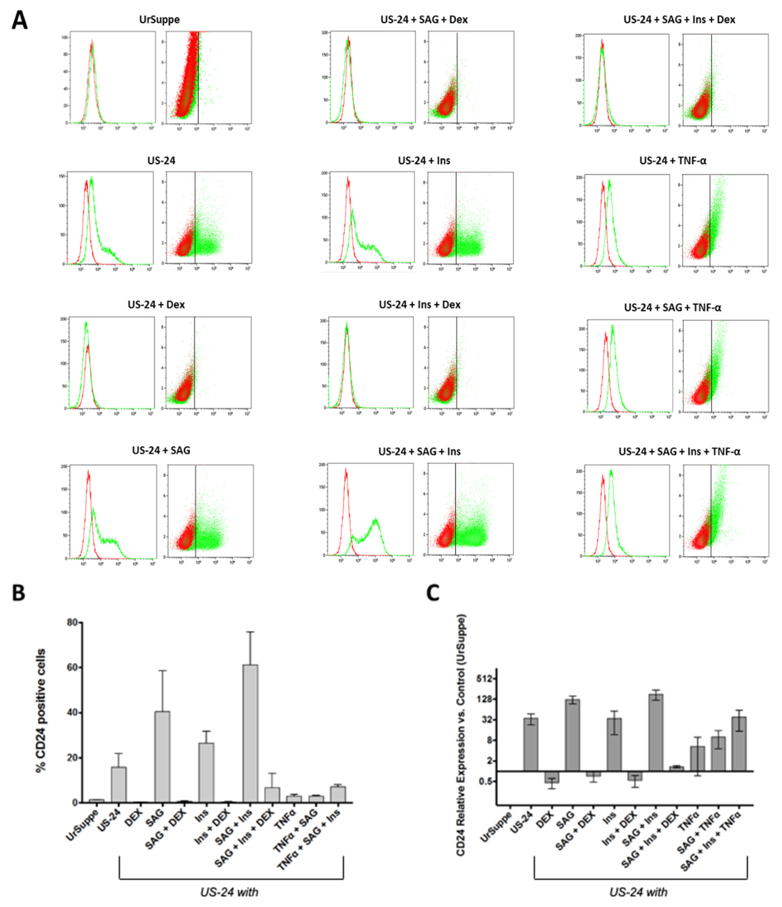
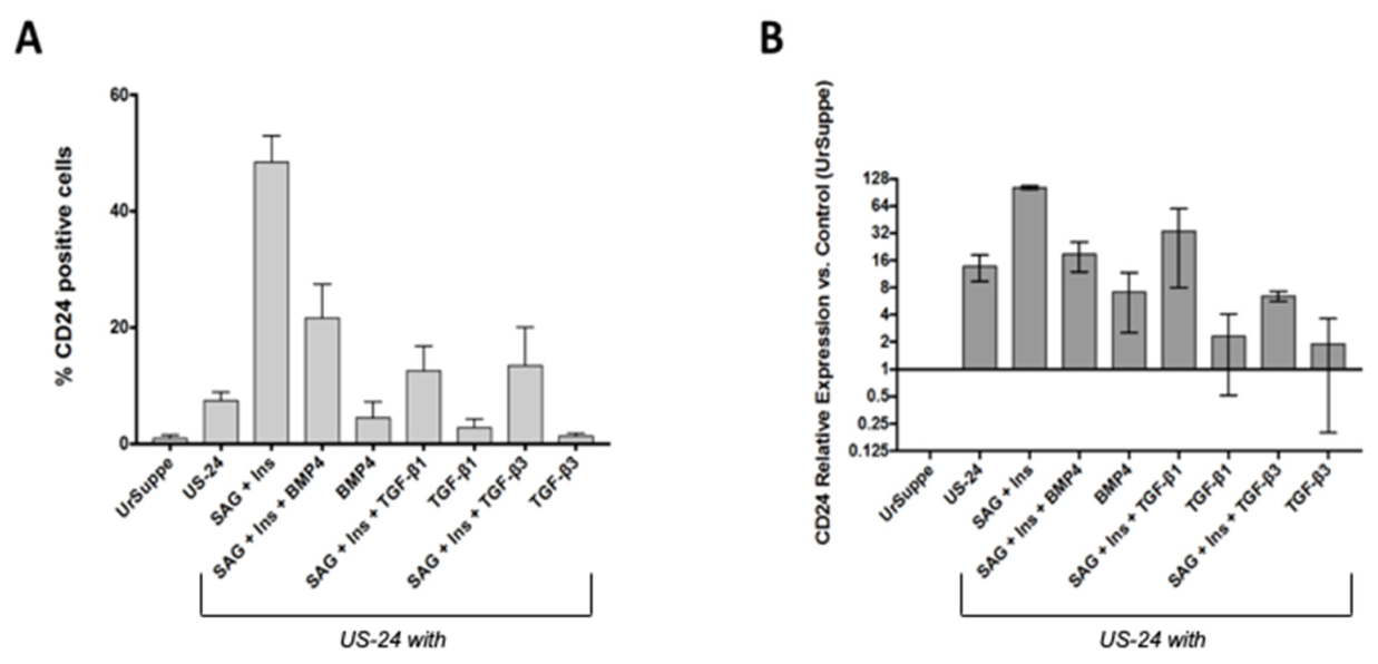
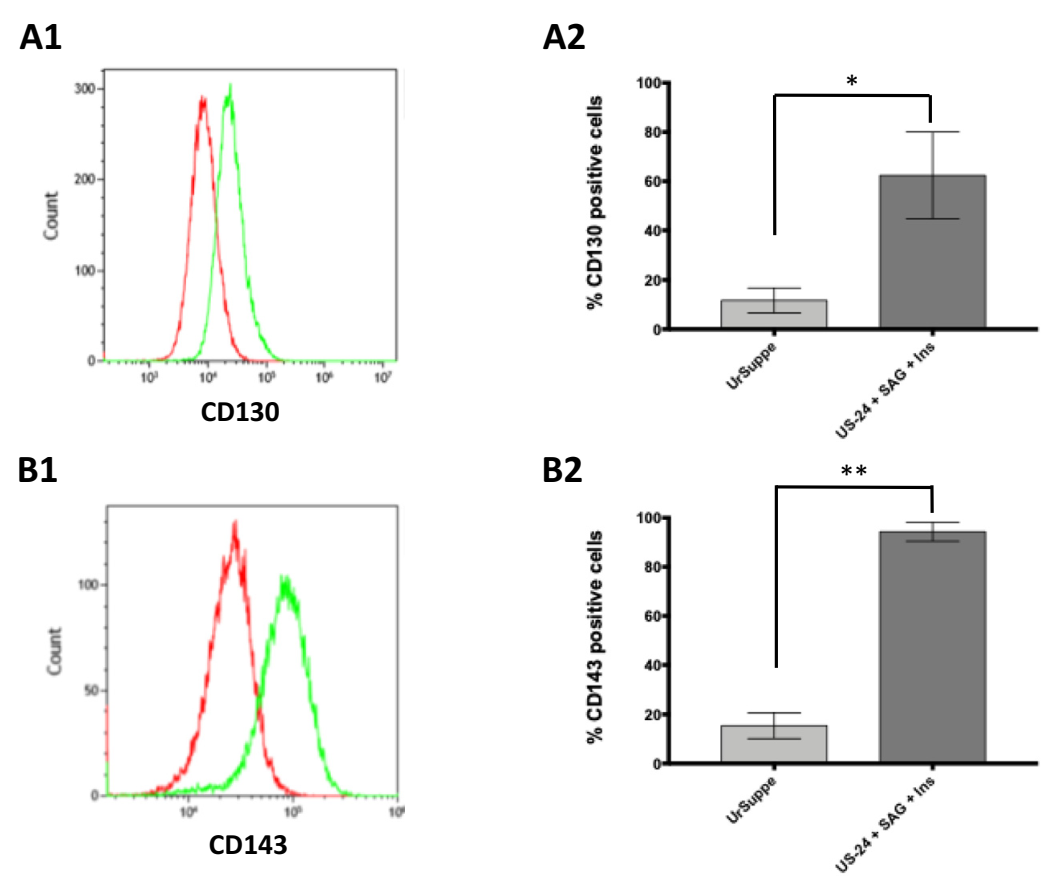
2.1. Generation In Vitro of CD24 Positive Cells from Primary hASCs

To give the Hh signaling pathway the possibility to operate without interference from other cell transduction circuits, we developed a basal-defined xeno- and serum-free medium called UrSuppe-24 (US-24). This latter is composed only of defined molecules appropriate to support cell viability without negatively interfering with the Hh signaling. We decided to use the synthetic agonist SAG (Smoothened agonist) at a concentration of 200 nM to activate this pathway. This molecule is a chlorobenzothiophene-containing compound that directly binds to Smoothened (Smo) and acts as an agonist for the Hh signaling pathway [50]. So, we analyzed by flow cytometry the hASCs obtained from six different donors for the expression of CD24 at passage 2 (P2) after growing them with the appropriate cell culture media combinations.
As shown in Figure 1A,B, the hASCs grown in the basal expansion and maintenance cell culture medium UrSuppe [51] did not express the CD24. However, we can modulate the CD24 expression on these cells when cultured in the US-24 medium with a combination of different treatments. When hASCs are grown with US-24 alone, about 20% of them are positive for CD24. With the addition of Insulin, the percentage rises to about 30%, and with SAG alone to about 40%. Finally, Insulin and SAG’s combination rises to about 60%, and some samples reach even about 80% of CD24 positive cells. Interestingly, the CD24 marker disappears from the cells’ surface by adding Dexamethasone (Dex) or TNFα (Figure 1A,B). These observations were confirmed when we analyzed the mRNA levels of CD24 by RT-qPCR analysis (Figure 1C).
These results demonstrate that it is possible to detect the CD24 on the surface of hASCs when cultured in defined conditions with a cell culture medium that does not contain inhibitors of the Hh signaling pathway. Furthermore, its expression can be enhanced by Insulin and SAG or entirely suppressed by Dexamethasone or TNFα.

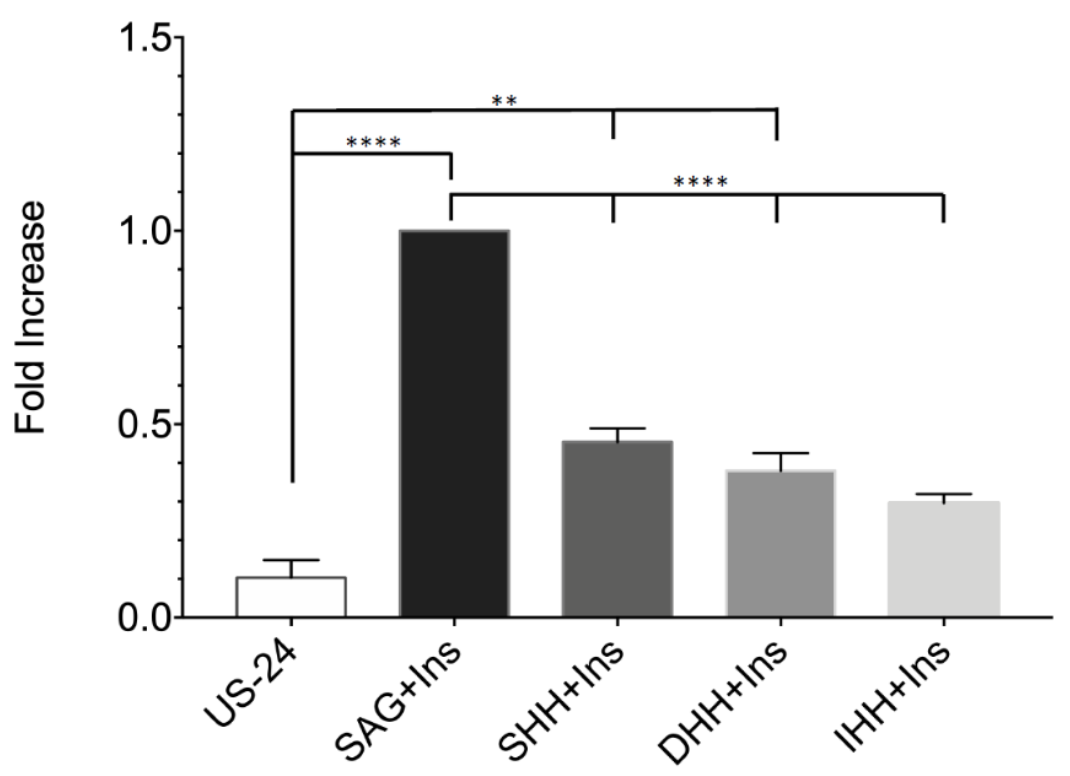
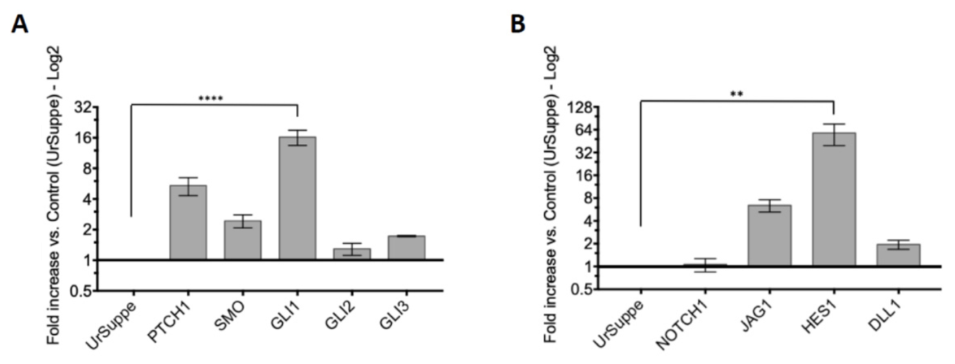
2.2. Induction of the Hedgehog Signaling and Detection by RT-qPCR of Its Core Transducing and Effector Genes

The Hh pathway is activated by the binding of one of the three natural ligands, Sonic Hedgehog (SHH), Desert Hedgehog (DHH), or Indian Hedgehog (IHH), to the receptor Patched-Smoothened [42]. As shown in Figure 2, the best performing recombinant ligand was SHH. However, compared to the inductions with SAG, recombinant SHH generated only about half of the CD24 positive cells. The percentage of CD24 positive cells relative to the value obtained with SAG as inducer decreases further with the recombinant ligands DHH: ~40%, and IHH: ~30%. The experiments performed by Robert Tokhunts et al. with processing deficient SHH mutants produced in mammalian cells also showed functional reductions between 60% and 30% compared to the processed wildtype ligand [52]. Thus, the post-transcriptional modifications of the natural Hh signaling proteins are very important to achieve full biological activity [53]. These alterations are lacking in the recombinant ligands produced in E. coli we used in these experiments. However, they can still trigger the Hh signaling pathway in hASC cultured under defined conditions in vitro. Therefore, these experiments show that it is possible to activate the Hh signaling with the three ultrapure recombinant ligands, confirming that the hASCs are receptive to this specific transduction pathway.
Next, we checked by RT-qPCR whether the hASCs grown in US-24 with SAG and Insulin expressed Hedgehog’s core transducing and effector genes. Relative to the measurements done with hASCs cultured in UrSuppe, the mRNA of the primary membrane receptors PTCH1 and SMO was upregulated following induction with US-24 with SAG and Insulin. The downstream effector transcription factor GLI1 was also significantly upregulated, while GLI2 and GLI3 less conspicuously. As shown in Figure 3A, these results confirmed that the induced hASCs were perfectly equipped to cope with ligands and transduce the Hh signaling. Interestingly, we could not detect mRNA of the three pathway ligands: SHH, DHH, and IHH in hASCs, while in SVF cells, we found SHH and DHH (data not shown). This suggests that the hASCs might be only Hh morphogen recipients and other cells in the adipose tissue secrete the ligands to trigger this signal pathway.
Some conserved signaling pathways interact at various levels to define tissue morphology, size, and differentiation during development. This is the case of Notch and Hh pathways, which have been shown to form a complex web of interaction throughout various developmental stages in different tissues. Indeed, some processes employ Notch signaling to regulate Hh response, while others utilize Hh signaling to modulate Notch response [54,55]. The Notch signaling pathway plays a crucial role in the adipose tissue because it regulates adipocyte progenitor cell proliferation and differentiation in vitro, and modulates adipose development and functions, beige adipocyte formation, and whole-body energy metabolism in vivo [56,57]. Thus, we checked by RT-qPCR analysis whether some essential genes of this pathway were also affected by the specially defined cell culture conditions favoring the Hh signaling. As shown in Figure 3B, while the expression of the Notch signaling receptor 1 (NOTCH1) did not change substantially between hASCs cultured in UrSuppe and those induced in US-24 with SAG and Insulin, the expression of two ligands of the Notch pathway, DLL1 and JAG1, significantly increased under conditions that trigger the Hh pathway. However, even more consistent was the increase in HES1, a transcriptional repressor protein classified as one of the many Notch target genes. Interestingly, it is known that HES1 is broadly detected in different tissue and cell types, including neuronal stem cells, embryonic stem cells, quiescent cells, and generally in cells at the precursor stage [58,59]. Therefore, the upregulation of HES1 is involved in maintaining stemness in mammals and reflects elevated Notch signaling pathway activity. In agreement with this notion, it was shown that HES1 blocked the differentiation of 3T3-L1 pre-adipocytes [60]. These results suggest that it is possible to observe the Notch and Hh signaling crosstalk in primary cells cultured in US-24 with SAG and Insulin. In the adipose tissue, both pathways are known as inhibitors of adipogenesis and promoters of the undifferentiated state of ASCs [42,45,56].

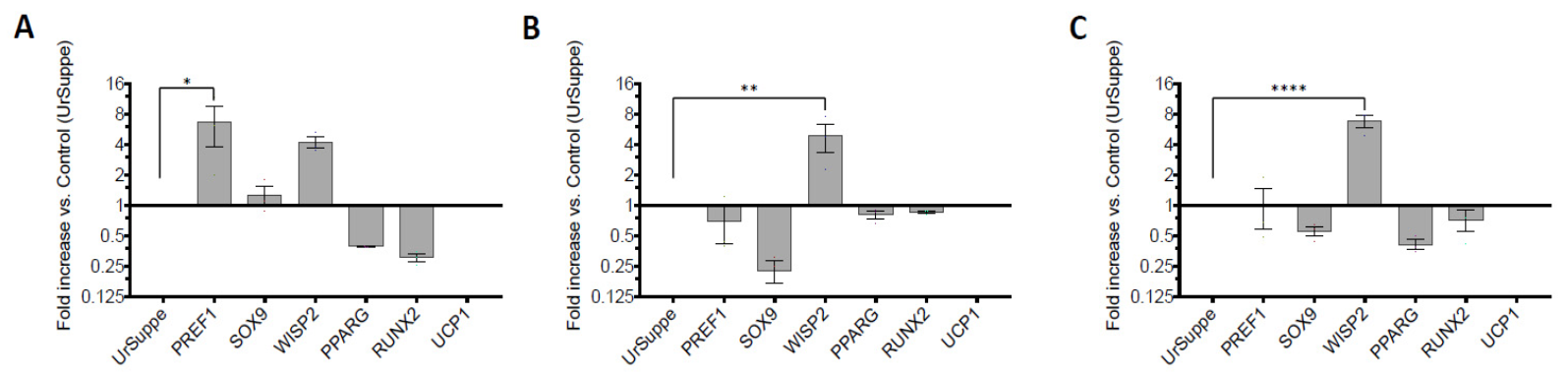
2.3. Monitoring the Expression of Selected Stemness or Cell Differentiation Genes by RT-qPCR

The cells used for this study were extracted from human subcutaneous adipose tissue. Thus, we reasoned that their default differentiation pathway would be mainly towards adipogenesis in case of spontaneous, unwanted, and uncontrolled maturation. In recent years, several essential genes have been discovered that are crucial for maintaining “stemness” or promote differentiation of hASCs. Thus, they can be used as markers in RT-qPCR tests to assess and compare the differentiation status of hASCs grown in UrSuppe medium or cultured for 5 days in US-24 with SAG and Insulin medium. We design a panel of genes to understand whether the cells are immature or heading towards differentiation. We chose three markers considered “stemness maintenance genes” for this analysis: PREF1 [61], SOX9 [62,63], and WISP2 [64]. On the other hand, we included in our analysis three genes classified as “differentiation regulators/markers”: PPARG [65,66,67], for white adipogenic differentiation; UCP1 [68], for brown adipogenic differentiation; and RUNX2 for endochondral bone development [69,70]. For more information regarding these genes, see Supplementary Materials Table S4.
Total RNAs were isolated from hASCs cultured in UrSuppe or the three different US-24 based media, and RT-qPCR was performed to analyze the expression of the six markers described above. The measured values were normalized to those obtained from hASCs grown in the UrSuppe medium. As shown in Figure 4A, the three markers signaling the cells’ undifferentiated status are significantly higher expressed in hASCs cultured with US-24 with SAG and Insulin than in UrSuppe medium. Simultaneously, the three markers signaling differentiation decreased or are not expressed (like UCP1). Interestingly, the addition of Dex (Figure 4B) or TNFα (Figure 4C) to US-24 with SAG and Insulin medium resulted in a change in the trend of marker genes expression: Those signaling an undifferentiated status slightly decreased, while those attributed to differentiation slightly increased. An exception was WISP2, whose expression was unaffected by Dex or TNFα addition in the cell culture medium.
So, triggering the Hh signaling led to reduced differentiation markers and enhanced expression of “stemness” maintenance genes in hASCs cultured in US-24 with SAG and Insulin medium. Furthermore, the cells did not choose alternative developmental pathways. Indeed, the expression of UCP1 (beige adipocytes) or RUNX2 (endochondral bone development) did not increase compared to the values found when hASCs were cultured in the UrSuppe medium.

2.4. Transforming Growth Factor Family Members Downregulate the Expression of the CD24 Antigen

The presence of CD24 on the surface of subcutaneous white adipose tissue cells has been associated by some researchers with adipose progenitor/stem cells [20], while by others, as preadipocytes committed to adipogenesis [71,72,73]. Bone morphogenic protein 4 (BMP4) plays a crucial role in the commitment of ASCs into the adipogenic lineage and in the terminal differentiation of preadipocytes into mature cells [74]. We reasoned that by adding BMP4 to hASCs cultured in US-24 with SAG and Insulin, we should have seen an increase or stabilization of CD24 expression if this surface protein was a marker of adipogenic commitment. However, as shown in Figure 5, this was not the case, and we found that BMP4 reduced the expression of CD24 on the surface of the cells. This effect was even more pronounced when we tested two BMP4-related factors: Transforming growth factor 1 (TGF-β1) and Transforming growth factor 3 (TGF-β3). The direct or indirect antagonistic crosstalk between the Hh and BMP/TGF signaling pathways is not new. For example, it has been well documented during limb development [75] and the neural tube’s dorsoventral patterning [76,77]. Thus, in the adipose tissue there may be active cell development regulatory mechanisms that have been previously described in other contexts.
Interestingly, we found that the addition of BMP4, TGF-β1, or TGF-β3 to the US-24 with SAG and Insulin medium also affected the expression of CD36 on the surface of hASCs, and BMP4 was the factor that generated the most significant increase of this marker (see Supplementary Materials Figure S1). CD36 is a practical surface antigen because it labels adipogenic progenitor cells [78,79]. Its strong upregulation in the presence of BMP4 fits with the role attributed to this factor as a promoter of adipogenesis and adipocyte maturation [74]. In the previous section, we showed that Dex, a well-known pro-adipogenic compound [80], can also downregulate the expression of CD24. Therefore, our data suggest that CD24 antigen is a marker that is most likely associated with adipose progenitor/stem cells and does not label preadipocytes committed to adipogenesis.

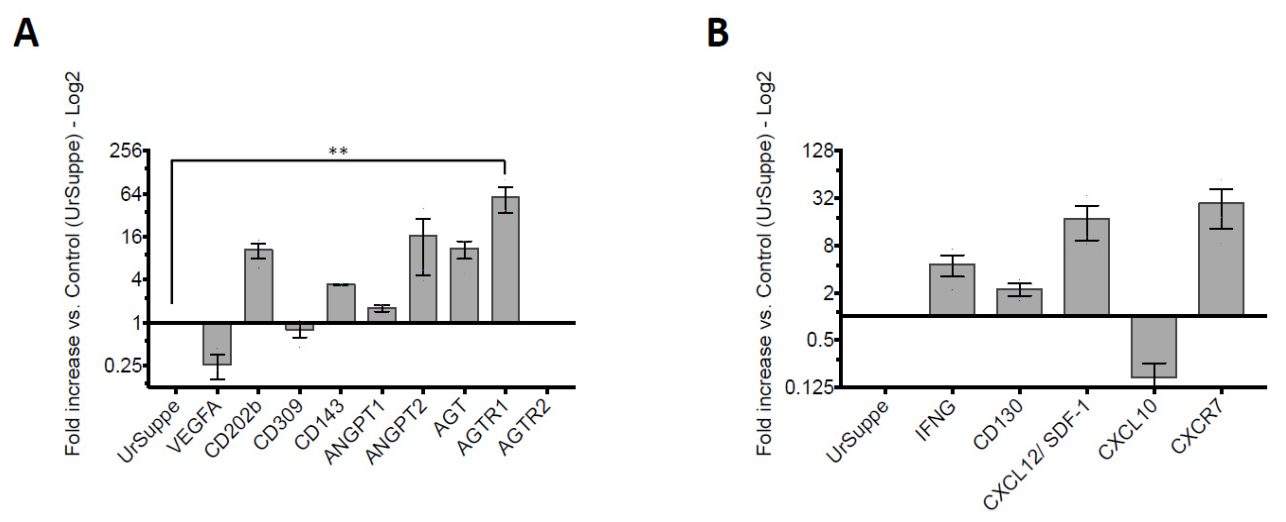
2.5. Monitoring Angiogenic Markers after Triggering the Hh Signaling Pathway in hASCs

Hh signaling is crucial for postnatal tissue repair; vascular development [81]; and in the adipose tissue, adipogenesis appears to be linked to angiogenesis. During this physiological process, the Hh signaling pathway orchestrates, together with the adipogenesis, the development of blood vessels [82,83]. This prompted us to investigate whether other surface receptors with an established angiogenic role are also induced, as the CD24 marker, by a Hh signaling pathway agonist such as SAG. After a simple screening by flow cytometry, we quickly found that CD130 (IL6-Rβ or GP130) and CD143 (ACE: angiotensin-converting enzyme) were induced when the hASCs were cultured for 5 days in US-24 with SAG and Insulin medium (Figure 6).
This result encouraged us to extend our analysis by using RT-qPCR assays to investigate other factors involved in angiogenesis that could be affected by the induction provided by the US-24 with SAG and Insulin medium. As shown in Figure 7, this was the case for most of the genes tested. The mRNA expression levels of Angiopoietin 2 (ANGPT2) and its receptor (CD202b) were upregulated when hASCs were cultured for 5 days in US-24 with SAG and Insulin. Interestingly, ANGPT2, known for its vascular remodeling property, was more induced than ANGPT1, linked to vessel maturation and stabilization. The renin-angiotensin system (RAS) was also upregulated: Starting with the mRNA coding for the ligand, angiotensinogen (AGT), up to the mRNAs coding for the receptors (AGTR1 and AGTR2) that mediate the biological effects triggered by the processed peptide hormones. Very intriguing is also the increase in the mRNA levels of CXCL12/SDF-1 and CXCR7. This constitutes a pair of ligand/receptor mRNAs that are simultaneously regulated. Activation of this pathway mediates chemotaxis, migration, and secretion of angiopoietic factors. The mRNA expression levels of IFNG and CD130 were also increased. Depending on the cellular, microenvironmental, and/or molecular context, IFNG can function as an angiogenesis inhibitor. CD130 is a transmembrane protein that can transduce signals from many different ligands, and therefore its activation can give rise to different biological responses. However, these signals transduced by CD130 often promote angiogenesis.
Only three genes were expressed at lower levels in cells cultured in US-24 with SAG and Insulin than in those grown in UrSuppe. So, the mRNA levels of the Vascular Endothelial Growth Factor A (VEGFA) and its receptor, Vascular Endothelial Growth Factor Receptor 2 or CD309, were reduced. This ligand/receptor pair clearly has to do with angiogenesis, but their signaling may also play an essential role in the tightly controlled “beiging” process of white adipocytes. Thus, it makes sense to find it inactive in the cells we analyzed. Finally, CXCL10 is an anti-angiogenic factor. We measured a decrease in its mRNA level in hASCs induced with the US-24 with SAG and Insulin medium compared to those grown in UrSuppe. This result is not surprising given that the cells were cultured in a medium supposed to promote angiogenesis.
In summary, these results suggest that triggering the Hh signaling pathway in hASCs under defined serum-free conditions leads to the expression of genes related to angiogenesis and vascular remodeling. Simultaneously, some of these genes are also known to be expressed especially by early progenitors. Thus, inducing the Hh signaling in hASCs might promote early precursors specialized in vascular morphogenesis and remodeling characterized by the expression of the CD24 surface antigen.
A list with descriptions and references regarding the 14 genes discussed in this paragraph is included in the Supplementary Materials List S1.

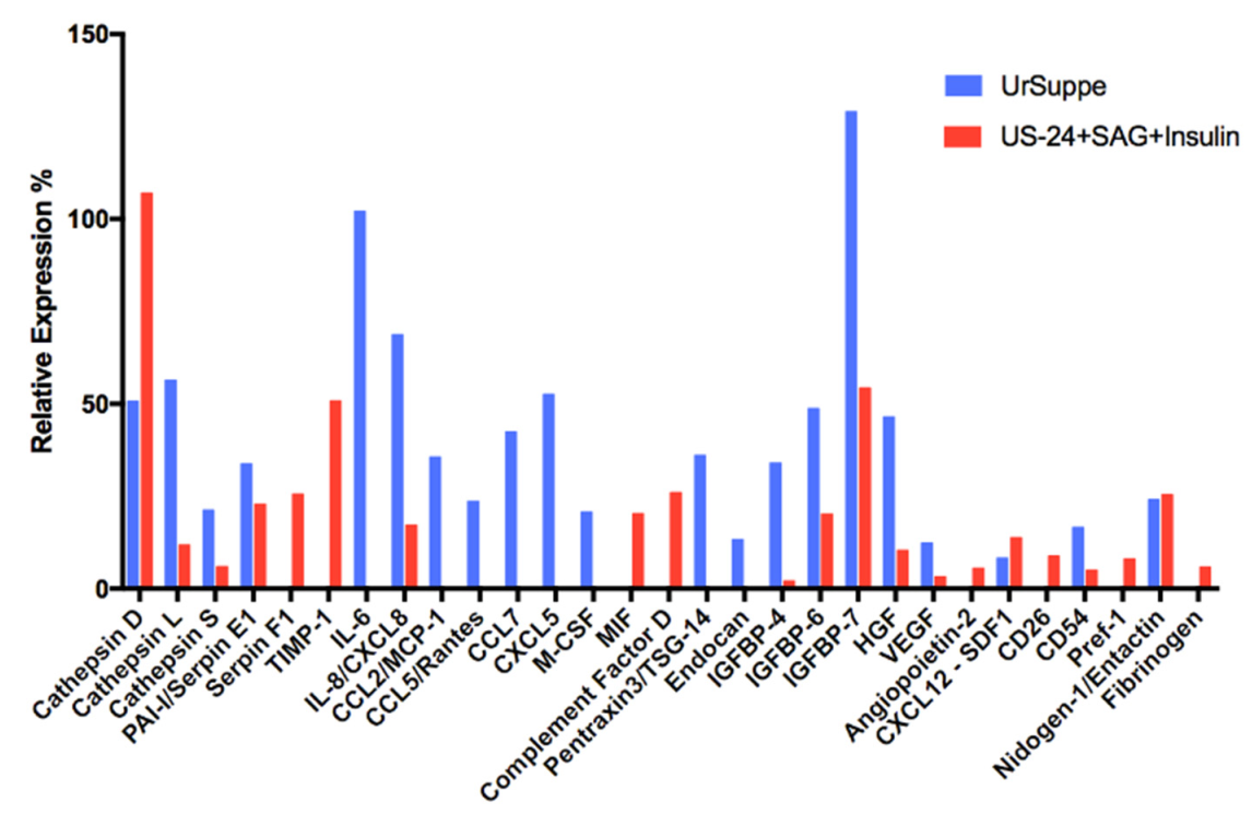
2.6. Secretome Analysis of hASCs Cultured in UrSuppe or Hedgehog Signaling Permissive Conditions

Adipose stem cells secrete many biologically active metabolic hormones, cytokines, chemokines, growth factors, extracellular matrix proteins, and tissue remodeling enzymes [6]. These polypeptides are produced and secreted only during certain precise phases of development. So, they can be considered markers and used to diagnose a specific metabolic state or a specific maturation degree of the cells under investigation. The supernatants of hASCs grown in UrSuppe or US-24 with SAG and Insulin medium were collected and subjected to an adipokine and chemokine proteome analysis using two commercial kits.
At first glance, as shown in Figure 8, the two secretion patterns were different from each other, suggesting that the cells that gave rise to the two profiles were no longer the same. Subsequently, we realized that the secreted polypeptides could be grouped based on some sheared features. Thus, the first group consists of proteases of the Cathepsin family and three protease inhibitors. Cathepsin D [84] (aspartyl protease) and Cathepsin L [85] (cysteine protease), as well as Cathepsin S (cysteine protease), play essential physiological roles in the adipose tissue and are essential for remodeling the extracellular matrix (ECM) [86]. The two last proteases are considered to be promoters of adipogenesis [84,85]. On the other hand, Cathepsin D seems to have more complex tasks and may exert mitogenic activity independent of its proteolytic activity [87]. This protease’s secretion increased when the hASCs were cultured in US-24 with SAG and Insulin medium, whereas Cathepsin L and Cathepsin S decreased. We also detected three protease inhibitors, PAI-I/Serpin E1, Serpin F1, and Timp-1 (tissue inhibitor of metalloproteases). This latter directs with the matrix metalloproteases (MMP) the regulated turnover of the ECM [88]. However, it also exerts multiple effects on biological processes [7,8]. Indeed, it was recently shown that TIMP-1 negatively regulated the adipogenesis of ASCs via the Wnt/β-catenin signaling pathway in an MMP-independent manner [89]. The two protease inhibitors PAI-I and Serpin F1 also belong to the Serpin gene superfamily [90] and are known for their anti-adipogenic effects. As far as PAI-I is concerned, one of its additional roles may be the stimulation of neovascularization in injured tissues [91]. Serpin F1 is a surprising member of this family: Indeed, it lacks a serine-reactive loop and has no function on protease inhibition. However, it plays a role in many important biological processes, such as being a negative regulator of angiogenesis, neuronal development, inflammation, and oxidative stress [92,93]. Interestingly, these two latter Serpins with clear anti-adipogenic and angiogenic effects were found only in the supernatant of cells where the Hh signaling pathway was triggered with SAG and Insulin.
The proteome profiler arrays also detected several cytokines and chemokines in the cell-conditioned media: IL-6, IL-8, CCL2/MCP-1, CCL5/Rantes, CCL7, CXCL5, M-CSF, MIF, Complement Factor D, and Pentraxin3/TSG-14. In addition to their roles as immunological playmakers, most of these factors have other regulatory functions that are often unknown or poorly characterized. However, we noted that most inflammatory cytokines and chemokines were reduced or missing in the supernatant of hASCs cultured in US-24 with SAG and Insulin medium. This also includes IL-6, which disappears, and IL-8, which is reduced. On the other hand, two factors are detected only when hASCs were cultured in the Hh signaling permissive medium. Macrophage migration inhibitory factor (MIF) is a pleiotropic factor with chemokine-like functions involved in cell-mediated immunity, immunoregulation, and inflammation [94]. Recently it was implicated as a mediator in neo-angiogenesis/vasculogenesis by endothelial cell activation and endothelial progenitor cell recruitment [95]. During adipogenesis, the role of MIF was investigated by Atsumi et al. and found to be anti-adipogenic [96]. This finding agrees with the known ability of MIF to counter-regulate the immunosuppressive effects of glucocorticoids [97], which are also well known for their capacity to induce adipocyte maturation. Conversely, Complement Factor D (adipsin) was described as a pro-adipogenic protein [98,99].
The next group of related polypeptides consists of the Insulin-like growth factor-binding proteins (IGFBPs). One of their primary function is to regulate Insulin-like growth factors (IGF-I, IGF-II) action by controlling the accessibility of these peptide hormones to their receptors [100]. Both IGF-I [101] and IGF-II [102,103] are pro-adipogenic. It has been shown that IGFBP-4 binds IGF-I and its expression negatively correlates with adipose tissue growth [104]. IGFBP-6 can bind IGF-II, while IGFBP-7 both IGF-I and II [105]. Thus, it is likely that these three members of the IGFBP family are anti-adipogenic. Interestingly, experimental evidence suggests that IGFBP-4 and IGFBP-6 are predominantly anti-angiogenic, whereas IGFBP-7 has pro and anti-angiogenic properties [106].
In the cell culture supernatants, we also detected four factors related to angiogenesis and cell proliferation: Hepatocyte Growth Factor (HGF) [107], Vascular Endothelial Growth Factor (VEGF) [108], Angiopoietin-2 [109], and CXCL12/SDF-1 [110]. In addition, we detected the soluble version of CD26, CD54, and Pref-1 as hASC-specific secretions. These proteins are encoded by genes classified as adipose progenitor cell markers [111,112]. Therefore, we consider their presence in the conditioned media of hASCs as a positive result that confirms the data obtained previously by flow cytometry or RT-qPCR. Nidogen/Entactin is a typical component of the ECM found in the adipose tissue [88] and, therefore, it is not surprising to find it in the cell supernatants.
The canonical role of Fibrinogen (Fg) is traditionally linked to its well-recognized role in blood clotting and hemostasis [113]. However, other functions for this protein were described over the years. For example, Fibrinogen (Fg) was shown to act as a bridging molecule to enhance leukocyte attachment to endothelial cells in vitro through a simultaneous interaction with CD11b/CD18 (LFA-1) on the leukocyte and intercellular adhesion molecule CD54 on the endothelium [114,115]. Furthermore, Fg and some of its fragments induced mitogenic activity in T and B lymphocyte cell lines and hematopoietic progenitor cells through the cell surface receptor CD54 [116]. This effect may explain why Fg has long been employed in the clinic as a synthetic matrix, providing a scaffold or a delivery system to facilitate tissue repair. Interaction between CD54 and Fg may also regulate endothelial cell survival [117]. Interestingly, Fg was detected only in the supernatant of cells cultured in US-24 with SAG and Insulin, even if its secretion was moderate.
The Endothelial cell-specific molecule 1 (ESM-1), also called endocan, described initially as an endothelial-specific molecule, was later found to also be expressed by adipocytes [118]. In vitro, endocan interferes with the molecular mechanisms of immune cell migration by binding to adhesion molecules. Indeed, David Béchard et al. showed that endocan directly binds to the integrin CD11a/CD18 (LFA-1) on lymphocytes and monocytes’ cell surface, inhibiting the interaction of CD54/ICAM-1 with CD11a/CD18 binding sites [119]. We detected endocan only in the conditioned medium of hASCs grown in UrSuppe medium and not in cells cultured in US-24 with SAG and Insulin medium. As described before, Fg and endocan target the same cell surface receptor system with opposite results: Fg acts as an adapter between LFA-1 and CD54, while endocan prevents this interaction. Therefore, it appears that there is a coordinated regulation of these two proteins in hASCs grown in US-24 with SAG and Insulin medium: Endocan is downregulated and Fg upregulated.
As a preliminary observation, the secretion profile of hASCs grown in UrSuppe medium seems different from that obtained with the same cells cultured in defined Hh signaling permissive conditions. The secretion of inflammatory mediators is reduced in the supernatants of hASCs cultured in US-24 with SAG and Insulin medium. Conversely, some anti-adipogenic or angiogenic factors and proteins related to early progenitor cells are mainly present in the conditioned media when hASCs are grown with an active Hh signaling pathway.

3. Discussion

Ryan Berry and Matthew S. Rodeheffer showed that the expression of the cell surface molecule CD24 was critically important for the reconstitution of murine WAT function in vivo [19]. These authors also proposed a model where the CD24-positive adipocyte progenitors become further committed to the adipocyte lineage as CD24 expression is lost, generating CD24-negative preadipocytes, which express late markers of adipogenesis [19,20]. Thus, CD24 has all the credentials to consider it an indicator of cell stemness and an essential factor in adipogenesis. In these last years, we developed a defined serum- and xeno-free cell culture medium called UrSuppe, specifically formulated to grow primary hASCs [51]. A crucial peculiarity for a stem cell culture medium is its ability to preserve the cells’ undifferentiated status and prevent spontaneous maturation during the in vitro amplification phase. So, we thought that obtaining CD24 positive hASCs in vitro would confirm their immature status. Furthermore, since the functional importance of CD24 in human ASCs is still unclear, an in vitro model could help understand its role during adipogenesis.
The scientific literature gave us a hint to start our studies: The Hedgehog signaling pathway could be responsible for the upregulation of the CD24 antigen [38,39]. Furthermore, Nicole C. Smith et al. also showed that the addition of 3-isobutyl-1-methylxanthine (IMBX) and dexamethasone (Dex) increases the expression level of CD24 mRNA and protein in both the murine 3T3-L1 cell line and in primary mouse preadipocyte [72]. The cells used for these experiments were grown in non-defined media containing 10% of new-born calf serum [72]. Recently, Florian M. Hatzmann et al. found that approximately 3% of human SVF ASCs were positive for CD24. Furthermore, they could induce CD24 expression in hASCs using a serum-free medium without apparently activating the Hh signaling pathway [33]. These two studies indicate that CD24 may also be upregulated by other, currently non-characterized, signal transduction circuits.
We noticed that triggering the Hh signaling pathway in hASCs under defined serum-free conditions leads to gene expression related to angiogenesis and vascular remodeling. CD24 is a GPI-linked sialoglycoprotein that was initially related to B lymphocytes and hematopoietic lineages [35]. In general, it is highly expressed on stem, and to a lesser extent, on terminally mature cells [21,37]. It gained prominence as a marker of cancer stem cells linked to metastatic progression. The relationship between CD24 and metastasis probably relates to the ability of CD24 to facilitate interactions with the vasculature. This ability of CD24 to alter adhesive properties may also explain why putative stem cells expressing CD24 can engraft and reconstitute diverse organ systems, whereas those lacking CD24 do not [19]. The role of Hh signaling has been well documented not only for cancer vascularization and metastasis but also in normal vascular development, maturation, and maintenance [81]. Thus, we suggest that CD24-positive hASCs may characterize early rare progenitor cells specialized in vascular morphogenesis and remodeling. Angiogenesis plays a crucial role in the modulation of adipogenesis: When the adipose tissue expands, both processes must perfectly time the development of microvasculature and new adipocytes’ maturation [83]. So, it could be possible that CD24-positive hASCs and the Hh signaling play an instructive role and orchestrate the expansion and metabolism of fat mass. Hh signaling has a known and evolutionary conserved role in preventing preadipocyte differentiation into mature fat cells [44,45]. However, also profound differences in the function of Hh signaling in rodent and human ASCs have been reported. Indeed, in humans, triggering the Hh transduction system in ASCs leads to adipogenesis and osteogenesis inhibition. On the contrary, in rodent cell lines, adipogenesis is blocked while osteogenesis is stimulated by the expression of osteoblast markers, such as Runx2, and mineralization enzymes, such as alkaline phosphatase [44,46,120].
Interestingly, the CD24 marker disappears from the cells’ surface by adding to the US-24 medium either Dex or TNFα. Matthew S. Rodeheffer et al. performed in vivo fat mass reconstitution assays using murine early adipocyte CD24-positive or CD24-negative progenitor cells. Only CD24-positive cells were able to proliferate, form an adipose tissue depot, and correct blood glucose levels in the transplanted lipodystrophic mice. It is known that TNFα plays a crucial role in the onset and perpetuation of obesity and insulin resistance [121,122]. Indeed, the expression of this inflammatory cytokine is elevated in the adipose tissue of multiple experimental models of obesity [123]. Therefore, it could be possible that one TNFα target in the adipose tissue is the CD24-positive ASC. Downregulation of CD24 could point out, as demonstrated by Matthew S. Rodeheffer et al. [18], that this progenitor cell has lost the ability to differentiate appropriately, forcing adipose tissue to become hypertrophic and triggering the metabolic pathologies typical of obesity. Growing evidence has given rise to the idea that obesity could be defined as a disease caused by adipose stem cells’ dysfunction [124]. We may now have the chance to test this hypothesis in vitro with our defined cell culture platform since we can grow significant numbers of human CD24-positive ASCs and study at the molecular level the effects on these cells of two obesogenic molecules like TNFα and Dex.
TGF-β1, TGF-β3, and BMP4 were also able to counteract the expression of CD24 in hASCs cultured in US-24 with SAG and Insulin. Simultaneously, the expression of CD36, a reliable marker signaling adipocyte maturation [79], was upregulated on the surface of hASCs. BMP4 plays a crucial role in ASCs’ commitment to adipogenic lineage and terminal differentiation of mature adipocytes [74]. Therefore, we concluded that CD24-positive hASCs do not represent preadipocytes committed to adipogenesis but a particular stem cell type of the subcutaneous adipose tissue. However, it is still unknown if these cells are the most potent stem cells in the adipose tissue or the CD24-positive population is maintained by another, currently unidentified, adult population. So, in the future, it will be essential for the CD24-positive hASCs to find their place inside the adipose tissue progenitor cells hierarchy, which is beginning to emerge from recent studies [111,112,125].
The Hh signaling pathway is crucial for growth and patterning during embryonic development. However, it is mostly quiescent in adults, regulating tissue homeostasis and stem cell behavior [49]. This is probably why not many genes are known in the adult organism, whose expression is regulated by the Hh signaling pathway. This study found several important genes, such as CD130, CD143, CD202b, SDF-1, and CXCR7, which the Hh signaling pathway could directly or indirectly regulate. Further specific studies are needed to distinguish between these two possibilities. Single-cell RNA sequencing (scRNAseq) technology was recently used to find new gene markers useful to characterize the cellular subpopulation involved in adipogenesis [112,126,127]. This method helped David Merrick et al. [111] identify distinct types of progenitor cells in murine subcutaneous adipose tissue, which were subdivided into three hierarchical groups based on their gene expression patterns [112,125,128,129]. Unfortunately, the authors of these studies did not attempt to assign a precise collocation in one of these three groups for the CD24-positive ASCs originally described by Matthew S. Rodeheffer et al. [18]. This is perhaps because CD24-positive ASCs are very rare in mouse tissue and have escaped the scRNAseq analysis. The gene expression pattern we detected by RT-qPCR assays indicated that these cells were undifferentiated and most likely classifiable as progenitors positioned high in the subcutaneous adipose tissue stem cell hierarchy.
CD24 could turn out as an exciting and valuable marker for the study of adipose tissue. Indeed, in mice, only less than 1% of the SVF cells that express CD24 also display the complete cell surface marker profile of adipocyte progenitors [19]. In the human SVF, CD24-positive ASCs are also rare [33]. The properties and functions attributed to this surface protein definitively promote it as a marker of hASCs and other types of stem cells. Surprisingly, Nika Shakiba et al. found that CD24 acts as an excellent marker to track the initiation of reprogramming following the exogenous overexpression of the four transcription factors (OCT4, KLF4, C-MYC, SOX2) that allow somatic cells to be induced to a pluripotent state and during which the cells acquire a CD24high/SSEA1 state [37].

4. Materials and Methods

4.1. Isolation and Culture of Human Adipose Stem Cells (hASCs)

4.1.1. Extraction of Adipo-Cutaneous Tissue

For studies involving human tissues, we obtained ethical approval from the local Ethics Committee of the Canton Ticino (Switzerland), which approved the project and its procedures (project reference number: CE 2915). A written agreement in compliance with the Ethics Committee’s directives was obtained from all patients who donated their spare adipo-cutaneous tissue, and all personal information was made anonymous by encryption. Human adipose tissue samples were obtained from tissue excess originating from surgical interventions performed in the Department of Plastic, Reconstructive and Aesthetic Surgery at the “Ospedale Regionale di Lugano.” Briefly, this study’s cellular source originated from adipose tissue harvested from the abdominal region of female patients undergoing autologous breast reconstruction with general anesthesia [51]. Adipose tissue biopsies were kept at room temperature and processed within 24 h to obtain the Stromal Vascular Fraction (SVF) [130].

4.1.2. Isolation of Stromal Vascular Fraction (SVF)

Extraction of the SVF from human adipose tissue, in vitro expansion of ASCs, and their cryopreservation conformed to the ethical principles outlined in the Declaration of Helsinki and were in compliance with the directives of the Ethics Committee of the Canton of Ticino (Switzerland).
SVF was isolated from fresh subcutaneous adipose tissue in four steps: homogenization, enzymatic digestion, hydrophilic separation, and filtration. (1) Homogenization: Adipose tissue was washed with DPBS with Ca2+ and Mg2+ (Bioconcept AG, Allschwil, Switzerland) in a Gosselin sterile container (Corning, New York, NY, USA), chopped into smaller pieces, and homogenized with a handheld blender. (2) Enzymatic digestion: The homogenized fat was then poured into an Omnifix 100 mL Syringe (BBraun, Melsungen, Germany) for dissociation with Collagenase Type B, Animal Origin Free (Worthington, Biochemical Corp., Lakewood, NJ, USA) at a final concentration of 0.28 Wünsch U/mL for 45 min at 37 °C under constant but gentle agitation. (3) Hydrophilic separation: 30 mL of an injectable 1% human albumin solution (CLS Behring, Bern, Switzerland) in DPBS without Ca2+ and Mg2+ (Biowest, Nuaillé, France) were aspirated and the syringes were thoroughly shaken to extract the cells. The syringes were placed vertically to allow the separation of the two phases. The lower layer (hydrophilic phase), which contains the SVF, was carefully poured into a conical 50 mL tube (TPP, Trasadingen, Switzerland). The extraction step was repeated with 20 mL on 1% human albumin solution, and subsequently, the hydrophilic phase was centrifuged (600× g for 10 min at Room Temperature, RT). (4) Filtration: The dissociated cells were resuspended and filtered through a 100 µm and then through a 40 µm sieve cell strainer (Becton Dickinson, Franklin Lakes, NJ, USA). At the end of the final centrifugation (600× g for 10 min at RT), the cellular pellet was resuspended in an injectable 1% human albumin solution or cell culture medium.
The SVF extraction and characterization by flow cytometry were fully described in our previous study [51].

4.1.3. Seeding and Culture of hASCs

Cells were always and exclusively cultivated using our proprietary defined serum-and xeno-free cell culture medium called “UrSuppe” [51]. The medium contains only defined molecules, some recombinant human growth factors, and injectable human albumin in the µg range. After the characterization and enumeration of the cells of the SVF by flow cytometry, the cells were seeded at a density of ~30,000 ASCs/cm2 in Fibronectin pre-coated vessels [131] (Corning PureCoat™ ECM Mimetic Fibronectin Peptide, Corning Inc., Corning, NY, USA) and kept in a humidified incubator at 37 °C with 5% CO2 at 90% humidity. This initial passage of the primary cell culture was referred to as passage 0 (P0). Cells were maintained in culture until they achieved 75–90% confluence. They were then either collected and cryopreserved or passaged in new Fibronectin pre-coated tissue culture vessels at a density of ~5000–10,000 ASCs/cm2. The medium was changed every two days, always keeping 20–50% of the conditioned medium until the cell reached 75–90% confluency. Cells were detached with TrypLEe Select [132] (Life Technologies, Thermo Fisher Scientific, Waltham, MA, USA) for 2 min at 37 °C, then washed with PBS supplemented with 1% injectable human albumin, and pelleted by centrifugation at 400× g for 5 min. After discarding the supernatant, the cells were counted with Trypan Blue (Thermo Fisher Scientific, Waltham, MA, USA), resuspended in UrSuppe, and passaged or used for various tests. Characterization by flow cytometry of passaged hASCs was shown in our previous study [51].

4.2. Induction and Characterization of CD24 Positive Cells

4.2.1. Generation of CD24 Positive Cells

hASCs were expanded in the defined xeno- and serum-free UrSuppe medium up to passage P2. UrSuppe contains only defined molecules, some recombinant human growth factors, and injectable human albumin in the μg range. Once the confluence was reached, the usual cell culture medium was replaced by UrSuppe-24 (US-24), a precisely defined xeno- and serum-free medium derived from UrSuppe, which we developed to induce the CD24 antigen in hASCs. This medium contains human Insulin (at 5 µg/mL, Sigma, St. Louis, MO, USA) since this hormone’s canonical signaling mechanism involves the activation of the phosphoinositide-3-kinase (PI3K) AKT pathway, which acts as an agonist with the Hh signaling components [101,102] and additionally has positive effects on the vitality of the cells.
The ready-to-use US-24 medium also incorporates the synthetic Hh agonist called Smoothened Agonist (SAG-dihydrochloride, 3-Chloro-N-[trans-4-(methylamino)cyclohexyl]-N-[3-(4-pyridinyl)benzyl]-1-benzothiophene-2-carboxamide·2HCl, water-soluble; AdipoGen, San Diego, CA, USA) at the final concentration of 200 nM [50,133].
The reason that led us to formulate US-24 lies in the fact that undefined media contain factors that could activate antagonistic pathways to the Hh signaling. Indeed, several antagonistic interactions between the Hh and other signaling pathways have been described in numerous studies. For example, inducers of intracellular cAMP and Protein Kinase A [134,135], the Wnt and Notch signaling pathways [136], the Bone Morphogenetic Proteins (BMPs) group of signaling molecules [75], Glucocorticoids [137], Retinoic Acid [138], and Interferon-gamma [139]. Inevitably, these Hh signaling antagonists (or agonists) can be found in large and unknown quantities in all undefined cell culture systems based on fetal bovine serum (FBS) or platelet lysate (PL). Glucocorticoids, BMPs, and other growth factors are ordinarily present in FBS and PL [140,141,142].
To activate the Hh signaling, in some experiments, we replaced the synthetic agonist SAG with the natural ligands of this pathway: Human recombinant Sonic Hedgehog (SHH); Human recombinant Desert Hedgehog (DHH); and Human recombinant Indian Hedgehog (IHH). SHH, DHH, and IHH were used at the final concentration of 50 ng/mL and purchased from ProSpec-Tany Ltd., Rechovot, Israel.
Human ASCs cultured in normal UrSuppe or US-24 without SAG and Insulin were used as negative controls.
In some experiments, primary hASCs were treated with 50 nM Dexamethasone (DEX) (Cayman Chemical, Ann Arbor, MI, USA) or 2 ng/mL human TNFα, or with members of the TGF-β superfamily: TGF-β1, TGF-β3, and BMP4, all three at the concentration of 10 ng/mL and purchased from ProSpec-Tany Ltd., Rechovot, Israel. The experiments shown in this study were done with hASCs samples that had at least 60% of CD24-positive cells on flow cytometric analysis.
Interestingly, we found that the coating of the cell culture vessels used to grow and test the hASCs played a crucial role. When Fibronectin was used as coating substrate (Corning PureCoat™ ECM Mimetic Fibronectin Peptide, Corning Inc., Corning, NY, USA), it was possible to reach a significant expression of the CD24 antigen with the US-24 with SAG and Insulin medium (data not shown). This was not the case when, for example, Collagen I coating was employed instead.

4.2.2. Flow Cytometry Analysis

hASCs were detached, washed, and resuspended with FACS buffer (PBS supplemented with 1% injectable human albumin and 50 ng/µL of human immunoglobulins, CLS Behring, Bern, Switzerland). All centrifugation steps were performed at 400× g for 5 min. For each sample, 50,000 cells (in 100 µL FACS Buffer) were used and incubated with appropriate fluorochrome-conjugated antibodies for 20 min a RT in the dark. Finally, the sample was diluted with 100 µL FACS Buffer before the acquisition. Cells were analyzed using a CytoFLEX Flow Cytometer (Beckman Coulter Inc., Pasadena, CA, USA), recording 30,000 cells/test. Data analysis was performed with Kaluza Flow Cytometry Analysis Software (Beckman Coulter Inc., Pasadena, CA, USA). All antibodies were titrated to optimize the signal-to-noise ratio and used at a specific concentration. Isotype controls and specific mAbs were used at the same final concentrations; further information about the different antibodies can be found in Supplementary Materials, Table S9.

4.2.3. RNA Preparation, cDNA Synthesis and Quantitative Real-Time PCR (RT-qPCR) Analysis

Total RNAs were purified using the Nucleospin® RNA kit (Macherey-Nagel, Düren, Germany) according to the manufacturer’s instructions. The purification procedure includes an on-column digestion step with DNase I. cDNA was obtained from 400 ng RNA using GoScriptTM Reverse Transcription System (Promega, Madison, WI, USA) and T100TM Thermal Cycler (BIO-RAD, Hercules, CA, USA); the protocol can be found in Supplementary Materials, Table S10. RNA and cDNA concentration and purity were determined by measuring the absorption at 260/280 nm with NanoDropTM 2000/2000c Spectrophotometers (Thermo Fisher Scientific, Waltham, USA). RT-qPCR was done using SsoAdvancedTMUniversal SYBR® Green Supermix kit (Biorad, Hercules, CA, USA) and detected with the CFX Connect Real-Time System (Biorad, Hercules, CA, USA). RT-qPCR was performed using 20 ng of cDNA for each sample, and RPL13A was used as an internal control gene. Each primer pair product was examined for proper amplification by agarose gel electrophoresis. We used exclusively primers, which gave rise to single sharp bands of the expected size. Primer sequences, temperature, and cycle conditions can be found in Supplementary Materials, Tables S11 and S12. Relative mRNA levels were normalized to RPL13A mRNA by the ∆∆Ct method. The mean Ct was converted to relative expression value by the equation: 2−∆∆Ct. All experiments were done in triplicate.

4.2.4. Secretome Profiler

The conditioned cell culture medium of hASCs cultured for 5 days in US-24 with SAG and Insulin medium was analyzed to detect secreted cellular factors. The medium was filtered through a 0.22 µm syringe mesh (Jet Biofil, Guangzhou, China) to remove cellular debris, then 1 mL of supernatant was incubated overnight with a nitrocellulose membrane containing many different capture antibodies. The array procedure was carried out according to the instructions provided with the Proteome Profiler Human Adipokine Array Kit (#ARY024) and Proteome Profiler Human Chemokine Array Kit (#ARY017), both manufactured by R&D Systems, Minneapolis, MN, USA. The secondary biotinylated detection antibodies were highlighted using Streptavidin conjugated IRDye 800 CW (Li-Cor Corporate, Lincoln, NE, USA). Positive signals were detected after scanning the membrane using an Odyssey® CLX Imaging System (Li-Cor Corporate, Lincoln, NE, USA).

4.3. Statistical Analysis

Statistical analyses were performed using one-way ANOVA in Prism 7 (Graph Pad Software Inc., San Diego, CA, USA). A p-value lower than 0.05 is considered statistically significant. More information about the statistical analysis can be found in the Supplementary Materials Tables S1–S3, S5, and S6.
A detailed description of all the chemicals and reagents used in this study is shown in the online Supplementary Materials Table S13.

5. Conclusions

The CD24 glycosylphosphatidylinositol (GPI)-linked cell surface receptor is an intriguing marker used to characterize early murine adipocyte progenitors [19,20]. However, CD24-positive ASCs are elusive, and the presence of these cells in human adipose tissue is poorly documented in the literature. Thus, in the present study we investigated how to upregulate CD24 expression in primary human ASCs and its possible functional meanings. We showed for the first time that this marker can be induced in primary human ASCs by the Hh signaling pathway. This was only possible thanks to the development and employment of defined serum-free media. Indeed, we observed that many growth factors and hormones, commonly contained in fetal bovine serum or platelet lysate, are antagonists of the Hh signaling. Surprisingly, even small biochemical molecules and the type of coating used in the experiments can modulate the Hh signaling pathway.
We show that in hASCs, the CD24 marker can be induced by the synthetic Hh signaling pathway agonist SAG or with the three natural ultrapure human recombinant ligands (SHH, DHH, IHH, produced in E. coli). So, triggering the Hh signaling in hASCs with SAG brought the CD24 marker to appear on their surface and led to reduced differentiation markers, enhanced expression of “stemness” maintenance genes, and the emergence of genes involved in angiogenesis and vascular remodeling. The Hh signaling is known mainly for its role in embryonic development, while few target genes for this pathway have been described in the adult organism. This study found several important genes, such as CD130, CD143, CD202b, SDF-1, and CXCR7, in which the Hh signaling pathway could directly or indirectly regulate in adult cells.
Two studies suggest that CD24 may also be upregulated by other, currently non-characterized, signal transduction circuits different from the Hh signaling pathway [33,72]. It is proposed that CD24 may act as a membrane-organizing factor that regulates the association and localization of cell surface receptors with their canonical ligands [30]. The fact that different signaling circuits can induce it is compatible with the postulated role for this marker. Indeed, different transduction systems could use CD24 as an adapter, and the specificity of the interactions might be determined by the different glycosylation patterns of this GPI-anchored protein. Therefore, it will be interesting to check whether the glycosylation pattern of the CD24 surface antigen changes according to the transduction signaling used for its upregulation.
Taken together, we believe that CD24 is an exciting multitasking protein that could help better understand adipose tissue biology in health and disease. With this new cell culture platform described in this study, we can now grow large numbers of CD24-positive hASCs in vitro. Based on the data from this study, we think that CD24-positive hASCs may characterize early rare progenitor cells specialized in vascular morphogenesis and remodeling and do not represent preadipocytes committed to adipogenesis. Therefore, it will be exciting to apply single-cell RNA sequencing to identify and profile these cells and find their collocation in the subcutaneous adipose tissue stem cell hierarchy, which is still yet to be clarified [111,112,125].

Supplementary Materials

The following are available online at https://0-www-mdpi-com.brum.beds.ac.uk/article/10.3390/biologics1020008/s1, Table S1: Generation in vitro of CD24 positive cells from primary hASCs: Statistical significance for flow cytometer analysis, Table S2: Generation in vitro of CD24 positive cells from primary hASCs: Statistical significance for RT-qPCR analysis, Table S3 CD24 upregulation in hASCs mediated by recombinant natural Hh ligands and synthetic SAG agonist, Table S4: Overview of measured genes, Table S5: CD24 positive hASCs & transforming growth factor β family: Statistical significance for flow cytometer analysis, Table S6: CD24 positive hASCs & transforming growth factor β family: Statistical significance for RT-qPCR analysis, Figure S1: Analysis of CD24 and CD36 expression in hASCs, List S1: Description and references regarding the 14 genes, Figure S2: Comparing the adipokines and chemokines secretion profile of hASCs, Table S7: Human adipokine array, Table S8: Human chemokine array, Table S9: Detailed information of the antibodies used for the flow cytometric measurements, Table S10: Reverse transcription detailed procedure, Table S11: RT-qPCR cycle conditions, Table S12: Primer sequences, Table S13: Materials. References [143,144,145,146,147,148,149,150,151,152,153,154,155,156,157,158,159,160,161,162,163,164,165,166,167,168,169,170,171,172] are cited in the Supplementary Materials.

Author Contributions

T.T. conceived and designed the experiments; Y.H. selected the adipose tissue donors and collected the biopsies; F.M. and S.P. processed the human adipose tissue samples; F.M. performed all experiments; F.M., S.P. and T.T. analyzed and interpreted the results; F.M. curated the data; F.M. performed the statistical analysis; F.M. wrote the original draft of the paper; F.M., S.P. and T.T. wrote, reviewed, and edited the original manuscript. All authors have read and agreed to the published version of the manuscript.

Funding

This work was supported by the Cardiocentro Ticino Foundation (CCTF) and the Foundation for Cardiological Research and Education (FCRE).

Institutional Review Board Statement

Extraction of the SVF from human adipose tissue, in vitro expansion of ASCs, and their cryopreservation were in line with the ethical principles outlined in the Declaration of Helsinki and in compliance with the directives of the Ethics Committee of the Canton of Ticino (Switzerland). The Canton of Ticino’s Local Ethics Committee approved the project and its procedures (project reference number: CE 2915).

Informed Consent Statement

All patients who donated their spare adipo-cutaneous tissue provided written agreement in compliance with the directives of the local Ethics Committee of the Canton of Ticino, which approved the project and its procedures (project reference number: CE 2915).

Data Availability Statement

All data for this study are contained within the article or Supplementary Materials.

Acknowledgments

Thanks to Fabio D’Auria, Fabio Ferri, Giorgio Gaiatto, Luca Perini, and Andreja Petkovic (Logistics Cardiocentro Ticino) for the collection and transport of the biopsies and the organization of all shipments. We also thank Christopher Kornfeld (Application Specialist Flow Cytometry, Beckman Coulter Switzerland) for his advice. We also thank all the anonymous patients who agreed to donate their adipose tissue that allowed this study to be carried out. Finally, we thank the anonymous reviewers for their constructive comment and the managing editorial staff of this journal who helped improve this manuscript and guided us well through the publishing process.

Conflicts of Interest

The authors declare no conflict of interest regarding this paper’s publication.

References

  1. Zuk, P.A.; Zhu, M.; Ashjian, P.; De Ugarte, D.A.; Huang, J.I.; Mizuno, H.; Alfonso, Z.C.; Fraser, J.K.; Benhaim, P.; Hedrick, M.H. Human Adipose Tissue Is a Source of Multipotent Stem Cells. Mol. Biol. Cell 2002, 13, 4279–4295. [Google Scholar] [CrossRef]
  2. Zuk, P.A. The Adipose-derived Stem Cell: Looking Back and Looking Ahead. Mol. Biol. Cell 2010, 21, 1783–1787. [Google Scholar] [CrossRef] [PubMed] [Green Version]
  3. Patrikoski, M.; Mannerström, B.; Miettinen, S. Perspectives for clinical translation of adipose stromal/stem cells. Stem Cells Int. 2019, 2019, 5858247. [Google Scholar] [CrossRef] [PubMed] [Green Version]
  4. Argentati, C.; Morena, F.; Bazzucchi, M.; Armentano, I.; Emiliani, C.; Martino, S. Adipose stem cell translational applications: From bench-to-bedside. Int. J. Mol. Sci. 2018, 19, 3475. [Google Scholar] [CrossRef] [Green Version]
  5. Gottipamula, S.; Chokalingam, K.; Sridhar, K.N. Major clinical application of adipose derived stem cells. J. Stem Cell Regen. Biol. 2018, 4, 4–19. [Google Scholar] [CrossRef]
  6. Kapur, S.K.; Katz, A.J. Review of the adipose derived stem cell secretome. Biochimie 2013, 95, 2222–2228. [Google Scholar] [CrossRef]
  7. Ceccarelli, S.; Pontecorvi, P.; Anastasiadou, E.; Napoli, C.; Marchese, C. Immunomodulatory Effect of Adipose-Derived Stem Cells: The Cutting Edge of Clinical Application. Front. Cell Dev. Biol. 2020, 8, 2222–2228. [Google Scholar] [CrossRef]
  8. Lombardi, F.; Palumbo, P.; Augello, F.R.; Cifone, M.G.; Cinque, B.; Giuliani, M. Secretome of adipose tissue-derived stem cells (ASCs) as a novel trend in chronic non-healing wounds: An overview of experimental in vitro and in vivo studies and methodological variables. Int. J. Mol. Sci. 2019, 20, 3721. [Google Scholar] [CrossRef] [Green Version]
  9. Dominici, M.; Le Blanc, K.; Mueller, I.; Slaper-Cortenbach, I.; Marini, F.C.; Krause, D.S.; Deans, R.J.; Keating, A.; Prockop, D.J.; Horwitz, E.M. Minimal criteria for defining multipotent mesenchymal stromal cells. The International Society for Cellular Therapy position statement. Cytotherapy 2006, 8, 315–317. [Google Scholar] [CrossRef]
  10. Bourin, P.; Bunnell, B.A.; Casteilla, L.; Dominici, M.; Katz, A.J.; March, K.L.; Redl, H.; Rubin, J.P.; Yoshimura, K.; Gimble, J.M. Stromal cells from the adipose tissue-derived stromal vascular fraction and culture expanded adipose tissue-derived stromal/stem cells: A joint statement of the International Federation for Adipose Therapeutics and Science (IFATS) and the International So. Cytotherapy 2013, 15, 641–648. [Google Scholar] [CrossRef] [Green Version]
  11. Ménard, C.; Dulong, J.; Roulois, D.; Hébraud, B.; Verdière, L.; Pangault, C.; Sibut, V.; Bezier, I.; Bescher, N.; Monvoisin, C.; et al. Integrated transcriptomic, phenotypic, and functional study reveals tissue-specific immune properties of mesenchymal stromal cells. Stem Cells 2020, 38, 146–159. [Google Scholar] [CrossRef] [Green Version]
  12. Cho, K.A.; Park, M.; Kim, Y.H.; Woo, S.Y.; Ryu, K.H. RNA sequencing reveals a transcriptomic portrait of human mesenchymal stem cells from bone marrow, adipose tissue, and palatine tonsils. Sci. Rep. 2017, 7, 17114. [Google Scholar] [CrossRef] [Green Version]
  13. Ferretti, E.; Hadjantonakis, A.K. Mesoderm specification and diversification: From single cells to emergent tissues. Curr. Opin. Cell Biol. 2019, 61, 110–116. [Google Scholar] [CrossRef]
  14. Loh, K.M.M.; Chen, A.; Koh, P.W.W.; Deng, T.Z.Z.; Sinha, R.; Tsai, J.M.M.; Barkal, A.A.A.; Shen, K.Y.Y.; Jain, R.; Morganti, R.M.M.; et al. Mapping the Pairwise Choices Leading from Pluripotency to Human Bone, Heart, and Other Mesoderm Cell Types. Cell 2016, 166, 451–467. [Google Scholar] [CrossRef] [Green Version]
  15. Mendjan, S.; Mascetti, V.L.; Ortmann, D.; Ortiz, M.; Karjosukarso, D.W.; Ng, Y.; Moreau, T.; Pedersen, R.A. NANOG and CDX2 pattern distinct subtypes of human mesoderm during exit from pluripotency. Cell Stem Cell 2014, 15, 310–325. [Google Scholar] [CrossRef] [Green Version]
  16. Sheng, G. The developmental basis of mesenchymal stem/stromal cells (MSCs). BMC Dev. Biol. 2015, 15, 26–31. [Google Scholar] [CrossRef] [PubMed] [Green Version]
  17. Sebo, Z.L.; Rodeheffer, M.S. Assembling the adipose organ: Adipocyte lineage segregation and adipogenesis in vivo. Development 2019, 146. [Google Scholar] [CrossRef] [PubMed] [Green Version]
  18. Rodeheffer, M.S.; Birsoy, K.; Friedman, J.M. Identification of White Adipocyte Progenitor Cells In Vivo. Cell 2008, 135, 240–249. [Google Scholar] [CrossRef] [PubMed] [Green Version]
  19. Berry, R.; Rodeheffer, M.S. Characterization of the adipocyte cellular lineage in vivo. Nat. Cell Biol. 2013, 15, 302–309. [Google Scholar] [CrossRef] [PubMed]
  20. Berry, R.; Jeffery, E.; Rodeheffer, M.S. Weighing in on adipocyte precursors. Cell Metab. 2014, 19, 8–20. [Google Scholar] [CrossRef] [Green Version]
  21. Ayre, D.C.; Pallegar, N.K.; Fairbridge, N.A.; Canuti, M.; Lang, A.S.; Christian, S.L. Analysis of the structure, evolution, and expression of CD24, an important regulator of cell fate. Gene 2016, 590, 324–337. [Google Scholar] [CrossRef]
  22. Kay, R.; Rosten, P.M.; Humphries, R.K. CD24, a signal transducer modulating B cell activation responses, is a very short peptide with a glycosyl phosphatidylinositol membrane anchor. J. Immunol. 1991, 147, 1412–1416. [Google Scholar] [PubMed]
  23. Eyvazi, S.; Kazemi, B.; Dastmalchi, S.; Bandehpour, M. Involvement of CD24 in Multiple Cancer Related Pathways Makes It an Interesting New Target for Cancer Therapy. Curr. Cancer Drug Targets 2018, 18, 328–336. [Google Scholar] [CrossRef]
  24. Weichert, W.; Denkert, C.; Burkhardt, M.; Gansukh, T.; Bellach, J.; Altevogt, P.; Dietel, M.; Kristiansen, G. Cytoplasmic CD24 expression in colorectal cancer independently correlates with shortened patient survival. Clin. Cancer Res. 2005, 11, 6574–6581. [Google Scholar] [CrossRef] [PubMed] [Green Version]
  25. Chou, Y.Y.; Jeng, Y.M.; Lee, T.T.; Hu, F.C.; Kao, H.L.; Lin, W.C.; Lai, P.L.; Hu, R.H.; Yuan, R.H. Cytoplasmic CD24 expression is a novel prognostic factor in diffuse-type gastric adenocarcinoma. Ann. Surg. Oncol. 2007, 14, 2748–2758. [Google Scholar] [CrossRef]
  26. Duex, J.E.; Owens, C.; Chauca-Diaz, A.; Dancik, G.M.; Vanderlinden, L.A.; Ghosh, D.; Leivo, M.Z.; Hansel, D.E.; Theodorescu, D. Nuclear CD24 drives tumor growth and is predictive of poor patient prognosis. Cancer Res. 2017, 77, 4858–4867. [Google Scholar] [CrossRef] [Green Version]
  27. Kristiansen, G.; Sammar, M.; Altevogt, P. Tumour biological aspects of CD24, a mucin-like adhesion molecule. J. Mol. Histol. 2004, 35, 255–262. [Google Scholar] [CrossRef]
  28. Altevogt, P.; Sammar, M.; Hüser, L.; Kristiansen, G. Novel insights into the function of CD24: A driving force in cancer. Int. J. Cancer 2020, 546–559. [Google Scholar] [CrossRef]
  29. Fischer, G.F.; Majdic, O.; Gadd, S.; Knapp, W. Signal transduction in lymphocytic and myeloid cells via CD24, a new member of phosphoinositol-anchored membrane molecules. J. Immunol. 1990, 144, 638–641. [Google Scholar]
  30. Ayre, D.C.; Christian, S.L. CD24: A rheostat that modulates cell surface receptor signaling of diverse receptors. Front. Cell Dev. Biol. 2016, 4, 146. [Google Scholar] [CrossRef] [PubMed] [Green Version]
  31. Haynes, B.A.; Huyck, R.W.; James, A.J.; Carter, M.E.; Gaafar, O.U.; Day, M.; Pinto, A.; Dobrian, A.D. Isolation, expansion, and adipogenic induction of CD34+CD31+ endothelial cells from human omental and subcutaneous adipose tissue. J. Vis. Exp. 2018, 2018, 57804. [Google Scholar] [CrossRef]
  32. Johal, K.S. Adipose-Derived Stem Cells; Selecting for Translational Success. Master’s Thesis, University of Manchester, Manchester, UK, 2014. Available online: https://www.escholar.manchester.ac.uk/uk-ac-man-scw:267164. (accessed on 15 July 2021).
  33. Hatzmann, F.M.; Ejaz, A.; Wiegers, G.J.; Mandl, M.; Brucker, C.; Lechner, S.; Rauchenwald, T.; Zwierzina, M.; Baumgarten, S.; Wagner, S.; et al. Quiescence, Stemness and Adipogenic Differentiation Capacity in Human DLK1−/CD34+/CD24+ Adipose Stem/Progenitor Cells. Cells 2021, 10, 214. [Google Scholar] [CrossRef] [PubMed]
  34. Fang, X.; Zheng, P.; Tang, J.; Liu, Y. CD24: From A to Z. Cell. Mol. Immunol. 2010, 7, 100–103. [Google Scholar] [CrossRef] [PubMed] [Green Version]
  35. Tan, Y.; Zhao, M.; Xiang, B.; Chang, C.; Lu, Q. CD24: From a Hematopoietic Differentiation Antigen to a Genetic Risk Factor for Multiple Autoimmune Diseases. Clin. Rev. Allergy Immunol. 2016, 50, 70–83. [Google Scholar] [CrossRef]
  36. Shackleton, M.; Vaillant, F.; Simpson, K.J.; Stingl, J.; Smyth, G.K.; Asselin-Labat, M.L.; Wu, L.; Lindeman, G.J.; Visvader, J.E. Generation of a functional mammary gland from a single stem cell. Nature 2006, 439, 84–88. [Google Scholar] [CrossRef]
  37. Shakiba, N.; White, C.A.; Lipsitz, Y.Y.; Yachie-Kinoshita, A.; Tonge, P.D.; Hussein, S.M.I.; Puri, M.C.; Elbaz, J.; Morrissey-Scoot, J.; Li, M.; et al. CD24 tracks divergent pluripotent states in mouse and human cells. Nat. Commun. 2015, 6. [Google Scholar] [CrossRef] [Green Version]
  38. Cao, X.; Geradts, J.; Dewhirst, M.W.; Lo, H.W. Upregulation of VEGF-A and CD24 gene expression by the tGLI1 transcription factor contributes to the aggressive behavior of breast cancer cells. Oncogene 2012, 31, 104–115. [Google Scholar] [CrossRef] [Green Version]
  39. Zeng, C.; Chen, T.; Zhang, Y.; Chen, Q. Hedgehog signaling pathway regulates ovarian cancer invasion and migration via adhesion molecule CD24. J. Cancer 2017, 8, 786–792. [Google Scholar] [CrossRef] [Green Version]
  40. Lee, R.T.H.; Zhao, Z.; Ingham, P.W. Hedgehog signalling. Development 2016, 143, 367–372. [Google Scholar] [CrossRef] [Green Version]
  41. Liu, H.; Gu, D.; Xie, J. Clinical implications of hedgehog signaling pathway inhibitors. Chin. J. Cancer 2011, 30, 13–26. [Google Scholar] [CrossRef]
  42. Kopinke, D.; Norris, A.M.; Mukhopadhyay, S. Developmental and regenerative paradigms of cilia regulated hedgehog signaling. Semin. Cell Dev. Biol. 2020, 1–15. [Google Scholar] [CrossRef]
  43. Ramsbottom, S.A.; Pownall, M.E. Regulation of hedgehog signalling inside and outside the cell. J. Dev. Biol. 2016, 4, 23. [Google Scholar] [CrossRef] [Green Version]
  44. Suh, J.M.; Gao, X.; McKay, J.; McKay, R.; Salo, Z.; Graff, J.M. Hedgehog signaling plays a conserved role in inhibiting fat formation. Cell Metab. 2006, 3, 25–34. [Google Scholar] [CrossRef] [PubMed] [Green Version]
  45. Cousin, W.; Fontaine, C.; Dani, C.; Peraldi, P. Hedgehog and adipogenesis: Fat and fiction. Biochimie 2007, 89, 1447–1453. [Google Scholar] [CrossRef]
  46. Fontaine, C.; Cousin, W.; Plaisant, M.; Dani, C.; Peraldi, P. Hedgehog Signaling Alters Adipocyte Maturation of Human Mesenchymal Stem Cells. Stem Cells 2008, 26, 1037–1046. [Google Scholar] [CrossRef]
  47. Plaisant, M.; Giorgetti-Peraldi, S.; Gabrielson, M.; Loubat, A.; Dani, C.; Peraldi, P. Inhibition of hedgehog signaling decreases proliferation and clonogenicity of human mesenchymal stem cells. PLoS ONE 2011, 6, e16798. [Google Scholar] [CrossRef]
  48. Pospisilik, J.A.; Schramek, D.; Schnidar, H.; Cronin, S.J.F.; Nehme, N.T.; Zhang, X.; Knauf, C.; Cani, P.D.; Aumayr, K.; Todoric, J.; et al. Drosophila Genome-wide Obesity Screen Reveals Hedgehog as a Determinant of Brown versus White Adipose Cell Fate. Cell 2010, 140, 148–160. [Google Scholar] [CrossRef] [Green Version]
  49. Varjosalo, M.; Taipale, J. Hedgehog: Functions and mechanisms. Genes Dev. 2008, 22, 2454–2472. [Google Scholar] [CrossRef] [Green Version]
  50. Stanton, B.Z.; Peng, L.F. Small-molecule modulators of the Sonic Hedgehog signaling pathway. Mol. Biosyst. 2009, 6, 44–54. [Google Scholar] [CrossRef]
  51. Panella, S.; Muoio, F.; Jossen, V.; Harder, Y.; Eibl-Schindler, R.; Tallone, T. Chemically Defined Xeno- and Serum-Free Cell Culture Medium to Grow Human Adipose Stem Cells. Cells 2021, 10, 466. [Google Scholar] [CrossRef]
  52. Tokhunts, R.; Singh, S.; Chu, T.; D’Angelo, G.; Baubet, V.; Goetz, J.A.; Huang, Z.; Yuan, Z.; Ascano, M.; Zavros, Y.; et al. The full-length unprocessed hedgehog protein is an active signaling molecule. J. Biol. Chem. 2010, 285, 2562–2568. [Google Scholar] [CrossRef] [PubMed] [Green Version]
  53. Heretsch, P.; Tzagkaroulaki, L.; Giannis, A. Modulators of the hedgehog signaling pathway. Bioorganic Med. Chem. 2010, 18, 6613–6624. [Google Scholar] [CrossRef] [PubMed]
  54. Borggrefe, T.; Lauth, M.; Zwijsen, A.; Huylebroeck, D.; Oswald, F.; Giaimo, B.D. The Notch intracellular domain integrates signals from Wnt, Hedgehog, TGFβ/BMP and hypoxia pathways. Biochim. Biophys. Acta Mol. Cell Res. 2016, 1863, 303–313. [Google Scholar] [CrossRef] [PubMed]
  55. Jacobs, C.T.; Huang, P. Complex crosstalk of Notch and Hedgehog signalling during the development of the central nervous system. Cell. Mol. Life Sci. 2020, 78, 635–644. [Google Scholar] [CrossRef]
  56. Shan, T.; Liu, J.; Wu, W.; Xu, Z.; Wang, Y. Roles of Notch Signaling in Adipocyte Progenitor Cells and Mature Adipocytes. J. Cell. Physiol. 2017, 232, 1258–1261. [Google Scholar] [CrossRef]
  57. Bi, P.; Kuang, S. Notch signaling as a novel regulator of metabolism. Trends Endocrinol. Metab. 2015, 26, 248–255. [Google Scholar] [CrossRef] [Green Version]
  58. Kageyama, R.; Ohtsuka, T.; Tomita, K. The bHLH gene Hes1 regulates differentiation of multiple cell types. Mol. Cells 2000, 10, 1–7. [Google Scholar] [CrossRef]
  59. Liu, Z.H.; Dai, X.M.; Du, B. Hes1: A key role in stemness, metastasis and multidrug resistance. Cancer Biol. Ther. 2015, 16, 353–359. [Google Scholar] [CrossRef] [Green Version]
  60. Ross, D.A.; Hannenhalli, S.; Tobias, J.W.; Cooch, N.; Shiekhattar, R.; Kadesch, T. Functional analysis of Hes-1 in preadipocytes. Mol. Endocrinol. 2006, 20, 698–705. [Google Scholar] [CrossRef] [Green Version]
  61. Hudak, C.S.; Gulyaeva, O.; Wang, Y.; Park, S.-M.; Lee, L.; Kang, C.; Sul, H.S. Pref-1 marks very early mesenchymal precursors required for adipose tissue development and expansion. Cell Rep. 2014, 8, 678–687. [Google Scholar] [CrossRef] [Green Version]
  62. Wang, Y.; Sul, H.S. Pref-1 Regulates Mesenchymal Cell Commitment and Differentiation through Sox9. Cell Metab. 2009, 9, 287–302. [Google Scholar] [CrossRef] [Green Version]
  63. Gulyaeva, O.; Nguyen, H.; Sambeat, A.; Heydari, K.; Sul, H.S. Sox9-Meis1 Inactivation Is Required for Adipogenesis, Advancing Pref-1+ to PDGFRα+ Cells. Cell Rep. 2018, 25, 1002–1017.e4. [Google Scholar] [CrossRef] [PubMed] [Green Version]
  64. Hammarstedt, A.; Hedjazifar, S.; Jenndahl, L.; Gogg, S.; Grünberg, J.; Gustafson, B.; Klimcakova, E.; Stich, V.; Langin, D.; Laakso, M.; et al. WISP2 regulates preadipocyte commitment and PPARγ activation by BMP4. Proc. Natl. Acad. Sci. USA 2013, 110, 2563–2568. [Google Scholar] [CrossRef] [Green Version]
  65. Tontonoz, P.; Hu, E.; Spiegelman, B.M. Stimulation of adipogenesis in fibroblasts by PPARγ2, a lipid-activated transcription factor. Cell 1994, 79, 1147–1156. [Google Scholar] [CrossRef]
  66. Barak, Y.; Nelson, M.C.; Ong, E.S.; Jones, Y.Z.; Ruiz-Lozano, P.; Chien, K.R.; Koder, A.; Evans, R.M. PPARγ is required for placental, cardiac, and adipose tissue development. Mol. Cell 1999, 4, 585–595. [Google Scholar] [CrossRef]
  67. Rosen, E.D.; Sarraf, P.; Troy, A.E.; Bradwin, G.; Moore, K.; Milstone, D.S.; Spiegelman, B.M.; Mortensen, R.M. PPARγ is required for the differentiation of adipose tissue in vivo and in vitro. Mol. Cell 1999, 4, 611–617. [Google Scholar] [CrossRef] [PubMed]
  68. Chouchani, E.T.; Kazak, L.; Spiegelman, B.M. New Advances in Adaptive Thermogenesis: UCP1 and Beyond. Cell Metab. 2019, 29, 27–37. [Google Scholar] [CrossRef] [PubMed]
  69. Komori, T. Runx2, an inducer of osteoblast and chondrocyte differentiation. Histochem. Cell Biol. 2018, 149, 313–323. [Google Scholar] [CrossRef]
  70. Toshihisa, K. Molecular Mechanism of Runx2-Dependent Bone Development. Mol. Cells 2020, 43, 168–175. [Google Scholar] [CrossRef]
  71. Cawthorn, W.P.; Scheller, E.L.; MacDougald, O.A. Adipose tissue stem cells meet preadipocyte commitment: Going back to the future. J. Lipid Res. 2012, 53, 227–246. [Google Scholar] [CrossRef] [Green Version]
  72. Smith, N.C.; Fairbridge, N.A.; Pallegar, N.K.; Christian, S.L. Dynamic upregulation of CD24 in pre-adipocytes promotes adipogenesis. Adipocyte 2015, 4, 89–100. [Google Scholar] [CrossRef] [Green Version]
  73. Sarantopoulos, C.N.; Banyard, D.A.; Ziegler, M.E.; Sun, B.; Shaterian, A.; Widgerow, A.D. Elucidating the Preadipocyte and Its Role in Adipocyte Formation: A Comprehensive Review. Stem Cell Rev. Rep. 2018, 14, 27–42. [Google Scholar] [CrossRef]
  74. Modica, S.; Wolfrum, C. The dual role of BMP4 in adipogenesis and metabolism. Adipocyte 2017, 6, 141–146. [Google Scholar] [CrossRef] [Green Version]
  75. Bastida, M.F.; Sheth, R.; Ros, M.A. A BMP-Shh negative-feedback loop restricts Shh expression during limb development. Development 2009, 136, 3779–3789. [Google Scholar] [CrossRef] [PubMed] [Green Version]
  76. Le Dréau, G.; Martí, E. Dorsal-ventral patterning of the neural tube: A tale of three signals. Dev. Neurobiol. 2012, 72, 1471–1481. [Google Scholar] [CrossRef] [PubMed] [Green Version]
  77. Jessell, T.M. Neuronal specification in the spinal cord: Inductive signals and transcriptional codes. Nat. Rev. Genet. 2000, 1, 20–29. [Google Scholar] [CrossRef]
  78. Christiaens, V.; Van Hul, M.; Lijnen, H.R.; Scroyen, I. CD36 promotes adipocyte differentiation and adipogenesis. Biochim. Biophys. Acta Gen. Subj. 2012, 1820, 949–956. [Google Scholar] [CrossRef] [PubMed]
  79. Gao, H.; Volat, F.; Sandhow, L.; Galitzky, J.; Nguyen, T.; Esteve, D.; Åström, G.; Mejhert, N.; Ledoux, S.; Thalamas, C.; et al. CD36 Is a Marker of Human Adipocyte Progenitors with Pronounced Adipogenic and Triglyceride Accumulation Potential. Stem Cells 2017, 35, 1799–1814. [Google Scholar] [CrossRef] [PubMed] [Green Version]
  80. Tomlinson, J.J.; Boudreau, A.; Wu, D.; Atlas, E.; Haché, R.J.G. Modulation of early human preadipocyte differentiation by glucocorticoids. Endocrinology 2006, 147, 5284–5293. [Google Scholar] [CrossRef] [PubMed] [Green Version]
  81. Chapouly, C.; Guimbal, S.; Hollier, P.L.; Renault, M.A. Role of hedgehog signaling in vasculature development, differentiation, and maintenance. Int. J. Mol. Sci. 2019, 20, 3076. [Google Scholar] [CrossRef] [PubMed] [Green Version]
  82. Hausman, G.J.; Richardson, R.L. Adipose tissue angiogenesis. J. Anim. Sci. 2004, 82, 925–934. [Google Scholar] [CrossRef] [PubMed]
  83. Cao, Y. Adipose tissue angiogenesis as a therapeutic target for obesity and metabolic diseases. Nat. Rev. Drug Discov. 2010, 9, 107–115. [Google Scholar] [CrossRef] [PubMed]
  84. Masson, O.; Prébois, C.; Derocq, D.; Meulle, A.; Dray, C.; Daviaud, D.; Quilliot, D.; Valet, P.; Muller, C.; Liaudet-Coopman, E. Cathepsin-D, a key protease in breast cancer, is up-regulated in obese mouse and human adipose tissue, and controls adipogenesis. PLoS ONE 2011, 6, e16452. [Google Scholar] [CrossRef] [PubMed] [Green Version]
  85. Yang, M.; Zhang, Y.; Pan, J.; Sun, J.; Liu, J.; Libby, P.; Sukhova, G.K.; Doria, A.; Katunuma, N.; Peroni, O.D.; et al. Cathepsin L activity controls adipogenesis and glucose tolerance. Nat. Cell Biol. 2007, 9, 970–977. [Google Scholar] [CrossRef] [PubMed]
  86. Vidak, E.; Javoršek, U.; Vizovišek, M.; Turk, B. Cysteine Cathepsins and their Extracellular Roles: Shaping the Microenvironment. Cells 2019, 8, 264. [Google Scholar] [CrossRef] [PubMed] [Green Version]
  87. Benes, P.; Vetvicka, V.; Fusek, M. Cathepsin D-Many functions of one aspartic protease. Crit. Rev. Oncol. Hematol. 2008, 68, 12–28. [Google Scholar] [CrossRef] [Green Version]
  88. Mariman, E.C.M.; Wang, P. Adipocyte extracellular matrix composition, dynamics and role in obesity. Cell. Mol. Life Sci. 2010, 67, 1277–1292. [Google Scholar] [CrossRef] [Green Version]
  89. Wang, L.; Zhang, C.-G.; Jia, Y.-L.; Hu, L. Tissue Inhibitor of Metalloprotease-1 (TIMP-1) Regulates Adipogenesis of Adipose-derived Stem Cells (ASCs) via the Wnt Signaling Pathway in an MMP-independent Manner. Curr. Med. Sci. 2020, 40, 989–996. [Google Scholar] [CrossRef]
  90. Silverman, G.A.; Bird, P.I.; Carrell, R.W.; Church, F.C.; Coughlin, P.B.; Gettins, P.G.W.; Irving, J.A.; Lomas, D.A.; Luke, C.J.; Moyer, R.W.; et al. The serpins are an expanding superfamily of structurally similar but functionally diverse proteins. Evolution, mechanism of inhibition, novel functions, and a revised nomenclature. J. Biol. Chem. 2001, 276, 33293–33296. [Google Scholar] [CrossRef] [Green Version]
  91. Isogai, C.; Laug, W.E.; Shimada, H.; Declerck, P.J.; Stins, M.F.; Durden, D.L.; Erdreich-Epstein, A.; DeClerck, Y.A. Plasminogen activator inhibitor-1 promotes angiogenesis by stimulating endothelial cell migration toward fibronectin. Cancer Res. 2001, 61, 5587–5594. [Google Scholar]
  92. Wang, M.; Wang, J.J.; Li, J.; Park, K.; Qian, X.; Ma, J.X.; Zhang, S.X. Pigment epithelium-derived factor suppresses adipogenesis via inhibition of the MAPK/ERK pathway in 3T3-L1 preadipocytes. Am. J. Physiol. Endocrinol. Metab. 2009, 297, 1378–1387. [Google Scholar] [CrossRef] [PubMed] [Green Version]
  93. Huang, K.T.; Hsu, L.W.; Chen, K.-D.; Kung, C.P.; Goto, S.; Chen, C.L. Decreased PEDF expression promotes adipogenic differentiation through the up-regulation of CD36. Int. J. Mol. Sci. 2018, 19, 3992. [Google Scholar] [CrossRef] [PubMed] [Green Version]
  94. Calandra, T.; Roger, T. Macrophage migration inhibitory factor: A regulator of innate immunity. Nat. Rev. Immunol. 2003, 3, 791–800. [Google Scholar] [CrossRef]
  95. Asare, Y.; Schmitt, M.; Bernhagen, J. The vascular biology of macrophage migration inhibitory factor (MIF): Expression and effects in inflammation, atherogenesis and angiogenesis. Thromb. Haemost. 2013, 109, 391–398. [Google Scholar] [CrossRef] [PubMed]
  96. Atsumi, T.; Cho, Y.; Leng, L.; McDonald, C.; Yu, T.; Danton, C.; Hong, E.; Mitchell, R.A.; Metz, C.; Niwa, H.; et al. The Proinflammatory Cytokine Macrophage Migration Inhibitory Factor Regulates Glucose Metabolism during Systemic Inflammation. J. Immunol. 2007, 179, 5399–5406. [Google Scholar] [CrossRef] [PubMed] [Green Version]
  97. Calandra, T.; Bernhagen, J.; Metz, C.N.; Spiegel, L.A.; Bacher, M.; Donnelly, T.; Cerami, A.; Bucala, R. MIF as a glucocorticoid-induced modulator of cytokine production. Nature 1995, 377, 68–71. [Google Scholar] [CrossRef]
  98. Pattrick, M.; Luckett, J.; Yue, L.; Stover, C. Dual role of complement in adipose tissue. Mol. Immunol. 2009, 46, 755–760. [Google Scholar] [CrossRef] [Green Version]
  99. Song, N.J.; Kim, S.; Jang, B.H.; Chang, S.H.; Yun, U.J.; Park, K.M.; Waki, H.; Li, D.Y.; Tontonoz, P.; Park, K.W. Small molecule-induced complement factor D (Adipsin) promotes lipid accumulation and adipocyte differentiation. PLoS ONE 2016, 11, e0162228. [Google Scholar] [CrossRef]
  100. Haywood, N.J.; Slater, T.A.; Matthews, C.J.; Wheatcroft, S.B. The insulin like growth factor and binding protein family: Novel therapeutic targets in obesity & diabetes. Mol. Metab. 2019, 19, 86–96. [Google Scholar] [CrossRef]
  101. Blüher, S.; Kratzsch, J.; Kiess, W. Insulin-like growth factor I, growth hormone and insulin in white adipose tissue. Best Pr. Res. Clin. Endocrinol. Metab. 2005, 19, 577–587. [Google Scholar] [CrossRef]
  102. Wang, C.; Li, X.; Dang, H.; Liu, P.; Zhang, B.; Xu, F. Insulin-like growth factor 2 regulates the proliferation and differentiation of rat adipose-derived stromal cells via IGF-1R and IR. Cytotherapy 2019, 21, 619–630. [Google Scholar] [CrossRef]
  103. Bäck, K.; Brännmark, C.; Strålfors, P.; Arnqvist, H.J. Differential effects of IGF-I, IGF-II and insulin in human preadipocytes and adipocytes—Role of insulin and IGF-I receptors. Mol. Cell. Endocrinol. 2011, 339, 130–135. [Google Scholar] [CrossRef] [Green Version]
  104. Gealekman, O.; Gurav, K.; Chouinard, M.; Straubhaar, J.; Thompson, M.; Malkani, S.; Hartigan, C.; Corvera, S. Control of Adipose Tissue Expandability in Response to High Fat Diet by the Insulin-like Growth Factor-binding Protein-4. J. Biol. Chem. 2014, 289, 18327–18338. [Google Scholar] [CrossRef] [Green Version]
  105. Headey, S.J.; Leeding, K.S.; Norton, R.S.; Bach, L.A. Contributions of the N- and C-terminal domains of IGF binding protein-6 to IGF binding. J. Mol. Endocrinol. 2004, 33, 377–386. [Google Scholar] [CrossRef]
  106. Slater, T.; Haywood, N.J.; Matthews, C.; Cheema, H.; Wheatcroft, S.B. Insulin-like growth factor binding proteins and angiogenesis: From cancer to cardiovascular disease. Cytokine Growth Factor Rev. 2019, 46, 28–35. [Google Scholar] [CrossRef] [PubMed] [Green Version]
  107. Nakamura, T.; Sakai, K.; Nakamura, T.; Matsumoto, K. Hepatocyte growth factor twenty years on: Much more than a growth factor. J. Gastroenterol. Hepatol. 2011, 26, 188–202. [Google Scholar] [CrossRef] [PubMed] [Green Version]
  108. Apte, R.S.; Chen, D.S.; Ferrara, N. VEGF in Signaling and Disease: Beyond Discovery and Development. Cell 2019, 176, 1248–1264. [Google Scholar] [CrossRef] [PubMed] [Green Version]
  109. Augustin, H.G.; Young Koh, G.; Thurston, G.; Alitalo, K. Control of vascular morphogenesis and homeostasis through the angiopoietin—Tie system. Nat. Rev. Mol. Cell Biol. 2009, 10, 165–177. [Google Scholar] [CrossRef] [PubMed]
  110. Li, Q.; Zhang, A.; Tao, C.; Li, X.; Jin, P. The role of SDF-1-CXCR4/CXCR7 axis in biological behaviors of adipose tissue-derived mesenchymal stem cells in vitro. Biochem. Biophys. Res. Commun. 2013, 441, 675–680. [Google Scholar] [CrossRef]
  111. Merrick, D.; Sakers, A.; Irgebay, Z.; Okada, C.; Calvert, C.; Morley, M.P.; Percec, I.; Seale, P. Identification of a mesenchymal progenitor cell hierarchy in adipose tissue. Science 2019, 364. [Google Scholar] [CrossRef]
  112. Ferrero, R.; Rainer, P.; Deplancke, B. Toward a Consensus View of Mammalian Adipocyte Stem and Progenitor Cell Heterogeneity. Trends Cell Biol. 2020, 30, 937–950. [Google Scholar] [CrossRef]
  113. Göbel, K.; Eichler, S.; Wiendl, H.; Chavakis, T.; Kleinschnitz, C.; Meuth, S.G. The coagulation factors fibrinogen, thrombin, and factor XII in inflammatory disorders-a systematic review. Front. Immunol. 2018, 9. [Google Scholar] [CrossRef] [PubMed] [Green Version]
  114. Languino, L.R.; Plescia, J.; Duperray, A.; Brian, A.A.; Plow, E.F.; Geltosky, J.E.; Altieri, D.C. Fibrinogen mediates leukocyte adhesion to vascular endothelium through an ICAM-1-dependent pathway. Cell 1993, 73, 1423–1434. [Google Scholar] [CrossRef]
  115. Tsakadze, N.L.; Zhao, Z.; D’Souza, S.E. Interactions of intercellular adhesion molecule-1 with fibrinogen. Trends Cardiovasc. Med. 2002, 12, 101–108. [Google Scholar] [CrossRef]
  116. Gardiner, E.E.; D’Souza, S.E. A mitogenic action for fibrinogen mediated through intercellular adhesion molecule-1. J. Biol. Chem. 1997, 272, 15474–15480. [Google Scholar] [CrossRef] [Green Version]
  117. Pluskota, E.; D’Souza, S.E. Fibrinogen interactions with ICAM-1 (CD54) regulate endothelial cell survival. Eur. J. Biochem. 2000, 267, 4693–4704. [Google Scholar] [CrossRef] [PubMed] [Green Version]
  118. Wellner, M.; Herse, F.; Janke, J.; Gorzelniak, K.; Engeli, S.; Bechart, D.; Lasalle, P.; Luft, F.C.; Sharma, A.M. Endothelial cell specific molecule-1—A newly identified protein in adipocytes. Horm. Metab. Res. 2003, 35, 217–221. [Google Scholar] [CrossRef]
  119. Béchard, D.; Scherpereel, A.; Hammad, H.; Gentina, T.; Tsicopoulos, A.; Aumercier, M.; Pestel, J.; Dessaint, J.-P.; Tonnel, A.-B.; Lassalle, P. Human Endothelial-Cell Specific Molecule-1 Binds Directly to the Integrin CD11a/CD18 (LFA-1) and Blocks Binding to Intercellular Adhesion Molecule-1. J. Immunol. 2001, 167, 3099–3106. [Google Scholar] [CrossRef] [Green Version]
  120. Plaisant, M.; Fontaine, C.; Cousin, W.; Rochet, N.; Dani, C.; Peraldi, P. Activation of Hedgehog Signaling Inhibits Osteoblast Differentiation of Human Mesenchymal Stem Cells. Stem Cells 2009, 27, 703–713. [Google Scholar] [CrossRef]
  121. Nieto-Vazquez, I.; Fernández-Veledo, S.; Krämer, D.K.; Vila-Bedmar, R.; Garcia-Guerra, L.; Lorenzo, M. Insulin resistance associated to obesity: The link TNF-alpha. Arch. Physiol. Biochem. 2008, 114, 183–194. [Google Scholar] [CrossRef]
  122. Akash, M.S.H.; Rehman, K.; Liaqat, A. Tumor Necrosis Factor-Alpha: Role in Development of Insulin Resistance and Pathogenesis of Type 2 Diabetes Mellitus. J. Cell. Biochem. 2018, 119, 105–110. [Google Scholar] [CrossRef]
  123. Alzamil, H. Elevated Serum TNF- α Is Related to Obesity in Type 2 Diabetes Mellitus and Is Associated with Glycemic Control and Insulin Resistance. J. Obes. 2020, 2020, 5–9. [Google Scholar] [CrossRef] [Green Version]
  124. Oestreich, A.K.; Collins, K.H.; Harasymowicz, N.S.; Wu, C.L.; Guilak, F. Is Obesity a Disease of Stem Cells? Cell Stem Cell 2020, 27, 15–18. [Google Scholar] [CrossRef]
  125. Pollard, A.E.; Carling, D. Thermogenic adipocytes: Lineage, function and therapeutic potential. Biochem. J. 2020, 477, 2071–2093. [Google Scholar] [CrossRef]
  126. Rondini, E.A.; Granneman, J.G. Single cell approaches to address adipose tissue stromal cell heterogeneity. Biochem. J. 2020, 477, 583–600. [Google Scholar] [CrossRef] [PubMed]
  127. Deutsch, A.; Feng, D.; Pessin, E. The Impact of Single-Cell Genomics on Adipose Tissue Research. Int. J. Mol. Sci. 2020, 21, 4773. [Google Scholar] [CrossRef]
  128. Vishvanath, L.; Gupta, R.K. Contribution of adipogenesis to healthy adipose tissue expansion in obesity. J. Clin. Invest. 2019, 129, 4022–4031. [Google Scholar] [CrossRef] [PubMed]
  129. Schwalie, P.C.; Dong, H.; Zachara, M.; Russeil, J.; Alpern, D.; Akchiche, N.; Caprara, C.; Sun, W.; Schlaudraff, K.U.; Soldati, G.; et al. A stromal cell population that inhibits adipogenesis in mammalian fat depots. Nature 2018, 559, 103–108. [Google Scholar] [CrossRef] [PubMed]
  130. Carvalho, P.P.; Wu, X.; Yu, G.; Dias, I.R.; Gomes, M.E.; Reis, R.L.; Gimble, J.M. The effect of storage time on adipose-derived stem cell recovery from human lipoaspirates. Cells Tissues Organs 2011, 194, 494–500. [Google Scholar] [CrossRef] [PubMed]
  131. Melkoumian, Z.; Weber, J.L.; Weber, D.M.; Fadeev, A.G.; Zhou, Y.; Dolley-Sonneville, P.; Yang, J.; Qiu, L.; Priest, C.A.; Shogbon, C.; et al. Synthetic peptide-acrylate surfaces for long-term self-renewal and cardiomyocyte differentiation of human embryonic stem cells. Nat. Biotechnol. 2010, 28, 606–610. [Google Scholar] [CrossRef]
  132. Carvalho, P.P.; Wu, X.; Yu, G.; Dietrich, M.; Dias, I.R.; Gomes, M.E.; Reis, R.L.; Gimble, J.M. Use of animal protein-free products for passaging adherent human adipose-derived stromal/stem cells. Cytotherapy 2011, 13, 594–597. [Google Scholar] [CrossRef]
  133. Hadden, M.K. Hedgehog pathway agonism: Therapeutic potential and small-molecule development. ChemMedChem 2014, 9, 27–37. [Google Scholar] [CrossRef] [PubMed]
  134. Tschaikner, P.; Enzler, F.; Torres-Quesada, O.; Aanstad, P.; Stefan, E. Hedgehog and Gpr161: Regulating cAMP Signaling in the Primary Cilium. Cells 2020, 9, 118. [Google Scholar] [CrossRef] [PubMed] [Green Version]
  135. Montagnani, V.; Stecca, B. Role of protein kinases in hedgehog pathway control and implications for cancer therapy. Cancers 2019, 11, 449. [Google Scholar] [CrossRef] [PubMed] [Green Version]
  136. Ulloa, F.; Martí, E. Wnt won the war: Antagonistic role of Wnt over Shh controls dorso-ventral patterning of the vertebrate neural tube. Dev. Dyn. 2010, 239, 69–76. [Google Scholar] [CrossRef] [PubMed]
  137. Chahal, K.K.; Parle, M.; Abagyan, R. Dexamethasone and Fludrocortisone Inhibit Hedgehog Signaling in Embryonic Cells. ACS Omega 2018, 3, 12019–12025. [Google Scholar] [CrossRef] [PubMed] [Green Version]
  138. El Shahawy, M.; Reibring, C.-G.; Neben, C.L.; Hallberg, K.; Marangoni, P.; Harfe, B.D.; Klein, O.D.; Linde, A.; Gritli-Linde, A. Cell fate specification in the lingual epithelium is controlled by antagonistic activities of Sonic hedgehog and retinoic acid. PLoS Genet. 2017, 13, e1006914. [Google Scholar] [CrossRef] [PubMed] [Green Version]
  139. Todoric, J.; Strobl, B.; Jais, A.; Boucheron, N.; Bayer, M.; Amann, S.; Lindroos, J.; Teperino, R.; Prager, G.; Bilban, M.; et al. Cross-talk between interferon-γ and hedgehog signaling regulates adipogenesis. Diabetes 2011, 60, 1668–1676. [Google Scholar] [CrossRef] [Green Version]
  140. Price, P.J.; Gregory, E.A. Relationship between in vitro growth promotion and biophysical and biochemical properties of the serum supplement. In Vitro 1982, 18, 576–584. [Google Scholar] [CrossRef]
  141. Zheng, X.; Baker, H.; Hancock, W.S.; Fawaz, F.; McCaman, M.; Pungor, E. Proteomic analysis for the assessment of different lots of fetal bovine serum as a raw material for cell culture. Part IV. Application of proteomics to the manufacture of biological drugs. Biotechnol. Prog. 2006, 22, 1294–1300. [Google Scholar] [CrossRef]
  142. Shih, D.T.B.; Burnouf, T. Preparation, quality criteria, and properties of human blood platelet lysate supplements for ex vivo stem cell expansion. Nat. Biotechnol. 2015, 32, 199–211. [Google Scholar] [CrossRef] [PubMed]
  143. Sul, H.S. Minireview: Pref-1: Role in Adipogenesis and Mesenchymal Cell Fate. Mol. Endocrinol. 2009, 23, 1717–1725. [Google Scholar] [CrossRef] [Green Version]
  144. Da Silva, C.; Durandt, C.; Kallmeyer, K.; Ambele, M.A.; Pepper, M.S. The Role of Pref-1 during Adipogenic Differentiation: An Overview of Suggested Mechanisms. Int. J. Mol. Sci. 2020, 21, 4104. [Google Scholar] [CrossRef] [PubMed]
  145. Kamachi, Y.; Kondoh, H. Sox proteins: Regulators of cell fate specification and differentiation. Development 2013, 140, 4129–4144. [Google Scholar] [CrossRef] [Green Version]
  146. Grünberg, J.R.; Elvin, J.; Paul, A.; Hedjazifar, S.; Hammarstedt, A.; Smith, U. CCN5/WISP2 and metabolic diseases. J. Cell Commun. Signal. 2018, 12, 309–318. [Google Scholar] [CrossRef] [Green Version]
  147. Ahmadian, M.; Suh, J.M.; Hah, N.; Liddle, C.; Atkins, A.R.; Downes, M.; Evans, R.M. PPARγ signaling and metabolism: The good, the bad and the future. Nat. Med. 2013, 19, 557–566. [Google Scholar] [CrossRef] [Green Version]
  148. Sun, K.; Asterholm, I.W.; Kusminski, C.M.; Bueno, A.C.; Wang, Z.; Pollard, J.W.; Brekken, R.A.; Scherer, P.E. Dichotomous effects of VEGF-A on adipose tissue dysfunction. Proc. Natl. Acad. Sci. USA 2012, 109, 5874–5879. [Google Scholar] [CrossRef] [Green Version]
  149. Park, J.; Kim, M.; Sun, K.; An, Y.; Gu, X.; Scherer, P.E. VEGF-A–Expressing Adipose Tissue Shows Rapid Beiging and Enhanced Survival After Transplantation and Confers IL-4–Independent Metabolic Improvements. Diabetes 2017, 66, 1479–1490. [Google Scholar] [CrossRef] [Green Version]
  150. Dallabrida, S.M.; Zurakowski, D.; Shih, S.-C.; E Smith, L.; Folkman, J.; Moulton, K.S.; A Rupnick, M. Adipose tissue growth and regression are regulated by angiopoietin-1. Biochem. Biophys. Res. Commun. 2003, 311, 563–571. [Google Scholar] [CrossRef] [PubMed]
  151. Pahlavani, M.; Kalupahana, N.S.; Ramalingam, L.; Moustaid-Moussa, N. Regulation and Functions of the Renin-Angiotensin System in White and Brown Adipose Tissue. Compr. Physiol. 2017, 7, 1137–1150. [Google Scholar] [CrossRef] [PubMed]
  152. Tyurin-Kuzmin, P.A.; Kalinina, N.I.; Kulebyakin, K.Y.; Balatskiy, A.V.; Sysoeva, V.; Tkachuk, V.A. Angiotensin receptor subtypes regulate adipose tissue renewal and remodelling. FEBS J. 2020, 287, 1076–1087. [Google Scholar] [CrossRef] [Green Version]
  153. Burrell, J.A.; Boudreau, A.; Stephens, J.M. Latest advances in STAT signaling and function in adipocytes. Clin. Sci. 2020, 134, 629–639. [Google Scholar] [CrossRef] [Green Version]
  154. Castro, F.; Cardoso, A.P.; Goncalves, R.M.; Serre, K.; Oliveira, M.J. Interferon-Gamma at the Crossroads of Tumor Immune Surveillance or Evasion. Front. Immunol. 2018, 9, 847. [Google Scholar] [CrossRef] [PubMed] [Green Version]
  155. Waite, K.J.; Floyd, E.; Arbour-Reily, P.; Stephens, J.M. Interferon-γ-induced Regulation of Peroxisome Proliferator-activated Receptor γ and STATs in Adipocytes. J. Biol. Chem. 2001, 276, 7062–7068. [Google Scholar] [CrossRef] [PubMed] [Green Version]
  156. McGillicuddy, F.C.; Chiquoine, E.H.; Hinkle, C.C.; Kim, R.J.; Shah, R.; Roche, H.; Smyth, E.M.; Reilly, M.P. Interferon γ Attenuates Insulin Signaling, Lipid Storage, and Differentiation in Human Adipocytes via Activation of the JAK/STAT Pathway. J. Biol. Chem. 2009, 284, 31936–31944. [Google Scholar] [CrossRef] [PubMed] [Green Version]
  157. Ivashkiv, L.B. IFNγ: Signalling, epigenetics and roles in immunity, metabolism, disease and cancer immunotherapy. Nat. Rev. Immunol. 2018, 18, 545–558. [Google Scholar] [CrossRef] [PubMed]
  158. Bravo, J.; Heath, J.K. Receptor recognition by gp130 cytokines. EMBO J. 2000, 19, 2399–2411. [Google Scholar] [CrossRef] [Green Version]
  159. Cron, L.; Allen, T.; Febbraio, M.A. The role of gp130 receptor cytokines in the regulation of metabolic homeostasis. J. Exp. Biol. 2016, 219, 259–265. [Google Scholar] [CrossRef] [Green Version]
  160. Meijer, K.; De Vries, M.; Al-Lahham, S.; Bruinenberg, M.; Weening, D.; Dijkstra, M.; Kloosterhuis, N.; Van Der Leij, R.J.; Van Der Want, H.; Kroesen, B.-J.; et al. Human Primary Adipocytes Exhibit Immune Cell Function: Adipocytes Prime Inflammation Independent of Macrophages. PLoS ONE 2011, 6, e17154. [Google Scholar] [CrossRef] [PubMed] [Green Version]
  161. Kabir, S.M.; Lee, E.-S.; Son, D.-S. Chemokine network during adipogenesis in 3T3-L1 cells. Adipocyte 2014, 3, 97–106. [Google Scholar] [CrossRef] [Green Version]
  162. Angiolillo, A.L.; Sgadari, C.; Taub, D.D.; Liao, F.; Farber, J.M.; Maheshwari, S.; Kleinman, H.K.; Reaman, G.H.; Tosato, G. Human interferon-inducible protein 10 is a potent inhibitor of angiogenesis in vivo. J. Exp. Med. 1995, 182, 155–162. [Google Scholar] [CrossRef] [PubMed] [Green Version]
  163. Herder, C.; Hauner, H.; Kempf, K.; Kolb, H.; Skurk, T. Constitutive and regulated expression and secretion of interferon-γ-inducible protein 10 (IP-10/CXCL10) in human adipocytes. Int. J. Obes. 2006, 31, 403–410. [Google Scholar] [CrossRef] [PubMed] [Green Version]
  164. Balabanian, K.; Lagane, B.; Infantino, S.; Chow, K.Y.C.; Harriague, J.; Moepps, B.; Arenzana-Seisdedos, F.; Thelen, M.; Bachelerie, F. The Chemokine SDF-1/CXCL12 Binds to and Signals through the Orphan Receptor RDC1 in T Lymphocytes. J. Biol. Chem. 2005, 280, 35760–35766. [Google Scholar] [CrossRef] [Green Version]
  165. Sánchez-Martín, L.; Sánchez-Mateos, P.; Cabañas, C. CXCR7 impact on CXCL12 biology and disease. Trends Mol. Med. 2013, 19, 12–22. [Google Scholar] [CrossRef] [PubMed]
  166. Wang, C.; Chen, W.; Shen, J. CXCR7 Targeting and Its Major Disease Relevance. Front. Pharmacol. 2018, 9, 641. [Google Scholar] [CrossRef] [Green Version]
  167. Baer, P.C.; Kuci, S.; Krause, M.; Kuçi, Z.; Zielen, S.; Geiger, H.; Bader, P.; Schubert, R. Comprehensive Phenotypic Characterization of Human Adipose-Derived Stromal/Stem Cells and Their Subsets by a High Throughput Technology. Stem Cells Dev. 2013, 22, 330–339. [Google Scholar] [CrossRef]
  168. Gilliam, D.T.; Menon, V.; Bretz, N.P.; Pruszak, J. The CD24 surface antigen in neural development and disease. Neurobiol. Dis. 2017, 99, 133–144. [Google Scholar] [CrossRef]
  169. Deng, W.; Gu, L.; Li, X.; Zheng, J.; Zhang, Y.; Duan, B.; Cui, J.; Dong, J.; Du, J. CD24 associates with EGFR and supports EGF/EGFR signaling via RhoA in gastric cancer cells. J. Transl. Med. 2016, 14, 1–13. [Google Scholar] [CrossRef] [Green Version]
  170. Song, B.-Q.; Chi, Y.; Li, X.; Du, W.; Han, Z.-B.; Tian, J.-J.; Li, J.-J.; Chen, F.; Wu, H.; Han, L.-X.; et al. Inhibition of Notch Signaling Promotes the Adipogenic Differentiation of Mesenchymal Stem Cells through Autophagy Activation and PTEN-PI3K/AKT/mTOR Pathway. Cell. Physiol. Biochem. 2015, 36, 1991–2002. [Google Scholar] [CrossRef]
  171. Kang, S.; Akerblad, P.; Kiviranta, R.; Gupta, R.K.; Kajimura, S.; Griffin, M.; Min, J.; Baron, R.; Rosen, E.D. Regulation of Early Adipose Commitment by Zfp521. PLoS Biol. 2012, 10, e1001433. [Google Scholar] [CrossRef]
  172. Mitterberger, M.C.; Lechner, S.; Mattesich, M.; Kaiser, A.; Probst, D.; Wenger, N.; Pierer, G.; Zwerschke, W. DLK1(PREF1) is a negative regulator of adipogenesis in CD105+/CD90+/CD34+/CD31−/FABP4− adipose-derived stromal cells from subcutaneous abdominal fat pats of adult women. Stem Cell Res. 2012, 9, 35–48. [Google Scholar] [CrossRef] [PubMed] [Green Version]
Figure 1. Analysis of CD24 expression in hASCs. The cells were expanded in UrSuppe up to passage P2. Once the confluence was reached, the usual cell culture medium was replaced by US-24. Additionally, the following factors were added, as indicated: Ins (Insulin), Dex (Dexamethasone), SAG, and TNFα. (A) Cells were analyzed by flow cytometry: Representative histograms and plots depicting the CD24 expression in response to different treatments. Red line and spot: the isotype control; Green line and spot: Specific anti-CD24 antibody. (B) Quantification of the flow cytometry analysis of CD24 expression. Percentage of CD24 positive cells depending on the cell culture conditions indicated on the histogram’s x-axis (n = 3, error bars represents S.E.M., complete statistical significance reported in Supplementary Materials Table S1). (C) Quantification of CD24 expression by RT-qPCR in response to the different treatments indicated on the x-axis. The histogram shows the relative fold-expression change related to the CD24 expression value measured in hASCs grown in UrSuppe (n = 3, error bars represent Standard Error of the Mean (S.E.M.), complete statistical significance reported in Supplementary Materials Table S2).
Biologics 01 00008 g001
Figure 2. The activity of the recombinant natural Hh ligands was reduced compared to that achieved with the synthetic agonist SAG. Data represent the mean of CD24 expression measured by flow cytometry. The percentage of CD24 positive cells obtained with the US-24 medium with SAG and Insulin (SAG + Ins) was set as 1. The results obtained with the other three conditions are related to the CD24 induction achieved with SAG + Ins. The agonists’ concentrations were: SAG, 200 nM; Recombinant natural Hh ligands (E. Coli), 50 ng/mL. (n = 3, error bars represent S.E.M., p value; **: p value < 0.01; ****: p value < 0.0001, complete statistical significance reported in Supplementary Materials Table S3).
Biologics 01 00008 g002
Figure 3. Detection by RT-qPCR of the Hh signaling core transducing and effector genes. Human ASCs were cultured for 5 days in the Hh signaling pathway permissive US-24 with SAG and Insulin medium. (A) Hh signaling core transducing genes. (B) Some Notch signaling core transducing genes and crosstalk between Hh and Notch. The calculated expression levels of the different markers of induced hASCs are related to the value obtained from the same cells cultured in the UrSuppe medium. We do not show the Hedgehog acyltransferase (HHAT) mRNA expression in the graphs because did not change between the two conditions, while the Delta-like 4 (DLL4) gene is undetectable in both cell culture media (n = 3, error bars represent S.E.M., p value; **: p value < 0.01; ****: p value < 0.0001).
Biologics 01 00008 g003
Figure 4. Relative expression levels measured by RT-qPCR of some essential genes influencing positively or negatively the differentiation process of hASCs. (A) hASCs cultured in US-24 with SAG and Insulin. (B) hASCs cultured in US-24 with SAG, Insulin, and Dex. (C) hASCs cultured in US-24 with SAG, Insulin, and TNFα. The normalized expression ratios of the six markers measured for hASCs cultured with the three US-24 media were related to the values obtained with cells grown in UrSuppe medium (n = 3, error bars represent S.E.M., p value; *: p value < 0.05; **: p value < 0.01; ****: p value < 0.0001). UCP1 mRNA was undetectable in the three experiments.
Biologics 01 00008 g004
Figure 5. TGF family members downregulate the CD24 antigen. Analysis by flow cytometry and RT-qPCR of the expression of this surface protein. Human ASCs were cultured for 5 days in the US-24 medium combined with the factors depicted on the x-axis. (A) Flow cytometry analysis: Percentage of CD24-positive hASCs depending on the factors displayed on the x-axis (n = 3, error bars represent S.E.M., complete statistical significance reported in Supplementary Materials Table S5). (B) CD24 mRNA expression measured by RT-qPCR. The cells were cultured and treated as in (A), the normalized expression ratios found with the eight cell culture conditions depicted on the x-axis were related to the value obtained with hASCs grown in UrSuppe medium (n = 3, error bars represent S.E.M., complete statistical significance reported in Supplementary Materials Table S6).
Biologics 01 00008 g005
Figure 6. Analysis of CD130 and CD143 expression in hASCs. Cells were analyzed by flow cytometry for the expression of CD130 (A1,A2) and CD143 (B1,B2). (A1,B1) Exemplary mono-parametric histograms showing the expression of CD130 or CD143 in hASCs grown in UrSuppe or US-24 with SAG and Insulin medium: Red line: hASCs grown in UrSuppe medium stained with anti-CD130-APC or anti-CD143-PE (control samples). Green line: hASCs induced with US-24 with SAG and Insulin medium and stained with anti-CD130-APC or anti-CD143-PE (test samples). (A2,B2) Quantification of flow cytometry analysis, percentage of CD130 or CD143 positive cells in UrSuppe, or US-24 with SAG and Insulin medium (n = 3, error bars represents S.E.M., p value; *: p value < 0.05; **: p value < 0.01).
Biologics 01 00008 g006
Figure 7. Human ASCs were cultured for 5 days in US-24 with SAG and Insulin medium, and then the mRNA expression levels of the 14 genes indicated on the x-axes were measured by RT-qPCR. Each gene’s normalized expression ratio was correlated to the corresponding value obtained with the hASCs grown in UrSuppe medium (n = 3, error bars represent S.E.M., p value; **: p value < 0.01). (A) Angiogenesis genes; (B) Cytokines and chemokines genes. AGTR2 is not expressed; mRNA not detectable.
Biologics 01 00008 g007
Figure 8. Proteome profiler of hASCs growth in UrSuppe (blue bar) or US-24 with SAG and Insulin medium (red bar). Data are represented as Mean Pixel Intensity (MPI) of fluorescence measured by the Licor acquisition system. Measured values were normalized on background fluorescence and on positive control to obtain a Relative MPI. This experiment was performed with hASCs.
Biologics 01 00008 g008
Publisher’s Note: MDPI stays neutral with regard to jurisdictional claims in published maps and institutional affiliations.

