Article

Effects of Varying Concentrations of Eimeria Challenge on the Intestinal Integrity of Broiler Chickens

by
Giovana Camargo de Souza
1,
Giovanna Fernandes Esteves
2,
Franciana Aparecida Volpato
3,
Rovian Miotto
3,
Marcos Antônio Zanella Mores
4,
Adriana Mércia Guaratini Ibelli
1,4 and
Ana Paula Bastos
1,4,*
1
Programa de Pós-Graduação em Ciências Veterinárias, Universidade Estadual do Centro-Oeste do Paraná, Campus Guarapuava, Guarapuava 85000-000, PR, Brazil
2
Centro de Amparo à Pesquisa Veterinária—CAPEV, Amparo 13900-000, SP, Brazil
3
Curso de Pós-Graduação em Produçã e Sanidade Animal (PPGPSA), Instituto Federal Catarinense, Campus Concórdia, Concórdia 89715-899, SC, Brazil
4
Embrapa Suínos e Aves, Concórdia 89715-899, SC, Brazil
*
Author to whom correspondence should be addressed.
Submission received: 6 December 2023 / Revised: 11 January 2024 / Accepted: 18 January 2024 / Published: 25 January 2024
(This article belongs to the Special Issue Poultry Infectious Diseases)

Abstract

:
The objective of the current investigation was to evaluate several Eimeria challenges and the resulting alterations in intestinal permeability, intestinal morphology, and intestinal lesion scores in broiler chickens. This study included four groups with ten replicate cages per treatment, in which each group received a different treatment dosage of Eimeria, characterizing high, medium-high, and medium-low challenges. Five days after the challenge, intestinal lesions and permeability were assessed. The results showed that the increase in Eimeria challenge led to a considerable decrease in the height of intestinal villosities, in the ratio between villosity size and crypt depth, and in goblet cells. Moreover, after the challenge, there was a considerable increase in intestinal permeability. In conclusion, the medium-low, medium-high, and high-challenge models can be utilized for experimental infection. In the context of clinical studies, it has been observed that the administration of medium-high and high-challenge doses has proven to be adequate. However, it is advisable to utilize a medium-low challenge level to develop a subclinical challenge model for forthcoming investigations that aim to evaluate nutritional recommendations.

1. Introduction

Coccidiosis is a disease that exerts a substantial influence on the global industrial poultry farming sector, affecting both broilers and laying hens [1]. The etiology of this intestinal disease can be attributed to pathogenic Eimeria species, belonging to the phylum Apicomplexa, which can develop both in wild animals and in domestic animals, whether mammals or birds. Presently, seven species of Eimeria have been identified as infecting hens, each exhibiting varying levels of pathogenicity. These species include E. acervulina, E. brunetti, E. maxima, E. mitis, E. necatrix, E. praecox, and E. tenella. The dissimilarities among these Eimeria species encompass variations in terms of intestinal invasion, pathogenicity, and lesion produced. As they develop in the intestinal tract, in addition to mucosa lesions, infection by coccidia causes decreases in zootechnical performance in farmed animals since nutrient absorption is affected. In some severe cases, there is a high mortality rate in the herd [2,3,4].
This illness mainly affects young birds since their immunity is still developing, though the possibility of other age groups becoming contaminated cannot discarded, and the main form of contamination begins after oocysts are ingested. It is noteworthy that low-level Eimeria challenges have the capacity to elicit a protective immunological response in the host, akin to the mechanism observed in vaccination [5]. Clinical disease in broilers is closely linked to large amounts of sporulated oocysts eaten by chickens that are susceptible, that is, if the host’s immunity is very low. Eimeria spp. show high degrees of specificity regarding hosts and locations. Multiple species of Eimeria spp. can infect chickens at the same time because they have different host–pathogen interactions, different levels of specificity, and different locations in the gut of birds. As a result, the manifestation of coccidiosis symptoms might vary significantly [6]. The clinical illness in broilers included diarrhea (mucoid and watery to hemorrhagic), an abrupt decline in weight growth, reduced feed conversion efficiency, and, in severe cases, mortality [7].
In order to mitigate the adverse outcomes associated with mild Eimeria infection, various approaches, such as nutritional interventions, the administration of anticoccidial medications, or implementation of vaccination protocols, have been employed [8,9]. In order to assess the efficacy of a nutritional approach, it is important to develop a preclinical or clinical model of coccidiosis. Therefore, determining the appropriate challenge dose and its effect on zootechnical performance and the chicken intestine is vital for subclinical and clinical model studies. In a previous study, Rochell et al. [10] discovered that the inoculation of elevated doses of Eimeria challenge caused a continuous decline in growth performance and ileal amino acid digestibility. It is crucial to comprehend the relationship between the doses of Eimeria infection and indices of intestinal integrity in order to ascertain appropriate challenge dosages in preclinical and clinical challenge models. The primary aim of this study was to assess the effects of varying levels of Eimeria challenge on the intestinal integrity of broiler chickens.

2. Materials and Methods

2.1. Ethical Aspects

The research was carried out at Embrapa Suínos e Aves and received approval from the Ethics Committee on Animal Use (CEUA) of Embrapa Suínos e Aves, with the assigned number 002/2018. Furthermore, adherence to the rules set forth by the National Council for the Control of Animal Experimentation (CONCEA) was observed during both the maintenance and handling procedures of the avian subjects.

2.2. Challenge

The detection methods, retrieval of oocysts from infected hens, sporulation of oocysts, and fabrication of challenge doses were conducted in accordance with the previously published procedures [11,12]. The broiler chicks (Cobb 500 lineage) used for oocyte propagation were raised in a sterilized isolator environment and subsequently infected with oocysts of E. acervulina, E. maxima, and E. tenella when they reached 14 days of age. Fecal samples were obtained from the isolator at a post-challenge time point of 7 days post-infection (dpi). The oocysts were isolated from the fecal samples using a saline flotation technique and then subjected to sporulation by means of an air pump under ambient conditions. To ensure an adequate supply of E. acervulina, E. maxima, and E. tenella oocysts for the present study, three rounds of passages were conducted.

2.3. Experimental Design

A total of 400 one-day-old, male broiler chickens (males from a female Cobb 500 lineage) were randomly allocated into four groups with different challenge doses. Overall, there were four treatment groups with ten replicate cages per treatment and ten birds per cage. The treatments included an unchallenged negative control group (U), a group with a medium-low challenge dose (G1), a medium-high challenge dose (G2), and a high challenge dose (G3). Four sets of battery cages were used in the study. The birds were weighed on the day they were accommodated (D1) and were kept in cages (0.049 m2 per bird). On day 16, the birds were challenged through gavage with 1 mL. The number of oocysts used in each challenge group is described in Table 1. The dosage levels for the present study were chosen based on previous studies [11]. The birds were fed with a starter feed composed of corn and soy bran (21% crude protein and 2975 kcal/kg) and did not receive coccidia vaccine and coccidiostats from day 0 to 21 days of age. Feed and water were supplied ad libitum, and the room temperature program was followed according to the recommendations of the Cobb Broiler Management Guide. At 5 dpi, the chicks (n = 10/group) were euthanized through cervical dislocation. Blood, feces, and 3 cm-long sections of the duodenum, jejunum, and cecum were collected. Sections of the duodenum were promptly identified and excised from the lower portion of the duodenal loop. The jejunum was excised 2 cm above Meckel’s diverticulum, and the cecum was excised from the middle section of the organ. They were excised and cut longitudinally. To eliminate the intestinal contents, fragments were rinsed with phosphate-buffered saline (PBS). The intestinal tissue was then immediately fixed in 4% paraformaldehyde (24 h) for analyses on intestinal morphology.

2.4. Oocyst Count (OPG) and Lesion Score

The number of oocysts was determined from feces excreted on days 15, 17, 19, and 21 of age. In general terms, the excretions within each cage were gathered and mixed (pool of 10 cages). The excretions/cage (approximately 500 mg) were weighed then passed and agitated using a sieve and 300 mL of a saturated solution of NaCl. A sample of the mixture was transferred to the two chambers of a McMaster slide [6]. After 5 min, the three species of Eimeria oocysts were counted using a microscope with a 10× increase. The number of oocysts was expressed as oocysts per gram of feces (OPG). Moreover, macroscopic lesions (one bird/cage at 21 days of age) were identified from the duodenum, medium jejunum, and cecum, according to Conway et al. [13], since the three species present in the challenge target the duodenal loop (E. acervulina), jejunum (E. maxima), and cecum (E. tenella).

2.5. Real-Time PCR Analysis

Before the challenges, the feces were collected and qPCR analyses for Eimeria spp. and oocyst count were carried out. The same analyses were carried out 1, 3, and 5 dpi. To identify the Eimeria spp., prime and probes for 5S rRNA gene sequence were used for real-time PCR assays (F: TCATCACCCAAAGGGATT; R:TTCATACTGCGTCTAATGCAC; P: [6FAM] CGCCGCTTAACTTCGGAGTTCAGATGGGAT [BHQ1]) [14]. The qPCR reactions were carried out using Quantstudio 6 real-time PCR equipment (Applied Biosystems, Waltham, MA, USA) with 15 μL containing 2X GoTaq Probe qPCR Master Mix with ROX as the passive reference dye (Promega, Madison, WI, EUA), 0.13 μM of each primer, 0.09 μM of the probe, and 1.5 μL of DNA. The cycling conditions used were 95 °C for 2 min followed by 40 cycles at 95 °C for 15 s and at 60 °C for 60 s. The samples were executed in duplicate and with negative and positive controls. After the qPCR reactions, the cycle thresholds (Ct) were identified. Standard curves were carried out using a Gblock gene fragment (IDT, Coralville, IA, USA) containing the assay sequence (Eimeria spp.). A ten-times dilution (107–1010 copies/μL) was carried out and performed for each qPCR reaction. The efficiency of the PCR and the absolute quantification were based on 10 (−1/slope).

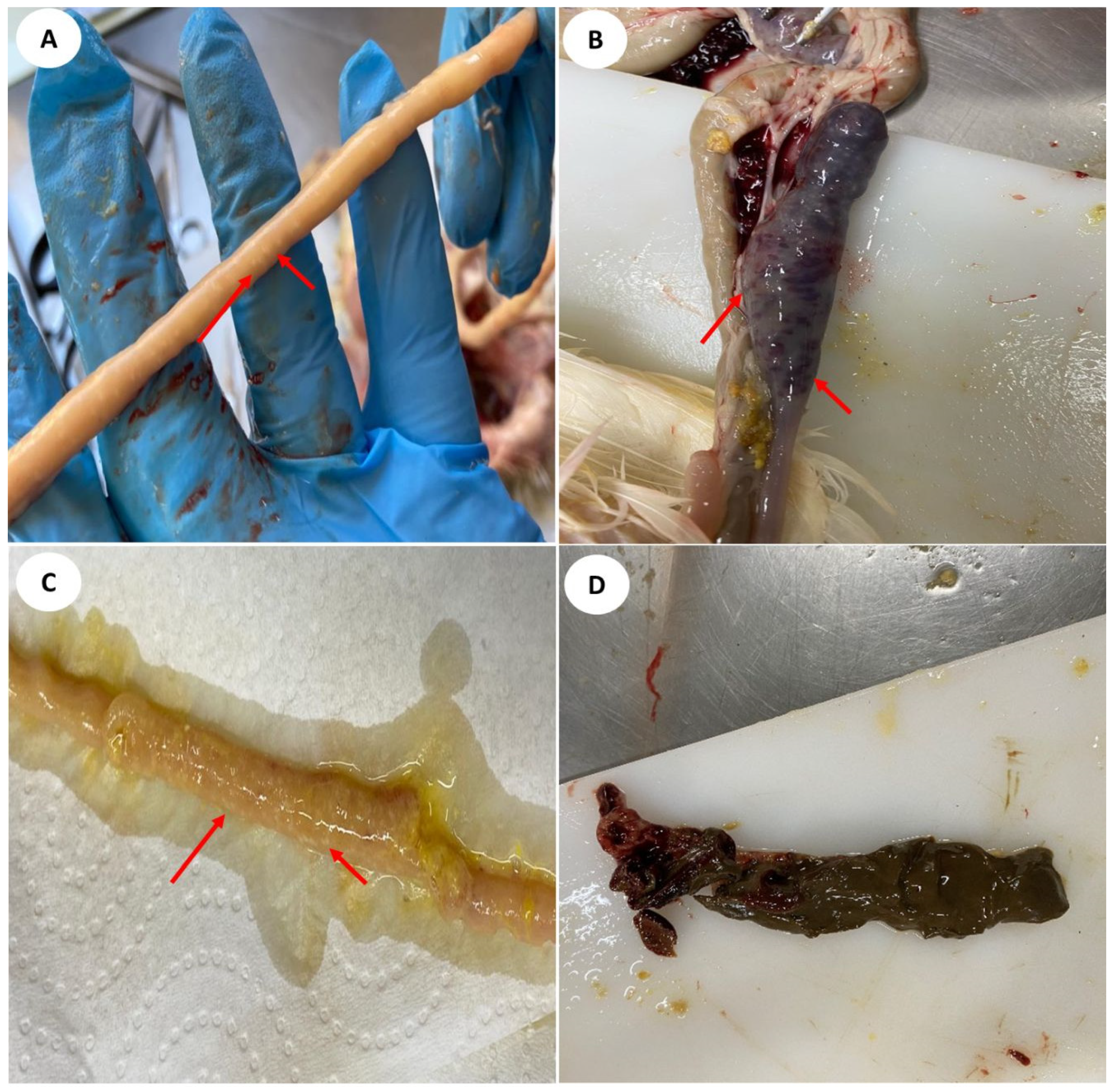
2.6. Macroscopic Score

The scoring of macroscopic lesions in the intestinal tract of birds was conducted in the duodenum, middle jejunum, and cecum using the methodology described by Conway, Mckenzie and Dayton [13]. This scoring system was used because the three species included in the challenge inoculum specifically affect different regions of the intestinal tract: Eimeria acervulina targets the duodenal loop, Eimeria maxima affects the midgut or jejunum region, and Eimeria tenella impacts the cecum. The macroscopic lesions were assessed individually for lesions generated by coccidia, following the scoring method described by Johnson and Reid [15] (Table 2). The scoring system ranged from zero to four, with zero indicating the absence of lesions and four representing the highest possible lesion score.

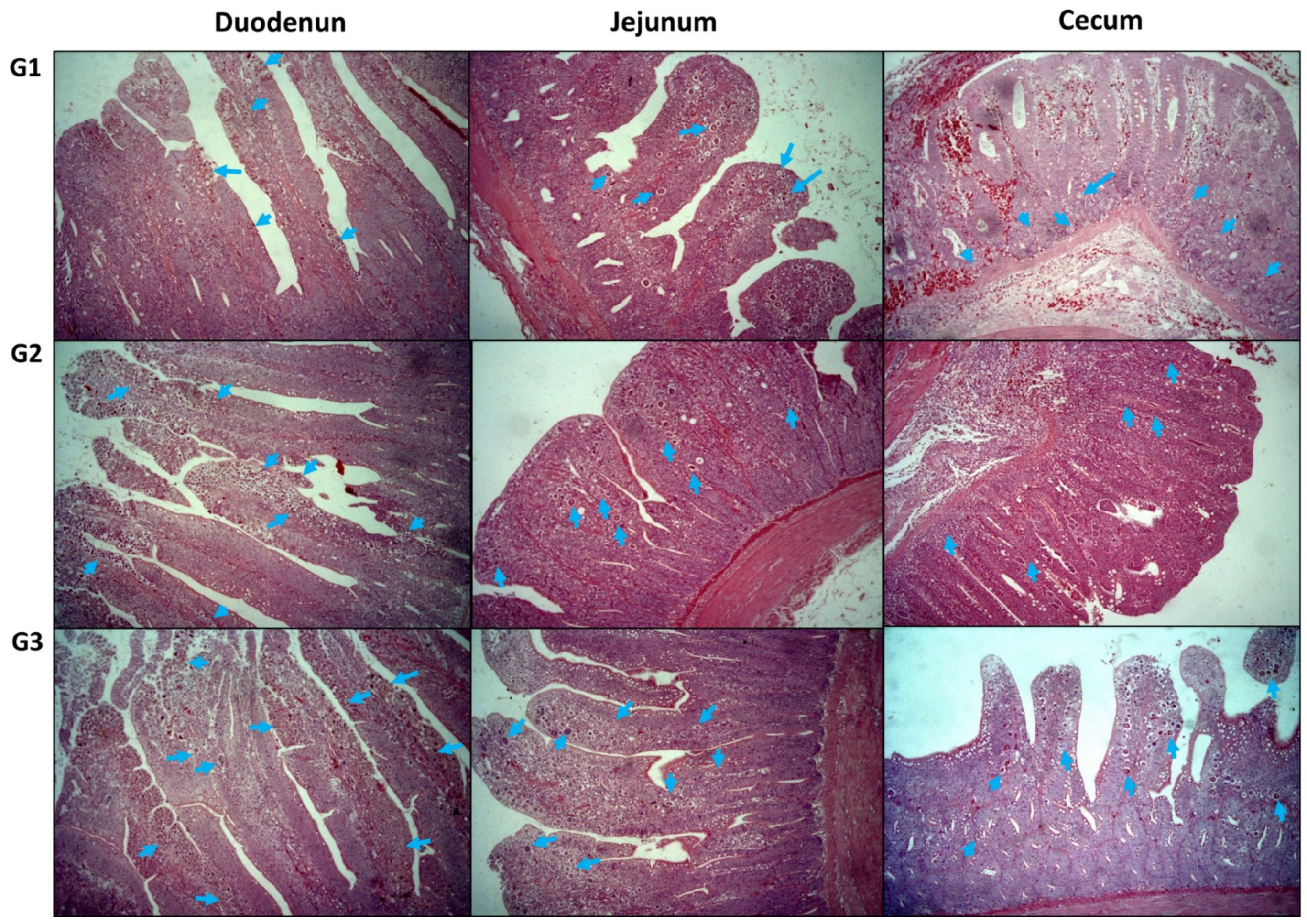
2.7. Histological Analysis

Histological analysis was carried out for the duodenum, jejunum, and cecum of the birds by staining the slides with hematoxylin and eosin (H&E). After 24 h of fixation with 4% paraformaldehyde, tissue samples were dehydrated using gradient alcohol solutions, transferred to xylene, embedded in paraffin, and sectioned into 5 μm-thickness sections. Following deparaffinization and rehydration in water, the sections were stained with H&E. The lesions were classified according to Goodwin et al. [16]. As such, a score for the microscopic lesions (MLS) was established in which MLS is the sum of A plus B. Variable A represents the distribution of the developmental stages of Eimeria along the intestinal segment examined (0 = no parasites, 1 = parasites in one field ×0; 2 = parasites in two fields ×10; 3 = parasites in three fields ×10; 4 = parasites in all four fields ×10), and B represents the severity of the infection by Eimeria in the fields examined (0 = parasites in 0% of villosities; 1 = parasites in <25% of villosities; 2 = parasites in 25 to 50% of villosities; 3 = parasites in 51 to 75% of villosities; 4 = parasites in >75% of villosities). For example, if A = 2 (parasites in two fields X10) and B = 2 (parasites in 25 to 50% of villosities), then A + B = 4. The total initial MLS can vary from 0 to 8.

2.8. Histomorphometric Analysis

The duodenum, jejunum, and cecum of the birds from all groups were fixed in situ with 4% buffered paraformaldehyde, paraffin-embedded, sectioned at 5 µm, staining with Alcian blue periodic acid-Schiff staining (G1049) and periodic acid-Schiff staining (G1008) commercial kits (Servicebio) according to the manufacturer’s instructions. The parameters analyzed were as follows: villosity height, crypt depth, thickness of villosity, thickness of mucosa, amount of goblet cells in the villus, and villosity height/crypt depth ratio. The villi measured were those that presented a whole body that could be measured. Height was measured from their peak to their base, limited by the area adjacent to the crypt zone. The quantification was carried out with common optical microscopy, at a 10× increase, in which each parameter was quantified and measured three times in ten different fields of the slide, for each intestinal segment per animal. After selecting the villi that were viable for measurements, the goblet cells present in them were counted. The count was carried out throughout the whole extension of the villus, on both sides, starting at the base to the peak and followed by the opposite side.

2.9. Intestinal Permeability

Isothiocyanate-dextran fluorescence (FITC-d, MW 3–5 KDa; Sigma Aldrich Co., St. Louis, MI, USA) was administered orally, and its presence in blood was used as a marker of paracellular transport and mucosal barrier dysfunction. The FITC-dextran was dissolved in sterile water, resulting in a final concentration of 20 mg/mL. At 21 days post-hatch, an oral administration of FITC-d (2.2 mg/bird; molecular weight, 3000–5000; Sigma Aldrich) was administered orally 2 h before euthanizing the chicks. Following euthanasia, blood was obtained via the jugular vein. The blood was kept at room temperature for 3 h to facilitate the process of clotting. After the clotting period, the samples were centrifuged (1000× g for 15 min) to separate the serum. A standard curve was prepared using serial dilutions of FITC-dextran in PBS (0, 125, 250, 375, 500, 750, 1000 µg/mL) [17,18]. A volume of 100 µL of diluted serum was added to a 96-well flat bottom plate in duplicate. The negative control involved the utilization of chicken serum that was not administered to the FITC-dextran solution. The fluorescence levels of diluted serum (1:1 in PBS) were measured at an excitation wavelength of 485 nm and an emission wavelength of 528 nm (Synergy HT, multimode microplate reader; BioTek Instruments, Winooski, VT, USA). The concentration of FITC-d per mL of serum was calculated based on a standard curve.

2.10. Mucosal Sample Analysis

Production of mucosal secretory immunoglobulin A ([sIgA (Bethyl Laboratories, Montgomery, TX, USA)] and polymeric Ig receptor/secretory component (pIgR/SC; MyBioSource, number MBS2512419)) was determined by ELISA following the manufacturer’s protocol. The total protein in the mucosal homogenates was measured colorimetrically based on the Bradford dye-binding method using a commercially available kit (BioRad, Hercules, CA, USA) with bovine serum albumin as the standard [14]. Collected explants were homogenized in ice-cold PBS and centrifuged for 15 min at 1500× g to obtain the supernatant.

2.11. Statistics

The data were initially analyzed through a K-S distance test. For non-parametric data, the Friedman test was used, with a Student–Newman–Keuls post-test. The Kruskal–Wallis test was employed for conducting histomorphometric analysis. This test was applied using Prism 4.00 (GraphPad Software, San Diego, CA, USA) and Sigma-Stat (SPSS Corporation, Chicago, IL, USA). We accepted in the present study an alpha risk lower than or equal to 5% and a beta risk lower than or equal to 20%.

3. Results

3.1. Oocyst Count (OPG) and qPCR

No oocysts were detected in chicken excreta by the 15th day of age, for all groups. Oocysts were not detected in unchallenged birds (U) on days 17, 19, and 21 of age. However, in groups G1, G2, and G3, oocysts were found in feces every day assessed after the challenge. As expected, the animals that received a higher concentration of inoculum in the challenge presented the greatest amounts of oocyst in their feces (Table 3). The challenged birds (G1, G2, and G3) presented a decrease in feed consumption and presented apathy and diarrhea as clinical signs; these occurred with greater intensity on the fourth and fifth days after inoculation. On the fifth day after the challenge, the birds of all groups challenged presented bloody diarrhea. Regarding the molecular identification of Eimeria using qPCR, all positive controls (CP) were amplified in the species assay, while the negative control (U, unchallenged group) was not amplified. In the challenged groups (G1, G2, and G3), DNA was detected in the feces on days 1, 3, and 5 after the challenge (Table 3), but the excretion of oocysts was lower in the groups challenged and infected at a lower challenge dose.

3.2. Mucosa Parameters

The concentrations of pIgR and sIGA in the ileum of broilers were observed at 21 days (5 dpi), as shown in Table 4. The levels of pIgR were found to be significantly greater in the challenged groups (G1, G2, and G3). The animals subjected to the medium-high challenge (G2, 39.45 ± 1.98 ng/mg protein) and high challenge (G3, 36.42 ± 2.31 ng/mg protein) exhibited a significantly greater level of pIgR compared to the animals subjected to the medium-low challenge (G1, 23.13 ± 1.23 ng/mg protein) (p ≤ 0.0001). Nevertheless, there were no significant differences observed across the challenge groups in terms of secretory immunoglobulin A (sIgA) levels. The experimental groups (G1, G2, and G3) exhibit a significantly elevated concentration of sIgA compared to the unchallenged group (p ≤ 0.0001).

3.3. Intestinal Permeability

The results of intestinal permeability are represented by the levels of FITC-d retrieved from the serum of the birds at 5 dpi. Higher concentrations of FITC-d in the serum represent an increase in intestinal permeability. More severe intestinal permeability (300 ± 14.1 ng/mL) was observed in the high-challenged group (G3). However, birds challenged in the medium-high dose group (G2) did not differ from G3 and presented 250 ± 18.4 ng/mL of FITC-dextran in their serum. However, birds from G3 and G2 presented higher permeability than the medium-low challenge group (G1), which presented 73.94 ± 4.3 ng/mL of FITC-dextran in its serum (p ≤ 0.05), and the unchallenged group (U), which presented less than 7.7 ± 2.8 ng/mL (p ≤ 0.0001).

3.4. Assessment of Macroscopic and Histopathological Lesions

All birds challenged (G1, G2, and G3) were positive for coccidiosis with specific macroscopic lesions in their intestine regarding all strains present in the challenge (E. acervulina, E. maxima, and E. tenella) (Figure 1). All groups challenged had higher scores for macroscopic and microscopic lesions than the control group (U, Table 5) in the upper and middle portions of the intestine and cecum. The macroscopic lesions with least extension were observed in the jejunum (E. maxima). The results demonstrated that the birds of group G3 (high challenge) presented the most severe macroscopic cecal lesion in comparison with birds challenged in groups G2 and G1. However, there was no significant difference among the groups challenged (G1, G2, and G3) in the macroscopic analysis of the upper (duodenum) and middle (jejunum) intestine.
The histopathological analysis of the small intestine (duodenum and jejunum) and cecum is presented in Figure 2 and Table 5. No obvious lesions to the small intestine and cecum were observed in group U (unchallenged birds). In turn, petechia and bleeding (light to moderate) in the small intestine and cecum were detected in all groups challenged (G1, G2, and G3), indicating that the birds in the present study presented moderate/severe coccidia infection. Birds that were challenged (G1, G2, and G3) presented moderate to severe lesions (score 6–7 in the duodenum, score 6–8 in the jejunum, and score 3–6 in the cecum). The main alterations found in this evaluation were hemorrhages in the mucosa and submucosa, sometimes accompanied by parasitic structures compatible with Eimeria spp., with no evidence of necrosis (Figure 2). The jejunum cuts that received a score of zero during the macroscopic evaluation presented, in the histopathologic analysis, E. maxima parasite structures in all birds (G1, G2 and G3). As anticipated, the jejunum sections obtained from the animals in the U group were assigned a score of zero during the macroscopic and histological assessment.

3.5. Histomorphometric Measurements of the Duodenum, Jejunum, and Cecum

The histomorphometry of the duodenum, jejunum, and cecum of the chickens at the age of 21 days is presented in Table 6. It is expected that a healthy chicken would present a villus height/crypt depth ratio (VH:CD) of at least five (five parts villus for one part crypt) [19], as observed in the control group (U). The challenges (G1, G2, and G3) resulted in a significant decrease in duodenal, jejunal, and cecal VH:CD ratios (p ≤ 0.05) in comparison with unchallenged birds (U). Moreover, the challenge presented a significant reduction in villi height and crypt depth (p ≤ 0.05) compared to unchallenged birds. The broiler chickens challenged presented a significant reduction in the density of goblet cells in comparison to the control group (p < 0.001), and interaction between the challenge dose was also observed in the amount of goblet cells in the villosities of the duodenum. The groups challenged (G1, G2, and G3) presented a decrease in villosity size (VH) and an increase in villus thickness when compared to the birds of the control group (U) in the duodenum, jejunum, and cecum.

4. Discussion

Coccidia infections are typically self-limiting and their severity is primarily determined by the quantity of sporulated oocysts that have been ingested [5]. Multiple studies have provided evidence supporting the existence of an ideal dose at which the parasite’s reproductive potential is achieved and it replicates efficiently within the epithelial cells. The use of too large quantities might result in the occurrence of the “crowding effect,” which disrupts the parasite’s life cycle and concurrently induces intestinal harm [19,20]. Several studies have examined the reproductive capacity of various Eimeria species under controlled experimental conditions. These experiments involved administering different amounts of sporulated oocysts from E. acervulina, E. maxima, E. tenella, E. mitis, E. necatrix, E. brunetti, or E. praecox [11,21]. The use of Eimeria spp. challenges in studies about infection or that assess additives and nutrients in chicken feed is common [21,22]. We reinforce the importance of studies about challenge dose. The intraspecific competition in coccidiosis infections has been reported to rely on the number of sporulated inoculum (oocysts) ingested [23,24,25].
In the current study, we observed that the severity of infection by Eimeria linearly regulated the macroscopic and microscopic lesions in the duodenum, jejunum, and cecum and altered the dynamics of intestinal permeability, which is in accordance with previous reports [11]. In fact, the coccidia utilized in the experiment infiltrate the intestinal mucosa and elicit a certain degree of injury and inflammation in the epithelial cells. In the three challenges applied, we observed oocysts causing noticeable histological alterations to intestinal epithelial cells of chickens, including distortion, rupture, separation of adjacent cells, and desquamation. Birds infected by the challenge presented the clinical disease with ruffled feathers, apathy, and diarrhea. Moreover, the consumption of feed and water decreased, the feces were aqueous over the course of the first days after the challenge, and, on the fifth day post-challenge, we observed hemorrhagic diarrhea in the three groups challenged. These alterations may be associated with poor absorption due to decreased activity of brush border enzymes [26] and to the rupture of intestinal integrity [27]. Coccidiosis can also cause other important intestinal histomorphological alterations; for example, challenges with E. acervulina and E. maxima showed increased size and number of goblet cells along the ileal crypts in broiler chickens [4].
The growth and feed efficiency of broiler chickens is closely linked to the functioning of the intestinal tract. This site is additionally accountable for the provision of a naturally occurring protective barrier. This barrier is upheld by a balanced arrangement of mucosal epithelial cells, secretory components (including mucus and sIgA), immune cells, and the microbiota [28]. The primary role of pIgR, which is found on the basolateral membrane of intestinal epithelial cells, is to facilitate the transportation of IgA antibodies through the epithelial cells and into the lumen of the intestinal tract [29]. The seeming increase in sIgA and pIgR in challenged groups suggested a damaged mucosal barrier in those groups. The damaged mucosa was more critical in medium-high and high challenges, with higher concentrations of pIgR in these challenge groups.
The term “intestinal integrity” refers to the measure of effectiveness in which the protective barrier hinders the movement of unwanted substances through the paracellular route. Upon being breached, this barrier will facilitate the movement of significantly large components from the lumen into the circulatory system, including microorganisms residing in the intestinal lumen [30]. Following the invasion of intestinal cells, sporozoites undergo a developmental process wherein they transform into trophozoites within 12 to 48 h [31]. Subsequently, these trophozoites undergo numerous nuclear divisions, resulting in the formation of schizonts. The schizonts, upon reaching maturity three days after infection, discharge merozoites into the intestine [31]. Merozoite infiltration into intestinal epithelial cells is proportional to the severity of tissue injury and the degree of inflammatory response [32]. The presence of a large number of merozoites has a detrimental impact on the intestinal health of chickens. Therefore, the Eimeria cycle can be associated with the impairment of intestinal barrier integrity (or intestinal barrier dysfunction), leading to increased intestinal permeability. Consequently, intestinal injury scores in Eimeria-infected animals were also correlated with intestinal permeability. Our results showed an increase in intestinal permeability after Eimeria-induced infection. Furthermore, there was a tendency for intestinal permeability to increase in response to the progressive inoculation dose in the groups challenged with medium-low challenge doses (G1) compared to the groups with medium-high challenge (G2) and high challenge (G3). However, the serum concentrations of FITC-d exhibited a notable resemblance between the high and medium-high groups of challenges; it may be that intestinal permeability may reach a maximum or a plateau at some point. In turn, there was no significant difference among the group’s challenges when evaluating the lesion scores of the duodenum and jejunum. Therefore, drawing from the findings regarding intestinal permeability, it is postulated that escalating doses may lead to an augmentation in intestinal permeability, as a larger number of sporozoites during the initial stages of infection can result in increased schizont and merozoite reproduction in succeeding stages.
The macroscopic and microscopic lesions of the upper and middle intestine did not differ significantly among the groups challenged. The crowding effect could be responsible for this finding; this effect occurs when a high concentration of oocysts in the gut results in self-inhibition of reproductive capacity and a reduction in oocyst excretion [11,19,24]. The chickens challenged with fewer oocysts provide more space for Eimeria spp. going through a third or fourth asexual cycle, resulting in a late permeability peak. Chickens with the high challenge (G3) had the highest level of FITC-d throughout the infection period. Since the high challenge triggered a more severe intestinal damage, the birds would present delays in recovery.
According to our findings on intestinal morphology, medium- and high-challenge poultry exhibited villous damage (G2 and G3). The higher the number of oocysts in the challenge, the lower the height of villosities in the duodenum, jejunum, and cecum of the chickens. The challenges reduced the height of villosities in the duodenum (>20%), as well as in the jejunum and cecum (>30%), and increased the depth of the crypts in comparison with birds that were not challenged. However, it is desirable that villosities remain high while crypts are shorter, as the decrease in the height of villosities is responsible for a reduction in digestibility of the birds challenged [33]. A decreased villus height/crypt depth ratio is indicative of heightened metabolic demands in birds that are challenged by infection. This leads to increased utilization of energy and nutrients, hence expediting the turnover of intestinal epithelial cells as a means to eliminate parasites from the digestive tract [21,25]. The challenge, regardless of the dose used, was related to a reduction in the amount of goblet cells. The goblet cells represent an important defense mechanism in the intestinal tract [34], secreting mucins that constitute a layer of mucus, acting as a physical barrier, and presenting immunological functions to inhibit the entry of pathogens in epithelial cells [35]. The mucins serve as the primary barrier against gut infections, so a reduction in goblet cells makes birds more susceptible to infections [34]. The dramatic reduction in mucin by goblet cells along with the increased gut permeability, particularly in scenarios involving medium-high and high challenges, would make the broilers more prone to coccidial infection and secondary infections caused by bacteria.
Our findings have collectively indicated the presence of the crowding effect in avian coccidia. We noticed a gradual increase in oocyst yields as the dosages of oocysts increased from medium-low challenge to high challenge. Nevertheless, despite the higher quantity of oocysts and parasite replication seen in the high-challenge dosage as opposed to the medium-high-challenge dose, there was no alteration in the clinical manifestation of the disease or the histological and histomorphometric lesions and mucosal permeability. These findings suggest that when the dosage of oocysts increases above the medium-high dose, the reproductive potential of oocysts tends to decrease while still potentially providing a crowding effect without altering the severity of disease, including the macroscopic, histological lesions, and intestinal permeability. To investigate the impact of chemotherapy or immunization on oocyst production, it is advisable to administer an infectious dosage lower than the crowding effect. To achieve optimal production of oocyst stocks in a laboratory or factory setting, it is recommended to employ the highest possible production dosage, which exceeds the crowding effect.
The coccidian life cycle lasts around 1 week [36]. The inflammatory response triggered the elimination of Eimeria spp. has substantial impacts on animal production performance. The damage to the intestinal mucosa caused by coccidiosis has a detrimental effect on the broiler’s performance [36,37]. The immune response and inflammatory process in the host also cause a reduction in feed intake and an increase in energy demand in affected sites, harming the flock’s zootechnical performance [38,39].
The current study employed several challenges utilizing sporulated oocysts of Eimeria, as they are well-suited for investigations targeting distinct levels of difficulty. The findings of our study reveal a noteworthy correlation between the severity of coccidiosis and its effect on the intestinal well-being of broiler chickens, as influenced by various Eimeria challenges. Nevertheless, the severity of the infection is not solely determined by the challenge dose but is also influenced by other factors such as the virulence of the Eimeria strain, the viability of oocysts, and the storage time and temperature of oocysts. The findings of our study offer valuable insights for developing clinical or subclinical challenge models in future studies. Additionally, this study provides support for determining optimal challenge dosages of E. acervulina, E. maxima, and E. tenella in future research endeavors that aim to assess the efficacy of feed ingredients or additives in enhancing gut health in broiler chickens.

5. Conclusions

The findings from our study indicate that the macroscopic, microscopic, and morphometric lesion data provide evidence that the three challenge models (medium-low, medium-high, and high) can serve as effective tests for experimental infection. Intestinal integrity in broiler chickens has been shown to be affected depending on the dosage of Eimeria acervulina, Eimeria tenella, and Eimeria maxima used for the challenge. Nevertheless, the assessment of intestinal permeability revealed significant fluctuations in intestinal leakage, particularly in scenarios involving medium-high and high levels of stress. In general, there was no significant difference observed in intestinal lesions and intestinal permeability between animals subjected to high challenge and those subjected to medium-high challenge, indicating a tendency for these outcomes to cluster together. Hence, in the context of nutritional studies, it would be more suitable to employ the medium-low and medium-high challenges, since they have effectively established a correlation between the dosage of the challenge and intestinal parameters, while avoiding confounding factors such as the crowding effect.

Author Contributions

Conceptualization, A.P.B.; methodology, G.C.d.S., G.F.E., F.A.V., R.M., A.M.G.I. and M.A.Z.M.; validation, G.C.d.S., F.A.V., R.M. and A.P.B.; formal analysis, A.M.G.I., M.A.Z.M. and A.P.B.; investigation, G.C.d.S. and A.P.B.; resources, A.P.B.; data curation, G.C.d.S.; writing—original draft preparation, G.C.d.S. and A.P.B.; writing—review and editing, A.M.G.I.; supervision, A.P.B.; project administration, A.P.B.; funding acquisition, A.P.B. All authors have read and agreed to the published version of the manuscript.

Funding

This research was funded by Embrapa Research Management System (SEG, grant number 20.18.03.004.00.00).

Institutional Review Board Statement

The animal study protocol was approved by the Ethics Committee of Embrapa Suínos e Aves (protocol 002/2018, 18 January 2018) for studies involved.

Informed Consent Statement

Not applicable.

Data Availability Statement

The data generated or analyzed during this study are included in this published article.

Acknowledgments

The authors are grateful to Alexandre Luis Tessmann (lab technician at Embrapa Suínos e Aves) for the support in experiments. The authors thank Embrapa Suínos e Aves for providing the facilities and crew for the in vivo experiments.

Conflicts of Interest

The authors declare that they have no known competing financial interests or personal relationships that could have appeared to influence the work reported in this paper.

References

  1. Shivaramaiah, C.; Barta, J.R.; Hernandez-Velasco, X.; Tellez, G.; Hargis, B.M. Coccidiosis: Recent advancements in the immunobiology of Eimeria species, preventive measures, and the importance of vaccination as a control tool against these Apicomplexan parasites. Vet. Med. 2014, 5, 23–34. [Google Scholar] [CrossRef]
  2. Muller, J.; Hemphill, A. In vitro culture systems for the study of apicomplexan parasites in farm animals. Int. J. Parasitol. 2013, 43, 115–124. [Google Scholar] [CrossRef] [PubMed]
  3. Yun, C.H.; Lillehoj, H.S.; Lillehoj, E.P. Intestinal immune responses to coccidiosis. Dev. Comp. Immunol. 2000, 24, 303–324. [Google Scholar] [CrossRef] [PubMed]
  4. Collier, C.T.; Hofacre, C.L.; Payne, A.M.; Anderson, D.B.; Kaiser, P.; Mackie, R.I.; Gaskins, H.R. Coccidia-induced mucogenesis promotes the onset of necrotic enteritis by supporting Clostridium perfringens growth. Vet. Immunol. Immunopathol. 2008, 122, 104–115. [Google Scholar] [CrossRef] [PubMed]
  5. Chapman, H.D.; Jeffers, T.K. Vaccination of chickens against coccidiosis ameliorates drug resistance in commercial poultry production. Int. J. Parasitol. Drugs Drug Resist. 2014, 4, 214–217. [Google Scholar] [CrossRef] [PubMed]
  6. Bortoluzzi, C.; Paras, K.L.; Applegate, T.J.; Verocai, G.G. Comparison between McMaster and Mini-FLOTAC methods for the enumeration of Eimeria maxima oocysts in poultry excreta. Vet. Parasitol. 2018, 254, 21–25. [Google Scholar] [CrossRef]
  7. Haug, A.; Gjevre, A.G.; Skjerve, E.; Kaldhusdal, M. A survey of the economic impact of subclinical Eimeria infections in broiler chickens in Norway. Avian Pathol. 2008, 37, 333–341. [Google Scholar] [CrossRef] [PubMed]
  8. Adedokun, S.A.; Adeola, O. The response in jejunal and ileal nutrient and energy digestibility and the expression of markers of intestinal inflammation in broiler chickens to coccidial vaccine challenge and phytase supplementation. Can. J. Anim. Sci. 2017, 97, 258–267. [Google Scholar] [CrossRef]
  9. Parker, J.; Oviedo-Rondon, E.O.; Clack, B.A.; Clemente-Hernandez, S.; Osborne, J.; Remus, J.C.; Kettunen, H.; Makivuokko, H.; Pierson, E.M. Enzymes as feed additive to aid in responses against Eimeria species in coccidia-vaccinated broilers fed corn-soybean meal diets with different protein levels. Poult. Sci. 2007, 86, 643–653. [Google Scholar] [CrossRef]
  10. Rochell, S.J.; Parsons, C.M.; Dilger, R.N. Effects of Eimeria acervulina infection severity on growth performance, apparent ileal amino acid digestibility, and plasma concentrations of amino acids, carotenoids, and alpha 1-acid glycoprotein in broilers. Poult. Sci. 2016, 95, 1573–1581. [Google Scholar] [CrossRef]
  11. Teng, P.Y.; Yadav, S.; Castro, F.L.S.; Tompkins, Y.H.; Fuller, A.L.; Kim, W.K. Graded Eimeria challenge linearly regulated growth performance, dynamic change of gastrointestinal permeability, apparent ileal digestibility, intestinal morphology, and tight junctions of broiler chickens. Poult. Sci. 2020, 99, 4203–4216. [Google Scholar] [CrossRef]
  12. Haug, A.; Williams, R.B.; Larsen, S. Counting coccidial oocysts in chicken faeces: A comparative study of a standard McMaster technique and a new rapid method. Vet. Parasitol. 2006, 136, 233–242. [Google Scholar] [CrossRef] [PubMed]
  13. Conway, D.P.; Mckenzie, M.E.; Dayton, A.D. Relationship of Coccidial Lesion Scores and Weight-Gain in Infections of Eimeria acervulina, E. maxima and E. tenella in Broilers. Avian Pathol. 1990, 19, 489–496. [Google Scholar] [CrossRef] [PubMed]
  14. Baron, L.F.; Fonseca, F.N.D.; Maciag, S.S.; Bellaver, F.A.V.; Ibeli, A.M.G.; Mores, M.A.Z.; Almeida, G.F.D.; Guterres, S.S.; Bastos, A.P.A.; Paese, K. Toltrazuril-Loaded Polymeric Nanocapsules as a Promising Approach for the Preventive Control of Coccidiosis in Poultry. Pharmaceutics 2022, 14, 392. [Google Scholar] [CrossRef] [PubMed]
  15. Johnson, J.; Reid, W.M. Anticoccidial drugs: Lesion scoring techniques in battery and floor-pen experiments with chickens. Exp. Parasitol. 1970, 28, 30–36. [Google Scholar] [CrossRef] [PubMed]
  16. Goodwin, M.A.; Brown, J.; Bounous, D.I. Use of microscopic lesion scores, gross lesion scores and oocyst count scores to detect in chickens. Avian Pathol. 1998, 27, 405–408. [Google Scholar] [CrossRef] [PubMed]
  17. Vicuña, E.A.; Kuttappan, V.A.; Tellez, G.; Hernandez-Velasco, X.; Seeber-Galarza, R.; Latorre, J.D.; Faulkner, O.B.; Wolfenden, A.D.; Hargis, B.M.; Bielke, L.R. Dose titration of FITC-D for optimal measurement of enteric inflammation in broiler chicks. Poult. Sci. 2015, 94, 1353–1359. [Google Scholar] [CrossRef]
  18. Baxter, M.F.A.; Merino-Guzman, R.; Latorre, J.D.; Mahaffey, B.D.; Yang, Y.C.; Teague, K.D.; Graham, L.E.; Wolfenden, A.D.; Hernandez-Velasco, X.; Bielke, L.R.; et al. Optimizing Fluorescein Isothiocyanate Dextran Measurement as a Biomarker in a 24-h Feed Restriction Model to Induce Gut Permeability in Broiler Chickens. Front. Vet. Sci. 2017, 4, 56. [Google Scholar] [CrossRef]
  19. Jenkins, M.C.; Dubey, J.P.; Miska, K.; Fetterer, R. Differences in fecundity of Eimeria maxima strains exhibiting different levels of pathogenicity in its avian host. Vet. Parasitol. 2017, 236, 1–6. [Google Scholar] [CrossRef]
  20. Williams, R.B. Quantification of the crowding effect during infections with the seven Eimeria species of the domesticated fowl: Its importance for experimental designs and the production of oocyst stocks. Int. J. Parasitol. 2001, 31, 1056–1069. [Google Scholar] [CrossRef]
  21. Teng, P.Y.; Choi, J.; Yadav, S.; Tompkins, Y.H.; Kim, W.K. Effects of low-crude protein diets supplemented with arginine, glutamine, threonine, and methionine on regulating nutrient absorption, intestinal health, and growth performance of Eimeria-infected chickens. Poult. Sci. 2021, 100, 101427. [Google Scholar] [CrossRef] [PubMed]
  22. Teng, P.Y.; Choi, J.; Tompkins, Y.; Lillehoj, H.; Kim, W. Impacts of increasing challenge with Eimeria maxima on the growth performance and gene expression of biomarkers associated with intestinal integrity and nutrient transporters. Vet. Res. 2021, 52, 81. [Google Scholar] [CrossRef] [PubMed]
  23. Jenkins, M.; Allen, P.; Wilkins, G.; Klopp, S.; Miska, K. Eimeria praecox infection ameliorates effects of Eimeria maxima infection in chickens. Vet. Parasitol. 2008, 155, 10–14. [Google Scholar] [CrossRef]
  24. Conway, D.P.; Sasai, K.; Gaafar, S.M.; Smothers, C.D. Effects of Different Levels of Oocyst Inocula of Eimeria acervulina, E. tenella, and E. maxima on Plasma Constituents, Packed Cell-Volume, Lesion Scores, and Performance in Chickens. Avian Dis. 1993, 37, 118–123. [Google Scholar] [CrossRef] [PubMed]
  25. Hernández-Velasco, X.; Chapman, H.D.; Owens, C.M.; Kuttappan, V.A.; Fuente-Martínez, B.; Menconi, A.; Latorre, J.D.; Kallapura, G.; Bielke, L.R.; Rathinam, T.; et al. Absorption and deposition of xanthophylls in broilers challenged with three dosages of oocysts. Brit Poult. Sci. 2014, 55, 167–173. [Google Scholar] [CrossRef] [PubMed]
  26. Adams, C.; Vahl, H.A.; Veldman, A. Interaction between nutrition and Eimeria acervulina infection in broiler chickens: Development of an experimental infection model. Brit J. Nutr. 1996, 75, 867–873. [Google Scholar] [CrossRef] [PubMed]
  27. Assis, R.C.L.; Luns, F.D.; Beletti, M.E.; Assis, R.L.; Nasser, N.M.; Faria, E.S.M.; Cury, M.C. Histomorphometry and macroscopic intestinal lesions in broilers infected with Eimeria acervulina. Vet. Parasitol. 2010, 168, 185–189. [Google Scholar] [CrossRef]
  28. Sugiharto, S.; Yudiarti, T.; Isroli, I.; Widiastuti, E.; Kusumanti, E. Dietary Supplementation of Probiotics in Poultry Exposed to Heat Stress—A Review. Ann. Anim. Sci. 2017, 17, 591–604. [Google Scholar] [CrossRef]
  29. Kaetzel, C.S. The polymeric immunoglobulin receptor: Bridging innate and adaptive immune responses at mucosal surfaces. Immunol. Rev. 2005, 206, 83–99. [Google Scholar] [CrossRef]
  30. Jeurissen, S.H.M.; Boonstra-Blom, A.G.; Al-Garib, S.O.; Hartog, L.; Koch, G. Defence mechanisms against viral infection in poultry: A review. Vet. Quart. 2000, 22, 204–208. [Google Scholar] [CrossRef]
  31. Mcdougald, L.R. Intestinal protozoa important to poultry. Poult. Sci. 1998, 77, 1156–1158. [Google Scholar] [CrossRef] [PubMed]
  32. Dubey, J.P.; Jenkins, M.C. Re-evaluation of the life cycle of Eimeria maxima Tyzzer, 1929 in chickens (Gallus domesticus). Parasitology 2018, 145, 1051–1058. [Google Scholar] [CrossRef] [PubMed]
  33. Bedrnik, P.; Jurkovic, P. An antigen from Eimeria tenella merozoites for the indirect fluorescence antibody test. Folia Parasitol. 1979, 26, 169–172. [Google Scholar]
  34. Bedrnik, P.; Jurkovic, P. Comparison of 2 Different Antigens of Eimeria tenella for Indirect Immunofluorescent Test (Ifat). J. Protozool. 1977, 24, A48–A49. [Google Scholar]
  35. Augustine, P.C. Cell: Sporozoite interactions and invasion by apicomplexan parasites of the genus Eimeria. Int. J. Parasitol. 2001, 31, 1–8. [Google Scholar] [CrossRef]
  36. Shirley, M.W.; Smith, A.L.; Tomley, F.M. The biology of avian with an emphasis on their control by vaccination. Adv. Parasit. 2005, 60, 285–330. [Google Scholar] [CrossRef]
  37. Belote, B.L.; Soares, I.; Tujimoto-Silva, A.; Sanches, A.W.D.; Kraieski, A.L.; Santin, E. Applying I see inside histological methodology to evaluate gut health in broilers challenged with Eimeria. Vet. Parasitol. 2019, 276S, 100004. [Google Scholar] [CrossRef]
  38. Klasing, K.C. Nutrition and the immune system. Br. Poult. Sci. 2007, 48, 525–537. [Google Scholar] [CrossRef] [PubMed]
  39. Kogut, M.H.; Klasing, K. An immunologist’s perspective on nutrition, immunity, and infectious diseases: Introduction and overview. J. Appl. Poult. Res. 2009, 18, 103–110. [Google Scholar] [CrossRef]
Figure 1. Duodenum and cecum evaluated macroscopically in a male, 21-day-old, broiler chicken. Broiler chickens were subjected to a high-challenge dosage consisting of 125,000 oocysts of E. acervulina, 80,000 oocysts of E. maxima, and 25,000 oocysts of E. tenella. (A): duodenum presenting whitish lesions (arrows) associated with Eimeria acervulina. (B): cecum, marked diffuse necrohemorrhagic typhlitis associated with Eimeria tenella, ceca presenting hemorrhagic lesions (petechia); (C): catarrhal duodenitis associated with Eimeria acervulina, and intestinal wall is thickened with whitish lesions (arrows); (D): cecum, marked diffuse necrohemorrhagic typhlitis associated with Eimeria tenella, cecum presenting clots and petechia in its mucosa.
Poultry 03 00001 g001
Figure 2. Effects of different amounts of E. acervulina, E. maxima, and E. tenella oocysts on the intestinal morphology of broilers (5 dpi). Arrows indicate gametocytes or oocysts. G1, G2, and G3: intestine presenting desquamation of its lining, cell necrosis, and different developmental stages of Eimeria spp. The Eimeria oocysts are within the enterocytes in the epithelium column. G1 (medium-low challenge), G2 (medium-high challenge), G3 (high challenge): intestine presenting severe contamination by Eimeria spp. (Arrows). The cecum presents a large presence of red blood cells. Amplification, 100×.
Poultry 03 00001 g002
Table 1. Challenge dose of E. acervulina, E. maxima, and E. tenella administered to each group (oocysts/chicks).
TreatmentE. acervulina
(No. Oocysts)
E. maxima
(No. Oocysts)
E. tenella
(No. Oocysts)
Unchallenged (U)zerozerozero
Medium-low challenge (G1)31,25020,0006250
Medium-high challenge (G2)62,50040,00012,500
High challenge (G3)125,00080,00025,000
Table 2. Lesion score determined by histological analysis of E. acervulina, E. maxima, and E. tenella strains in chickens.
Lesion ScoreMacroscopic Finding Observed
0No lesion
1Wall of the small intestine roughly thinner, easily torn under slight tension, but no rough evidence of necrosis of the mucosa or other abnormalities
2One or more necrotic focuses in areas of the mucosa measuring approximately 1 to 5 mm in total thickness
3Necrosis greater than 5 mm and consisting of orange/brown necrotic detritus
4Large confluent necrotic areas over the total thickness of the mucosa in the small intestine, affecting 25% or more of the small intestine
Table 3. Number of Eimeria spp. oocysts per gram of feces for each group, before the challenge and 1, 3, and 5 days after the challenge (dpi) following the McMaster chamber counting.
Oocysts g−1qPCR StatusMean Amount in qPCR
Negative control (U)Before the challenge0undetectable0
1 dpi0undetectable0
3 dpi0undetectable0
5 dpi0undetectable0
Medium-low challenge (G1)Before the challenge0undetectable0
1 dpi2.0 × 103detectable3167.545
3 dpi2.9 × 104detectable25,569.347
5 dpi5.7 × 105detectable250,501.568
Medium-high challenge (G2)Before the challenge0undetectable0
1 dpi3.0 × 103detectable8665.856
3 dpi3.6 × 104detectable31,751.037
5 dpi7.4 × 105detectable250,059.875
High challenge (G3)Before the challenge0undetectable0
1 dpi9.6 × 104detectable41,723.154
3 dpi6.7 × 103detectable8375.960
5 dpi3.4 × 106detectable417,284.204
Table 4. IgA and polymeric Ig receptor/secretory component (pIgR/SC) concentrations from ileal mucosa of broilers with or without challenge.
Group
VariableUG1G2G3
pIgR (ng/mg protein)12.04 ± 1.20 a23.13 ± 1.23 b39.45 ± 1.98 c36.42 ± 2.31 c
IgA (ng/mg protein)9.72 ± 0.69 a20.49 ± 0.65 b18.66 ± 0.64 b18.37 ± 0.84 b
The analysis was carried out applying the Kruskal–Wallis test and the data are presented as Mean ± SEM. a,b,c Different superscript letters indicate a significant statistical difference among the groups (p ≤ 0.0001).
Table 5. Scores of macroscopic and microscopic intestinal lesions in broiler chickens (5 dpi) experimentally infected with Eimeria spp. (U: unchallenged, G1: medium-low challenge, G2: medium-high challenge, G3: high challenge).
GroupUnchallenged
(U)
Medium-Low Challenge
(G1)
Medium-High Challenge
(G2)
High Challenge
(G3)
Pr > χ2
Macroscopic Score
Duodenum0 ± 0 a3.37 ± 0.52 b3.25 ± 0.71 b3.13 ± 0.64 b0.0017
Jejunum0 ± 0 a1.00 ± 0.75 b1.25 ± 0.25 b1.37 ± 0.38 b0.0091
Cecum0 ± 0 a2.00 ± 0.93 b3.12 ± 0.99 b.c3.63 ± 0.52 c0.0372
Microscopic Score
Duodenum0 ± 0 a6.13 ± 2.85 b7.38 ± 0.52 b7.00 ± 1.19 b0.0032
Jejunum0 ± 0 a6.87 ± 2.80 b8.00 ± 0.00 b6.75 ± 1.40 b0.0020
Cecum0 ± 0 a3.75 ± 2.24 b5.12 ± 2.47 b6.25 ± 1.16 b0.0082
The analysis was carried out applying the Kruskal–Wallis test and the data are presented as Mean ± SEM. a,b,c Different superscript letters indicate a significant statistical difference among the groups (p ≤ 0.05).
Table 6. Mean values (standard deviation) of intestinal morphometry (in microns) of broiler chickens experimentally infected with E. maxima, E. acervulina, and E. tenella and necropsied at 21 days of age (5 dpi) (U: negative control, G1: medium-low challenge, G2: medium-high challenge, G3: high challenge) in the intestinal morphology of broilers (5 DPI).
Group
VariableUG1G2G3
Duodenum
VH, µm852 ± 88.64 a586.5 ± 178.07 b576.9 ± 214.64 b636.6 ± 207.68 b
CD, µm145.4 ± 8.52 168.2 ± 21.36173.2 ± 67.72157.5 ± 20.20
MT, µm124.3 ± 30.68 a211.2 ± 43.73 b252.2 ± 66.30 b274.5 ± 65.71 b
V/C5.8 ± 0.69 a3.5 ± 0.88 b3.5 ± 1.36 b4.1 ± 1.36 b
GC126.8 ± 26.66 a56.29 ± 8.48 b34.13 ± 9.48 c34.89 ± 8.95 c
Jejunum
VH, µm739.7 ± 39.78 a389.9 ± 164.42 b440.1 ± 124.42 b429.4 ± 140.44 b
CD, µm124.1 ± 11.49 a163.4 ± 36.43 b152.7 ± 49.42 b146.1 ± 22.82 a.b
MT, µm110.1 ± 10.33 a227.8 ± 89.52 b198.3 ± 40.52 b235.2 ± 21.31 b
V/C6.0 ± 0.65 a2.3 ± 0.64 b2.9 ± 1.00 b3.1 ± 1.43 b
GC135.87 ± 22.00 a45.94 ± 12.00 b31.05 ± 7.50 b30.2 ± 4.30 b
Cecum
VH, µm583.5 ± 52.58 a256.5 ± 125.70 b183.0 ± 42.78 b182.0 ± 43.39 b
CD, µm109.8 ± 9.33 a145.4 ± 23.04 b137.8 ± 17.88 b136.0 ± 20.62 b
MT, µm101.8 ± 18.81131.3 ± 46.95122.2 ± 23.04102.2 ± 19.99
V/C5.3 ± 0.63 a1.7 ± 0.70 b1.3 ± 0.31 b1.3 ± 0.32 b
GC124.5 ± 13.86 a10.3 ± 3.50 b6.5 ± 2.76 c7.0 ± 2.86 b.c
VH: villosity height; CD: crypt depth; MT: mucosa thickness; V/C: ratio between villosity height/crypt depth; GC: goblet cells. The analysis was carried out applying the Kruskal–Wallis test. a,b,c Different superscript letters indicate a significant statistical difference among the groups (p ≤ 0.05).
Disclaimer/Publisher’s Note: The statements, opinions and data contained in all publications are solely those of the individual author(s) and contributor(s) and not of MDPI and/or the editor(s). MDPI and/or the editor(s) disclaim responsibility for any injury to people or property resulting from any ideas, methods, instructions or products referred to in the content.

