Article

GPR56 mRNA Expression Is Modulated by Acute and Chronic Training Variable Manipulations in Resistance-Trained Men

by
Vitor Angleri
1,
Felipe Damas
1,
Uliana Sbeguen Stotzer
2,
Heloisa Sobreiro Selistre-de-Araujo
2,
Natalia Santanielo
1,
Samuel Domingos Soligon
1,
Luiz Augusto Riani Costa
3,
Manoel Emílio Lixandrão
3,
Miguel Soares Conceição
3,
Felipe Cassaro Vechin
3,
Michael D. Roberts
4,
Carlos Ugrinowitsch
3 and
Cleiton Augusto Libardi
1,*
1
MUSCULAB—Laboratory of Neuromuscular Adaptations to Resistance Training, Department of Physical Education, Federal University of São Carlos, São Carlos 13565-905, Brazil
2
LBBM—Laboratory of Biochemistry and Molecular Biology, Department of Physiological Sciences, Federal University of São Carlos, São Carlos 13565-905, Brazil
3
School of Physical Education and Sport, University of São Paulo, São Paulo 05508-030, Brazil
4
School of Kinesiology, Auburn University, Auburn, AL 36849, USA
*
Author to whom correspondence should be addressed.
Submission received: 21 December 2021 / Revised: 25 January 2022 / Accepted: 27 January 2022 / Published: 30 January 2022

Abstract

:
Background: Skeletal muscle adaptations are affected by resistance training (RT)-induced acute and chronic transcriptional responses. An under-explored gene target involved in mechanotransduction is the expression of the G protein-coupled receptor 56 (GPR56). However, studies investigating the acute and chronic effects of RT manipulations on GPR56 mRNA are scarce. Methods: Twenty subjects had each leg randomly assigned to a standard ((CON) no specific manipulation) or a variable RT (manipulations in load, volume, muscle action, and pause in a session-by-session fashion (VAR)). GPR56 mRNA expression was assessed before and after 16 training sessions (chronic effect) and 24 h after a 17th session (acute effect). Results: Acute GPR56 mRNA expression increased at 24 h (p < 0.01) without differences between CON and VAR (p > 0.05). No differences were found in GPR56 mRNA expression when comparing each VAR condition (load vs. sets vs. eccentric actions vs. pause) nor with CON at 24 h (p > 0.05). Chronic GPR56 mRNA expression increased at Post compared with Pre (p < 0.02) for VAR only, with a tendency (p = 0.058) toward higher expression for VAR as compared with CON. Conclusion: GPR56 mRNA expression is acutely and chronically modulated by RT. Additionally, chronic GPR56 mRNA expression is modulated by RT variable manipulations.

1. Introduction

Resistance training (RT) induces acute (after a single bout) and chronic (after several bouts) transcriptional changes, which are suggested to affect skeletal muscle adaptations [1,2,3,4]. In recent years, several studies have explored novel transcriptional targets that could be modulated by RT [5,6,7,8]. More recently, the mechanisms involved in mechanotransduction have garnered interest in the field. In the mechanotransduction process, membrane proteins translate RT-induced mechanical tension in the cell membrane into chemical signaling, activating protein synthesis pathways [9,10]. Although the upstream regulators of mechanotransduction are currently unclear, the adhesion G protein-coupled receptor family (GPCR) may be a candidate activator of anabolic signaling pathways [11,12]. The expression of the G protein-coupled receptor 56 ((GPR56) a family member of the GPCR, characterized by a large extracellular N terminus) has been shown to be induced by chronic RT in animals and humans [11,13]. GPR56 mRNA expression is controlled by the transcriptional coactivator PGC-1α4 and seems to drive muscle hypertrophy downstream of a Gα12/13–Rho pathway, leading to mTOR activation and an increase in protein synthesis [11]. In fact, the role of this pathway in mediating overload-induced hypertrophy is supported by muscle hypertrophy being blunted in GPR56 mRNA knockout mice and primary myotubes [11]. However, evidence regarding both acute and chronic RT effects on GPR56 mRNA gene expression in humans is scarce.
Exercise training mode (e.g., resistance vs. endurance) differentially affects the acute and chronic expression of certain mRNA transcripts [14]. In this regard, GPR56 mRNA expression is also differentially affected by specific exercise modes; more specifically, RT stimulates mRNA expression more than endurance training [11]. Regarding RT in particular, the transcriptional responses greatly vary between studies. The manipulation of one or another RT variable (e.g., eccentric actions and load) may differentially modulate mRNA expression compared to traditional RT protocols [15,16], although these results are not universal [17]. However, no study has investigated how acute and chronic GPR56 mRNA expression respond to the manipulations of RT variables.
Therefore, the present study compared a protocol that frequently manipulated the RT variables (load, sets, eccentric actions, and pause in a session-by-session fashion (VAR)) with a standard progressive RT protocol (CON) and their effects on the acute and chronic expression of skeletal muscle GPR56 mRNA using a within-subject design in humans. We hypothesized that manipulations of RT variables would acutely and chronically induce higher GPR56 mRNA expression levels compared to a traditional RT protocol.

2. Results

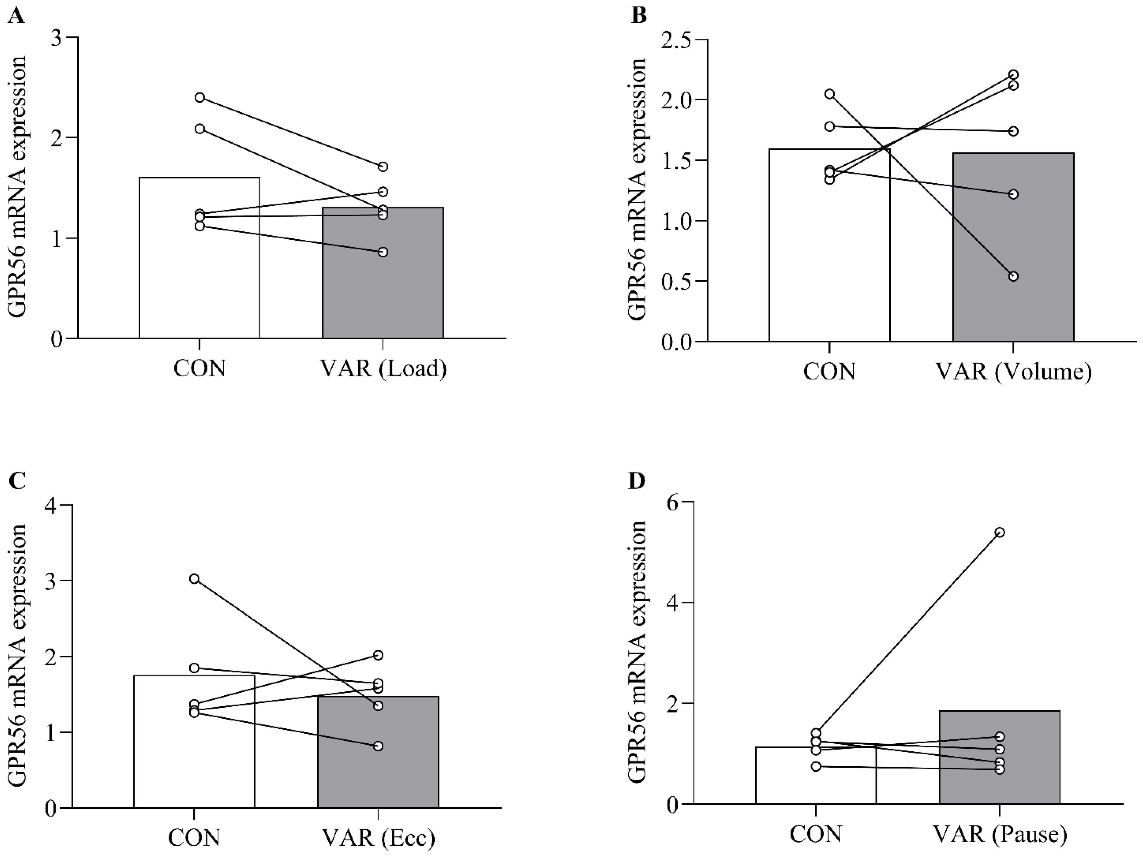
2.1. Acute GPR56 mRNA Expression

No significant differences in acute GPR56 mRNA expression were detected between CON and VAR at baseline (0 h) (p > 0.05) (Figure 1). Both CON and VAR significantly increased GPR56 mRNA expression (main time effect; p < 0.01) from 0 h to 24 h (Figure 2). No significant differences were found between CON and VAR at 24 h for GPR56 mRNA expression (p > 0.05) (Figure 2). No significant difference was observed when the four VAR conditions were compared to each other at 24 h (p > 0.05) (Figure 2). No significant difference was observed between each of the VAR conditions as compared with CON at 24 h (p > 0.05) (Figure 3A–D).

2.2. Chronic GPR56 mRNA Expression

GPR56 mRNA expression was significantly greater for VAR at Post compared to Pre (p < 0.02). No difference was found from Pre to CON at Post (p > 0.05) (Figure 4). We found a tendency for a higher increase in GPR56 mRNA expression for VAR as compared with CON at Post (p = 0.058) (Figure 4).

2.3. Correlation Analysis

No correlation was found between changes in fCSA (fCSA data from Fox et al. [18]) and acute or chronic GPR56 mRNA expression for CON (n = 19 for both acute and chronic) and VAR (n = 19 for acute and n = 18 for chronic) (p > 0.05).
As CON and VAR produced similar fCSA increases (see Fox et al. [18]), we collapsed the protocols (CON + VAR) to further the correlation analysis. Results showed no correlation between changes in fCSA and acute (n = 38) or chronic (n = 37) GPR56 mRNA expression (p > 0.05).

3. Discussion

This is the first study comparing the acute and chronic effects of manipulating RT variables on GPR56 mRNA expression. Acutely, CON and VAR increased GPR56 mRNA expression without differences between the protocols at 24 h. Additionally, no differences were found when comparing each VAR condition to the other (i.e., load, sets, eccentric action, and pause) or with CON at 24 h. Chronically, only VAR increased GPR56 mRNA expression after 16 RT sessions (Post) as compared with baseline (Pre). Additionally, GPR56 mRNA expression tended to be higher at Post for VAR as compared with CON.
The transcriptional changes induced by an acute resistance exercise bout is the initial physiological response to potential subsequent phenotypic adaptations. In this regard, investigations on novel transcriptional targets responsive to RT contributes knowledge to RT-induced phenotypic adaptations and provide future research directions. To the authors’ knowledge, no study has investigated the acute exercise effects, especially when utilizing RT, on GPR56 mRNA expression patterns in humans. We showed a significant increase in GPR56 mRNA expression at 24 h; however, the acute GPR56 mRNA response was not modulated by the frequent manipulation of several RT variables. Although some studies suggest that specific RT variable manipulations (e.g., eccentric action) may modulate acute transcriptional responses [15], these results are not universal [17]. Interestingly, no differences were found in GPR56 mRNA expression levels when comparing the VAR conditions to each other (i.e., load vs. sets vs. eccentric actions vs. pause), nor when compared with CON at 24 h. These results are in agreement with a previous study that showed similar acute transcriptional responses, regardless of the manipulation of training variables [17]. It is possible that training to (or close to) concentric muscle failure may maximally stimulate acute GPR56 mRNA expression, regardless of the manipulations of one or several RT variables simultaneously.
Robust transcriptional changes are commonly observed in novice RT practitioners, which result from the novelty of the training stimuli. However, chronic RT alters transcriptional responses toward a more refined response [15]. Moreover, the transcriptional response to one bout of exercise following chronic training alters genes involved in several physiological processes (e.g., muscle hypertrophy, energetic efficiency, extracellular matrix organization, as well as antioxidant and immune response) [15]. We sought to investigate the longer-term effects of manipulating RT variables on GPR56 mRNA expression, given that this target has been implied to be involved in the mechanotransduction process. We found that only VAR produced an increase in GPR56 mRNA expression after 16 RT sessions. Interestingly, there was a tendency (p = 0.058) for a higher GPR56 mRNA expression from VAR as compared with CON at Post. The only study that had investigated the chronic effects of training on GPR56 mRNA expression in humans showed that this gene is responsive to exercise and is specifically modulated by different exercise training modes (i.e., endurance, resistance, or combined) [11]. Therefore, we expanded the results from previous studies demonstrating that GPR56 mRNA expression may be differently modulated by specific RT manipulations. Studies that investigated the effects of RT manipulations in the chronic gene expression responses are scarce. Mallinson et al. [15] compared the effects of different RT manipulations (i.e., maximal concentric vs. maximal eccentric and concentric muscle actions) on chronic transcriptome responses in humans. Although results showed a higher transcriptional response for the protocol that performed both maximal contraction modes in the initial RT phase, these differences were attenuated after 28 and 84 days. This suggests that differences in transcriptional responses, probably induced by an “unaccustomed” stimulus from a specific RT protocol, are quickly redirected toward those observed in a traditional RT protocol. More importantly, our subjects had years of RT experience; therefore, it is unlikely that chronic increases in GPR56 mRNA expression induced by VAR were due to an “unaccustomed” RT effect. Collectively, these results allowed us to speculate that chronic GPR56 mRNA expression may be responsive to frequent manipulations of several RT variables. However, caution should be exercised when interpreting these results as no correlation was observed between fCSA changes and chronic GPR56 mRNA expression, as well as with acute expression. This lack of correlation may be due to: (i) subjects having different individual time-points to increase gene expression [19], and (ii) although qRT-PCR produces repeatable measurements at the group level, its sensitivity as a biomarker of adaptive potential and/or as an indicator of responsiveness at the individual level is low [20].
Our study is not without limitation. Caution should be exercised when interpreting results exclusively from qRT-PCR analyses, as the post-transcriptional responses may not be aligned with those observed at the transcriptional level. In this regard, future studies should investigate the GPR56 mRNA responses beyond gene expression (e.g., at the protein and phenotypic level). Additionally, comparisons with each VAR condition separately were performed with a sample size of n = 5. Future studies should investigate the effects of RT variables separately with a bigger sample size. This study provides important insights. To the authors’ knowledge, this is the first study demonstrating that GPR56 mRNA expression is responsive to acute and chronic RT in resistance-trained subjects. Additionally, we are the first to demonstrate that chronic GPR56 mRNA expression seems to be differently modulated by specific RT manipulations. The findings showed herein highlight the importance of investigating novel molecular targets responsive to RT. Surprisingly, the area of RT-induced mechanotransduction is still obscure, and more research needs to be conducted to address the following questions: (i) Which other targets are involved in RT-induced mechanotransduction? (ii) Which other RT manipulations could modulate GPR56 mRNA expression? (iii) Are the changes observed in chronic GPR56 mRNA expression sustained in an RT program for more than 8 weeks? (iv) Is the stimulation of these mechanisms accompanied by phenotypic muscle adaptations? Future studies that examine the aforementioned phenomena will be critical in furthering our knowledge in these areas.

4. Materials and Methods

4.1. Participants

Subjects and muscle samples included in the present study were the same as those from our previous study (n = 20, age: 26 ± 3 years, body mass index: 25.6 ± 2.1 kg/m2, and RT experience: 2.5 ± 1.1 years) (Fox et al. [18]). To be included, subjects had to be free from musculoskeletal disorders, had not taken anabolic steroids in the previous year, and had to sign an informed consent form. Our resistance-trained subjects presented baseline leg extension 1-RM values (58.6 kg) ~25% higher as compared with their untrained pairs from our laboratory (46.6 kg reported by Biazon et al. [21]). The study was conducted according to the Declaration of Helsinki, and the Human Research Ethics Committee of the local university approved the study (# 2.226.596).

4.2. Experimental Design

Participants had each leg assigned to a RT protocol that systematically manipulated several RT variables (load, sets, eccentric contractions, pause (VAR)) or a standard RT protocol (progressive traditional control (CON))–full descriptions of RT protocols below. Each protocol was composed of 10 dominant and 10 non-dominant legs. After 72 h without exercise, the baseline (Pre) vastus lateralis biopsy was performed unilaterally in a randomized fashion. Then, subjects performed 2 RT sessions weekly over 8 weeks. Ninety-six hours after the last RT session, bilateral vastus lateralis biopsies were performed (Post). Thereafter, subjects performed a 17th RT session, and bilateral biopsies were performed 24 h thereafter. Chronic GPR56 mRNA expression was assessed by Pre and Post biopsies. The acute effect of the 17th workout on GPR56 mRNA expression was assessed by Post (named 0 h for these analyses) and 24 h biopsies. Chronic and acute experimental designs are depicted in Figure 5. After each RT session (including the acute trial), participants ingested 30 g of isolated whey protein provided by the P.I.

4.3. Muscle Biopsy

The Vastus Lateralis was locally anesthetized with 2–3 mL of 1% Xylocaine. Then, muscle samples were harvested using percutaneous biopsy needles with a manual suction. A portion of ~50 mg of muscle tissue was separated for gene expression analyses. All samples were stored at −80 °C until analysis.

4.4. Resistance Training

Both RT protocols were performed to (or close to) concentric muscle failure using the leg press (Nakagym, Diadema, São Paulo, Brazil) followed by leg extension (Nakagym, Diadema, São Paulo, Brazil). CON performed 4 sets for each exercise, with 9–12 maximum repetitions with a 2 min rest interval between sets and exercises. VAR performed one of the following manipulations: (1) load: 4 sets for each exercise of 25–30 maximum repetitions, with a 2 min rest interval; (2) sets: 6 sets for each exercise of 9–12 maximum repetitions, with a 2 min rest interval; (3) muscle action: 4 sets for each exercise of 10 eccentric contractions at 110% of the CON load, with a 2 min rest interval; (4) rest: 4 sets for each exercise, with 9–12 maximum repetitions and a 4 min rest interval. These manipulations of VAR were repeated 4 times over the 8 weeks and performed in a randomized and balanced fashion by each participant. In the acute (17th) RT session, the leg utilized in the CON program performed the CON protocol (n = 20). The leg utilized in the VAR program was randomly assigned to one of the four VAR conditions (n = 20; n = 5 per VAR condition).

4.5. qRT-PCR

Approximately 15 mg of muscle tissue was incubated and homogenized in the Trizol reagent (Invitrogen Corporation, California, USA) to isolate the RNA. RNA concentration and purity were determined by absorbance (NanoDrop 2000) for each sample. RNA integrity was assessed by electrophoresis on 1% agarose gel for the detection of 28S and 18S ribosomal RNA bands. All samples were treated with DNase I (Invitrogen Corporation Carlsbad, California, USA), and 1 µg of treated RNA was reverse transcribed using GoScript™ Reverse Transcriptase (Promega Corporation, Madison, WI, USA). We utilized 20 ng of cDNA and 0.5 µM of forward and reverse primers in a 25 µL mix containing SoFastTM Eva Green (BioRad, San Francisco, CA, USA) to perform real-time PCR (CFX 96 real-time PCR–Bio-Rad, San Francisco, CA, USA). Samples were analyzed in duplicate. Settings for the reaction were as follows: (1) 95 °C for 10 min, (2) 40 cycles of 95 °C for 15 s, (3) 54.4–57.6 for 30 s, and (4) 72 °C for 30 s, respectively. A melt curve reaction was completed after PCR to ensure that only one PCR product was amplified per reaction. Relative gene expression was calculated using the delta–delta Ct method. We tested the genes RPLP0, RPL13A, TFRC, and GAPDH as potential housekeeping genes, and used the GAPDH to normalize Ct values (delta–Cts (ΔCt)) (Table 1). Then, the values were used to normalize the ΔCt values (delta–delta Cts (ΔΔCt)). The values were transformed out of the logarithmic scale using the equation: fold change = 2−ΔΔCt [22].

4.6. Statistical Analysis

To compare GPR56 mRNA expression between goups at 0 h a paired t test was used. Acute changes (from 0 h to 24 h) in GPR56 mRNA expression were analyzed using several mixed models, assuming time (0 h and 24 h) and protocol (CON and VAR) as fixed factors and subjects as a random factor. In the case of significant F-values, Tukey’s post hoc test was used for pairwise comparisons. We compared acute (from 0 h to 24 h) GPR56 mRNA expression between the 4 VAR conditions using a one-way ANOVA. To compare the GPR56 mRNA response separated by each VAR condition with CON at 24 h, a paired t-test was utilized. Chronic effects (from Pre to Post) of CON and VAR on GPR56 mRNA expression were analyzed through two procedures. As Pre data analyses were performed based on the muscle tissue harvested from just one leg, we used a one-way ANOVA and Dunnett post hoc to determine if the GPR56 mRNA expression at Post were different from Pre. Then, we compared the GPR56 mRNA expression between CON and VAR at Post using a paired t-test. Using Pearson’s product moment correlation, a series of correlations were determined between the acute and chronic GPR56 mRNA expression and the changes (∆%) in muscle fiber cross-sectional (fCSA) area (data from Fox et al. [18]). Correlations were performed for each condition separately (n = 19). Additionally, as both protocols produced similar fCSA increases, we collapsed the protocols (CON + VAR) (n = 38) to produce further correlations with acute and chronic GP56 mRNA expression. Significance level was set at p ± 0.05.

5. Conclusions

We conclude that GPR56 mRNA expression is acutely increased by a RT bout regardless of specific manipulations of one or several RT variables, at least when training to (or close to) concentric muscle failure. Chronically, GPR56 mRNA expression seems to be modulated only when frequent manipulations of several RT variables are performed.

Author Contributions

V.A., F.D., C.U. and C.A.L. designed the study; V.A., F.D., N.S., S.D.S., L.A.R.C., M.E.L., M.S.C. and F.C.V. conducted the experiments; V.A., F.D., U.S.S., H.S.S.-d.-A., M.S.C., F.C.V. and C.A.L. analyzed the data; V.A., F.D., U.S.S., H.S.S.-d.-A., N.S., S.D.S., M.E.L., M.S.C., F.C.V., M.D.R., C.U. and C.A.L. interpreted the results; V.A. and C.A.L. drafted the manuscript. V.A., F.D., U.S.S., H.S.S.-d.-A., N.S., S.D.S., L.A.R.C., M.E.L., M.S.C., F.C.V., M.D.R., C.U. and C.A.L. edited and revised the manuscript. All authors have read and agreed to the published version of the manuscript.

Funding

This work was supported by The São Paulo Research Foundation (#2017/05331-6 to V. Angleri; #2016/24259-1 and #2018/13064-0 to F. Damas; #2013/00789-2 to H. S. Selistre-de-Araujo; #2016/22635-6 to M. E. Lixandrão; #2017/04299-1 and #2020/13613-4 to C. A. Libardi). C. A. Libardi and C. Ugrinowitsch also were supported by the National Council for Scientific and Technological Development (C. A. Libardi #302801/2018-9; and C. Ugrinowitsch #303085/2015-0).

Institutional Review Board Statement

The study was conducted according to the guidelines of the Declaration of Helsinki, and approved by the Institutional Ethics Committee (#2.226.596).

Informed Consent Statement

Informed consent was obtained from all subjects involved in the study.

Data Availability Statement

The data that support the findings of this study are available on request from the corresponding author.

Acknowledgments

The authors acknowledge Supley Laboratório de Alimentos e Suplementos Nutricionais (Max Titanium, São Paulo, Brazil) for donating whey protein. Furthermore, we would like to express our gratitude to the subjects who participated in this study.

Conflicts of Interest

The authors declare no conflict of interest. The funders had no role in the design of the study; in the collection, analyses, or interpretation of the data; in the writing of the manuscript, or in the decision to publish the results.

References

  1. Gordon, P.M.; Liu, D.; Sartor, M.A.; IglayReger, H.B.; Pistilli, E.E.; Gutmann, L.; Nader, G.A.; Hoffman, E. Resistance exercise training influences skeletal muscle immune activation: A microarray analysis. J. Appl. Physiol. 2012, 112, 443–453. [Google Scholar] [CrossRef] [PubMed]
  2. Mackey, A.L.; Brandstetter, S.; Schjerling, P.; Bojsen-Moller, J.; Qvortrup, K.; Pedersen, M.M.; Doessing, S.; Kjaer, M.; Magnusson, S.P.; Langberg, H. Sequenced response of extracellular matrix deadhesion and fibrotic regulators after muscle damage is involved in protection against future injury in human skeletal muscle. FASEB J. 2011, 25, 1943–1959. [Google Scholar] [CrossRef] [PubMed] [Green Version]
  3. Mahoney, D.J.; Safdar, A.; Parise, G.; Melov, S.; Fu, M.; MacNeil, L.; Kaczor, J.; Payne, E.T.; Tarnopolsky, M.A. Gene expression profiling in human skeletal muscle during recovery from eccentric exercise. Am. J. Physiol. Integr. Comp. Physiol. 2008, 294, R1901–R1910. [Google Scholar] [CrossRef] [PubMed] [Green Version]
  4. Joisten, N.; Kummerhoff, F.; Koliamitra, C.; Schenk, A.; Walzik, D.; Hardt, L.; Knoop, A.; Thevis, M.; Kiesl, D.; Metcalfe, A.J.; et al. Exercise and the Kynurenine pathway: Current state of knowledge and results from a randomized cross-over study comparing acute effects of endurance and resistance training. Exerc. Immunol. Rev. 2020, 26, 24–42. [Google Scholar] [PubMed]
  5. Schoenfeld, B.J. Potential Mechanisms for a Role of Metabolic Stress in Hypertrophic Adaptations to Resistance Training. Sports Med. 2013, 43, 179–194. [Google Scholar] [CrossRef] [PubMed]
  6. Figueiredo, V.; Caldow, M.; Massie, V.; Markworth, J.F.; Cameron-Smith, D.; Blazevich, A. Ribosome biogenesis adaptation in resistance training-induced human skeletal muscle hypertrophy. Am. J. Physiol. Metab. 2015, 309, E72–E83. [Google Scholar] [CrossRef] [PubMed]
  7. Mobley, C.B.; Haun, C.T.; Roberson, P.A.; Mumford, P.; Kephart, W.C.; Romero, M.A.; Osburn, S.C.; Vann, C.G.; Young, K.C.; Beck, D.T.; et al. Biomarkers associated with low, moderate, and high vastus lateralis muscle hypertrophy following 12 weeks of resistance training. PLoS ONE 2018, 13, e0195203. [Google Scholar] [CrossRef] [PubMed]
  8. Roberts, M.D.; Haun, C.T.; Mobley, C.B.; Mumford, P.W.; Romero, M.A.; Roberson, P.A.; Vann, C.G.; McCarthy, J.J. Physiological Differences Between Low Versus High Skeletal Muscle Hypertrophic Responders to Re-sistance Exercise Training: Current Perspectives and Future Research Directions. Front. Physiol. 2018, 9, 834. [Google Scholar] [CrossRef] [PubMed]
  9. Hornberger, T.A.; Esser, K.A. Mechanotransduction and the regulation of protein synthesis in skeletal muscle. Proc. Nutr. Soc. 2004, 63, 331–335. [Google Scholar] [CrossRef] [PubMed]
  10. Hornberger, T.A. Mechanotransduction and the regulation of mTORC1 signaling in skeletal muscle. Int. J. Biochem. Cell Biol. 2011, 43, 1267–1276. [Google Scholar] [CrossRef] [Green Version]
  11. White, J.P.; Wrann, C.D.; Rao, R.R.; Nair, S.K.; Jedrychowski, M.P.; You, J.-S.; Martinez-Redondo, V.; Gygi, S.P.; Ruas, J.; Hornberger, T.; et al. G protein-coupled receptor 56 regulates mechanical overload-induced muscle hypertrophy. Proc. Natl. Acad. Sci. USA 2014, 111, 15756–15761. [Google Scholar] [CrossRef] [PubMed] [Green Version]
  12. Schiaffino, S.; Reggiani, C.; Akimoto, T.; Blaauw, B. Molecular Mechanisms of Skeletal Muscle Hypertrophy. J. Neuromuscul. Dis. 2021, 8, 169–183. [Google Scholar] [CrossRef] [PubMed]
  13. Kitakaze, T.; Yoshikawa, M.; Kobayashi, Y.; Kimura, N.; Goshima, N.; Ishikawa, T.; Ogata, Y.; Yamashita, Y.; Ashida, H.; Harada, N.; et al. Extracellular transglutaminase 2 induces myotube hypertrophy through G protein-coupled receptor 56. Biochim. Biophys. Acta-Bioenerg. 2020, 1867, 118563. [Google Scholar] [CrossRef] [PubMed]
  14. Pillon, N.J.; Gabriel, B.M.; Dollet, L.; Smith, J.A.B.; Puig, L.S.; Botella, J.; Bishop, D.J.; Krook, A.; Zierath, J.R. Transcriptomic profiling of skeletal muscle adaptations to exercise and inactivity. Nat. Commun. 2020, 11, 470. [Google Scholar] [CrossRef] [PubMed] [Green Version]
  15. Mallinson, J.E.; Taylor, T.; Constantin-Teodosiu, D.; Billeter-Clark, R.; Constantin, D.; Franchi, M.V.; Narici, M.V.; Auer, D.; Greenhaff, P.L. Longitudinal hypertrophic and transcriptional responses to high-load eccentric-concentric vs concentric training in males. Scand. J. Med. Sci. Sports 2020, 30, 2101–2115. [Google Scholar] [CrossRef] [PubMed]
  16. Burd, N.; West, D.W.D.; Staples, A.W.; Atherton, P.J.; Baker, J.M.; Moore, D.; Holwerda, A.; Parise, G.; Rennie, M.J.; Baker, S.K.; et al. Low-Load High Volume Resistance Exercise Stimulates Muscle Protein Synthesis More Than High-Load Low Volume Resistance Exercise in Young Men. PLoS ONE 2010, 5, e12033. [Google Scholar] [CrossRef] [PubMed] [Green Version]
  17. Wilborn, C.D.; Taylor, L.W.; Greenwood, M.; Kreider, R.B.; Willoughby, D.S. Effects of Different Intensities of Resistance Exercise on Regulators of Myogenesis. J. Strength Cond. Res. 2009, 23, 2179–2187. [Google Scholar] [CrossRef] [Green Version]
  18. Fox, C.D.; Mesquita, P.H.C.; Godwin, J.S.; Angleri, V.; Damas, F.; Ruple, B.A.; Sexton, C.L.; Brown, M.D.; Kavazis, A.N.; Young, K.C.; et al. Frequent Manipulation of Resistance Training Variables Promotes Myofibrillar Spacing Changes in Re-sistance-Trained Individuals. Front. Physiol. 2021, 12, 2239. [Google Scholar]
  19. Nederveen, J.P.; Joanisse, S.; Snijders, T.; Thomas, A.C.Q.; Kumbhare, D.; Parise, G. The influence of capillarization on satellite cell pool expansion and activation following exercise-induced muscle damage in healthy young men. J. Physiol. 2018, 596, 1063–1078. [Google Scholar] [CrossRef] [Green Version]
  20. Islam, H.; Edgett, B.; Bonafiglia, J.T.; Shulman, T.; Ma, A.; Quadrilatero, J.; Simpson, C.A.; Gurd, B.J. Repeatability of exercise-induced changes in mRNA expression and technical considerations for qPCR analysis in human skeletal muscle. Exp. Physiol. 2019, 104, 407–420. [Google Scholar] [CrossRef]
  21. Biazon, T.M.P.C.; Ugrinowitsch, C.; Soligon, S.D.; Oliveira, R.M.; Bergamasco, J.G.; Borghi-Silva, A.; Libardi, C.A. The Association Between Muscle Deoxygenation and Muscle Hypertrophy to Blood Flow Restricted Training Performed at High and Low Loads. Front. Physiol. 2019, 10, 446. [Google Scholar] [CrossRef] [PubMed] [Green Version]
  22. Livak, K.J.; Schmittgen, T.D. Analysis of relative gene expression data using real-time quantitative PCR and the 2(-Delta Delta C(T)) Method. Methods 2001, 25, 402–408. [Google Scholar] [CrossRef] [PubMed]
Figure 1. Acute GPR56 mRNA expression at 0 h and 24 h after the acute CON and VAR protocols. N = 20 for each protocol and time point, except for VAR at 0 h (n = 19). * Significant difference from 0 h (main time effect; p < 0.01). Values presented as mean and SD.
Muscles 01 00002 g001
Figure 2. Acute GPR56 mRNA expression at 24 h, separated by each VAR condition (load (n = 5), sets (n = 5), eccentric actions (n = 5), and rest (n = 5) (see Methods for details)). Values presented as mean and SD.
Muscles 01 00002 g002
Figure 3. Acute GPR56 mRNA expression at 24 h for CON (n = 5) compared to each VAR condition (n = 5) (load (A), sets (B), eccentric action (C), and rest (D) (see Methods for details)). Values presented as mean and SD.
Muscles 01 00002 g003
Figure 4. Chronic GPR56 mRNA expression at baseline (Pre (n = 18)) and after 8 training weeks for CON (n = 20) and VAR (n = 19) protocols. Values presented as mean and SD.
Muscles 01 00002 g004
Figure 5. Experimental design. Single arrow indicates unilateral biopsy, and double arrows indicate bilateral muscle biopsies. RT: resistance training; CON: control RT; VAR: variable RT.
Muscles 01 00002 g005
Table 1. qRT-PCR primer sequences.
Gene NameForward SequenceReverse Sequence
RPLP0CGGTTTCTGATTGGCTACACGATGTCACTTCCACG
RPL13AGTCTGAAGCCTACAAGAAAGTGTCAATTTTCTTCTCCACG
TFRCAAGATTCAGGTCAAAGACAGCTTACTATACGCCACATAACC
GAPDHGATGCTGGTGCTGAGTATGTCGGTGGTGCAGGATGCATTGCTGA
GPR-56CTGTAATATGGAAACTGGGGAAACCGTAGCTAAACTGAAAACCACC
RPLP0: Ribosomal protein lateral stalk subunit P0; RPL13A: Ribosomal protein L13a; TFRC: Transferrin receptor; GAPDH: Glyceraldehyde 3-phosphate dehydrogenase; GPR-56: G protein-coupled receptor 56.
Publisher’s Note: MDPI stays neutral with regard to jurisdictional claims in published maps and institutional affiliations.

