Polycyclic Polyprenylated Acylphloroglucinol Derivatives from Hypericum acmosepalum


1
The Key Laboratory of Plant Stress Biology in Arid Land, College of Life Sciences, Xinjiang Normal University, Urumqi 830054, China
2
State Key Laboratory of Bioactive Substance and Function of Natural Medicines, Institute of Materia Medica, Chinese Academy of Medical Sciences & Peking Union Medical College, Beijing 100050, China
*
Authors to whom correspondence should be addressed.
Molecules 2019, 24(1), 50; https://0-doi-org.brum.beds.ac.uk/10.3390/molecules24010050
Submission received: 27 November 2018
/
Revised: 19 December 2018
/
Accepted: 20 December 2018
/
Published: 23 December 2018
/
Corrected: 21 June 2022
Abstract
:Hypericum acmosepalum belongs to the Hypericum genus of the Guttiferae family. The characteristic components in Hypericum are mainly a series of polycyclic polyprenylated acylphloroglucinols (PPAPs), flavonoids, and xanthones. Among them, the PPAPs have received much attention due to their novel structures and diverse pharmacological activities and have become hot spots in organic chemistry and medicinal chemistry. However, there are few reports about the chemical constituents of Hypericum acmosepalum at present, especially the PPAPs. This research is dedicated to the study of the air-dried aerial parts of Hypericum acmosepalum, which were extracted with 95% EtOH under reflux, then suspended and successively partitioned with petroleum ether and ethyl acetate. Five PPAP derivatives were obtained using various chromatographic techniques, and their structures were determined by NMR spectroscopic data, including two new phloroglucinol derivatives, hyperacmosin A (1) and hyperacmosin B (2). Those compounds were evaluated for their neuroprotective effect using two models.
1. Introduction
Plants of the genus Hypericum of the family Guttiferae, which includes 484 species, mostly herbs or shrubs, are widely distributed in various regions of the world. There are 55 species and eight subspecies in China, and there are 27 species and four subspecies in Yunnan Province alone [1,2,3]. Hypericum plants have diverse pharmacological applications as folk medicines due to the diverse chemical constituents [4,5,6]. As a classical natural product from this family, polycyclic polyprenylated acylphloroglucinols (PPAPs) have intriguing chemical structures [7,8,9] and various biological activities, such as anti-neurodegenerative, anti-HIV, tumor inhibitory, antioxidative, antidepressant, and so on [10,11,12,13,14,15].
Hypericum acmosepalum N. Robson, commonly known as Xiangzhen tree in Yunnan Province, belongs to the Hypericum genus [16]. It mostly grows in Yunnan, Guizhou, and Sichuan Province, and is used in folk medicine to treat inflammation and hepatitis [17]. Currently, there are few studies on the chemical constituents of H. acmosepalum [18]. As a part of an ongoing research program aimed at the isolation, structural characterization, and pharmacological evaluation of bioactive PPAPs from the Hypericum genus [6,19,20], a preliminary study was carried out on the petroleum ether-soluble part of the 95% EtOH extract from the air-dried aerial parts of H. acmosepalum, and obtained five PPAP derivatives. The neuroprotective effect of those compounds was evaluated in human neuroblastoma SK-N-SH cells.
2. Results and Discussion
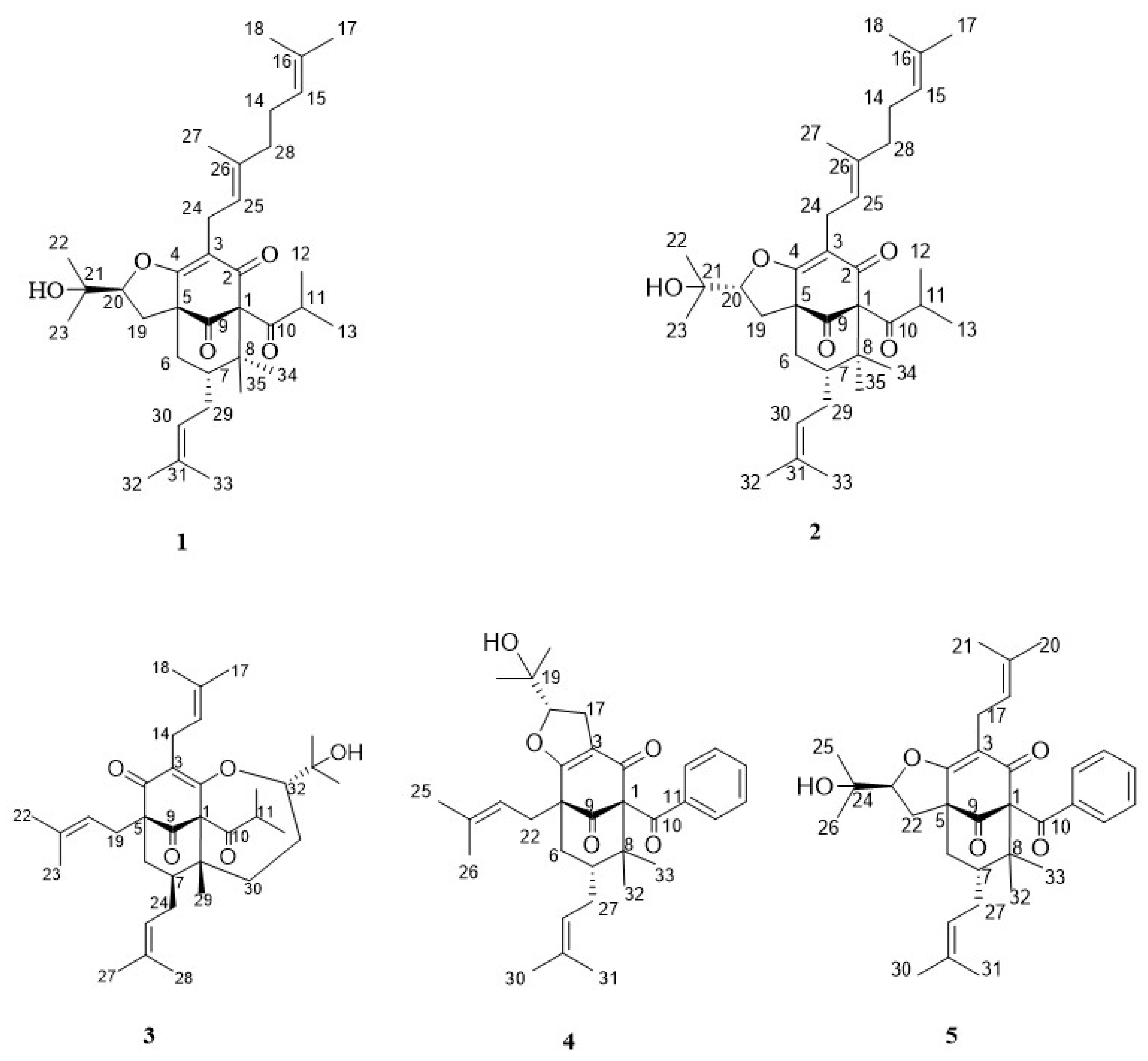
The 95% EtOH extract of the dried aerial parts of H. acmosepalum was partitioned successively with petroleum ether and ethyl acetate (EtOAc). The petroleum ether soluble part was subjected to silica gel column chromatography to give two new compounds (1–2) and three known compounds: Oxepahyperforin (3) [21], sampsonione O (4) [22], sampsonione L (5) [23,24] (Figure 1, 1H- and 13C-NMR data for 3–5 are found in the Supplementary Materials: Tables S1–S3). The structure of the new compounds were elucidated by the analysis of spectroscopic data and the known compounds were concluded by comparison of the NMR data with the published literature.
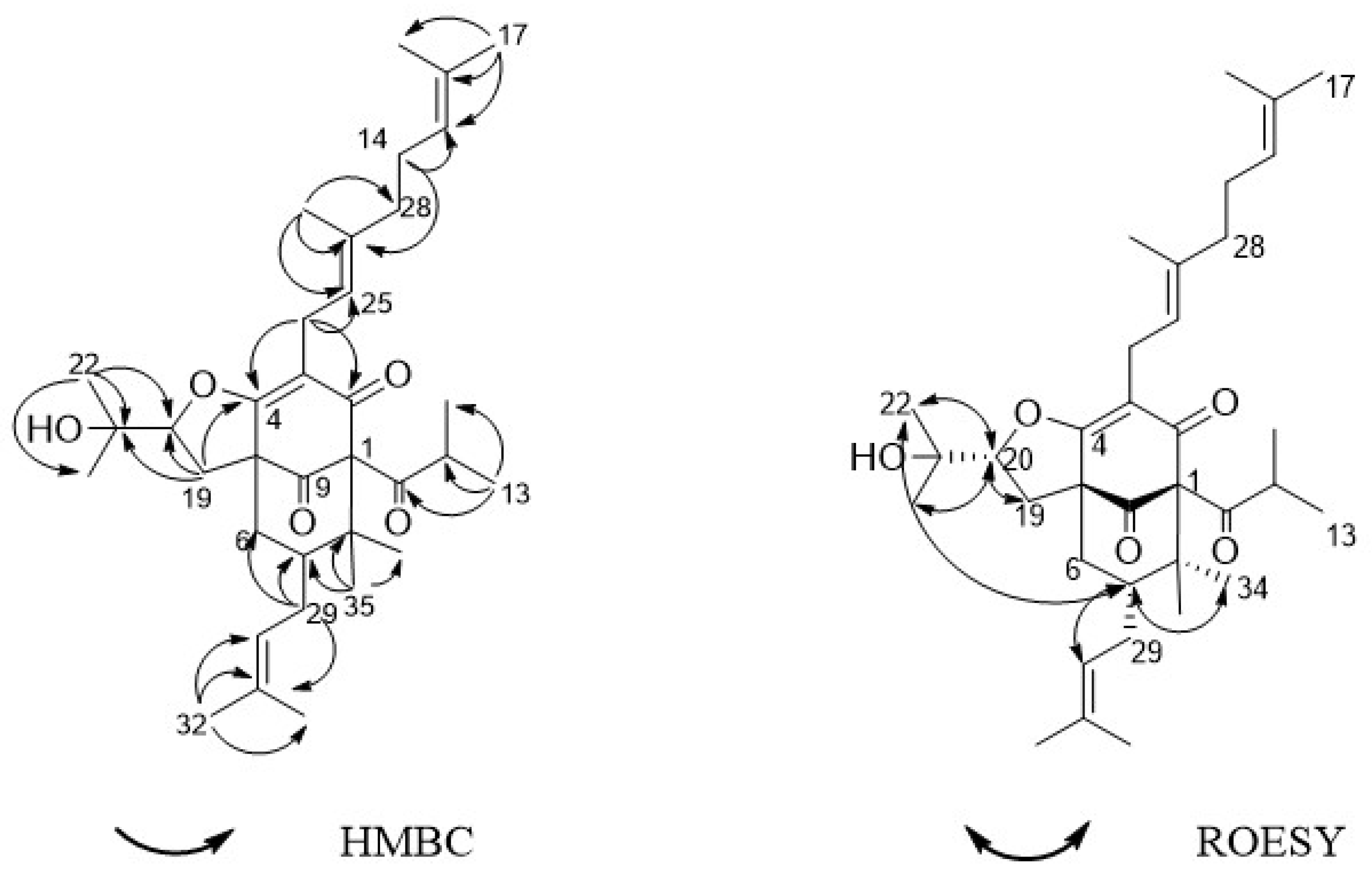
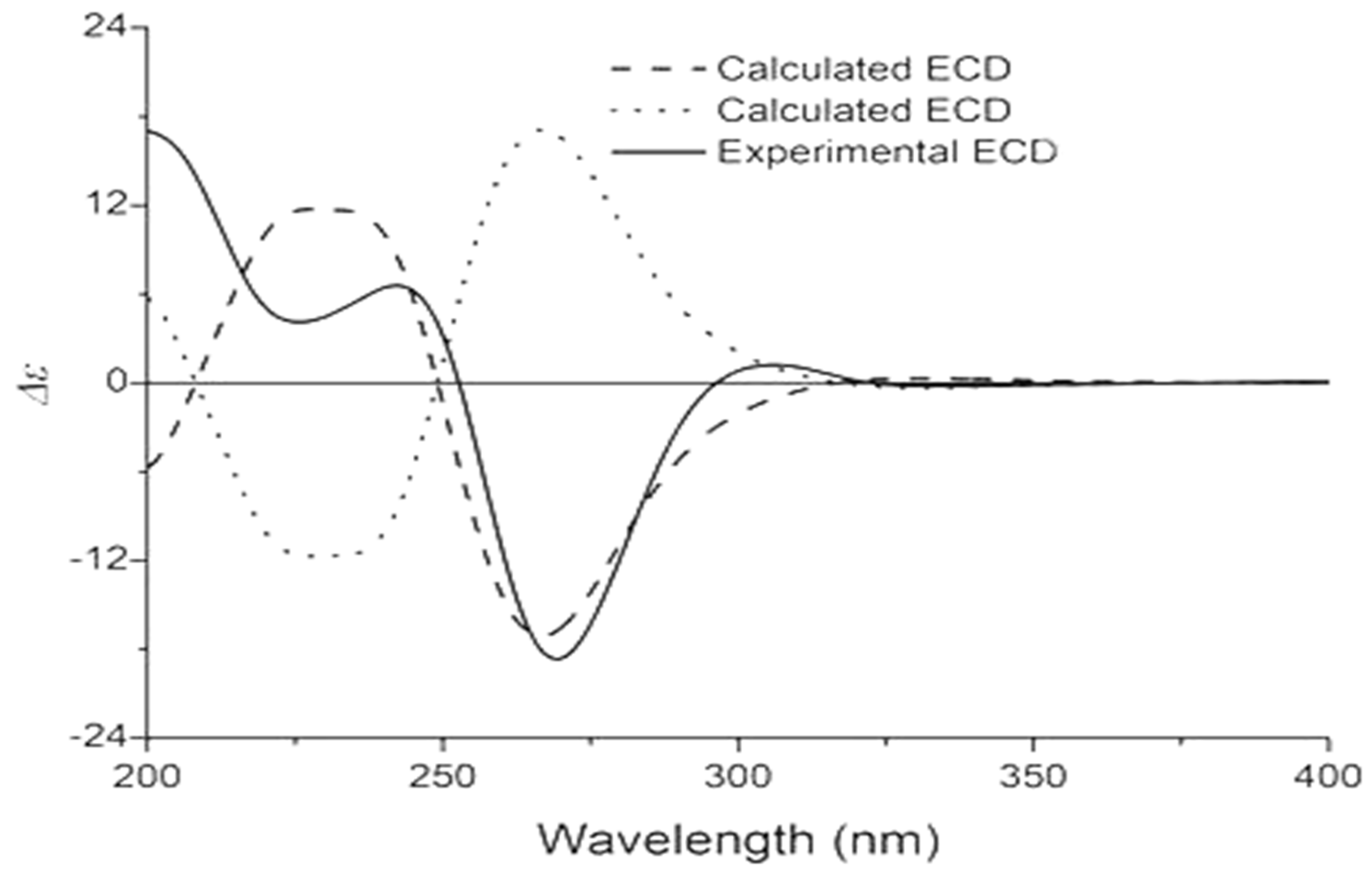
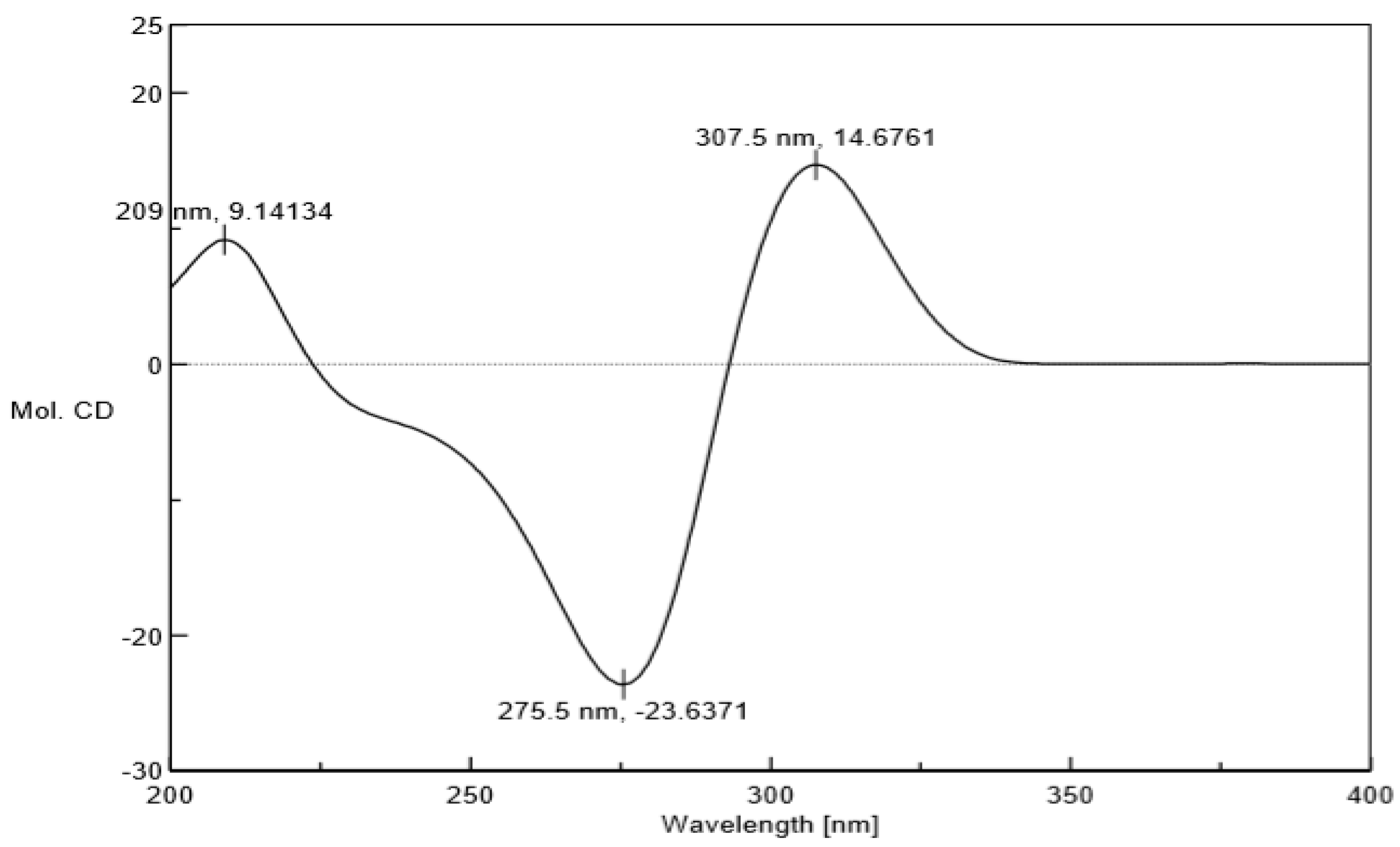
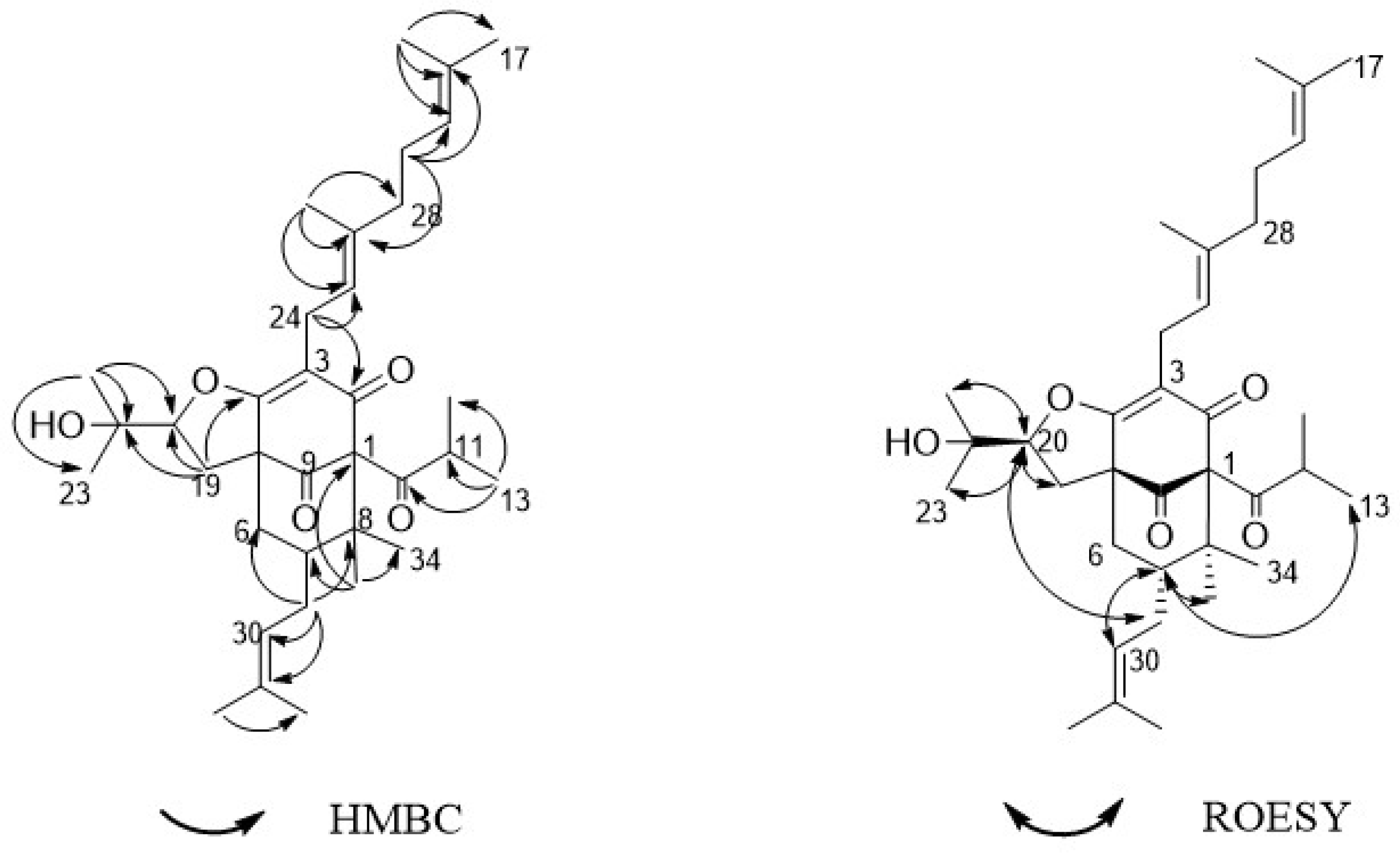
Compound 1 was acquired as a colorless oil. The molecular formula of 1 was determined as C35H52O5 on the basis of HRESIMS (high resolution electrospray ionization mass spectrum) data (m/z 553.3883 [M + H]+, calculated 553.3888, see Figure S7), with ten degrees of unsaturation. Its IR (infrared spectrum, see Figure S9) absorptions implied the presence of hydroxy (3440 cm−1) and carbonyl groups (1725, 1642, and 1618 cm−1). The 1H-NMR spectroscopic data (Table 1) of hyperacmosin A showed the presence of three olefinic protons signals of isopentenyl (δH 4.83 (1H, t, J = 7.0 Hz), 5.04 (1H, s), 5.04 (1H, s)), two doublet methyl signals of the isopropyl group (δH 1.16 (3H, d, J = 6.5 Hz), 1.03 (3H, d, J = 6.5 Hz)), nine singlet methyl signals. Additionally, the data showed characteristic signals of furan-type hyperforin compounds: An oxygen-bearing methine signal of furan nucleus (δH 4.62 (1H, dd, J = 11.5, 10.0 Hz)). The 13C-NMR data (Table 1) showed 35 carbon signals, which were similar to those of furohyperforin [19] except for the position of C-(14–18): The fragment C-(14–18) of furohyperforin was located at C-34, but the C-(14–18) of 1 was located at C-28. Three carbonyl carbons (δC 208.6, 206.8, 191.5), six olefinic carbons (δC 138.0, 132.9, 131.1, 124.5, 124.4, 119.5), the characteristic carbon signals of α, β-unsaturated carbonyl group (δC 191.5, 172.7, 119.5), an oxygen-bearing methine (δC 93.5) and three quaternary carbon signals on the skeleton (δC 72.0, 62.1, 46.5). These data indicated that 1 was a furan-type hyperforin derivative. In the HMBC (heteronuclear multiple bond correlation) and HSQC (heteronuclear single quantum correlation) spectra (Figure 2), based on two classic methyl signals in a double peak of the isopropyl group in 1H-NMR, the chemical shift of C-12 and C-13 were determined. H-11 was correlated with C-10, C-12, C-13; H-12 was correlated with C-10, C-11, C-13; H-13 was correlated with C-10, C-11, C-12; the fragment C-(10–13) was determined. Based on olefinic proton H-30, the chemical shift of C-30 was determined, H-30 was correlated with C-29, C-31, C-32; H-32 was correlated with C-30, C-31, C-33; H-33 was correlated with C-30, C-31, C-32, in addition, H-29 was correlated with C-6, C-7, C-8, C-30 and C-31, suggesting that the branch of C-(29–33) was connected to C-7. The correlations of H-34 with C-1, C-7, C-8, C-35 and H-35 with C-1, C-7, C-8, C-34 showed that two methyl groups were situated at C-8. Additionally, the correlations of H-22 with C-20, C-21, C-23; H-23 with C-20, C-21, C-22; H-20 with C-19, C-22, C-23; H-19 with C-4, C-20, C-21 determined the fragment of C-(19-23). The correlations of H-17 with C-15, C-16, C-18; H-18 with C-15, C-16, C-17; H-15 with C-14, C-18; H-14 with C-15, C-26 determined the fragment of C-(14-18); and the correlations of H-27 with C-25, C-26, C-28; H-28 with C-14, C-15, C-(25-27); H-25 with C-24, C-27, C-28; H-24 with C-(2–4), C-25, C-26 determined the fragment of C-(24-28), which showed that geranyl alkenyl was situated at C-3. In the ROESY (Rotating frame Overhauser Enhancement Spectroscopy) spectrum (see Figure S5) of 1, significant correlations were observed between H-20 (δH 4.62), H-13 (δH 1.03), H-19 (δH 2.88), H-22 (δH 1.36) and H-23 (δH 1.27); H-7 (δH 1.97), H-22 (δH 1.36), H-30 (δH 5.04) and H-34 (δH 1.38). Hence, the relative configuration was determined as shown in Figure 2. To assign the absolute configuration of 1, the electronic circular dichroism (ECD) experiment was conducted by the time-dependent density functional theory (TDDFT) method [20,25]. Based on a large number of previous studies on PPAPs in our laboratory, the chirality of C-1, 5, 7 in the skeleton structure of 1 had been assigned to be (1R, 5S, 7S) by comparing the ECD spectra (Figure 3) to that of hyperscabrones A [9]. In the ROESY spectrum of 1, the correlations between H-20 (δH 4.62) and H-13 (δH 1.03) indicated that the orientation of the H-20 was β-oriented. Therefore, the absolute configuration of 1 was determined to be (1R, 5S, 7S, 20R) as shown in Figure 4, and was named hyperacmosin A.
Compound 2 was obtained as a colorless oil. The molecular formula of 2 was determined as C35H52O5 on the basis of HRESIMS data (m/z 553.3892 [M + H]+, calculated 553.3888, see Figure S16), with ten degrees of unsaturation. Its IR (see Figure S18) absorptions implied the presence of hydroxy (3467 cm−1) and carbonyl groups (1726 and 1618 cm−1). The 1H-NMR and 13C-NMR data of 2 were similar to those of 1, except for H-15 and 30 (2: δH 5.05 and 4.88, 1: δH 4.83 and 5.04), 22 and 34 (2: δH 1.38 and 1.36, 1: δH 1.36 and 1.38), 23 and 35 (2: δH 1.25 and 1.28, 1: δH 1.27 and 1.25). In the HMBC spectra, H-11 was correlated with C-10, C-12, C-13; H-32 was correlated with C-7, C-30, C-31, C-33; H-29 was correlated with C-6, C-8, C-30 and C-31. In addition, the correlations of H-34 with C-1, C-7, C-35; H-20 with C-3, C-4, C-19, C-21, C-22, C-23; H-19 with C-2, C-3, C-4, C-8, C-10, C-20, C-21. The correlations of H-15 with C-17, C-27, C-28; H-14 with C-15, C-16, C-26; and the correlations of H-28 with C-14, C-15, C-(25-27); H-24 with C-2, C-5, C-6, C-25, C-26. The ROESY spectra (see Figure S14) revealed the correlations (Figure 5) between H-20 (δH 4.80), H-19 (δH 2.94), H-22 (δH 1.38), H-23 (δH 1.25) and H-29 (δH 2.21); H-7 (δH 1.96), H-13 (δH 1.15), H-22 (δH 1.38), H-27 (δH 1.67) and H-35 (δH 1.28). Thus, the relative configuration was determined as shown in Figure 5. In the ECD spectra, the chirality of C-1, 5, 7 in the skeleton structure of 2 was consistent with compound 1 because the spectra of 2 had a high degree of similarity to the spectra of 1 (Figure 6). Furthermore, the ROESY correlation between H-20 (δH 4.62) and H-29 (δH 2.21) indicated that the orientation of the H-20 was α-oriented. Therefore, the absolute configuration of 2 was determined to be (1R, 5S, 7S, 20S), and was named hyperacmosin B.
The five compounds were evaluated for neuroprotective effect in two models: l-glutamic acid (L-Glu) and oxygen-glucose deprivation (OGD) by the MTT method [26]. Donepezil and PHPB (potassium 2-(1-hydroxypentyl)-benzoate) were used as the positive controls. l-glutamic acid and sodium hyposulfite were used as the damage agents. In the model of l-Glu, the cell survival rate of the positive control groups had increased compared with the control group. In experiment groups, hyperacmosin A (1) and sampsonione O (4) increased the survival rates of the SK-N-SH cells to 68.20%, 80.90% compared with the control group respectively (Table 2), which showed remarkable neuroprotection activity. In the model of OGD, PHPB increased the survival rates of the SK-N-SH cells to 80.30% compared with the control group (Table 2). While the cell survival rate of hyperacmosin B, oxepahyperforin and sampsonione L had increased, those compounds did not exhibit obvious neuroprotective activity against OGD induced-injury on SK-N-SH cells compared with the control group.
3. Materials and Methods
3.1. General Information
Optical rotations were measured on a JASCO P-2000 polarimeter (Jasco Inc., Tokyo, Japan) using methanol as a solvent. UV spectra were determined with a JASCO V-650 spectrophotometer (Jasco Inc., Tokyo, Japan). IR spectra were recorded on a Nicolet 5700 FT-IR spectrometer (Thermo Fisher Scientific, Waltham, MA, USA). ECD and NMR spectra were obtained on Varian Inova-500 spectrometer (Varian Inc., Palo Alto, CA, USA) and a Mecury-400 spectrometer (Varian Inc., Palo Alto, CA, USA) with trimethylsilane (TMS) as an internal standard and CDCl3 as a solvent. HRESIMS spectra were performed on an Agilent 1100 LC/MSD Trap SL mass spectrometer (Agilent Technologies Ltd., Santa Clara, CA, USA). Preparative HPLC experiments were carried out on a preparative YMC-Pack ODS-A column (250 × 20 mm, 5 μm, YMC, Kyoto, Japan). Silica gel (100–200 mesh and 200–300 mesh, Qingdao Haiyang Chemistry Company, Qingdao, China), silica gel H (Qingdao Haiyang Chemistry Company, Qingdao, China), MCI gel CHP20P (35−75 μm, Mitsubishi Chemical Corp, Japan), ODS (50 μm, YMC, Kyoto, Japan), and Sephadex LH-20 (40−70 μm, Healthcare Bio-Sciences AB, Uppsala, Sweden) were used for column chromatography and silica gel GF-254 (Qingdao Haiyang Chemistry Company, Qingdao, China) was used for TLC. 3-(4,5-dimethylthiazol-2-yl)-2,5-diphenyltetrazolium bromide (MTT) were purchased from Sigma-Aldrich (Saint Louis, MO, USA). Other chemicals used in the study were of analytical grade (Beijing Chemical works, Beijing, China). Fetal bovine serum (FBS) was obtained from Invitrogen Corporation (Carlsbad, CA, USA). Penicillin and streptomycin were purchased from Sigma (St. Louis, MO, USA).
3.2. Plant Material
The air-dried aerial parts of H. acmosepalum were collected from the Wenshan, Yunnan Province, China, in July 2016. The plant was identified by Prof. Lin Ma. A voucher specimen (No. ID-S-2764) was deposited in the herbarium of the Institute of Materia Medica, Chinese Academy of Medical Sciences.
3.3. Extraction and Isolation
Air-dried and powdered aerial parts of H. acmosepalum (17.0 kg) were extracted three times, with 95% EtOH (170 L per extraction) under reflux. The 95% EtOH extracts were concentrated in vacuo to provide a crude extract (1.8 kg), which was suspended in water and then partitioned with petroleum ether and EtOAc successively. The petroleum ether soluble part (500.0 g) was separated by silica gel column chromatography (200−300 mesh) in reduced pressure and eluted with a gradient of petroleum ether–EtOAc (1:0 to 0:1) to obtain 11 fractions (Fr. 1–Fr. 11).
Fr. 1 (254.0 g) was subjected to column chromatography over MCI gel in reduced pressure, eluting with EtOH-H2O (75% to 95%), to give 6 fractions (Fr. 1A–Fr. 1F). Fr. 1C (94.0 g) was loaded onto silica gel column chromatography, eluting with petroleum ether–EtOAc (1:0 to 0:1), to give 20 fractions (Fr. A–Fr. t), Fr. m (5.7 g) was further separated by silica gel H column chromatography and eluted with petroleum ether-CH2Cl2 (9:1) and petroleum ether-Et2O (20:1) repeatedly to give compound 2 (17.0 mg). Fr. r (9.1 g) was further separated by silica gel H column chromatography and eluted with petroleum ether-EtOAc (1:0 to 0:1) and petroleum ether-Et2O (20:1) to yield 14 fractions (Fr. r1–Fr. r14). Compound 1 (9.4 mg) was gained from both Fr. r5 and Fr. r6 (2.4 g) by ODS column chromatography (MeOH-H2O), silica gel H column chromatography (petroleum ether–EtOAc) and semipreparative HPLC.
Fr. 3–5 (104.0 g) were subjected to column chromatography over MCI gel in reduced pressure, eluting with EtOH–H2O (70% to 95%), to give 12 fractions (Fr. A–Fr. L). Fr. A (22.0 g) was loaded onto MPLC (octa decylsilyl silicion, ODS) column chromatography eluted by MeOH-H2O (70% to 100%), which gave 7 fractions (Fr. A1–Fr. A7). Fr. A4 (7.5 g) and Fr. A5 (6.0 g) were further separated by silica gel H column chromatography and eluted with petroleum ether-EtOAc (1:0 to 0:1) respectively to give several subfractions. Each subfraction of Fr. A4 was further purified by silica gel H column chromatography with petroleum ether-EtOAc (9:1), by HPLC (RP C18, 85% MeOH) and by Sephadex LH-20 (MeOH) to give compound 4 (20.0 mg) and compound 5 (30.0 mg). Each subfraction of Fr. A5 was further purified by Sephadex LH-20 (MeOH), silica gel H column chromatography with petroleum ether-EtOAc (9:1), and recrystallization to give compound 3 (27.0 mg).
3.4. Spectroscopic Data
Hyperacmosin A (1): Colorless oil, [α]20D = +87.8 (c = 0.337, MeOH). UV: (MeOH) λmax (log ε) 209.8 (1.96), 280.0 (2.64) nm. IR (FT-IR Microscope Transmission method): υmax 3440, 3229, 2973, 2927, 1726, 1642, 1619, 1444, 1405, 1249, 1149, 1092, 969, 846, 802 cm−1. 1H-NMR (500 MHz, CDCl3) and 13C-NMR (125 MHz, CDCl3) data are shown in Table 1. HRESIMS (MeOH): 553.3883 ([M + H]+, C35H52O5; calculated 553.3888). HSQC, HMBC and ROESY (500 MHz, CDCl3) spectroscopic data can be found in the Supplementary Materials (Figures S3–S5). ECD (MeOH) λ(Δ ε) 209 (+9.14), 275.5 (−23.64), 307.5 (+14.68) nm.
Hyperacmosin B (2): Colorless oil, [α]20D = −53.0 (c = 0.502, MeOH). UV: (MeOH) λmax (log ε) 203.4 (0.57), 282.2 (0.28) nm. IR (FT-IR Microscope Transmission method): υmax 3467, 2971, 2926, 1727, 1619, 1447, 1381, 1240, 1092, 966, 842 cm−1. 1H-NMR (400 MHz, CDCl3) and 13C-NMR (125 MHz, CDCl3) data are shown in Table 1. HRESIMS (MeOH): 553.3892 ([M + H]+, C35H52O5; calculated 553.3888). HSQC, HMBC and ROESY (500 MHz, CDCl3) spectroscopic data can be found in the Supplementary Materials (Figures S12–S14). ECD (MeOH) λ(Δ ε) 209 (+8.03), 278 (−27.48), 310 (+12.08) nm.
3.5. Neuroprotection Bioassays
The neuroprotection of compounds was determined by the MTT method in SK-N-SH cells, grown in Dulbeco’s modified containing 10% FBS, penicillin (100 U/mL) and streptomycin (100 μg/mL). Cell cultures were incubated at 37 °C under a 5% CO2 atmosphere. The compounds and positive control were dissolved with DMSO (10 μmol/L) respectively. The cells were seeded onto 96-well microplates at a density of 1 × 10⁵ cells/well. After incubation at 37 °C for 24 h under a 5% CO2 atmosphere, the cells were pre-incubated with compounds and positive controls respectively for 1 h. In the l-Glu, the DMEM (Dulbecco’s modified Eagle’s medium) without l-Glu was added to the cells of the normal group, the DMEM containing l-Glu (the final concentration of 27 mM) was added to the cells of other groups. After 4 h of co-incubation, 100 μL of MTT (0.5 mg/mL) were added to each well after the withdrawal of the culture medium and were incubated for an additional 4 h at 37 °C. The resulting formazan crystals were dissolved in 150 μL of DMSO after aspiration of the culture medium. The optical density (OD) of the formazan solution was measured on a microplate reader at 570 nm. In the OGD, the l-DMEM (low-sugar Dulbecco’s modified Eagle’s medium) without sodium hyposulfite was added to the cells of the normal group, the l-DMEM containing sodium hyposulfite (the final concentration of 3.5 mM) was added to the cells of other groups, and then incubated for 24 h. After 24 h, MTT solution (0.5 mg/mL) was added for 4 h at 37 °C. Finally, the formazan crystals were solubilized by DMSO and were spectrophotometrically measured at 570 nm. All data presented in our study were obtained from three independent experiments. Survival rate (%) was obtained by the following formula: Improved survival rate (%) = (survival rate of the experimental group-survival rate of the control group)/Survival rate of the control group.
4. Conclusions
In this study, five phloroglucinols were isolated from the petroleum ether soluble part of the 95% EtOH extract from H. acmosepalum, including two new phloroglucinols and three known compounds. In the neuroprotection screening, the compound 1 (hyperacmosin A) and compound 4 (sampsonione O) showed significant neuroprotective activity in L-Glu induced-injury on human neuroblastoma SK-N-SH cells. However, those compounds did not exhibit neuroprotective activity in the OGD.
In summary, PPAPs research is currently in focus, but the low polarity and instability of the PPAPs greatly increase the difficulty in their separation and structure determination. Additionally, this work provides preliminary evidence for the neuroprotective function of these compounds, but further investigations of this neuroprotective function and other activities are warranted. By elucidating how these molecules interact, this work provides the necessary data to explore the relationship between neuroprotective activity and molecular structure.
Supplementary Materials
The following are available online: Figures S1–S27, Tables S1–S3.
Author Contributions
The contributions of respective authors were as follows: J.W. (Jiao Wang) performed the elucidation of the chemical constituents from the plant, the interpretation of the spectra and prepared the manuscript; M.S. performed the isolation, purification, identification this chemical constituents; J.W. (Jiajia Wang), J.L. and T.J. contributed to the manuscript of review and editing. The research was performed based on the plan of the corresponding authors, J.L. and T.J.
Funding
This research was funded by CAMS Innovation Fund for Medical Sciences (CIFMS), CAMS Initiative for Innovative Medicine (CAMS-I2M, No. 2016-I2M-1-010) and supported by Scientific Research Project of Xinjiang Normal University (No. XJNUSYS112017A01).
Acknowledgments
The authors acknowledge all contributors for their valuable time and commitment to the study.
Conflicts of Interest
The authors declare no conflict of interest.
References
- Crockett, S.L.; Robson, N.K.B. Taxonomy and Chemotaxonomy of the Genus Hypericum. Med. Aromat. Plant Sci. Biotechnol. 2011, 5, 1–13. [Google Scholar] [PubMed]
- Rouis, Z.; Abid, N.; Aouni, M.; Faiella, L.; Dal Piaz, F.; De Tommasi, N.; Braca, A. Benzophenone glycosides from Hypericum humifusum ssp. austral. J. Nat. Prod. 2013, 76, 979–982. [Google Scholar] [CrossRef] [PubMed]
- Gao, W.; Hu, J.W.; Hou, W.Z.; Xu, F.; Zhao, J.; Xu, F.; Sun, H.; Xing, J.G.; Peng, Y.; Wang, X.L.; et al. Four new prenylated phloroglucinol derivatives from Hypericum scabrum. Tetrahedron Lett. 2016, 57, 2244–2248. [Google Scholar] [CrossRef]
- Avato, P. A Survey on the Hypericum Genus: Secondary Metabolites and Bioactivity. Stud. Nat. Prod. Chem. 2005, 30, 603–634. [Google Scholar]
- Chen, J.J.; Chen, H.J.; Lin, Y.L. Novel Polyprenylated Phloroglucinols from Hypericum sampsonii. Molecules 2014, 19, 19836–19844. [Google Scholar] [CrossRef] [PubMed]
- Hu, J.W.; Gao, W.; Xu, F.; Wei, C.J.; Shi, M.J.; Sun, H.; Zhen, B.; Wang, J.J.; Ji, T.F.; Jiang, J.D. Polycyclic polyprenylated acylphloroglucinol derivatives from Hypericum scabrum. Bioorgan. Med. Chem. Lett. 2017, 27, 4932–4936. [Google Scholar] [CrossRef] [PubMed]
- Yang, X.W.; Grossman, R.B.; Xu, G. Research Progress of Polycyclic Polyprenylated Acylphloroglucinols. Chem. Rev. 2018, 118, 3508–3558. [Google Scholar] [CrossRef]
- Liao, Y.; Liu, X.; Yang, J.; Lao, Y.Z.; Yang, X.W.; Li, X.N.; Zhang, J.J.; Ding, Z.J.; Xu, H.X.; Xu, G. Hypersubones A and B, New Polycyclic Acylphloroglucinols with Intriguing Adamantane Type Cores from Hypericum subsessile. Org. Lett. 2015, 17, 1172–1175. [Google Scholar] [CrossRef]
- Gao, W.; Hou, W.Z.; Zhao, J.; Xu, F.; Li, L.; Xu, F.; Sun, H.; Xing, J.G.; Peng, Y.; Wang, X.L.; et al. Polycyclic Polyprenylated Acylphloroglucinol Congeners from Hypericum scabrum. J. Nat. Prod. 2016, 79, 1538–1547. [Google Scholar] [CrossRef]
- Lundstrom, K.; Pham, H.T.; Dinh, L.D. Interaction of Plant Extracts with Central Nervous System Receptors. Medicines 2017, 4, 12. [Google Scholar] [CrossRef]
- Gao, W.; Hu, J.W.; Xu, F.; Wei, C.J.; Shi, M.J.; Zhao, J.; Wang, J.J.; Zhen, B.; Ji, T.F.; Xing, J.G.; et al. Polyisoprenylated benzoylphloroglucinol derivatives from Hypericum scabrum. Fitoterapia 2016, 115, 128–134. [Google Scholar] [CrossRef] [PubMed]
- Singh, I.P.; Sidana, J.; Bharate, S.B.; Foley, W. Phloroglucinol compounds of natural origin: Synthetic aspects. J. Nat. Prod. Rep. 2010, 27, 393–416. [Google Scholar] [CrossRef] [PubMed]
- Hu, L.Z.; Zhang, Y.; Zhu, H.C.; Liu, J.J.; Li, H.; Li, X.N.; Sun, W.G.; Zeng, J.F.; Xue, Y.B.; Zhang, Y.H. Filicinic Acid Based Meroterpenoids with Anti-Epstein−Barr Virus Activities from Hypericum japonicum. Org. Lett. 2016, 18, 2272–2275. [Google Scholar] [CrossRef] [PubMed]
- Naonobu, T.; Yuka, K.; Haruaki, I.; Takaaki, K.; Jun’ichi, K. Yezo’otogirins A–C, new tricyclic terpenoids from Hypericum yezoense. Tetrahedron Lett. 2009, 50, 4747–4750. [Google Scholar]
- Imreova, P.; Feruszova, J.; Kyzek, S.; Bodnarova, K.; Zduriencikova, M.; Kozics, K.; Mucaji, P.; Galova, E.; Sevcovicova, A.; Miadokova, E.; et al. Hyperforin Exhibits Antigenotoxic Activity on Human and Bacterial Cells. Molecules 2017, 22, 167. [Google Scholar] [CrossRef] [PubMed]
- Flora of China Editorial Committee of Chinese Academy of Sciences. Flora of China; Science Press: Beijing, China, 1990; Volume 50, pp. 18–19. [Google Scholar]
- Li, Z.Q.; Luo, L.; Ma, G.Y.; Chen, X. Phloroglucinol and flavonoid constituents of Hypericum acmosepaium. J. Yunnan Univ. 2004, 26, 162–167. [Google Scholar]
- Luo, L.; Zhang, Y.; Pan, Z.H.; Li, Z.Q. Xanthones from Hypericum Acmosepaium. J. Yunnan Univ. 2006, 28, 337–339. [Google Scholar]
- Shi, M.J.; Hu, J.W.; Wang, J.J.; Zhen, B.; Wang, X.; Ji, T.F. Chemical constituents of Hypericum perforatum. Chain J. Chin. Mater. Med. 2018, 43, 2726–2731. [Google Scholar]
- Hu, J.W.; Shi, M.J.; Wang, J.J.; Li, L.; Jiang, J.D.; Ji, T.F. Methylated Polycyclic Polyprenylated Acylphloroglucinol Derivatives from Hypericum ascyron. J. Nat. Prod. 2018, 81, 2348–2356. [Google Scholar] [CrossRef]
- Verotta, L.; Appendino, G.; Jakupovic, J.; Bombardelli, E. Hyperforin Analogues from St. John’s Wort (Hypericum perforatum). J. Nat. Prod. 2000, 63, 412–415. [Google Scholar] [CrossRef]
- Xiao, Z.Y.; Mu, Q.; Shiu, W.K.; Zeng, Y.H.; Gibbons, S. Polyisoprenylated Benzoylphloroglucinol Derivatives from Hypericum sampsonii. J. Nat. Prod. 2007, 70, 1779–1782. [Google Scholar] [CrossRef] [PubMed]
- Ciochina, R.; Grossman, R.B. Polycyclic polyprenylated acylphloroglucinols. Chem. Rev. 2006, 106, 3963–3986. [Google Scholar] [CrossRef] [PubMed]
- Fan, Y.M.; Tian, D.S.; Gu, W.; Huang, L.J.; Yuan, C.M.; Hao, X.J. Chemical Constituents from the Leaves and Twigs of Garcinia multiflora and Their Cytotoxic as well as Anti-inflammatory Activity. Nat. Prod. Res. Dev. 2016, 28, 222–227. [Google Scholar]
- Bringmann, G.; Bruhn, T.; Maksimenka, K.; Hemberger, Y. The assignment of absolute stereostructures through quantum chemical circular dichroism calculations. Eur. J. Org. Chem. 2009, 17, 2717–2727. [Google Scholar] [CrossRef]
- Lei, H.; Zhao, C.Y.; Wang, X.L.; Peng, Y.J. L-3-n-Butylphthalide attenuates β-amyloid-induced toxicity in neuroblastoma SH-SY5Y cells through regulating mitochondrion-mediated apoptosis and MAPK signaling. Asian Nat. Prod. Res. 2014, 16, 854–864. [Google Scholar] [CrossRef] [PubMed]
Sample Availability: Samples of the compounds 1–5 are available from the authors. |
Figure 1.
Structure of compounds 1–5.

Figure 2.
Selected key HMBC and ROESY correlations for 1.

Figure 3.
Calculated and experimental ECD spectra of hyperscabrones A in methanol [9].
Figure 3.
Calculated and experimental ECD spectra of hyperscabrones A in methanol [9].

Figure 4.
Experimental ECD spectra of 1 in methanol.

Figure 5.
Selected key HMBC and ROESY correlations of 2.

Figure 6.
Experimental ECD spectra of 2 in methanol.


Table 1.
1H- and 13C-NMR data for 1 and 2 in CDCl3.
| Position | Type | 1 | 2 | ||
|---|---|---|---|---|---|
| δC a | δH (J in Hz) b | δC a | δH (J in Hz) c | ||
| 1 | C | 72.0 | - | 71.7 | - |
| 2 | C | 191.5 | - | 191.2 | - |
| 3 | C | 119.5 | - | 120.1 | - |
| 4 | C | 172.7 | - | 173.1 | - |
| 5 | C | 62.1 | - | 62.3 | - |
| 6 | CH2 | 38.6 | 2.05, m 1.97, m | 38.7 | 2.13, m 1.96, m |
| 7 | CH | 48.1 | 1.97, m | 48.2 | 1.96, m |
| 8 | C | 46.5 | - | 46.9 | - |
| 9 | C | 206.8 | - | 206.5 | - |
| 10 | C | 208.6 | - | 208.6 | - |
| 11 | CH | 39.9 | 2.39, m | 39.8 | 2.49, m |
| 12 | CH3 | 20.9 | 1.16, d (6.5) | 21.1 | 1.11, d (5.2) |
| 13 | CH3 | 20.6 | 1.03, d (6.5) | 21.0 | 1.15, d (5.2) |
| 14 | CH2 | 26.7 | 2.03, m | 26.7 | 2.01, m |
| 15 | CH | 124.4 | 4.83, t (7.0) | 124.3 | 5.05, m |
| 16 | C | 131.1 | - | 131.2 | - |
| 17 | CH3 | 25.7 | 1.64, s | 25.7 | 1.64, s |
| 18 | CH3 | 17.6 | 1.57, s | 17.6 | 1.57, s |
| 19 | CH2 | 27.6 | 2.88, m | 27.2 | 2.94, m |
| 20 | CH | 93.5 | 4.62, dd (11.5, 10.0) | 93.9 | 4.80, t (8.0) |
| 21 | C | 70.7 | - | 71.2 | - |
| 22 | CH3 | 26.5 | 1.36, s | 26.7 | 1.38, s |
| 23 | CH3 | 25.1 | 1.27, s | 25.3 | 1.25, s |
| 24 | CH2 | 29.2 | 2.46, m | 29.3 | 2.49, m |
| 25 | CH | 119.3 | 5.04, s | 119.3 | 5.05, m |
| 26 | C | 138.0 | - | 138.0 | - |
| 27 | CH3 | 16.4 | 1.68, s | 16.3 | 1.67, s |
| 28 | CH2 | 40.0 | 1.97, m | 40.0 | 1.96, m |
| 29 | CH2 | 29.6 | 2.17, m 1.80, m | 29.5 | 2.21, m 1.96, m |
| 30 | CH | 124.5 | 5.04, s | 124.5 | 4.88, t (5.2) |
| 31 | C | 132.9 | - | 132.7 | - |
| 32 | CH3 | 25.8 | 1.64, s | 25.8 | 1.66, s |
| 33 | CH3 | 17.9 | 1.52, s | 17.9 | 1.55, s |
| 34 | CH3 | 23.7 | 1.38, s | 23.2 | 1.36, s |
| 35 | CH3 | 26.3 | 1.25, s | 26.7 | 1.28, s |
a Recorded at 125 MHz; b Recorded at 500 MHz; c Recorded at 400 MHz.
Table 2.
Neuroprotection activity of 1 to 5 on human neuroblastoma SK-N-SH cells.
| Compounds | L-Glu Model | Compounds | OGD Model | ||||
|---|---|---|---|---|---|---|---|
| Mean ± SD (n = 3) | Survival Rate (%) | Improved Survival Rate (%) | Mean ± SD (n = 3) | Survival Rate (%) | Improved Survival Rate (%) | ||
| Normal a | 0.901 ±0.05 | Normal b | 1.565 ± 0.04 | ||||
| Control c | 0.555 ± 0.02 *** | 61.70 | Control d | 1.071 ± 0.04 *** | 68.40 | ||
| Donepezil e | 0.559 ± 0.03 | 62.10 | 0.66 | Donepezil e | 1.107 ± 0.04 | 70.70 | 3.33 |
| PHPB e | 0.569 ± 0.02 | 63.20 | 2.46 | PHPB e | 1.257 ± 0.09 # | 80.30 | 17.34 |
| 1 | 0.614 ± 0.01 # | 68.20 | 10.62 | 1 | 0.850 ± 0.07 ## | 54.30 | −20.67 |
| 2 | 0.577 ± 0.04 | 64.00 | 3.84 | 2 | 1.202 ± 0.07 | 76.80 | 12.23 |
| 3 | 0.567 ± 0.00 | 62.90 | 2.04 | 3 | 1.091 ± 0.18 | 69.70 | 1.84 |
| 4 | 0.729 ± 0.03 ## | 80.90 | 31.27 | 4 | 0.904 ± 0.19 | 57.80 | −15.56 |
| 5 | 0.528 ± 0.02 | 58.60 | −4.92 | 5 | 1.109 ± 0.05 | 70.90 | 3.58 |
a DMEM (Dulbecco’s modified Eagle’s medium) without l-glutamic acid was added. b l-DMEM (low-sugar Dulbecco’s modified Eagle’s medium) without sodium hyposulfite was added. c l-glutamic acid was added as the damage agent. d Sodium hyposulfite was added as the damage agent. e Donepezil and PHPB were added as the positive controls. *** p < 0.001 vs. Normal; # p < 0.05 versus Control, ## p < 0.01 versus Control.
© 2018 by the authors. Licensee MDPI, Basel, Switzerland. This article is an open access article distributed under the terms and conditions of the Creative Commons Attribution (CC BY) license (http://creativecommons.org/licenses/by/4.0/).
Share and Cite
MDPI and ACS Style
Wang, J.; Shi, M.; Wang, J.; Li, J.; Ji, T. Polycyclic Polyprenylated Acylphloroglucinol Derivatives from Hypericum acmosepalum. Molecules 2019, 24, 50. https://0-doi-org.brum.beds.ac.uk/10.3390/molecules24010050
AMA Style
Wang J, Shi M, Wang J, Li J, Ji T. Polycyclic Polyprenylated Acylphloroglucinol Derivatives from Hypericum acmosepalum. Molecules. 2019; 24(1):50. https://0-doi-org.brum.beds.ac.uk/10.3390/molecules24010050
Chicago/Turabian StyleWang, Jiao, Mengjiao Shi, Jiajia Wang, Jin Li, and Tengfei Ji. 2019. "Polycyclic Polyprenylated Acylphloroglucinol Derivatives from Hypericum acmosepalum" Molecules 24, no. 1: 50. https://0-doi-org.brum.beds.ac.uk/10.3390/molecules24010050