Article

Structural and Photophysical Properties of 2,1,3-Benzothiadiazole-Based Phosph(III)azane and Its Complexes

Nikolaev Institute of Inorganic Chemistry, Siberian Branch of the Russian Academy of Sciences, 630090 Novosibirsk, Russia
*
Author to whom correspondence should be addressed.
Submission received: 28 April 2020 / Revised: 17 May 2020 / Accepted: 17 May 2020 / Published: 22 May 2020
(This article belongs to the Special Issue Polysulfur- and Sulfur-Nitrogen Heterocycles)

Abstract

:
Here we describe the synthesis of a novel N,N’-bis(2,1,3-benzothiadiazol-4-yl)-1-phenylphosphanediamine (H2L) and its zinc (II) and copper (I) coordination compounds [Zn2L2]·nC7H8 (1·nC7H8), [Zn2(H2L)2Cl4]·nC7H8 (2·nC7H8), and [Cu(H2L)Cl]n·nTHF (3·THF). According to single crystal X-ray diffraction analysis, H2L ligand and its deprotonated species exhibit different coordination modes. An interesting isomerism is observed for the complexes [Zn2(H2L)2Cl4] (2a and 2b) that differ by the arrangement of H2L. Both complexes possess internal cavities capable of incorporating toluene molecules. Upon toluene release, the geometry of 2b changes substantially, while that of 2a changes slightly. Due to the diverse structures, the compounds 13 reveal different photophysical properties. These results are discussed based on previously reported studies and DFT (density functional theory) calculations.

Graphical Abstract

1. Introduction

Photoluminescent organic compounds and transition metal complexes with organic ligands are important components in various scientific applications, including optoelectronics, photocatalysis, and photodynamic therapy. Heteroaromatic units, in particular 2,1,3-benzothiadiazole (btd) derivatives, are often used as building blocks of optical materials [1,2,3]. Generally, btd acts as an acceptor in polymer molecules [4,5]. However, btd-based small molecules are also being actively studied [6,7,8,9,10]. The photophysical behavior of such derivatives can be tuned by two ways; (1) functionalization of btd; (2) coordination to suitable metal ions. From the viewpoint of coordination chemistry, the high binding affinity of ligands towards transition metals is very important. By choosing appropriate donor groups capable to binding with metal, one can construct defined species with particular ligand arrangement. The presence of different types of donor atoms in one ligand opens up the possibility to interplay with various metal ions as well as to perform a wide variety of coordination modes of this ligand [11]. In this context, P,N-containing ligands are highly attractive, as they combine both strong and weak Lewis base centers. A number of btd derivatives bearing N-donor functional groups were recently studied [2,10,11,12,13]. Phosphorus-containing btds are much less abundant; they are mainly limited to derivatives with phosphate groups [14,15,16,17,18].
Compared with btds, the luminescence properties of organophosphorus derivatives are relatively poorly explored. However, the latter have been studied as optical materials in the last decade. Phosphorus (V) compounds have been tested as acceptor blocks and hosts in optical materials [19,20,21], while reports on P(III)-based compounds in the field of photophysical applications [22], e.g., in organic electronics [23] are scarce, due to their air-sensitivity. Meanwhile, phosphorus (III) derivatives are interesting objects in terms of the ability of the P atom to coordinate metals, allowing the design of various coordination compounds. In fact, coordination of a metal protects P atom from its interaction with oxygen, which increases the stability of a complex to oxidize with respect to free P(III) derivative.
Recently, we reported the study of the phosphorylated 4-amino-2,1,3-benzothiadiazole derivative − N,N-bis(diphenylphosphanyl)-2,1,3-benzothiadiazol-4-amine ((Ph2P)2N-btd) (Scheme 1) and its transition-metal complexes [24]. This ligand contains one btd moiety and tends to coordinate in a bidentate manner via two P atoms. In the present work, we synthesized a novel amino-benzothiadiazole derivative bearing two secondary amino groups and a phosphino group, namely, N,N’-bis(2,1,3-benzothiadiazol-4-yl)-1-phenylphosphanediamine (H2L). Contrary to (Ph2P)2N-btd, H2L contains acidic hydrogen atoms and thus can act as anionic ligand. In addition, H2L bears two btd moieties that provide a possibility to bridging coordination via two N atoms. The coordination chemistry of H2L was demonstrated by means of few examples of its metal complexes, viz. Cu(I) and Zn(II). These central metals are frequently used for luminescent applications as earth-abounded non-precious transition elements. Structural aspects of the compounds prepared were studied using single crystal X-ray diffraction (XRD) analysis and quantum chemical calculations. Photophysical properties of the compounds were also studied.

2. Results and Discussion
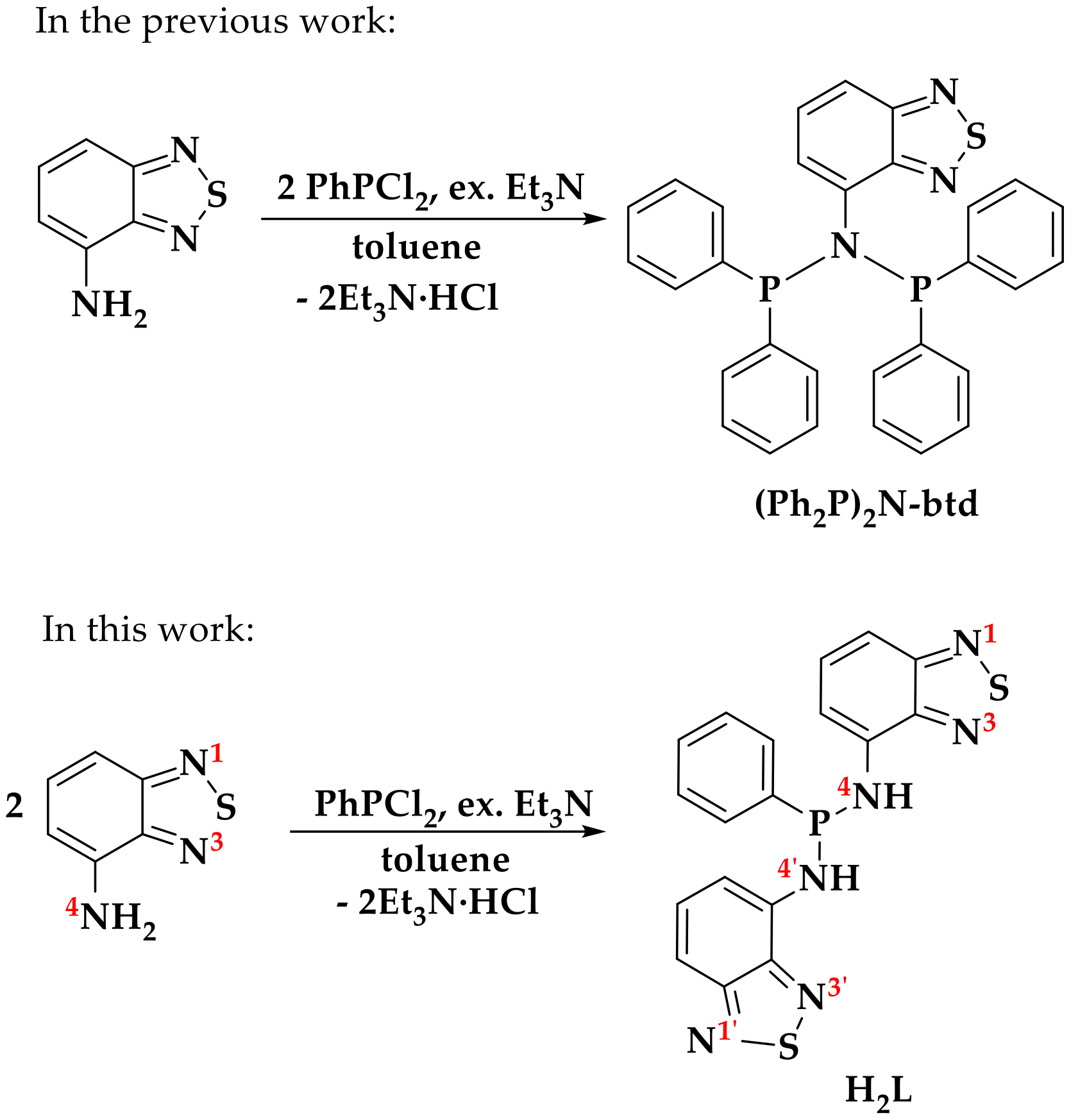
2.1. Synthesis

A novel N,N′-bis(2,1,3-benzothiadiazol-4-yl)-1-phenylphosphanediamine (H2L) can be prepared from 4-amino-2,1,3-benzothiadiazole (NH2-btd) by its modification at amino group using phosphorylation method. The reaction of the benzothiadiazole derivative and dichlorophenylphosphine in a mole ratio 2:1 in the presence of triethylamine gave the title phosphazane in the 80% isolated yield (Scheme 1). The phosphorus reagent, as well as the reaction product, is water and air sensitive; therefore, the reactions were performed under strong anaerobic conditions. The product as a yellow crystalline solid was purified of unreacted amine, phosphine and other by-products by washing with diethyl ether, as the target phosphazane is poorly soluble in it. One of the by-products, namely N-[1,3-bis(2,1,3-benzothiadiazol-4-yl)-2,4-diphenyl-1,3,2λ5,4-diazadiphosphetidin-2-ylidene]-2,1,3-benzothiadiazol-4-amine (4) was isolated by cooling of the ether solution as a few crystals analyzed by single crystal XRD (Figure 1). It can be assumed that the formation of 4 is a result of a side condensation reaction [25]. Most probably, phosph(III)azene PhP=Nbtd was formed and condensed with H2L in the reaction mixture. To establish the exact formation pathway of 4, a further research is required.
H2L contains two acidic hydrogens and thus can act as one- or dibasic acid in the presence of strong bases. Thus, we studied the coordination chemistry of the phosphazane behaving as both anionic and neutral ligand.
The complex [Zn2L2] (1) was obtained by treatment of H2L with ZnCl2 and KHMDS (potassium bis(trimethylsilyl)amide) in THF in a mole ratio 1:1:2 (Scheme 2) as dark red powder. The complex was recrystallized from toluene and isolated as its solvate [Zn2L2]·nC7H8 (1·nC7H8) with the yield of 40%. In 1, the ligand L2– is twice deprotonated. We also attempted to synthesize a complex with partially deprotonated HL ligand by using other reagents ratio, namely H2L:KHMDS = 1:1. However, the reaction gave the complex 1. Upon drying in vacuum, the compound loses almost all lattice toluene; according to the elemental analysis, its content n equals to circa 0.1.
The synthesis of zinc (II) and copper (I) complexes with neutral H2L was achieved by the treatment of the phosphazane with ZnCl2 and CuCl, respectively (Scheme 2). The high solubility of the zinc complex in THF prevented the obtaining of crystals suitable for XRD analysis. Therefore, in the synthesis of the zinc complex, the solvent was changed to toluene, in which it is slightly soluble. The slightly different habitus of the two types of crystals allowed for the harvesting of the samples for single crystal XRD analysis. According to the data, two isomeric complexes of the formula [Zn2(H2L)2Cl2]·nC7H8 (2a·3C7H8 and 2b·2.5C7H8) were formed. Having similar chemical-physical properties, the compounds were not separated from each other. The total yield was 95%. Upon drying, part of the solvate molecules is lost, resulting in a structural transformation of both compounds (Figure S1). The elemental analysis data of the mixture of phases (2·nC7H8) indicate the toluene content n of 1.1.
The complex [Cu(H2L)Cl]n·nTHF (3·THF) is insoluble in THF. To obtain a crystalline product, the reaction was carried out without stirring by adding a solution of H2L in THF to a solid CuCl. After a week, the heterogeneous reaction was completed with crystalline complex 3·THF formation. The isolated yield was 60%. According to XRD analysis, the complex is polymeric, which explains its insolubility in THF. Both XRD and elemental analysis indicate the compound has one THF per formula unit.

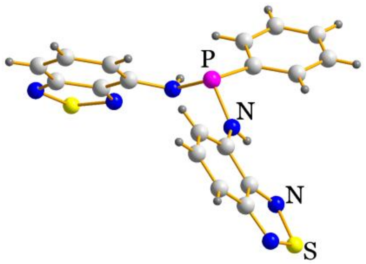
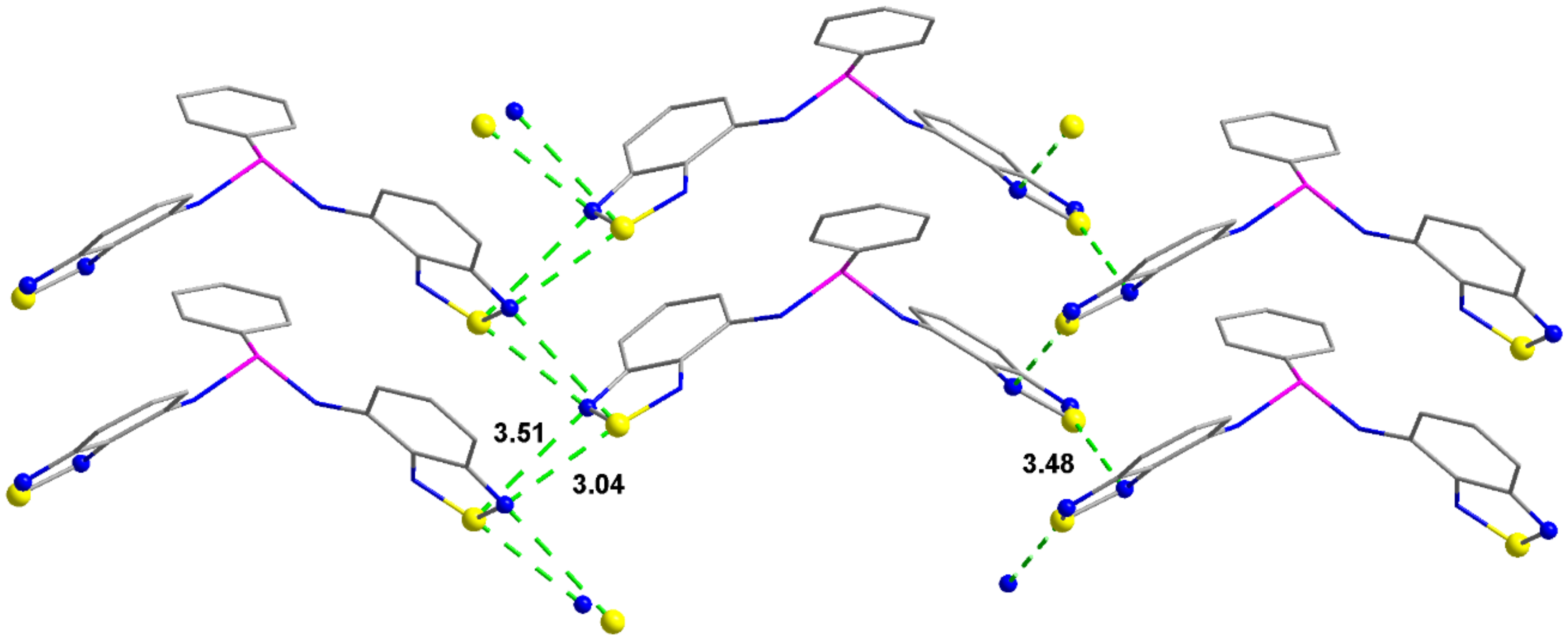
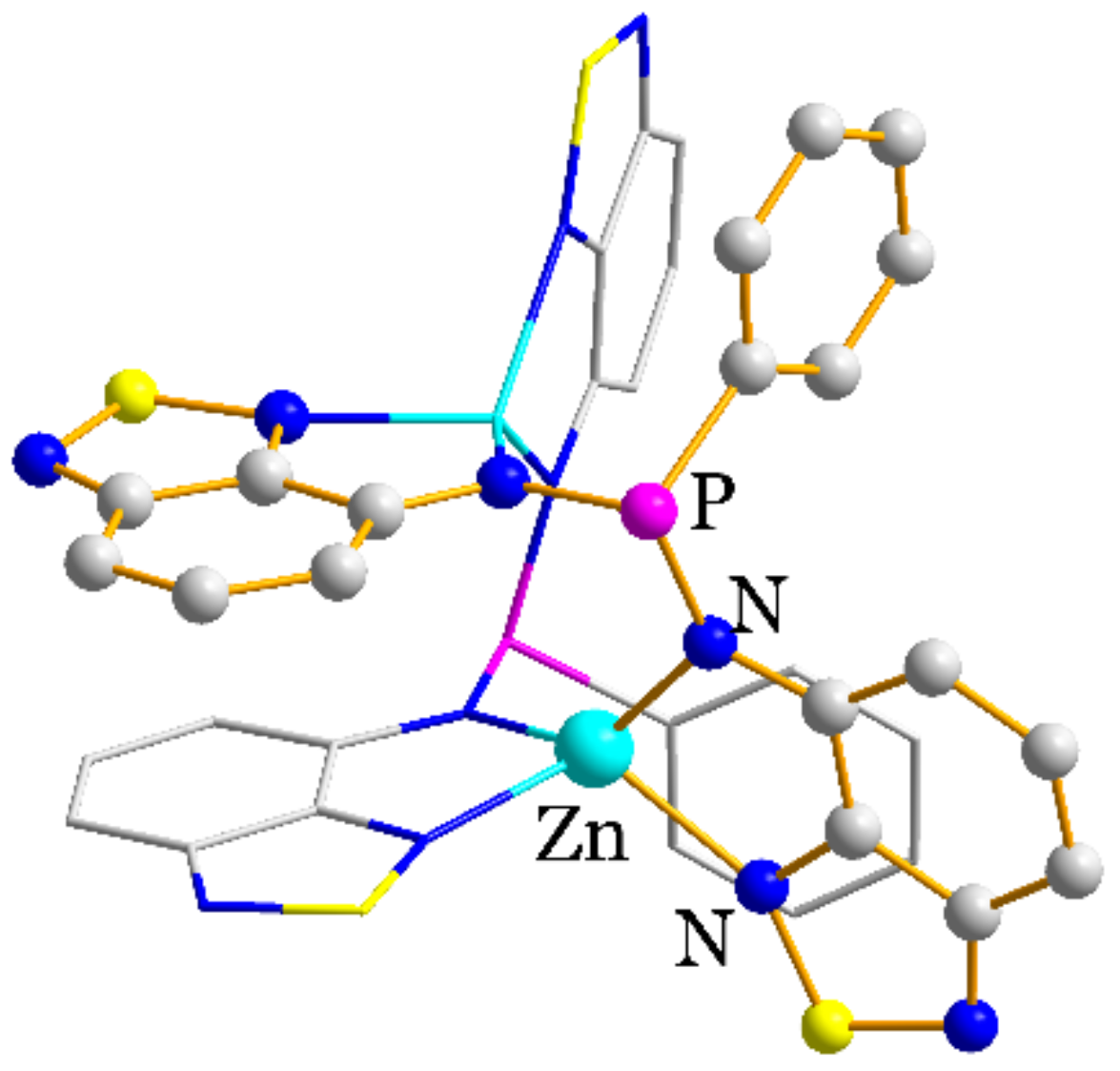
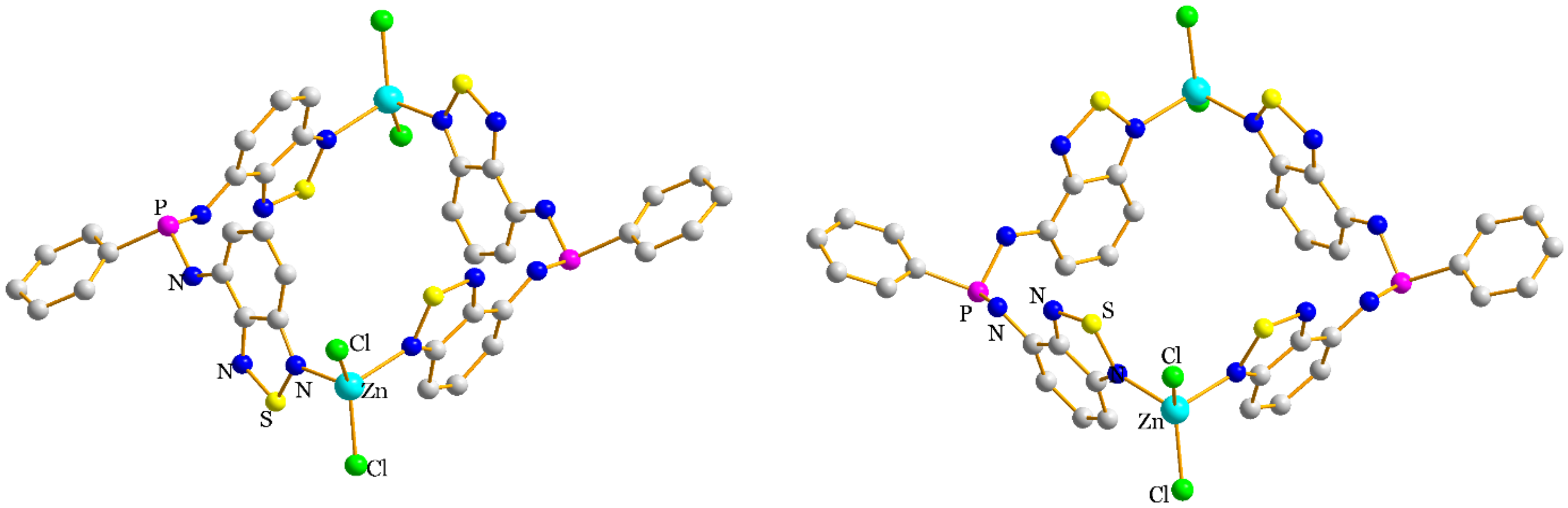
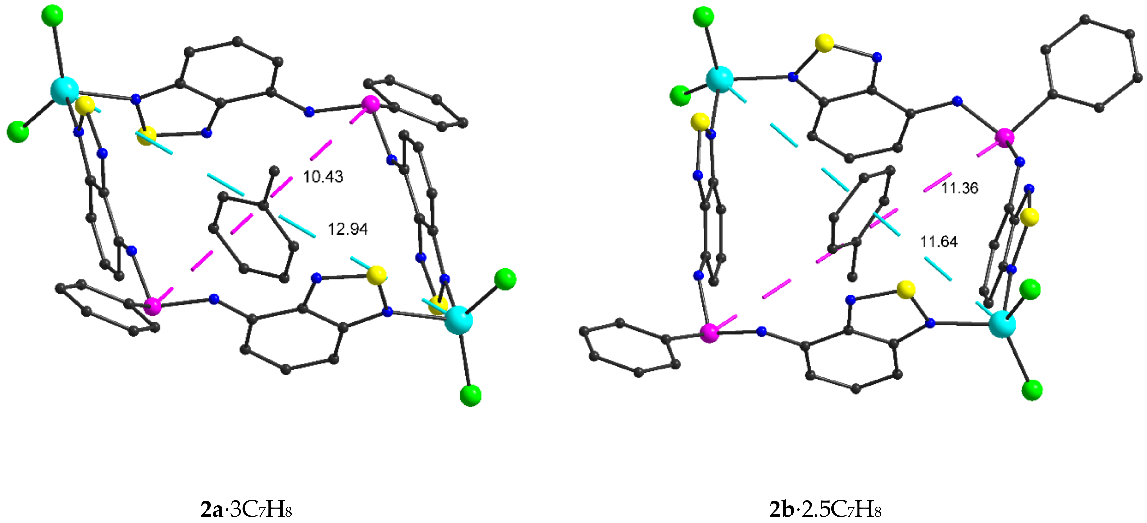
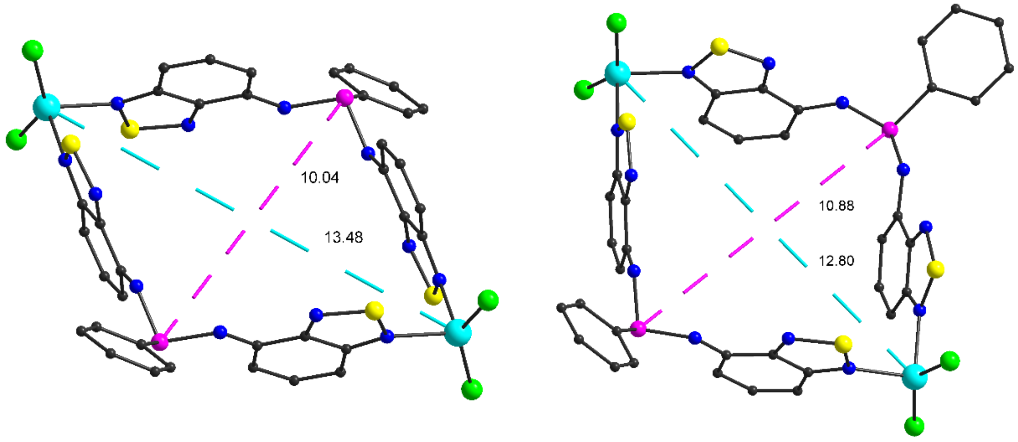
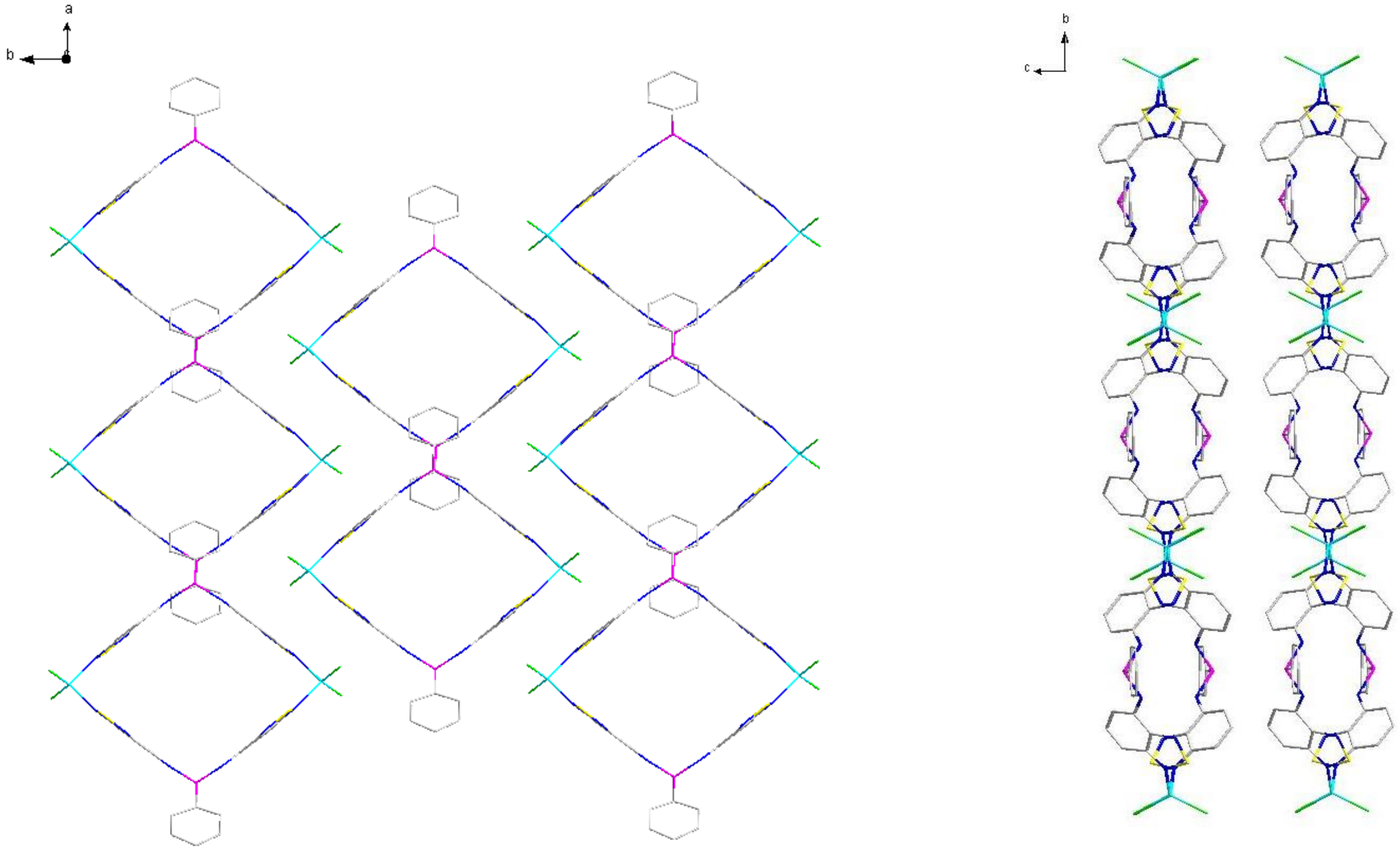
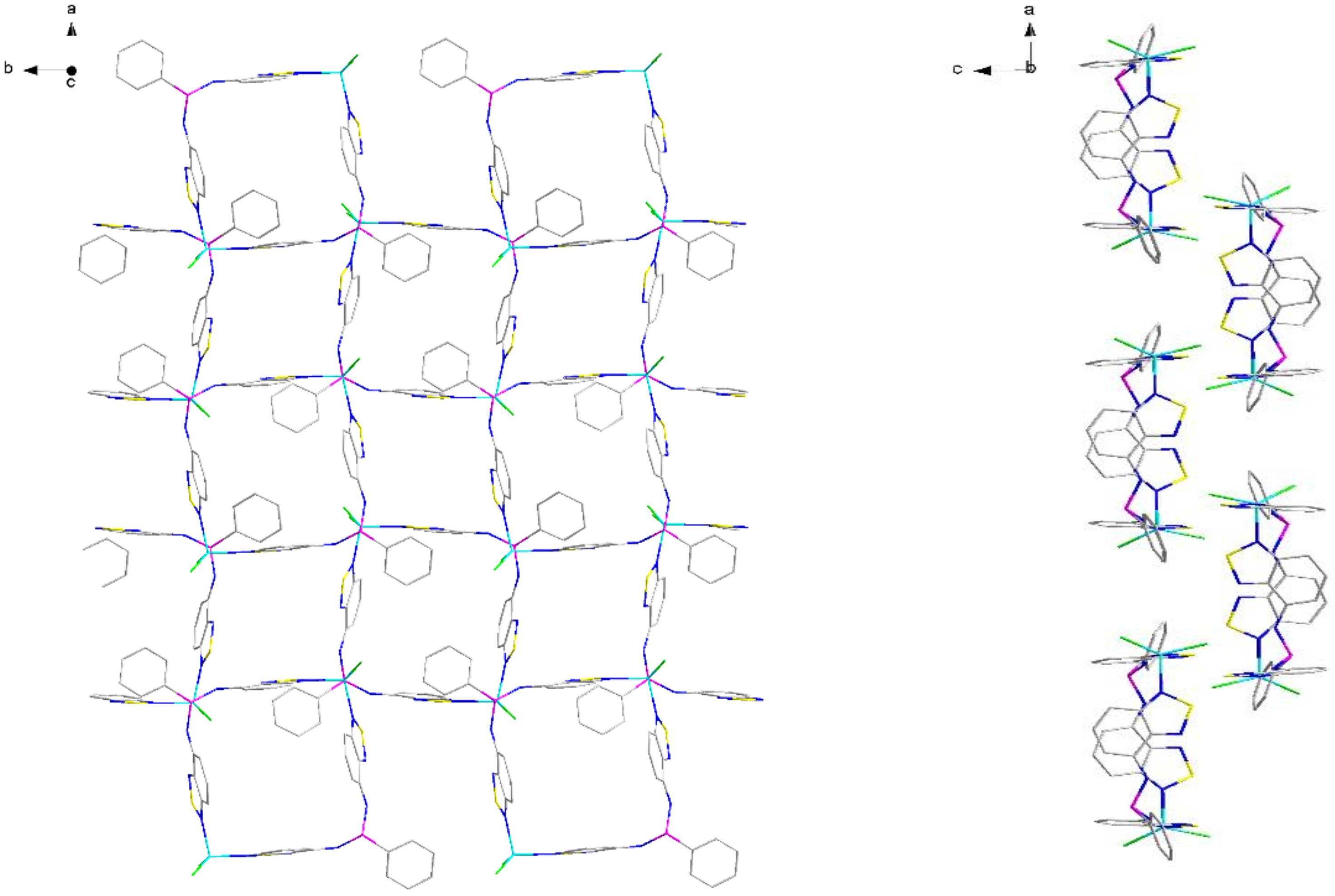
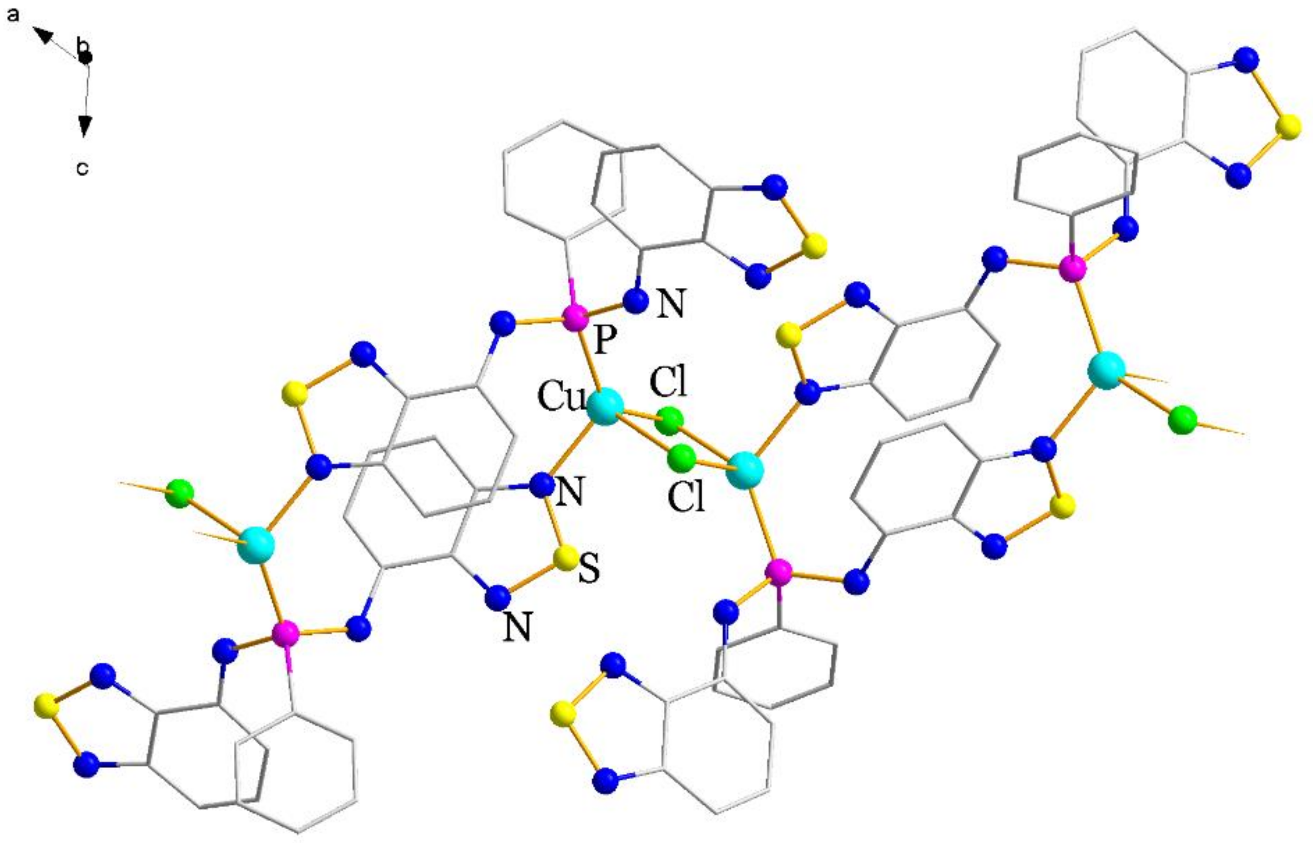
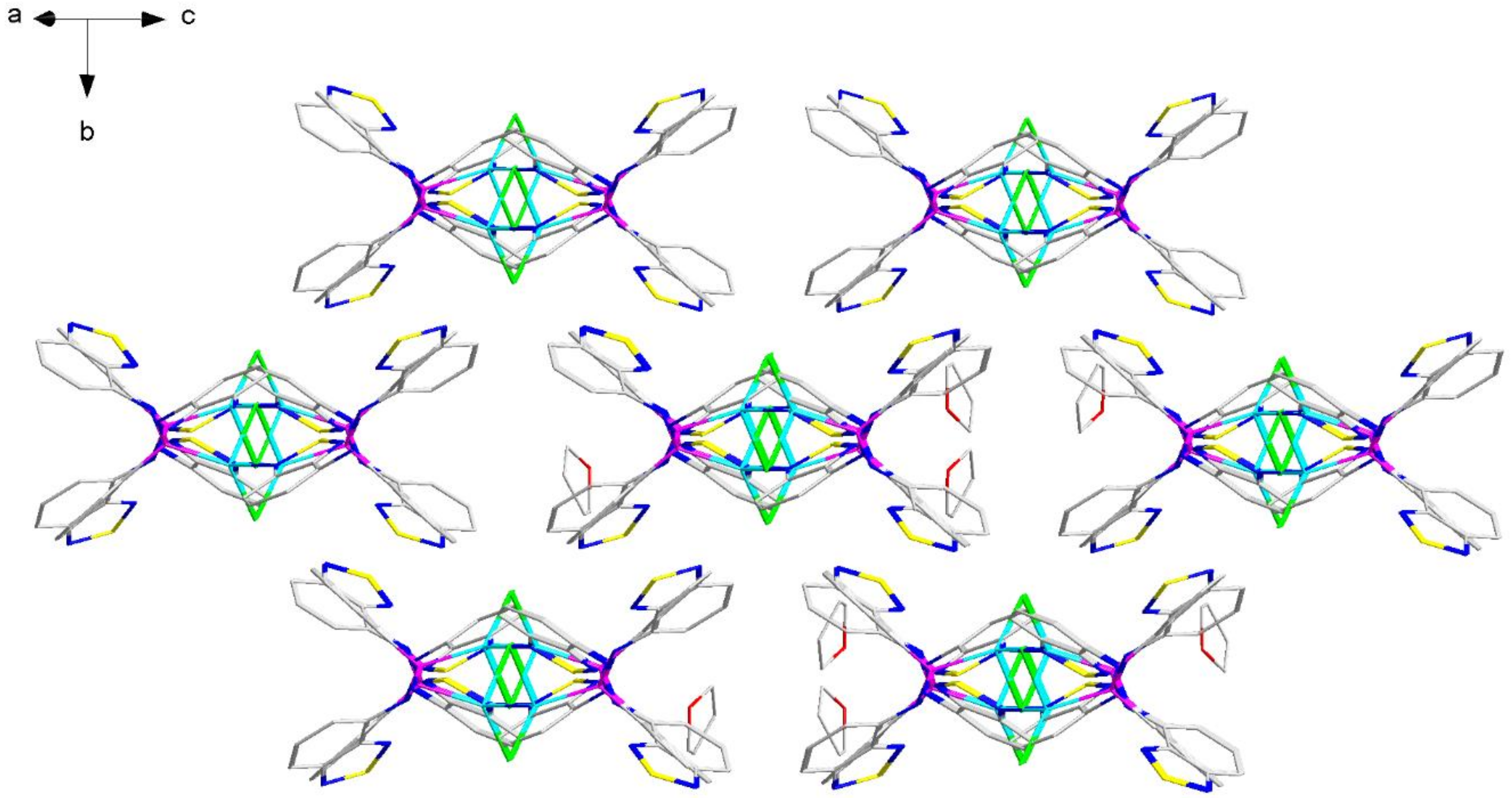
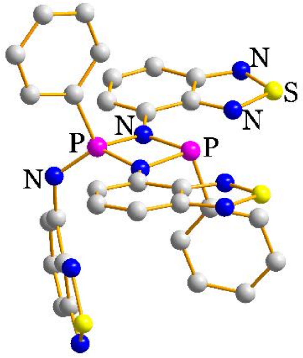
2.2. Structural Characterization

Figure 2 shows the crystal structure of H2L. Both amine nitrogen-centered fragments N4 and N4′ (for atom numbering, see Scheme 2) have a trigonal planar geometry assuming strong conjugation of the lone pairs of the N atoms with the aromatic system of the btd. The molecule H2L approximately belongs to Cs symmetry (Schoenflies notation) with the mirror plane going through P atom. Small differences in the arrangement of btd moieties relative to PPh violate this symmetry. For instance, the P–N–C valent angles equal to 127.3° and 125.7°, while those of P–N–H equal to 124° and 128°. The torsion angles C–P–N–C slightly differ from one another (Table 1, Figure S2). The DFT-optimized molecule has similar geometry with the symmetry closer to Cs (Table 1).
The crystal packing of H2L shows N···S intermolecular interactions between btd moieties (Figure 3). The intermolecular distances between adjacent N and S atoms lie in the range 3.04–3.51 Å. These distances are consistent with the literature data for secondary bonding interactions between btd moieties [24,26,27]. In H2L, two crystallographically independent types of N···S contacts are observed: the first type links the molecules into a chain via single interactions (Figure 3, on the right), while the second links via double ones (Figure 3, on the left).
XRD molecular structure of 1 is shown in Figure 4. The compound 1 is a binuclear complex in which zinc atoms are linked by two chelate-bridging L2– ligands. The coordination environment of Zn is tetrahedral. In accordance with the strong and weak Lewis acids and bases concept, the ligands coordinate the metals via N3 and N4 atoms, while P atoms remain free. The complex has C2 symmetry with two-fold axis going parallel to two btd moieties. N4 atoms of deprotonated amine groups have trigonal planar coordination environment of P, C and Zn atoms. One of C–P–N–C torsion angles significantly differ by circa 70° from that of free H2L; this implies that the HN-btd unit can rotate freely along the P–N bond adjusting to the metal environment. The N–P–N angle is smaller than in H2L, likely due to the attraction interaction of N4 with the neighboring Zn atom at a distance of 3.03 Å (Figure S3). Two out of four btd moieties enter a π-π stacking between two ligands in the complex molecule.
The isomeric complexes 2a·3C7H8 and 2b·2.5C7H8 differ from each other by the ligand arrangement (Figure 5). In 2a·2.5C7H8, the btd fragments oriented in different directions (head-to-tail manner), while in 2b·2.5C7H8, they oriented in one direction (head-to-head manner). As a result, the first complex molecule has C2h symmetry, while the second one has C2 symmetry. Zn atoms reveal similar tetrahedral coordination environment of two N and two Cl atoms. In contrast to L2– in 1·C7H8, H2L acts as a bridging ligand, coordinating only via N1 and N1′ atoms. Geometry of the ligand in the complexes resembles that in free H2L. The complex molecules in both structures have tetragonal prismatic cavities filled by toluene molecules (Figure 6, top). The phenyl planes of the toluene molecules are not arranged parallel to any btd aromatic system. The corresponding planes are perpendicular to P···P (in the case of 2a·3C7H8) or Zn···Zn (in the case of 2b·2.5C7H8) diagonals within the complex molecules. The particular arrangement of the toluene causes quantitative differences in the geometry of the cavities. The structure of 2b·2.5C7H8 reveals quite similar Zn···Zn (of 11.6 Å) and P···P (of 11.4 Å) intramolecular distances, while those in 2a·3C7H8 are different, being 12.9 and 10.4 Å respectively (Figure 6, top). In the structure of 2a·3C7H8, the solvent molecule is disordered over four positions due to proximity to the two-fold rotation axis and the mirror plane, while in 2b·2.5C7H8, the molecule has a single position.
Upon DFT-optimization in the absence of the toluene molecule, both molecules 2a and 2b exhibit distorted geometry with elongated Zn···Zn and shortened P···P distances (Figure 6, bottom). The main structural changes occur due to reducing N–Zn–N angles (Table 1). We conclude that the geometry of 2b changes more strongly with the removal of the toluene than that of 2a, while the cavities of free 2a and 2b become similar. This implies that the particular conformation of complex 2a with similar Zn···Zn and P···P diagonal distances is stabilized by the inclusion of the toluene molecule, but it is not related to the “head-to-tail” arrangement of the ligands. The total energy of 2a and 2b is similar (the difference of 4.10 kJ/mol), which suggests equally probable formation of the “head-to-head” or “head-to-tail” isomers from a solution. Analysis of Hirshfeld surfaces [28] of the crystal structures did not revealed the presence of specific interactions between the complexes and toluene (Figure S4). In principle, other molecules forming specific interactions with the complex could enter the cavities. This is a possible way of stabilizing one or another isomer, which allows one to isolate 2a or 2b as a single phase.
The difference in geometry of the molecules leads to different packing in the crystal. The complex molecules in 2a·3C7H8 are located one above another and form stacks along c direction (Figure 7). In contrast, in 2b molecules are staggered along c direction (Figure 8).
Unlike the abovementioned molecular complexes, complex 3·THF is a coordination polymer (Figure 9). Cu(I) as a weak Lewis acid has P atom in its environment; further coordination places are occupied by one N and two Cl atoms, forming a tetrahedral coordination polyhedron. Both H2L and Cl ligands act as bridging ones, thus forming a polymer chain. btd moieties not bonded directly to copper atoms enter interchain π-stacking (Figure 10). Notably, the conformation of H2L ligand in 3 significantly deviates from that of free H2L with reduced C–P–N–C and P–N–C–C(N) angles (by circa 150° and 35° respectively; Table 1). This conformation is obviously implemented to avoid unfavorable Cu···HC repulsive interactions (Figure S5).
The cyclodiphosphazane 4 has an unusual structure; it formally contains both P(V) and P(III) atoms in a four-membered P,N-cycle (Figure 11). In general, such cyclophosphazanes contain either trivalent [29] or pentavalent [30] phosphorus atoms. The P(V)–N bonds (of 1.69 Å) in the cycle are shorter by 0.08 Å compared to the P(III)–N ones. The P(V) atom attaches a pendent N-btd fragment; the corresponding P(V)–N bond is the shortest (of 1.54 Å), indicating the absence of hydrogen at the N atom. The corresponding bonds agree well with the literature data (see, for instance, those in t-butylamido-arylimino-derivatives [31]) and with the DFT-optimized molecule. Two btd fragments are located nearly in the same plane with the P,N-cycle. In the crystal packing, an intermolecular π- stacking is observed between these btd fragments.

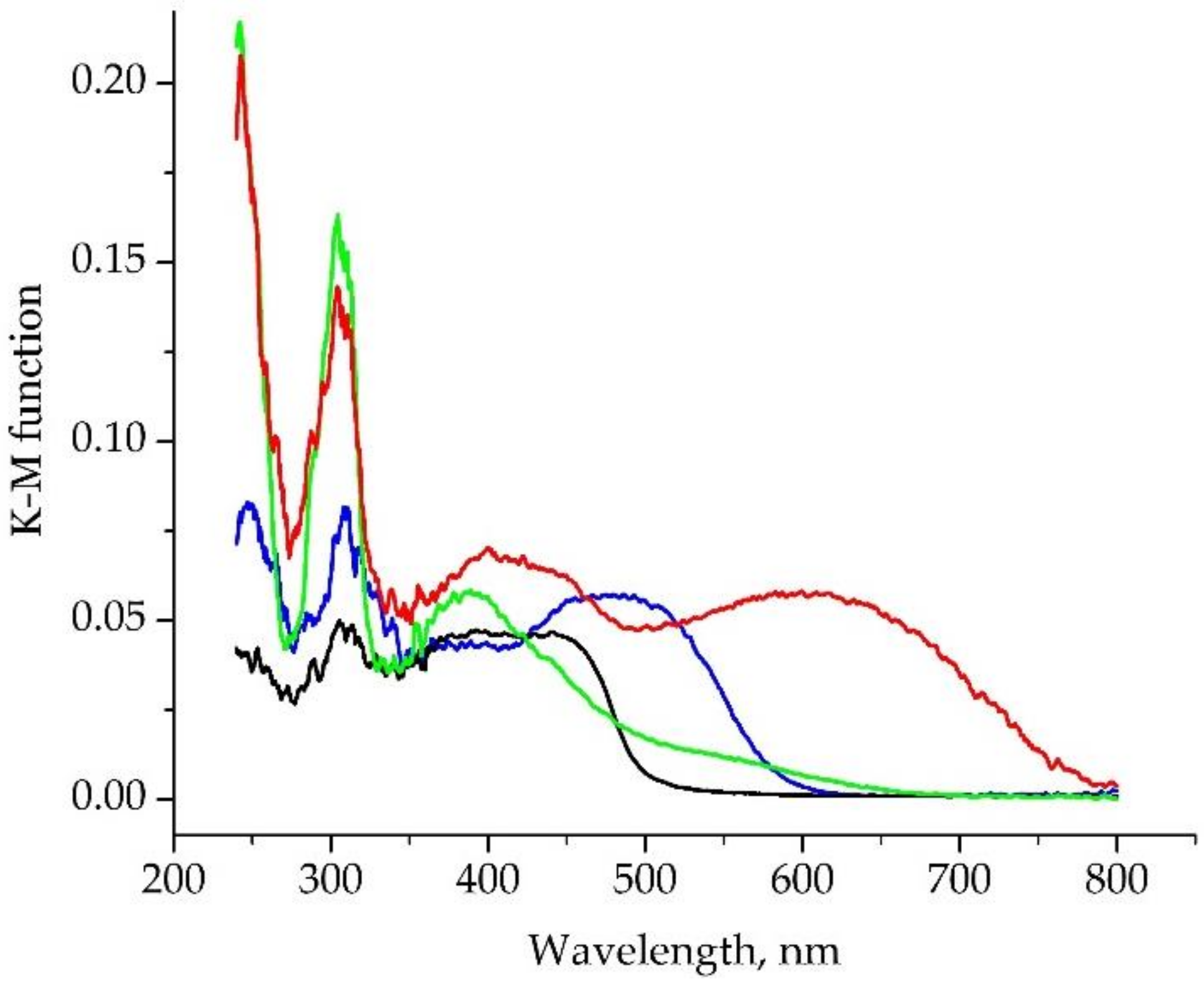
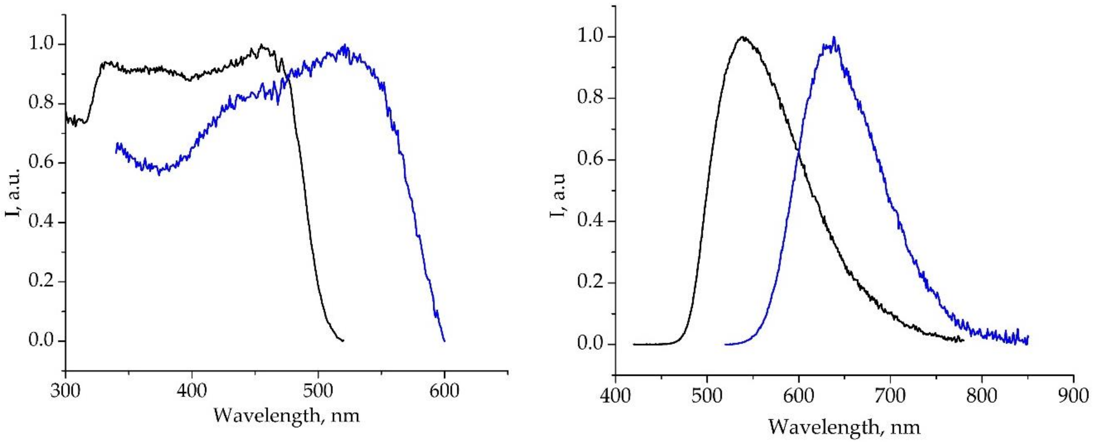
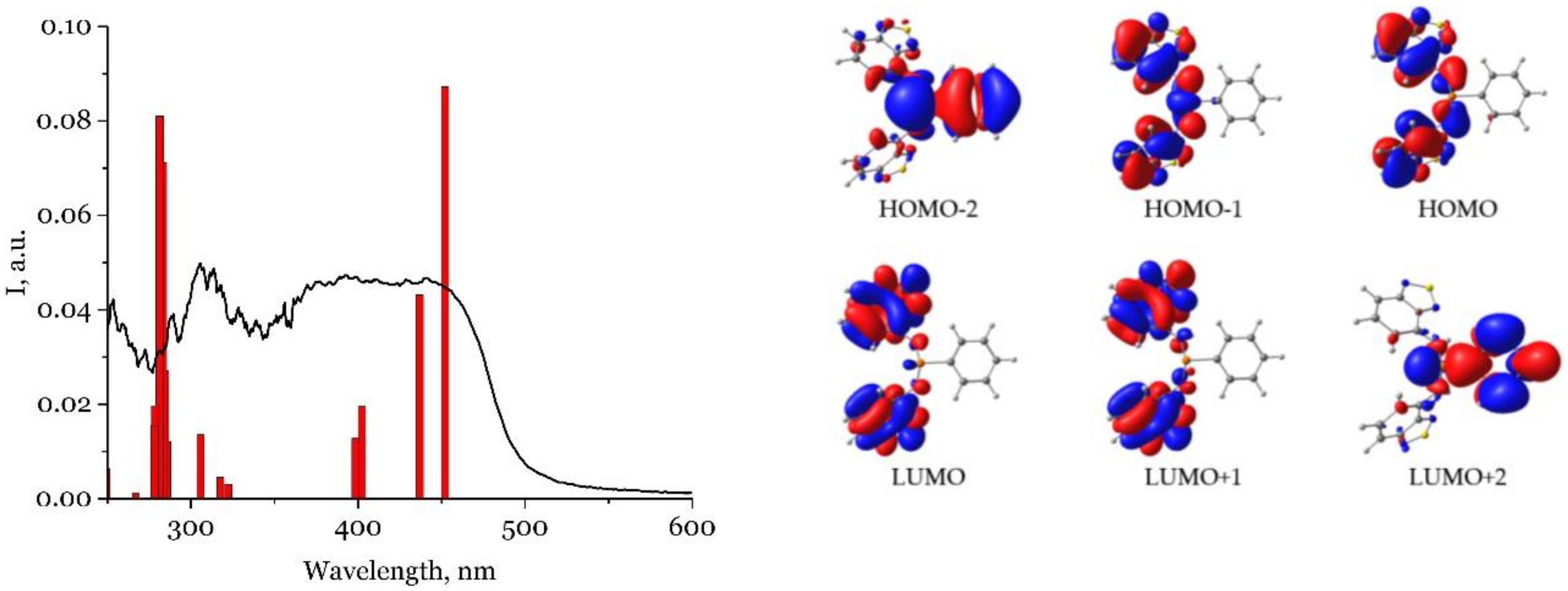
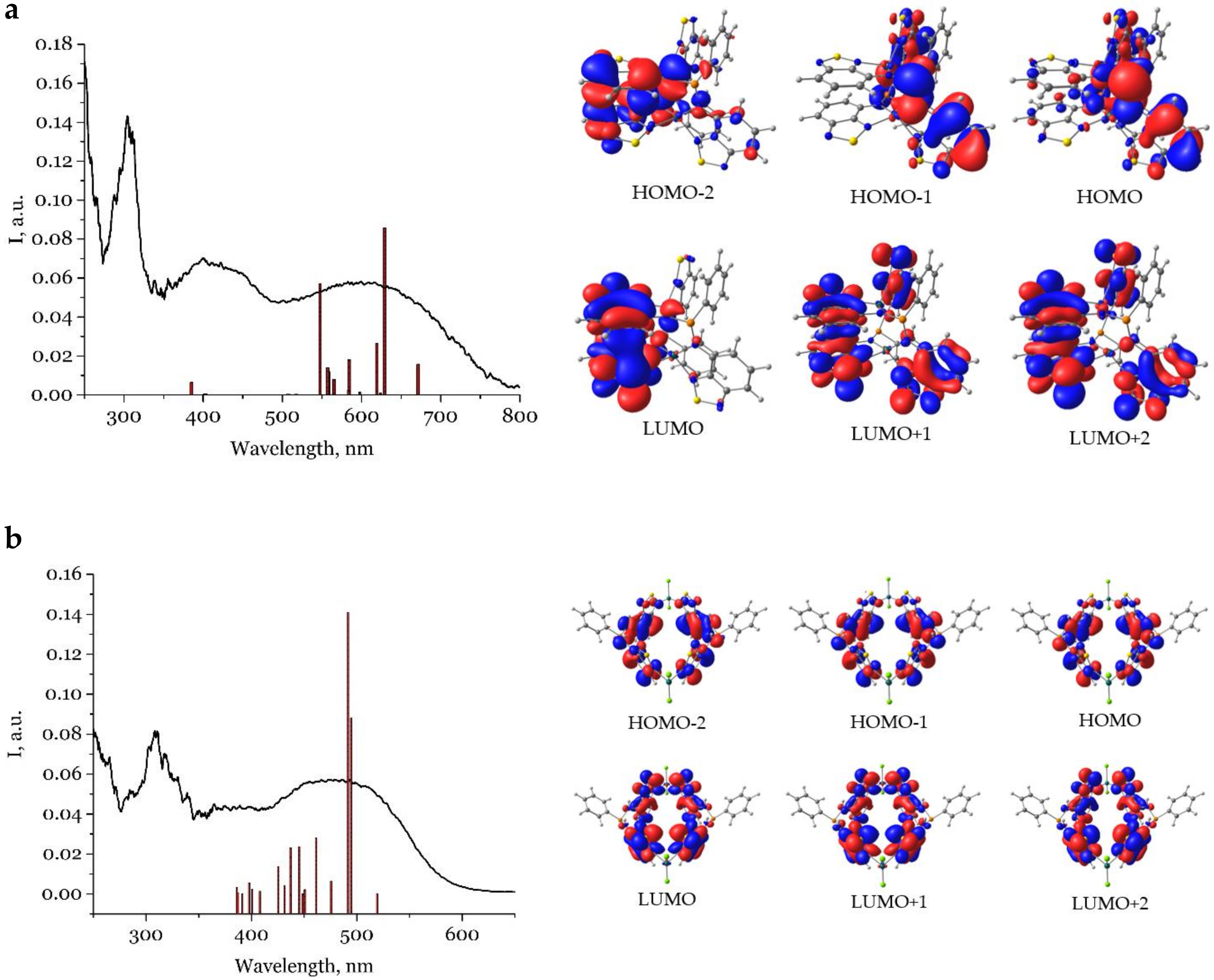
2.3. Photophysical Properties

For all compounds in the solid state (polycrystalline samples for luminescence studies and a mixture with BaSO4 for UV-vis), the absorption (Figure 12) and photoluminescence (Figure 13) spectra were measured. Only H2L and 2·nC7H8 show radiation in the visible range, while 1·nC7H8 and 3·THF do not emit in this range. For the luminescent compounds, lifetimes and quantum yields were also measured. All investigated spectroscopic and photophysical data are summarized in Table 2.
The UV-vis spectra of H2L, 1-3 in the solid state have similar peaks at around 305 nm. H2L, 2·nC7H8 and 3·THF have a long-wavelength absorption band lying in the range of 380–480 nm. Compared to H2L, this band is bathochromically shifted in the case of compounds 2·nC7H8, while it is hypsochromically shifted for compound 3·THF, although the latter features a broad low-intensity shoulder spanning the range of 500–600 nm. This behavior can be explained by different coordination types of H2L. Recently we have shown that for compounds with NH2-btd and (Ph2P)2N-btd ligands coordinated via N1 atom, the absorption and emission band maxima are bathochromically shifted compared to the corresponding free btds [11,24]. This tendency is observed for a number of Cu(I), Zn(II), Ag(I), Cd(II), Pd(II) and Pt(II) complexes studied; the nature of the metal and conformation of the ligand affects the position of the bands to a lesser extent. Compound 3·THF somewhat breaks the strong correlation between the coordination via N1 and the bathochromic shift (if the shoulder is not considering). Probably, this is due to cooperative effects of significantly distorted conformation of the ligand compared to free H2L and the presence of the uncoordinated btd unit, which manifests itself in a hypsochromic shift and/or a decrease in the intensity of the band.
The absorption spectrum of compound 1·nC7H8 reveals two absorption bands in the visible region. The band peaked at 600 nm is the most long-wavelength one among the compounds. Possible reasons of this behavior are as follows: (1) contrary to the others, the ligand in 1·nC7H8 is deprotonated and thus possess a different electronic structure; (2) the coordination of the ligand via N3 atom can result in a bathochromic shift of the band. To date, only two compounds with anionic ligand containing the btd-N unit and their photophysical studies have been reported, namely, Zn and Sm complexes with 4-(2,1,3-benzothiadiazol-4-ylamino)pent-3-en-2-onate [32]. They fit to the second proposed hypothesis: the Zn complex reveals coordination of the ligand only via O and N4 atoms and exhibits position of the absorption and emission bands similar to free neutral btd derivative. In the Sm complex, the ligand further coordinates via N3, while the emission band is bathochromically shifted, as in the case of 1·nC7H8. TD-DFT (vide infra) calculations reveal the frontier molecular orbitals in complex 1 significantly differ from the orbitals in the H2L and 2.
According the TD-DFT calculation on B3LYP theory level, the absorption band of H2L at 305 nm corresponds to a transition between the HOMO and LUMO + 2, which can be described as a charge transfer from btd to PPh fragments (Table S1). Long-wavelength bands mainly correspond to the HOMO → LUMO (452.1 nm), HOMO → LUMO + 1 (436.8 nm), HOMO – 1 → LUMO (402.3 nm) and HOMO – 1 → LUMO + 1 (398.5 nm) transitions. According to the depicted frontier molecular orbitals (Figure 14), the LUMO and LUMO + 1 orbitals are preferably located on the btd fragments, while the HOMO and HOMO – 1 orbitals have significant localization on the amino groups. Consequently, the low-energy transitions can be described as a charge transfer from the amino group to the btd fragments. The position of the long wavelength absorption and emission bands of H2L and (Ph2P)2N-btd is similar, although the latter is rather characterized by a charge transfer from PPh2 to btd [24].
According TD-DFT calculations, the low-energy bands of the coordination compounds arise from ligand-centered transitions. In 1, three long-wavelength transitions mainly correspond to the promotions HOMO → LUMO (671.4 nm), HOMO → LUMO + 1 (629.3 nm) and HOMO → LUMO + 2 (623.8 nm) (Table S2). Contrary to free H2L, different btd moieties of each of the two ligands contribute to the orbitals (Figure 15, Figure S7): LUMO orbitals are preferably localized at the stacked btd moieties and HOMO—at PPh and pendant btd moieties. This is manifested in a large charge transfer. In 2b, three first intense transitions correspond to the HOMO → LUMO (532.2 nm), HOMO → LUMO + 1 (506.8 nm) and HOMO – 1 → LUMO (503.7 nm) (Table S3). They are also characterized by charge transfer; however, the orbitals resemble that of free H2L: the LUMO orbitals are mainly localized on the thiadiazole moiety, while the HOMO orbitals—on the amino group and the benzene ring of btd. Thus, as expected, the bathochromic shift of the long-wavelength band in 1 and 2b occurs due to a decrease of the HOMO-LUMO gap from 3.28 eV for H2L to 2.35 eV for 1 and 2.85 eV for 2b. We assume that 2a shows similar to 2b electronic structure and thus shows similar photophysics; for this reason, TD-DFT calculations for 2a were not performed. The calculations for 3 were also not performed due to its complicated polymeric structure.
The emission band of the compounds 2·nC7H8 is significantly bathochromically shifted by 2770 cm−1 compared to H2L. The excitation spectra are slightly batchochromically shifted compared to the corresponding absorption spectra. The absolute quantum yield of emission (QY) upon transition from H2L to the complex decreases from 8% to 3% (Table 2). The photoluminescence lifetimes for H2L and 2·nC7H8 belong to the nanosecond time scales, indicating that emission occurs by fluorescence mechanism from the singlet excited state (Table 2). Note that the mixture of compounds 2·nC7H8 has both the single-peak band and the single-exponential decay kinetics. This means that either these isomers exhibit similar photophysical properties, or one of them has a much lower emission intensity compared to the other.

3. Materials and Methods

3.1. General Methods

Starting materials, except for solvents, were used as received from suppliers. 4-amino-2,1,3-benzothiadiazole (4-NH2-btd) [33] was synthesized as reported previously. The synthesis of novel compounds was carried out in evacuated vessels by using Schlenk techniques at room temperature unless otherwise specified. The solvents were distilled in inert atmosphere over common drying agents. Elemental analysis was performed with a Eurovector EuroEA3000 analyzer (Eurovector SPA, Redavalle, Italy). 1H NMR spectra (500.13 MHz) and 31P NMR spectra (202.45 MHz) were taken with a Bruker DRX-500 spectrometer (Bruker Corporation, Billerica, MA, USA) in C6D6 at room temperature; the solvent peak was used as internal reference. The IR spectra were recorded in KBr pellets at room temperature by means of a FT-801 Fourier spectrometer (Simex, Novosibirsk, Russia). The diffuse reflectance UV-vis spectra were obtained with a Shimadzu UV-3101 spectrophotometer (Shimadzu, Kioto, Japan) at room temperature. Samples for the diffuse reflectance measurements were prepared by a thorough grinding of a mixture of the compounds under study (about 0.005 mole fraction) with BaSO4, which was used also as a standard. Spectral dependences of the diffuse reflectance were converted into spectra of a Kubelka–Munk function [34]. Emission and excitation spectra were recorded on a Fluorolog 3 spectrometer (Horiba Jobin Yvon, Edison, NJ, USA) equipped with cooled PC177CE-010 photon detection module with a PMT R2658 photomultiplier. Excitation and emission spectra were corrected for source intensity (lamp and grating) and emission spectral response (detector and grating) by standard correction curves. For the measurements, powdered samples were placed between two nonfluorescent quartz plates. Absolute quantum yields were determined using Quanta-phi integrating sphere (Horiba Jobin Yvon, Edison, NJ, USA). Luminescence decay kinetics was recorded by a time-correlated single photon counting (TCSPC) technique using a NanoLED pulsed light source and a NanoLED-C2 controller on a Fluorolog 3 spectrometer (Horiba Jobin Yvon, Edison, NJ, USA).
Hirshfeld promolecular surface mapped over dnorm plots of the complexes were built using Crystal Explorer (version 17.5) program [28].

3.2. Quantum Chemical Calculations

Quantum chemical calculations were performed using the ORCA v. 4.2.1 computational package [35,36].
The full geometry optimization of H2L, 1, 2a, 2b and 4 molecules was carried in the gas phase without symmetry constraint at DFT level with B3LYP functional and Ahlrichs def2-SVP basis set. Atom-pairwise dispersion correction D3 with Becke–Johnson damping were used for geometry optimization [37]. Optimized geometries were submitted to numerical frequency analysis at the same level of theory to check whether the stationary point were “genuine” minima; no imaginary frequencies were found. The resolution of identity chain-of-spheres module, RIJCOSX was used to reduce the computational cost of the calculations [38].
We performed several TD-DFT H2L excitation energies calculations with various functionals in the gas phase and def2-TZVPP basis set. B3LYP, B97XD, BH&HLYP and CAM-B3LYP. B97XD, BH&HLYP and CAM-B3LYP gave poor results, underestimating the transitions energy (Figure S6). The B3LYP calculation results were closer to the experimental data. For 1 and 2b, TD-DFT calculations were performed with B3LYP functional and def2-TZVPP basis set in the gas phase.

3.3. X-ray Structure Determination

Single crystal XRD data for the compounds H2L, 13 were collected with a Bruker Apex DUO diffractometer (Bruker Corporation, Billerica, MA, USA) equipped with a 4K CCD area detector and a graphite-monochromated sealed tube (Mo Kα radiation, λ = 0.71073 Å). The data for 4 were collected with a Bruker D8 Venture diffractometer (Bruker Corporation, Billerica, MA, USA) with a CMOS PHOTON III detector and IµS 3.0 source (Mo Kα radiation) (Table S4). All measurements were conducted at 150 K, the φ- and ω-scan techniques were employed. Absorption corrections were applied with the use of the SADABS program [39]. The crystal structures were solved using the SHELXT [40] and were refined using SHELXL [41] programs with OLEX2 GUI [42]. Atomic displacement parameters for non-hydrogen atoms were refined anisotropically, with the exception of some disordered molecules. Hydrogen atoms were refined in riding model with the exception of those of the amino group, which were refined freely with the DFIX restraint on the corresponding N–H bonds. Phenyl groups in 2b·2C7H8 were disordered over two positions due to the proximity to the 2-fold rotation axis.
CCDC 1996456–1996460 and 1998636 contain the supplementary crystallographic data for this paper. These data can be obtained free of charge from the Cambridge Crystallographic Data Centre via www.ccdc.cam.ac.uk/data_request/cif [43].
The powder XRD analysis of the compounds was performed using a Shimadzu XRD-7000 diffractometer at room temperature (CuKα radiation, Ni filter) (Shimadzu, Kioto, Japan) Powder samples were slightly ground with heptane in an agate mortar and deposited on the polished side of a quartz-glass holder. Powder diffraction patterns were collected in 5–30° 2θ range.

3.4. Syntheses

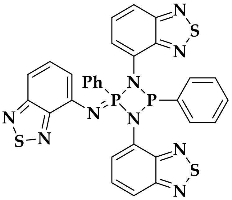
3.4.1. Synthesis of H2L

A solution of PhPCl2 (0.179 mL, 1.32 mmol) in 10 mL of toluene was slowly added to a cooled to 0 °C solution of 4-amino-2,1,3-benzothiadiazole (0.400 g, 2.65 mmol) and Et3N (0.6 mL, 4.3 mmol) in 10 mL of toluene. A precipitate formed gradually in the solution. The resulting suspension was stirred overnight, followed by filtration to remove triethylamine hydrochloride. Slow evaporation of the solvent gave yellow plate crystals. The crystalline product was washed with ether (3 × 2 mL) and dried under vacuum. Yield 0.420 g (78%). Calc. for C18H13N6PS2 (%): C 52.9; H 3.3; N 20,0; found: C 52.9; H 3.2; N 20.6. 1H NMR (C6D6, δ, ppm): 6.37 (s, 2H); 7.08 (m, 2H); 7,15–7,21 (m, 3H, Ph-H + solvent); 7,23 (s, 1H); 7,26 (dd, 2H); 7.38 (d, 2H); 7,66–7,69 (m, 2H). 31P{H} NMR (C6D6, δ, ppm): 43,5 (s). IR (cm−1): 3337 (m), 3049 (w), 2922 (w), 2851 (w), 1972 (w), 1911 (w), 1829 (w), 1718 (w), 1604 (m), 1547 (s), 1484 (s), 1437 (s), 1373 (s), 1274 (s), 1189 (w), 1161 (w), 1082 (s), 1038 (m), 999 (w), 905 (s), 849 (s), 808 (s), 740 (s), 703 (m), 653 (w), 543 (s), 508 (s).

3.4.2. [Zn2L2]·nC7H8 (1)

To a solid H2L (0.150 g, 0.367 mmol), anhydrous ZnCl2 (0.500 g, 0.367 mmol) and KHMDS (0.146 g, 0.734 mmol), THF (10 mL) was added. The mixture was stirred overnight under heating at 60 °C. The THF was removed by evaporation. The substance was dissolved in toluene and the KCl precipitate was removed by centrifugation. Evaporation of the solvent to a minimum volume and periodic heating and cooling of mixture led to the formation of suitable for XRD analysis dark red crystals. The solid was centrifuged and washed with toluene (3 × 2 mL) and dried under vacuum. Yield 0.0715 g (40%). Calc. for C36H22N12P2S4Zn2·0.1C7H8 (%): C 46.3; H 2.4; N 17.6; S 13.5; found: C 46.5; H 2.6; N 17.5; S 13.6. 1H NMR (C6D6, δ, ppm): 6.36 (d, 1H), 6.53 (t, 1H), 6.65 (d, 1H), 6.75 (t, 2H), 6.83 (dd, 1H), 6.95 (d, 1H), 7.03 (dd, 1H), 7.21 (qu, 1H), 7.76 (t, 2H). 31P{H} NMR (C6D6, δ, ppm): 74.1 (s). IR (cm−1): 3044 (m), 2850 (w), 1603 (w), 1568 (m), 1528 (s), 1473 (s), 1437 (w), 1374 (s), 1286 (s), 1170 (w), 1099 (s), 1041 (w), 905 (s), 885 (s), 851 (m), 806 (m), 739 (s), 699 (w), 671 (w), 543 (w), 508 (m).

3.4.3. [Zn2(H2L)2Cl4]·nC7H8 (2a·nC7H8 and 2b·nC7H8)

To a solid H2L (0.0749 g, 0.183 mmol) and anhydrous ZnCl2 (0.0250 g, 0.183 mmol), THF (10 mL) was added. The red solution was stirred overnight and the solvent was removed off by evaporation. The red fine-crystalline precipitate was obtained by slow extraction with toluene (5 mL) in a two-sector ampoule. The obtained crystals were suitable for XRD analysis. The solution was decanted, the crystals were washed with toluene (3 × 2 mL) and dried under vacuum. Yield 0.1067 (95 %). Calc. for C36H26Cl4N12P2S4Zn2·1.1C7H8 (%): C 44.1; H 2.9; N 14.1; found: C 43.7; H 3.1; N 13.5. IR (cm−1): 3353 (m), 3290 (m), 3055(w), 2918 (m), 2849 (m), 1602 (w), 1549 (s), 1487 (s), 1364 (m), 1289 (m), 1086 (s), 1077 (w), 1047 (w), 916 (w), 871 (w), 825 (w), 744 (m), 702 (w).

3.4.4. [Cu(H2L)Cl]n nTHF (3)

A solution of H2L (0.0202 g, 0.0495 mmol) in THF (5 mL) was added to a vial with powder of CuCl (0.0049 g, 0.049 mmol). The mixture was left without stirring in the closed vial. A week later, yellow plate crystals formed. The obtained crystals were suitable for XRD analysis. The solution was decanted, the crystals were washed with THF (3 × 2 mL) and dried under vacuum. Yield 0.0176 g (60%). Calc. for C18H13ClCuN6PS2·C4H8O (%): C 45.6; H 3.6, N 14.5; found: C 45.4; H 3.7; N 14.4. IR (cm−1): 3309 (w), 3071 (w), 2973 (w), 2856 (w), 1609 (m), 1539 (s), 1490 (s), 1435 (m), 1379 (s), 1299 (m), 1161 (w), 1082 (m), 1049 (w), 972 (w), 907 (m), 857 (m), 828 (w), 804 (w), 747 (s), 694 (w), 667 (w), 545 (w), 520 (w).

4. Conclusions

To conclude, we present the synthesis of the novel phosph(III)azane (H2L) based on 2,1,3-benzothiadiazole. The presence of accessible lone pairs on both P and N atoms makes this phosph(III)azane a promising multidentate ligand for transition metal complexes. The coordination ability of this derivative and its deprotonated species (L2–) is demonstrated on the example of Zn and Cu(I) complexes 13 that were isolated as the solvates with toluene or THF. According to single crystal XRD analysis, the complexes of the formula [Zn2(H2L)2Cl2] can exist in two isomeric species, viz. 2a and 2b, that differ by the arrangement of one of H2L ligand. The quantum chemical calculations reveal these complexes are thermodynamically equally favorable. Inside the molecules 2a and 2b, tetragonal prismatic cavities are observed that large enough for the inclusion of solvate toluene molecule. The toluene arranged differently inside the complex molecules defining their geometry. With the removal of toluene, the geometry of 2b changes substantially, while that of 2a changes slightly. The side product of the synthesis of H2L, viz. four-membered cyclophosphazane 4, has been characterized by single crystal XRD analysis. The photophysical properties of H2L 13 were studied. H2L and the mixture of the phases 2·nC7H8 reveal fluorescence in the visible range; the single-peak band and the single-exponential decay kinetics for 2·nC7H8 indicates the species 2a and 2b possess similar photophysical properties. The difference in the absorption spectra of H2L, 1 and 2b is discussed using TD-DFT calculations. These compounds reveal ligand-centered low-energy transitions; their charge transfer nature in H2L and 2b is similar, while that in 1 differs significantly from them due to anionic nature of L2– and different coordination type of the ligand.

Supplementary Materials

The following are available online: Figure S1: Experimental and simulated powder patterns of the compounds 2a·3C7H8 and 2b·2.5C7H8; Figure S2: Representation of selected torsion angles in H2L or L2– fragments; Figure S3: Molecular structure of 1 showing attraction interaction Zn–N4; Figure S4: The dnorm Hirshfeld surface of the complexes in crystal structures 2a·3C7H8 and 2b·2.5C7H8; Figure S5: Representation of a Cu–H2L model with the geometry derived from XRD data for free H2L and the corresponding fragment from XRD data for 3; Figure S6: Overlaid TD-DFT calculations results and experimental UV-vis spectrum of H2L; Table S1: Calculated properties of the first singlet excited states S0 → Sn of H2L; Table S2: Calculated properties of the first singlet excited states S0 → Sn of 1; Table S3: Calculated properties of the first singlet excited states S0 → Sn of 2b; Table S4: Crystal data and structure refinement for H2L 14; Figure S7: Frontier molecular orbitals of 1.

Author Contributions

Conceptualization, S.K.; funding acquisition, T.S.; investigation, R.K., A.R. and T.S.; methodology, D.B. and T.S.; supervision, S.K.; visualization; R.K. and T.S.; writing—original draft, R.K.; writing—review and editing: D.B., S.K. and T.S. All authors have read and agree to the published version of the manuscript.

Funding

This research was funded by the Russian Science Foundation (project no. 19-73-00030).

Acknowledgments

We are grateful to Siberian Supercomputer Center of Institute of Computational Mathematics and Mathematical Geophysics for computational capability and thank the technical staff of the Institute for the assistance.

Conflicts of Interest

The authors declare no conflict of interest.

References

  1. Gutierrez, G.D.; Sazama, G.T.; Wu, T.; Baldo, M.A.; Swager, T.M. Red Phosphorescence from Benzo[2,1,3]thiadiazoles at Room Temperature. J. Org. Chem. 2016, 81, 4789–4796. [Google Scholar] [CrossRef] [Green Version]
  2. Paisley, N.R.; Tonge, C.M.; Mayder, D.M.; Thompson, K.A.; Hudson, Z.M. Tunable benzothiadiazole-based donor–acceptor materials for two-photon excited fluorescence. Mater. Chem. Front. 2020, 4, 555–566. [Google Scholar] [CrossRef]
  3. Neto, B.A.D.; Carvalho, P.H.P.R.; Correa, J.R. Benzothiadiazole Derivatives as Fluorescence Imaging Probes: Beyond Classical Scaffolds. Acc. Chem. Res. 2015, 48, 1560–1569. [Google Scholar] [CrossRef] [PubMed]
  4. Schill, J.; Ferrazzano, L.; Tolomelli, A.; Schenning, A.P.H.J.; Brunsveld, L. Fluorene benzothiadiazole co-oligomer based aqueous self-assembled nanoparticles. RSC Adv. 2020, 10, 444–450. [Google Scholar] [CrossRef] [Green Version]
  5. Kini, G.P.; Choi, J.Y.; Jeon, S.J.; Suh, I.S.; Moon, D.K. Effect of mono alkoxy-carboxylate-functionalized benzothiadiazole-based donor polymers for non-fullerene solar cells. Dye. Pigment. 2019, 164, 62–71. [Google Scholar] [CrossRef]
  6. Prima, D.O.; Makarov, A.G.; Bagryanskaya, I.Y.; Kolesnikov, A.E.; Zargarova, L.V.; Baev, D.; Eliseeva, T.F.; Politanskaya, L.V.; Makarov, A.Y.; Slizhov, Y.G.; et al. Fluorine-Containing n-6 and Angular and Linear n-6-n′ (n, n′ = 5, 6, 7) Diaza-Heterocyclic Scaffolds Assembled on Benzene Core in Unified Way. Chemistry 2019, 4, 2383–2386. [Google Scholar] [CrossRef]
  7. Qian, G.; Wang, X.; Wang, S.; Zheng, Y.; Wang, S.; Zhu, W.; Wang, Y. Polymorphous Luminescent Materials Based on ’T’-Shaped Molecules Bearing 4,7-Diphenylbenzo[c][1,2,5]thiadiazole Skeletons: Effect of Substituents on the Photophysical Properties. Chem. A Eur. J. 2019, 25, 15401–15410. [Google Scholar] [CrossRef]
  8. Mikhailov, M.S.; Gudim, N.S.; Knyazeva, E.A.; Tanaka, E.; Zhang, L.; Mikhalchenko, L.V.; Robertson, N.; Rakitin, O.A. 9-(p-Tolyl)-2,3,4,4a,9,9a-hexahydro-1H-carbazole—A new donor building-block in the design of sensitizers for dye-sensitized solar cells. J. Photochem. Photobiol. A Chem. 2020, 391, 112333. [Google Scholar] [CrossRef]
  9. Ansell, J.; Wills, M. Enantioselective catalysis using phosphorus-donor ligands containing two or three P-N or P-O bonds. Chem. Soc. Rev. 2002, 31, 259–268. [Google Scholar] [CrossRef]
  10. Martín, R.; Prieto, P.; Carrillo, J.R.; Rodríguez, A.M.; De Cozar, A.; Boj, P.G.; Díaz-García, M.A.; Ramírez, M.G. Design, synthesis and amplified spontaneous emission of 1,2,5-benzothiadiazole derivatives. J. Mater. Chem. C 2019, 7, 9996–10007. [Google Scholar] [CrossRef]
  11. Sukhikh, T.S.; Ogienko, D.S.; Bashirov, D.A.; Konchenkoa, S.N. Luminescent complexes of 2,1,3-benzothiadiazole derivatives. Russ. Chem. Bull. 2019, 68, 651–661. [Google Scholar] [CrossRef]
  12. Ishi-I, T.; Tanaka, H.; Youfu, R.; Aizawa, N.; Yasuda, T.; Kato, S.-I.; Matsumoto, T. Mechanochromic fluorescence based on a combination of acceptor and bulky donor moieties: Tuning emission color and regulating emission change direction. N. J. Chem. 2019, 43, 4998–5010. [Google Scholar] [CrossRef]
  13. Rakitin, O.A.; Zibarev, A.V. Synthesis and Applications of 5-Membered Chalcogen-Nitrogen π-Heterocycles with Three Heteroatoms. Asian J. Org. Chem. 2018, 7, 2397–2416. [Google Scholar] [CrossRef]
  14. Islam, A.; Akhtaruzzaman, M.; Chowdhury, T.H.; Qin, C.; Han, L.; Bedja, I.M.; Stalder, R.; Schanze, K.; Reynolds, J.R. Enhanced Photovoltaic Performances of Dye-Sensitized Solar Cells by Co-Sensitization of Benzothiadiazole and Squaraine-Based Dyes. ACS Appl. Mater. Interfaces 2016, 8, 4616–4623. [Google Scholar] [CrossRef] [PubMed]
  15. Page, Z.A.; Liu, Y.; Puodziukynaite, E.; Russell, T.P.; Emrick, T. Hydrophilic Conjugated Polymers Prepared by Aqueous Horner–Wadsworth–Emmons Coupling. Macromolecules 2016, 49, 2526–2532. [Google Scholar] [CrossRef]
  16. Boucard, J.; Boudjemaa, R.; Steenkeste, K.; Jacqueline, C.; Stephant, N.; Lefèvre, F.-X.; Laurent, A.D.; Lartigue, L.; Hulin, P.; Nedellec, S.; et al. Phosphonic Acid Fluorescent Organic Nanoparticles for High-Contrast and Selective Staining of Gram-Positive Bacteria. ACS Omega 2018, 3, 17392–17402. [Google Scholar] [CrossRef]
  17. Ren, Y.; Sezen, M.; Guo, F.; Jäkle, F.; Loo, Y.-L. [d]-Carbon–carbon double bond engineering in diazaphosphepines: A pathway to modulate the chemical and electronic structures of heteropines. Chem. Sci. 2016, 7, 4211–4219. [Google Scholar] [CrossRef] [Green Version]
  18. Zhang, Z.; Jin, Z.; Fu, W.; Shi, M.; Chen, H. Phosphate ester side-chain-modified conjugated polymer for hybrid solar cells. J. Appl. Polym. Sci. 2017, 134, 134. [Google Scholar] [CrossRef]
  19. Li, Y.; Liu, J.-Y.; Zhao, Y.-D.; Cao, Y.-C. Recent advancements of high efficient donor–acceptor type blue small molecule applied for OLEDs. Mater. Today 2017, 20, 258–266. [Google Scholar] [CrossRef]
  20. Duan, C.; Li, J.; Han, C.; Ding, D.; Yang, H.; Wei, Y.; Xu, H. Multi-dipolar Chromophores Featuring Phosphine Oxide as Joint Acceptor: A New Strategy toward High-Efficiency Blue Thermally Activated Delayed Fluorescence Dyes. Chem. Mater. 2016, 28, 5667–5679. [Google Scholar] [CrossRef]
  21. Li, H.; Hong, M.; Scarpaci, A.; He, X.; Risko, C.; Sears, J.S.; Barlow, S.; Winget, P.; Marder, S.R.; Kim, D.; et al. Chemical Stabilities of the Lowest Triplet State in Aryl Sulfones and Aryl Phosphine Oxides Relevant to OLED Applications. Chem. Mater. 2019, 31, 1507–1519. [Google Scholar] [CrossRef] [Green Version]
  22. Jia, W.; Wang, Q.; Shi, H.; An, Z.; Huang, W. Manipulating the Ultralong Organic Phosphorescence of Small Molecular Crystals. Chem. A Eur. J. 2020, 26, 4437–4448. [Google Scholar] [CrossRef] [PubMed]
  23. Joly, D.; Bouit, P.-A.; Hissler, M. Organophosphorus derivatives for electronic devices. J. Mater. Chem. C 2016, 4, 3686–3698. [Google Scholar] [CrossRef] [Green Version]
  24. Sukhikh, T.S.; Khisamov, R.M.; Bashirov, D.A.; Komarov, V.Y.; Molokeev, M.S.; Ryadun, A.A.; Benassi, E.; Konchenko, S.N. Tuning of the Coordination and Emission Properties of 4-Amino-2,1,3-Benzothiadiazole by Introduction of Diphenylphosphine Group. Cryst. Growth Des. 2020. [Google Scholar] [CrossRef]
  25. Burford, N.; Cameron, T.S.; Conroy, K.D.; Ellis, B.; Lumsden, M.; Macdonald, C.L.B.; McDonald, R.; Phillips, A.D.; Ragogna, P.J.; Schurko, R.W.; et al. Transformations between Monomeric, Dimeric, and Trimeric Phosphazanes: Oligomerizing NP Analogues of Olefins. J. Am. Chem. Soc. 2002, 124, 14012–14013. [Google Scholar] [CrossRef]
  26. Ams, M.; Trapp, N.; Schwab, A.; Milić, J.V.; Diederich, F. Chalcogen Bonding “2S–2N Squares” versus Competing Interactions: Exploring the Recognition Properties of Sulfur. Chem. A Eur. J. 2018, 25, 323–333. [Google Scholar] [CrossRef]
  27. Sukhikh, T.S.; Bashirov, D.; Shuvaev, S.; Komarov, V.; Kuratieva, N.; Konchenko, S.; Benassi, E. Noncovalent interactions and photophysical properties of new Ag(I) complexes with 4-amino-2,1,3-benzothiadiazole. Polyhedron 2018, 141, 77–86. [Google Scholar] [CrossRef]
  28. CrystalExplorer, version 17; University of Western: Perth, Australia, 2017.
  29. Bond, A.D.; Doyle, E.L.; García, F.; Kowenicki, R.A.; Moncrieff, D.; McPartlin, M.; Riera, L.; Woods, A.D.; Wright, D.S. Thermodynamic/Kinetic Control in the Isomerization of the[{tBuNP(μ-NtBu)}2]2− Ion. Chem. A Eur. J. 2004, 10, 2271–2276. [Google Scholar] [CrossRef]
  30. Tirreé, J.; Gudat, D.; Nieger, M.; Niecke, E. Reversible Tautomeric Transformation between a Bis(amino)cyclodiphosph(V)azene and a Bis(imino)cyclodiphosph(V)azane. Angew. Chem. Int. Ed. 2001, 40, 3025–3028. [Google Scholar] [CrossRef]
  31. Moser, D.F.; Carrow, C.J.; Stahl, L.; Staples, R.J. Titanium complexes of bis(1°-amido)cyclodiphosph(III)azanes and bis(1°-amido)cyclodiphosph(V)azanes: Facial versus lateral coordination. J. Chem. Soc. Dalton Trans. 2001, 1246–1252. [Google Scholar] [CrossRef]
  32. Sukhikh, T.S.; Ogienko, D.S.; Bashirov, D.A.; Kurat’Eva, N.V.; Smolentsev, A.I.; Konchenko, S.N. Samarium, Europium, and Gadolinium Complexes with 4-(2,1,3-Benzothiadiazol-4-ylamino)pent-3-en-2-onate. Russ. J. Coord. Chem. 2019, 45, 30–35. [Google Scholar] [CrossRef]
  33. Liu, Y.; Lu, Y.; Prashad, M.; Repič, O.; Blacklock, T.J. A Practical and Chemoselective Reduction of Nitroarenes to Anilines Using Activated Iron. Adv. Synth. Catal. 2005, 347, 217–219. [Google Scholar] [CrossRef]
  34. Kubelka, P. New contributions to the optics intensely light scattering materials. J. Opt. Soc. Am. 1948, 38, 448–457. [Google Scholar] [CrossRef] [PubMed]
  35. Neese, F. Software update: The ORCA program system, version 4.0. Wiley Interdiscip. Rev. Comput. Mol. Sci. 2017, 8, e1327. [Google Scholar] [CrossRef]
  36. Neese, F. The ORCA program system. Wiley Interdiscip. Rev. Comput. Mol. Sci. 2011, 2, 73–78. [Google Scholar] [CrossRef]
  37. Grimme, S.; Ehrlich, S.; Goerigk, L. Effect of the damping function in dispersion corrected density functional theory. J. Comput. Chem. 2011, 32, 1456–1465. [Google Scholar] [CrossRef]
  38. Neese, F.; Wennmohs, F.; Hansen, A.; Becker, U. Efficient, approximate and parallel Hartree–Fock and hybrid DFT calculations. A ‘chain-of-spheres’ algorithm for the Hartree–Fock exchange. Chem. Phys. 2009, 356, 98–109. [Google Scholar] [CrossRef]
  39. Bruker Apex3 Software Suite: Apex3, SADABS-2017/2 and SAINT, version 2018.7-2; Bruker AXS Inc.: Madison, WI, USA, 2017.
  40. Sheldrick, G.M. SHELXT—Integrated space-group and crystal-structure determination. Acta Crystallogr. Sect. A Found. Adv. 2015, 71, 3–8. [Google Scholar] [CrossRef] [Green Version]
  41. Sheldrick, G.M. Crystal structure refinement with SHELXL. Acta Crystallogr. Sect. C Struct. Chem. 2015, 71, 3–8. [Google Scholar] [CrossRef]
  42. Dolomanov, O.; Bourhis, L.J.; Gildea, R.; Howard, J.A.; Puschmann, H. OLEX2: A complete structure solution, refinement and analysis program. J. Appl. Crystallogr. 2009, 42, 339–341. [Google Scholar] [CrossRef]
  43. Groom, C.R.; Bruno, I.J.; Lightfoot, M.P.; Ward, S.C. The Cambridge Structural Database. Acta Crystallogr. Sect. Struct. Science 2016, B72, 171–179. [Google Scholar] [CrossRef] [PubMed]
Sample Availability: Samples used are available from the authors.
Scheme 1. Synthesis of (Ph2P)2N-btd [24] and H2L.
Molecules 25 02428 sch001
Figure 1. Skeletal formula of the by-product (4).
Molecules 25 02428 g001
Scheme 2. Synthesis of zinc and copper(I) complexes.
Molecules 25 02428 sch002
Figure 2. Molecular structure of H2L from XRD analysis.
Molecules 25 02428 g002
Figure 3. N···S intermolecular contacts in H2L (“ball and stick” model for atoms participating in the contacts and “wires and sticks” one for others) marked dashed green. Hydrogens are omitted.
Molecules 25 02428 g003
Figure 4. Molecular structure of 1. Solvate molecules and hydrogen atoms are not shown. Crystallographically independent fragment is shown in “ball and stick” model and a symmetry related fragment is shown in “wires and sticks” model.
Molecules 25 02428 g004
Figure 5. Molecular structures of 2a (left) and 2b (right). Hydrogen atoms are not shown.
Molecules 25 02428 g005
Figure 6. Arrangement of toluene molecules inside the cavity of 2a·3C7H8 and 2b·2.5C7H8 (top). DFT-optimized molecules of 2a and 2b (bottom). Hydrogen atoms are omitted.
Molecules 25 02428 g006aMolecules 25 02428 g006b
Figure 7. Crystal packing for 2a·3C7H8. Hydrogen atoms and solvate molecules are not shown.
Molecules 25 02428 g007
Figure 8. Crystal packing for 2b·2.5C7H8. Hydrogen atoms and solvate molecules are not shown.
Molecules 25 02428 g008
Figure 9. Molecular structure of fragment of 3. Solvate molecules and hydrogen atoms are not shown. Carbon atoms are shown in “wires and sticks’ mode.
Molecules 25 02428 g009
Figure 10. Crystal packing for 3·THF. Hydrogen atoms are not shown.
Molecules 25 02428 g010
Figure 11. Molecular structure of 4. Hydrogen atoms are not shown.
Molecules 25 02428 g011
Figure 12. Absorption spectra of the solid samples of H2L (black), 1·nC7H8 (red), 2·nC7H8 (blue) and 3 nTHF (green) mixed with BaSO4, presented in the form of Kubelka-Munk (K-M) functions.
Molecules 25 02428 g012
Figure 13. Normalized excitation (left) and emission (right) spectra of H2L (black) and 2·nC7H8 (blue).
Molecules 25 02428 g013
Figure 14. UV-vis spectrum and calculated transitions (left) and frontier molecular orbitals (right) of H2L based on TD-DFT at B3LYP/def2TZVPP level (isovalue = 0.02 a.u.).
Molecules 25 02428 g014
Figure 15. UV-vis spectrum and calculated transitions (left) and frontier molecular orbitals (right) of 1 (a) and 2b (b) based on TD-DFT at B3LYP/def2TZVPP level (isovalue = 0.02 a.u.).
Molecules 25 02428 g015
Table 1. Selected angles in single crystal XRD and DFT-optimized structures.
P–N–CN–P–NC–P–N–CP–N–C–C(N)N–Zn–N
H2L125.7, 127.3,107.4151.4, 145.7176.0, 171.8
H2L (optimized)125.6, 126.0105.5154.9, 156.4170.2, 164.9
1·C7H8122.0, 122.197.980.0, 145.9166.7, 177.884.3, 84.7
2a·3C7H8124.1106.2150.0177.496.2
2a (optimized)126.4, 126.4104.8152.0, 152.0176.592.6
2b·2.5C7H8122.2, 124.5105.2156.3, 154.9160.0, 165.9101.5
2b (optimized)125.4, 126.2104.5156.6, 151.2171.9, 178.594.6
3·THF120.9, 129.9109.1156.9, 49.2137.9, 169.2
Table 2. Wavelengths of the absorption (λabs) and emission (λEm) band maxima, excited states lifetimes and absolute quantum yields for H2L, 13 in the solid state at room temperature.
Compoundλabs, nmλEm, nmτQY, %
H2L305, 390-4405404.3 ns8
1·nC7H8304, 400, 600
2·nC7H8309, 4806353.1 ns3
3·THF304, 385, 550(sh)

