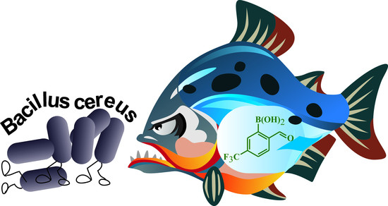
Synthesis, Properties and Antimicrobial Activity of 5-Trifluoromethyl-2-formylphenylboronic Acid

 , , and
Abstract
:
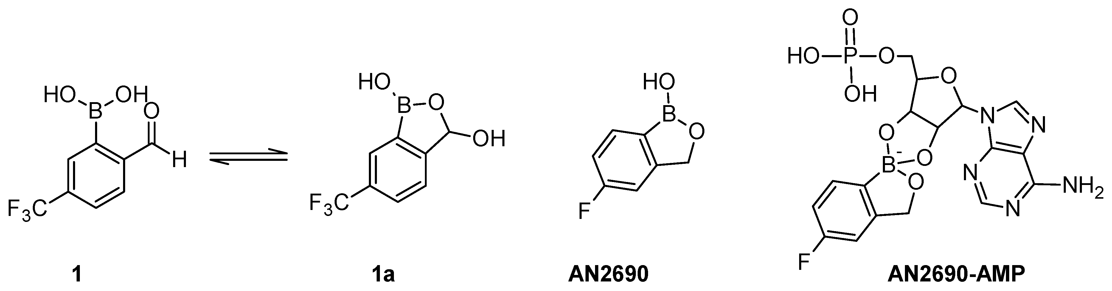
1. Introduction
2. Results and Discussion
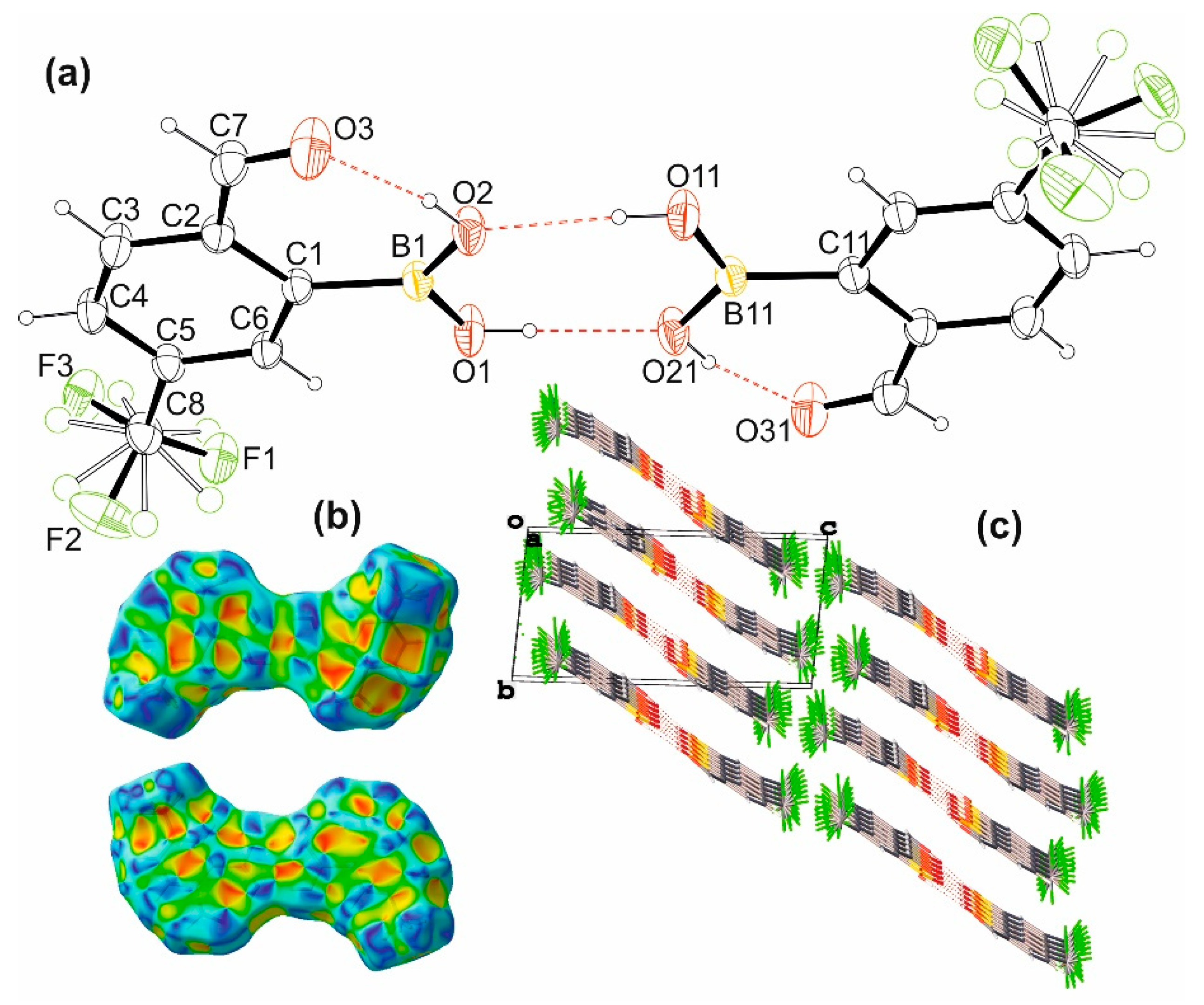
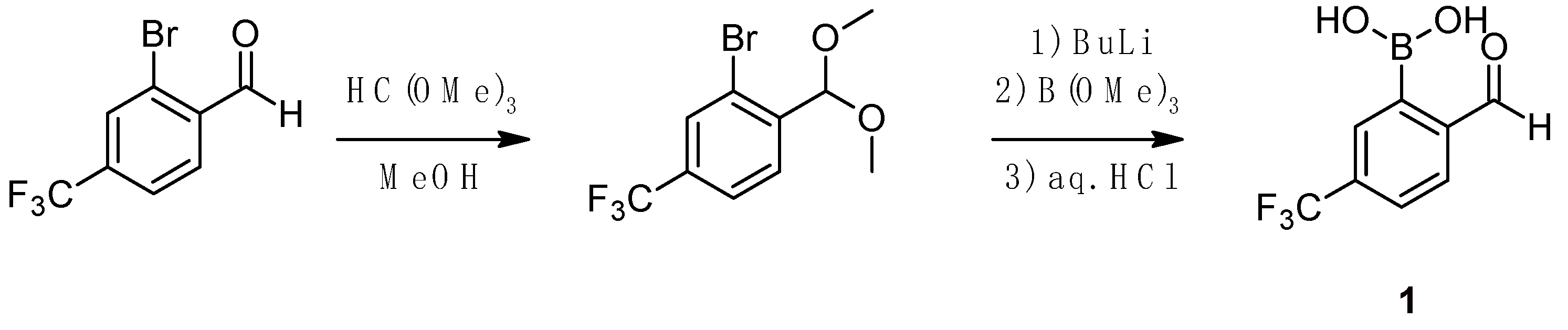
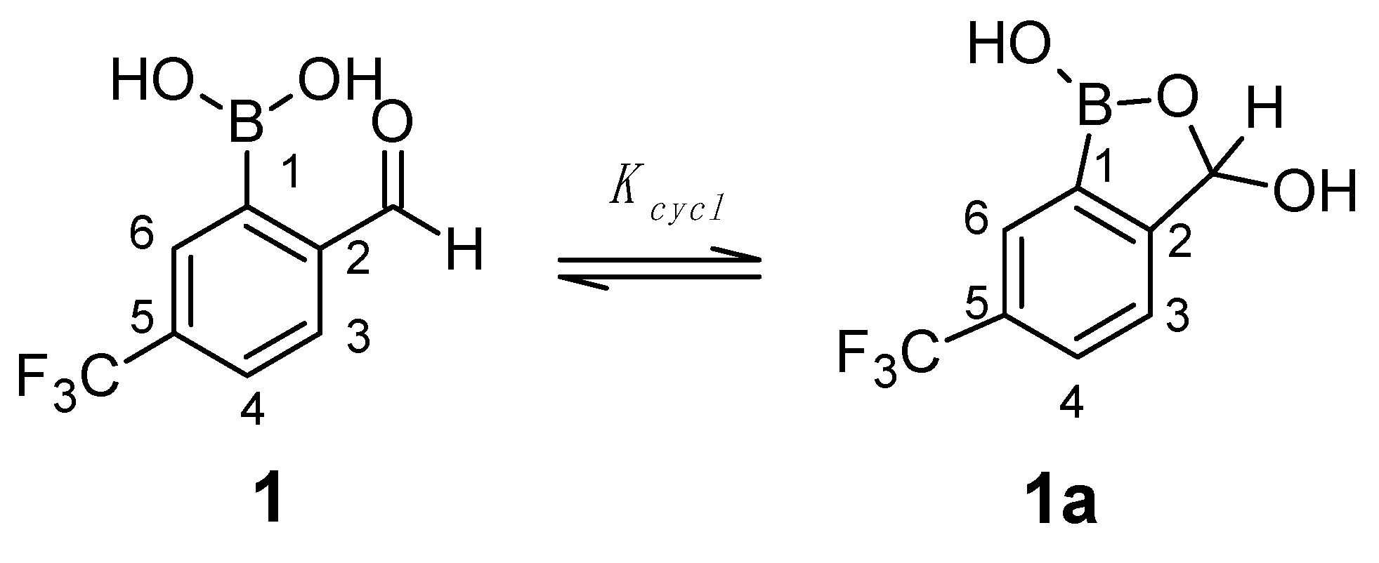
2.1. Synthesis, Molecular and Crystal Structure
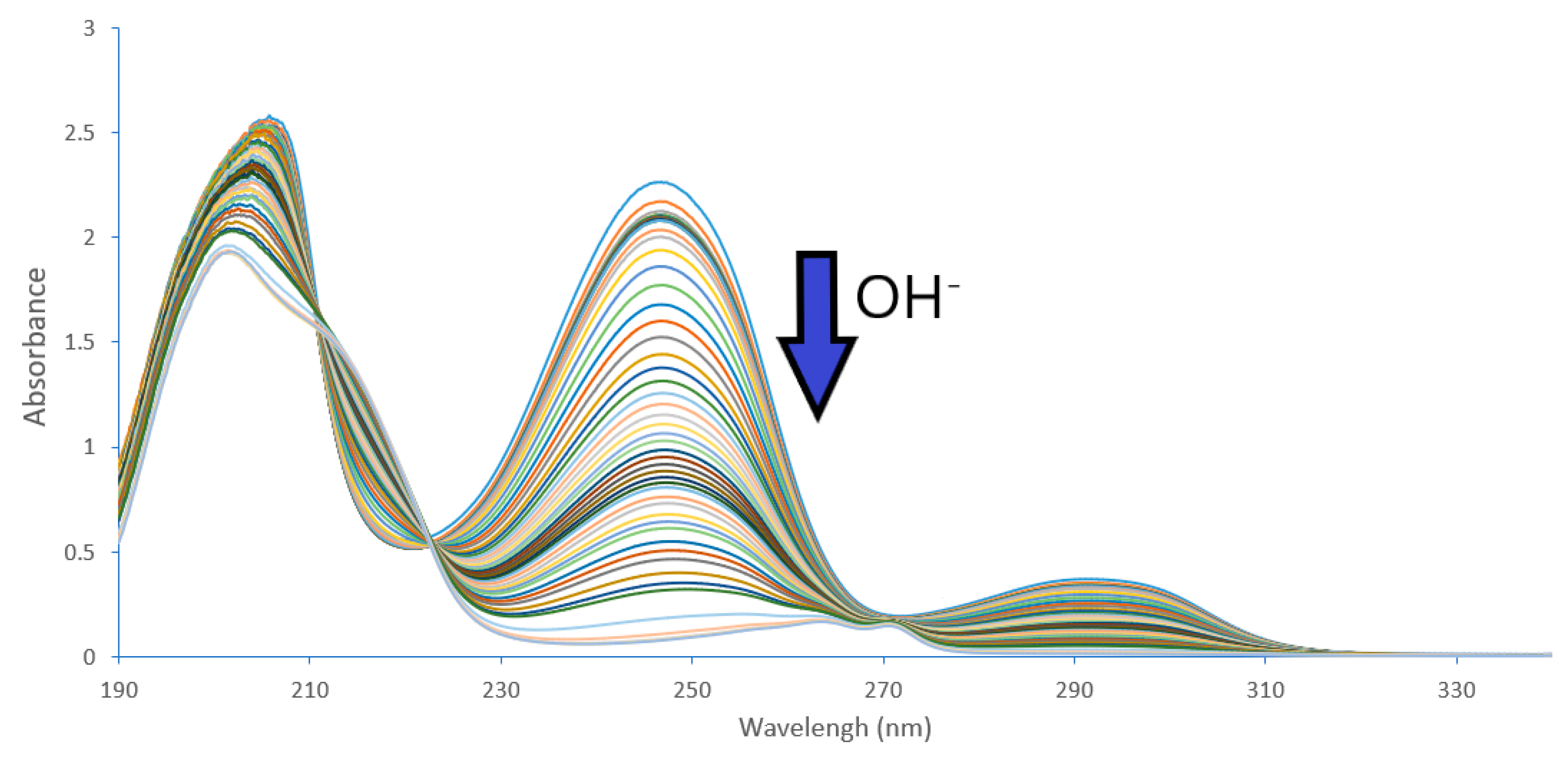
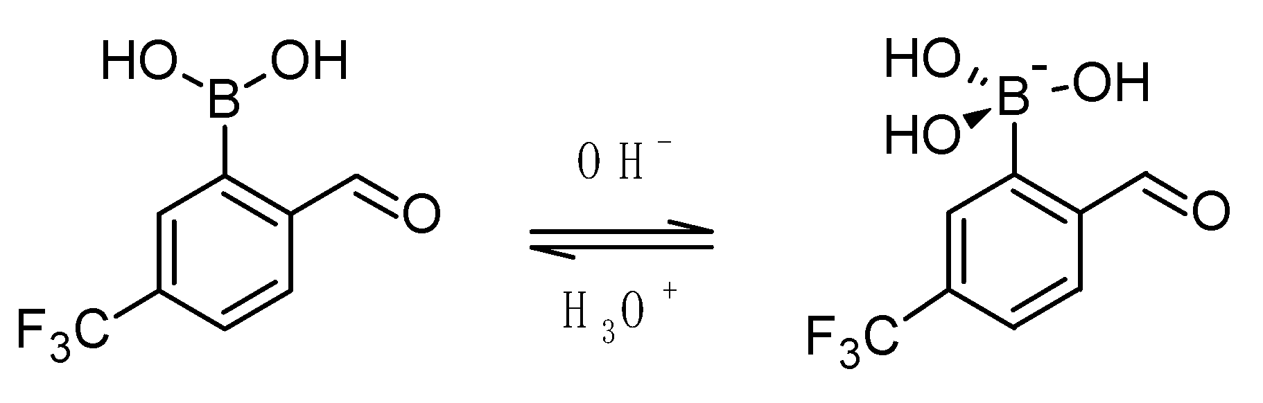
2.2. Acidity Constant Determination
2.3. Spectral Characterization, Cyclization Equilibrium
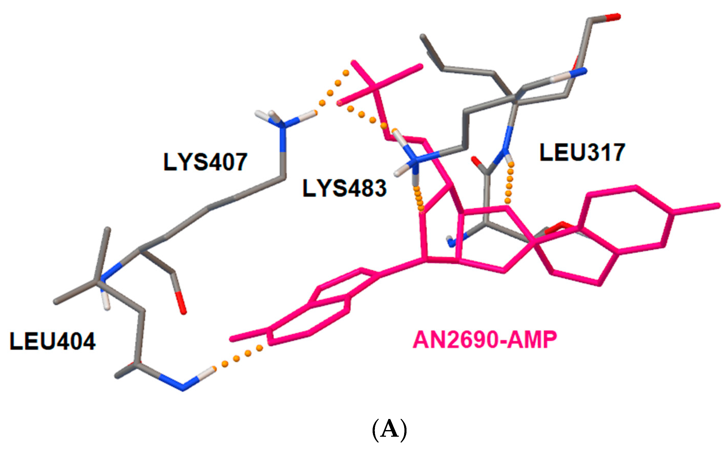
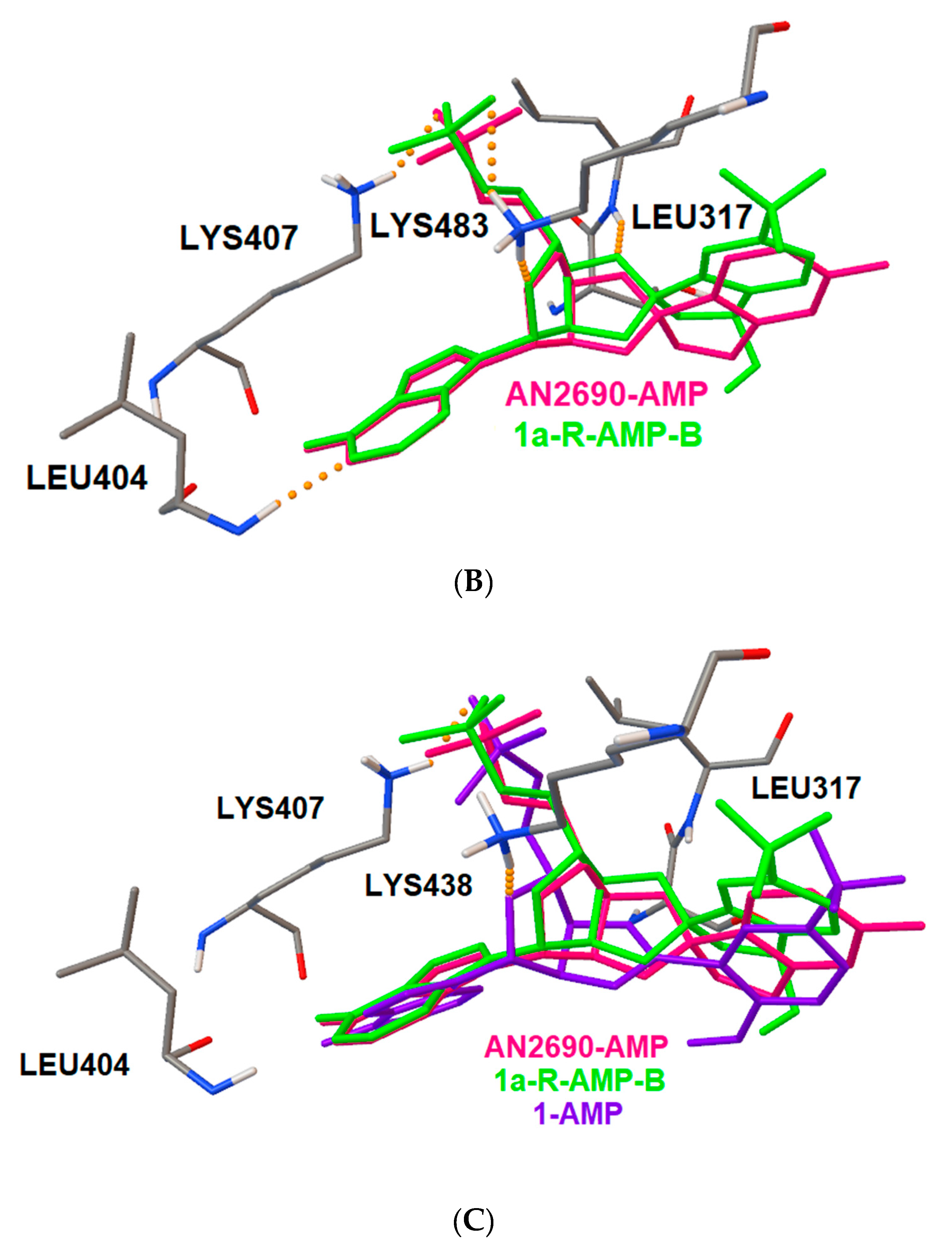
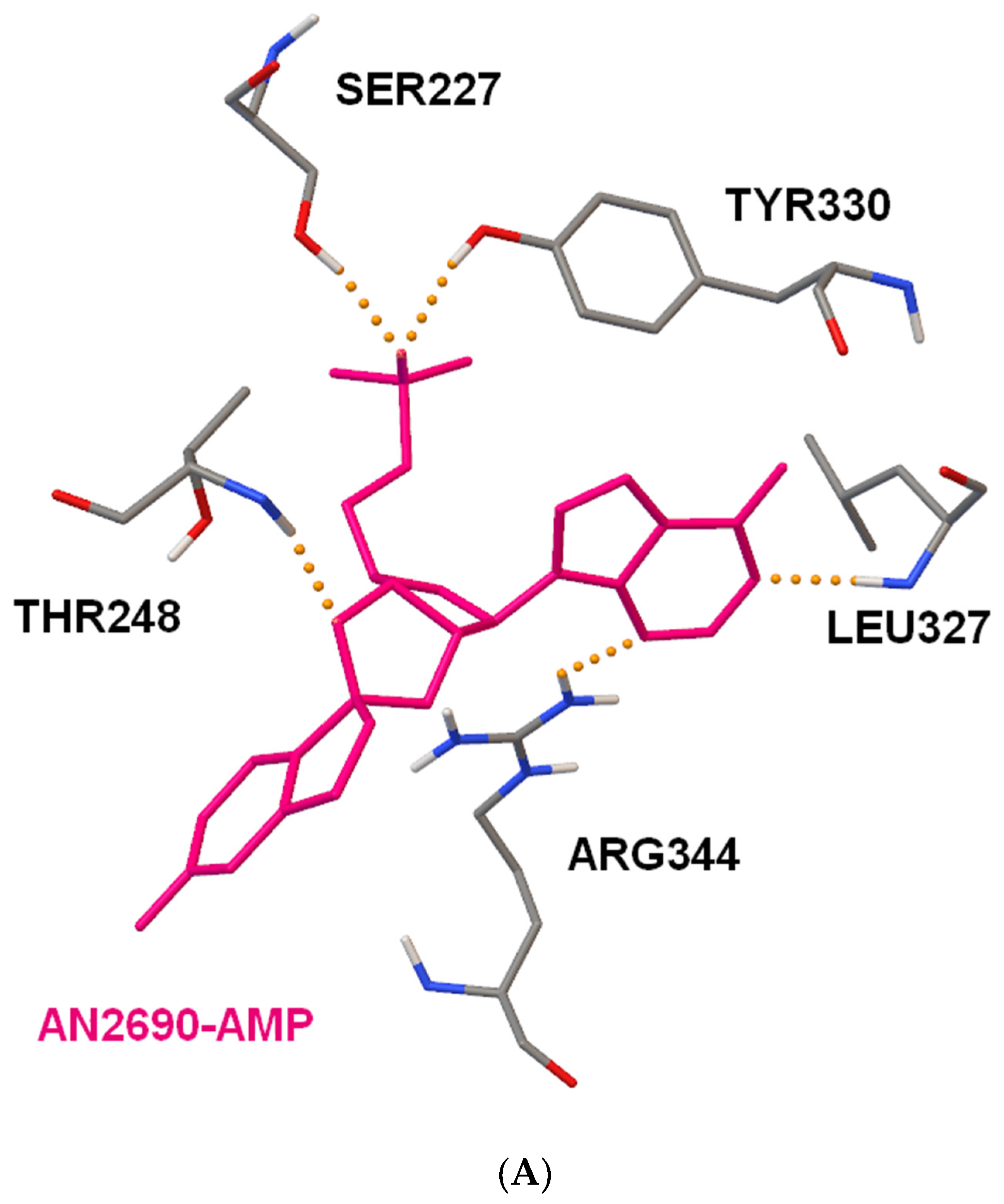
2.4. Docking Studies of Interactions of AN2690 and 1/1a with Candida albicans’ and Eschericha coli LeuRS
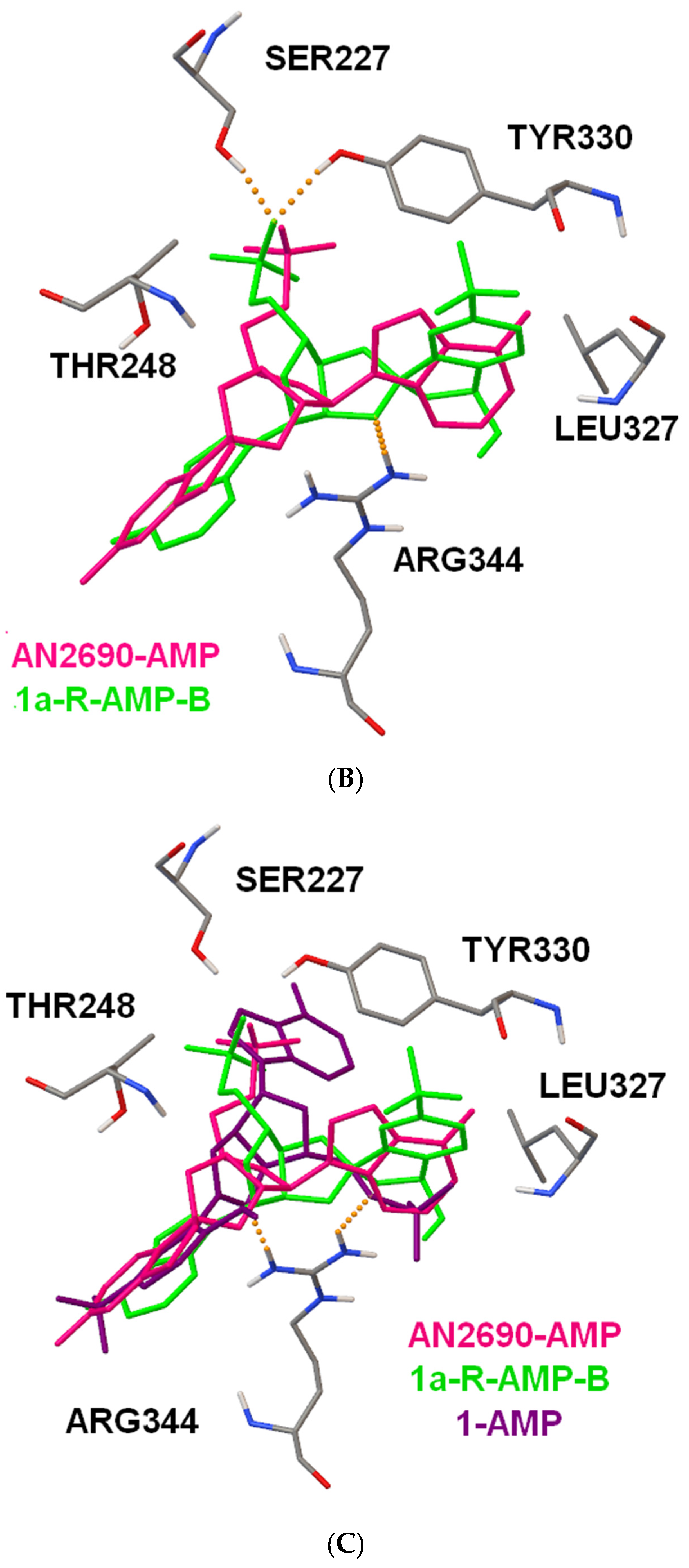
2.5. In Vitro Studies of Antimicrobial Activity of 1
3. Materials and Methods
3.1. Synthesis and Isolation
3.2. Single Crystal X-Ray Diffraction
3.3. Acidity Constant Determination
3.4. NMR Measurements
3.5. Docking Studies
3.6. Microbial Activity
4. Conclusions
Supplementary Materials
Author Contributions
Funding
Conflicts of Interest
References
- Gozdalik, J.T.; Adamczyk-Woźniak, A.; Sporzyński, A. Influence of fluorine substituents on the properties of phenylboronic compounds. Pure Appl. Chem. 2018, 90, 677–702. [Google Scholar] [CrossRef]
- Boron: Sensing, Synthesis and Supramolecular Self-Assembly. Meng, L.; Fossey, J.S.; James, T.D. (Eds.) Monographs in Supramolecular Chemistry; Royal Society of Chemistry: Cambridge, UK, 2015; ISBN 978-1-84973-674-9. [Google Scholar]
- Boronic Acids. Preparation and Applications in Organic Synthesis, Medicine and Materials, 2nd ed.; Hall, D.G., Ed.; WILEY-VCH: Weinheim, Germany, 2011. [Google Scholar]
- Cheng, T.; Li, H.; Ma, Y.; Liu, X.; Zhang, H. Synthesis of boronic-acid-functionalized magnetic attapulgite for selective enrichment of nucleosides. Anal. Bioanal. Chem. 2015, 407, 3525–3529. [Google Scholar] [CrossRef] [PubMed]
- Evangelista, L.; Jori, G.; Martini, D.; Sotti, G. Boron neutron capture therapy and 18F-labelled borophenylalanine positron emission tomography: A critical and clinical overview of theliterature. Appl. Radiat. Isot. 2013, 74, 91–101. [Google Scholar] [CrossRef] [PubMed]
- Adamczyk-Woźniak, A.; Borys, K.M.; Sporzyński, A. Recent Developments in the Chemistry and Biological Applications of Benzoxaboroles. Chem. Rev. 2015, 115, 5224–5247. [Google Scholar] [CrossRef] [PubMed]
- Madura, I.D.; Adamczyk-Woźniak, A.; Sporzyński, A. Diversified self-association through O–H∙∙∙O hydrogen bonds in crystals of formylphenylboronic acid isomers. J. Mol. Struct. 2015, 1083, 204–211. [Google Scholar] [CrossRef]
- Luliński, S.; Madura, I.; Serwatowski, J.; Szatyłowicz, H.; Zachara, J. A tautomeric equilibrium between functionalized 2-formylphenylboronic acids and corresponding 1,3-dihydro-1,3-dihydroxybenzo[c][2,1]oxaboroles. New J. Chem. 2007, 31, 144–154. [Google Scholar] [CrossRef]
- Borys, K.M.; Wieczorek, D.; Pecura, K.; Lipok, J.; Adamczyk-Woźniak, A. Antifungal activity and tautomeric cyclization equilibria of formylphenylboronic acids. Bioorg. Chem. 2019, 91, 103081. [Google Scholar] [CrossRef]
- Kowalska, K.; Adamczyk-Woźniak, A.; Gajowiec, P.; Gierczyk, B.; Kaczorowska, E.; Popenda, Ł.; Schroeder, G.; Sikorski, A.; Sporzyński, A. Fluoro-substituted 2-formylphenylboronic acids: Structures, properties and tautomeric equilibria. J. Fluor. Chem. 2016, 187, 1–8. [Google Scholar] [CrossRef]
- Das, S.; Alexeev, V.L.; Sharma, A.C.; Geib, S.J.; Asher, S.A. Synthesis and crystal structure of 4-amino-3-fluorophenylboronic acid. Tetrahedron Lett. 2003, 44, 7719–7722. [Google Scholar] [CrossRef]
- Alexeev, V.L.; Sharma, A.C.; Goponenko, A.V.; Das, S.; Lednev, I.K.; Wilcox, C.S.; Finegold, D.N.; Asher, S.A. High ionic strength glucose-sensing photonic crystal. Anal. Chem. 2003, 75, 2316–2323. [Google Scholar] [CrossRef]
- Kirsch, P. Modern Fluoroorganic Chemistry: Synthesis, Reactivity, Applications, 2nd ed.; WILEY-VCH: Darmstadt, Germany, 2013; ISBN 9783527603930. [Google Scholar]
- Purser, S.; Moore, P.R.; Swallow, S.; Gouverneur, V. Fluorine in medicinal chemistry. Chem. Soc. Rev. 2008, 37, 320–330. [Google Scholar] [CrossRef] [PubMed]
- Liu, F.; Tian, J.; Liu, Y.; Tao, C.; Zhu, H.; Zhang, A.; Xua, D.; Zhao, B. Decarboxylative Umpolung of conjugated enals to β-carbanions for intramolecular nucleophilic addition to an aldehyde. Org. Chem. Front. 2017, 4, 1586–1589. [Google Scholar] [CrossRef]
- Etter, M.C.; MacDonald, J.C.; Bernstein, J. Graph-set analysis of hydrogen-bond patterns in organic crystals. Acta Crystallogr. Sect. B Struct. Sci. 1990, 46, 256–262. [Google Scholar] [CrossRef]
- McKinnon, J.J.; Spackman, M.A.; Mitchell, A.S. Novel tools for visualizing and exploring intermolecular interactions in molecular crystals. Acta Crystallogr. Sect. B Struct. Sci. 2004, 60, 627–668. [Google Scholar] [CrossRef]
- Yan, J.; Springsteen, G.; Deeter, S.; Wang, B. The relationship among pKa, pH, and binding constants in the interactions between boronic acids and diols—It is not as simple as it appears. Tetrahedron 2004, 60, 11205–11209. [Google Scholar] [CrossRef]
- Yamamoto, Y.; Matsumura, T.; Takao, N.; Yamagishi, H.; Takahashi, M.; Iwatsuki, S.; Ishihara, K. Fast trigonal/tetragonal interconversion followed by slow chelate-ring closure in the complexation of boronic acids. Inorg. Chim. Acta 2005, 358, 3355–3361. [Google Scholar] [CrossRef]
- Westmark, P.R.; Gardiner, S.J.; Smith, B.D. Selective monosaccharide transport through lipid bilayers using boronic acid carriers. J. Am. Chem. Soc. 1996, 118, 11093–11100. [Google Scholar] [CrossRef] [Green Version]
- Siodła, T.; Ozimiński, W.P.; Hoffmann, M.; Koroniak, H.; Krygowski, T.M. Toward a physical interpretation of substituent effects: The case of fluorine and trifluoromethyl groups. J. Org. Chem. 2014, 79, 7321–7331. [Google Scholar] [CrossRef]
- Zarzeczańska, D.; Adamczyk-Woźniak, A.; Kulpa, A.; Ossowski, T.; Sporzyński, A. Fluorinated Boronic Acids: Acidity and Hydrolytic Stability of Fluorinated Phenylboronic Acids. Eur. J. Inorg. Chem. 2017, 2017, 4493–4498. [Google Scholar] [CrossRef] [Green Version]
- Gozdalik, J.T.; Marek, P.H.; Madura, I.D.; Gierczyk, B.; Popenda, Ł.; Schroeder, G.; Adamczyk-Woźniak, A.; Sporzyński, A. Structures and properties of trifluoromethylphenylboronic acids. J. Mol. Struct. 2019, 1180, 237–243. [Google Scholar] [CrossRef]
- Torssell, K.; McClendon, J.H. Chemistry of Arylboric Acids VIII. The Relationship between Physico-chemical Properties and Activity in Plants. Acta Chem. Scand. 1958, 12, 1373–1385. [Google Scholar] [CrossRef] [Green Version]
- Tomsho, J.W.; Pal, A.; Hall, D.G.; Benkovic, S.J. Ring Structure and Aromatic Substituent Effects on the pKa of the Benzoxaborole Pharmacophore. ACS Med. Chem. Lett. 2011, 3, 48–52. [Google Scholar] [CrossRef] [PubMed] [Green Version]
- Adamczyk-Woźniak, A.; Cabaj, M.K.; Dominiak, P.M.; Gajowiec, P.; Gierczyk, B.; Lipok, J.; Popenda, Ł.; Schroeder, G.; Tomecka, E.; Urbański, P.; et al. The influence of fluorine position on the properties of fluorobenzoxaboroles. Bioorg. Chem. 2015, 60, 130–135. [Google Scholar] [CrossRef] [PubMed]
- Adamczyk-Woźniak, A.; Ejsmont, K.; Gierczyk, B.; Kaczorowska, E.; Matuszewska, A.; Schroeder, G.; Sporzyński, A.; Zarychta, B. Novel 2,6-disubstituted phenylboronic compounds: Synthesis, crystal structures, solution behaviour and reactivity. J. Organomet. Chem. 2015, 788, 36–41. [Google Scholar] [CrossRef]
- Dolbier, W.R., Jr. Guide to Fluorine NMR for Organic Chemists, 2nd ed.; Wiley: Hoboken, NJ, USA, 2016. [Google Scholar]
- Rock, F.L.; Mao, W.; Yaremchuk, A.; Tukalo, M.; Crepin, T.; Zhou, H.; Zhang, Y.-K.; Hernandez, V.; Akama, T.; Baker, S.J.; et al. An Antifungal Agent Inhibits an Aminoacyl-tRNA Synthetase by Trapping tRNA in the Editing Site. Science 2007, 316, 1759–1761. [Google Scholar] [CrossRef]
- Palencia, A.; Crepin, T.; Vu, M.T.; Lincecum, T.L., Jr.; Martinis, S.A.; Cusack, S. Structural Dynamics of the Aminoacylation and Proofreading Functional Cycle of Bacterial Leucyl-tRNA Synthetase. Nat. Struct. Mol. Biol. 2012, 19, 677. [Google Scholar] [CrossRef] [Green Version]
- Gualerzi, C.O.; Brandi, L.; Fabbretti, A.; Pon, C.L. Antibiotics: Targets, Mechanism and Resistance; Wiley: Hoboken, NJ, USA, 2013. [Google Scholar]
- Kim, S. (Ed.) Aminoacyl-tRNA Synthetases in Biology and Medicine. In Topics in Current Chemistry; Springer Verlag: Berlin Heidelberg, Germany, 2014; p. 312. [Google Scholar]
- CrysAlisPro 1.171.38.46, Rigaku; Oxford Diffraction: Abingdon-on-Thames, UK, 2015.
- Dolomanov, O.V.; Bourhis, L.J.; Gildea, R.J.; Howard, J.A.K.; Puschmann, H. OLEX2: a complete structure solution, refinement and analysis program. J. Appl. Crystallogr. 2009, 42, 339–341. [Google Scholar] [CrossRef]
- Sheldrick, G.M. SHELXT–Integrated space-group and crystal-structure determination. Acta Crystallogr. Sect. A Found. Adv. 2015, 71, 3–8. [Google Scholar] [CrossRef] [Green Version]
- Sheldrick, G.M. Crystal structure refinement with SHELXL. Acta Crystallogr. Sect. C Struct. Chem. 2015, 71, 3–8. [Google Scholar] [CrossRef]
- Farrugia, L.J. Ortep-3 for Windows-a version of ORTEP-III with a Graphical User Interface (GUI). J. Appl. Crystallogr. 1997, 30, 565. [Google Scholar] [CrossRef]
- Burley, S.K.; Berman, H.M.; Bhikadiya, Ch.; Bi, Ch.; Chen, L.; Di Costanzo, L.; Christie, C.; Dalenberg, K.; Duarte, J.M.; Dutta, S.; et al. RCSB Protein Data Bank: biological macromolecular structures enabling research and education in fundamental biology, biomedicine, biotechnology and energy. Nucleic Acids Res. 2019, 47, D464–D474. Available online: www.rcsb.org (accessed on 11 December 2019). [CrossRef] [PubMed] [Green Version]
- Morris, G.M.; Huey, R.; Lindstrom, W.; Sanner, M.F.; Belew, R.K.; Goodsell, D.S.; Olson, A.J. Autodock4 and AutoDockTools4: automated docking with selective receptor flexiblity. J. Comput. Chem. 2009, 16, 2785–2791. Available online: http://autodock.scripps.edu/ (accessed on 11 December 2019). [CrossRef] [PubMed] [Green Version]


| H∙∙∙A | D∙∙∙A | D-H∙∙∙A | |
|---|---|---|---|
| Intramolecular | |||
| O2‒H2…O3 | 1.82(1) | 2.619(1) | 164(2) |
| O21‒H21…O31 | 1.81(1) | 2.605(1) | 164(2) |
| Intermolecular | |||
| O1‒H1…O21 | 1.95(1) | 2.774(1) | 177(2) |
| O11‒H11…O2 | 1.97(1) | 2.779(1) | 174(2) |
| C7‒H7…O1 i | 2.61 | 3.254(2) | 127 |
| C71‒H71…O11 ii | 2.56 | 3.220(2) | 128 |
| General Formula | Substituent | pKa Value | Reference |
|---|---|---|---|
 | - | 8.86 | [10] |
| 3-F | 7.50 ± 0.02 | [22] | |
| 3-CF3 | 7.85 ± 0.05 | [23] | |
| 2-CHO | 7.31 | [24] | |
| 2-CHO, 5-F | 6.72 ± 0.03 | [10] | |
| 2-CHO, 5-CF3 (1) | 5.67 ± 0.01 | This work |
| 1H NMR | 11B | 19F | ||||||
|---|---|---|---|---|---|---|---|---|
| Solvent (Form) | B(OH)2 | CHO | H3 | H4 | H6 | |||
| OH | H | |||||||
| CDCl3 (1) | 7.13 (s) | 10.04 (m) | 8.54 (m) | 7.97 (m) | 8.06 (d) 3JH5 = 7.8 | 27 (s) | −63.44 | |
| C6D6 (1) | 7.04 (s) | 9.04 (s) | 8.55 (m) | 7.20 (m) | 6.73 (d) 3JH5 = 7.8 | 27 (s) | −63.29 | |
| (CD3)2CO (1) | 7.84 (s) | 10.40 (s) | 8.10 (m) | 7.93 (m) | 8.15 (m) | 29 (s) | −62.94 | |
| (CD3)2CO (1a) | 8.50 (s) | 6.38 (d) 3JH = 7.9 | 6.22 (d) 3JOH = 8.1 | 8.01 (m) | 7.85 (m) | 7.70 (m) | 31 (s) | −61.98 |
| (CD3)2SO (1) | 8.47 (s) | 10.24 (s) | 7.89 (m) | 7.91 (m) | 8.07 (m) | 30 (s) | −61.67 | |
| (CD3)2SO (1a) | 9.57 (s) | 7.23 (d) 3JH = 8.1 | 6.25 (d) 3JOH = 8.1 | 8.01 (m) | 7.85 (m) | 7.64 (m) | 30 (s) | −60.73 |
| Solvent | Cyclization Constant | 1H/19F NMR Shift of the Cyclic Form (1a) | 1H/19F NMR Shift of The Opened Form (1) |
|---|---|---|---|
| (CD3)2SO | 0.59 ± 0.01 | 6.25/−60.73 | 10.24/−61.67 |
| (CD3)2CO | 0.24 ± 0.02 | 6.22/−61.98 | 10.40/−62.94 |
| D2O | 0.26 ± 0.03 * | 6.36/−62.22 | 10.06/−63.26 |
| Form | CHO/CH-OH | CF3 | C1 | C2 | C3 | C4 | C5 | C6 |
|---|---|---|---|---|---|---|---|---|
| 1 | 193.82 (s) | 124.5 (q) 1JF = 270.5 | 140.8 (bs) | 142.1 (d) 4JF = 1.1 | 129.5 (m) | 125.7 (m) | 132.2 (q) 2JF = 31.5 | 129.9 (s) |
| 1a | 97.06 (s) | Not Observed | Not Observed | 158.9 (s) | 126.8 (q) 3JF = 3.7 | 127.8 (m) | Not Observed | 123.8 (s) |
| Ligand | The Lowest Binding Energy [kcal/mol] | Number of Structures | Mean Binding Energy [kcal/mol] | Inhibition Constant | Number of Hydrogen Bonds | |
|---|---|---|---|---|---|---|
| Candida albicans | AN2690-AMP | −11.98 | 17 | −10.80 | 1.93 nM | 5 |
| 1a-R-AMP-A | −10.77 | 2 | −10.21 | 12.77 nM | 4 | |
| 1a-R-AMP-B | −11.83 | 3 | −11.66 | 2.12 nM | 5 | |
| 1a-S-AMP-A | −10.75 | 19 | −10.42 | 13.08 nM | 3 | |
| 1a-S-AMP-B | −11.59 | 4 | −10.94 | 3.17 nM | 4 | |
| 1-AMP | −11.05 | 2 | −10.77 | 7.95 nM | 2 | |
| Escherichia coli | AN2690-AMP | −8.27 | 22 | −7.55 | 865.19 nM | 5 |
| 1a-R-AMP-A | −7.54 | 21 | −6.90 | 2.99 μM | 2 | |
| 1a-R-AMP-B | −8.15 | 2 | −7.81 | 1.09 μM | 3 | |
| 1a-S-AMP-A | −7.54 | 12 | −6.98 | 2.96 μM | 2 | |
| 1a-S-AMP-B | −7.72 | 4 | −7.45 | 2.21 μM | 3 | |
| 1-AMP | −8.23 | 4 | −7.75 | 924.99 nM | 2 |
| 10 µg | 25 µg | 50 µg | 100 µg | AN2690 (50 µg) | Antibiotic (50 µg) | |
|---|---|---|---|---|---|---|
| Candida albicans | 0 | 9 | 13 ± 2 | 17 ± 3 (8 ± 1) | (53) | (10 ± 1) * |
| Aspergillus niger | 0 | 8 ± 1 | 13 ± 2 | 26 ± 4 (5 ± 1) | (62) | (9 ± 1) * |
| Escherichia coli | 2 ± 1 | 5 ± 1 | 7 ± 1 | 9 ± 1 | (22) | (16 ± 1) # |
| Bacillus cereus | 12 | 15 ± 1 | 18 | 19 ± 1 | (14) | (21 ± 1) # |
| MIC [µg/mL] | |||
|---|---|---|---|
| 1 | AN 2690 | Antibiotic | |
| Candida albicans | 250 | 2 | ≤ 1 * |
| Aspergillus niger | 32 | ≤ 1 | - |
| Escherichia coli | 125 | 7.8 | 2 # |
| Bacillus cereus | 8 | 62.5 | 4 # |
| * Amphotericin B, # Streptomycin | |||
© 2020 by the authors. Licensee MDPI, Basel, Switzerland. This article is an open access article distributed under the terms and conditions of the Creative Commons Attribution (CC BY) license (http://creativecommons.org/licenses/by/4.0/).
Share and Cite
Adamczyk-Woźniak, A.; Gozdalik, J.T.; Wieczorek, D.; Madura, I.D.; Kaczorowska, E.; Brzezińska, E.; Sporzyński, A.; Lipok, J. Synthesis, Properties and Antimicrobial Activity of 5-Trifluoromethyl-2-formylphenylboronic Acid. Molecules 2020, 25, 799. https://0-doi-org.brum.beds.ac.uk/10.3390/molecules25040799
Adamczyk-Woźniak A, Gozdalik JT, Wieczorek D, Madura ID, Kaczorowska E, Brzezińska E, Sporzyński A, Lipok J. Synthesis, Properties and Antimicrobial Activity of 5-Trifluoromethyl-2-formylphenylboronic Acid. Molecules. 2020; 25(4):799. https://0-doi-org.brum.beds.ac.uk/10.3390/molecules25040799
Chicago/Turabian StyleAdamczyk-Woźniak, Agnieszka, Jan T. Gozdalik, Dorota Wieczorek, Izabela D. Madura, Ewa Kaczorowska, Ewa Brzezińska, Andrzej Sporzyński, and Jacek Lipok. 2020. "Synthesis, Properties and Antimicrobial Activity of 5-Trifluoromethyl-2-formylphenylboronic Acid" Molecules 25, no. 4: 799. https://0-doi-org.brum.beds.ac.uk/10.3390/molecules25040799