Article

A Highly Selective Turn-On Fluorescent Probe for the Detection of Zinc

1
The Key Laboratory of Environmental Pollution Monitoring and Disease Control, School of Public Health, Ministry of Education, Guizhou Medical University, Guiyang 550014, China
2
School of Basic Medical Science, Guizhou Medical University, Guiyang 550004, China
3
Department of Chemistry, University of Hull, Cottingham Road, Hull, Yorkshire HU6 7RX, UK
*
Authors to whom correspondence should be addressed.
These authors contributed equally to this work.
Submission received: 23 May 2021 / Revised: 19 June 2021 / Accepted: 19 June 2021 / Published: 23 June 2021

Abstract

:
A novel turn-on fluorescence probe L has been designed that exhibits high selectivity and sensitivity with a detection limit of 9.53 × 10⁻⁸ mol/L for the quantification of Zn2+. 1H-NMR spectroscopy and single crystal X-ray diffraction analysis revealed the unsymmetrical nature of the structure of the Schiff base probe L. An emission titration experiment in the presence of different molar fractions of Zn2+ was used to perform a Job’s plot analysis. The results showed that the stoichiometric ratio of the complex formed by L and Zn2+ was 1:1. Moreover, the molecular structure of the mononuclear Cu complex reveals one ligand L coordinates with one Cu atom in the asymmetric unit. On adding CuCl2 to the ZnCl2/L system, a Cu-Zn complex was formed and a strong quenching behavior was observed, which inferred that the Cu2+ displaced Zn2+ to coordinate with the imine nitrogen atoms and hydroxyl oxygen atoms of probe L.

1. Introduction

Zinc is the second most abundant transition metal ion in the human body and is essential for various biochemical processes, such as neurotransmission, enzyme regulation, gene expression, and apoptosis [1,2,3,4]. In addition, zinc at normal concentrations controls many metabolic, biological, and environmental processes, while deficiency usually leads to the appearance of some clinical diseases, such as growth retardation, brain dysfunction, high blood cholesterol, Parkinson’s disease, ischemic stroke, Alzheimer’s disease, etc. [5,6,7,8,9]. On the other hand, excessive zinc will also cause problems for humans; for example, it will reduce soil microbial activity and cause phytotoxic effects [10,11,12,13,14]. Therefore, it is of great significance to be able to accurately detect zinc ion concentration [15,16,17,18].
At present, the main detection methods of zinc include spectrophotometry, electrochemical methods [19], atomic absorption methods [20], chromatographic methods, and mass spectrometry [21,22]. However, due to the high cost of equipment, cumbersome sample preparation, or prolonged testing time, the wide application of these methods in actual testing has been limited. Among various detection methods, metal ion fluorescent chemical sensors have attracted much attention because of the convenient use, high sensitivity, and ability to directly measure concentration through fluorescent signals [23,24,25,26,27]. Zn2+ fluorescent probes can be divided into three categories: fluorescence quenching, fluorescence enhanced type, and ratio type. There is still an urgent need to develop new small molecule probes that are easy to prepare, have high sensitivity, and can recognize Zn2+ with excellent selectivity.
Schiff bases can coordinate with a variety of metals due to their special chemical structure, so they are considered to be dominant ligands for metal ions [28]. Schiff bases and their transition metal complexes not only have a wide range of applications in synthesis, catalytic chemistry, and materials chemistry [29], but also have a wide range of applications in antibacterial, antifungal, anticancer [30], clinical, analytical, and pharmacological aspects [31,32,33,34]. Moreover, a series of Schiff base ligands have been reported to be fluorescence turn-on chemosensors for Zn2+ with high sensitivity and selectivity [35,36,37,38,39,40]. Such systems are based on several reported mechanisms of fluorescence enhancement behavior, including internal charge transfer (ICT) [41], chelating-enhanced fluorescence (CHEF) [42,43], photoinduced electron transfer (PET) [44,45], aggregation-induced emission (AIE) [46,47,48], and C=N isomerization mechanisms [49,50,51]. In the current work, we have designed an asymmetric Schiff base L that not only has the ability to chelate metals but also has lone pair electrons on nitrogen atoms. The synthesis of L is shown in Scheme 1, and its structure has been confirmed by NMR spectroscopy, mass spectrometry, single crystal X-ray diffraction, and UV-Vis spectroscopy.
The fluorescence spectrum indicated little fluorescence emission of the free probe L. However, after adding Zn2+, L exhibited a fluorescence emission peak at 475 nm and a 512-fold fluorescence enhancement. Meanwhile, qualitative and quantitative detection are achieved through the linear relationship between fluorescence intensity and zinc ion concentration. Additionally, most of the coexisting metal ions had little or negligible interference on the emission response of probe L toward Zn2+. Of particular note is Cd2+, which has very similar chemical properties to Zn2+, and most Zn2+ sensors tend to respond to both Zn2+ and Cd2+ [32,33,34,35]. Hence, the development of a Zn2+ selective fluorescence sensor that can discriminate Zn2+ from Cd2+ is a great challenge and is of great significance. The recognition mechanism of the probe for Zn2+ in ethanol solution is proposed to be C=N isomerization [49,50,51] and chelation-enhanced fluorescence [42,43]. The C=N isomerization is inhibited by the coordination of Zn2+ with the probe L, so the fluorescence is significantly enhanced. Moreover, both Zn2+ and Cu2+ were investigated as the metal ions to coordinate simultaneously with L in order to understand the strong quenching behavior of Cu2+.

2. Results and Discussion

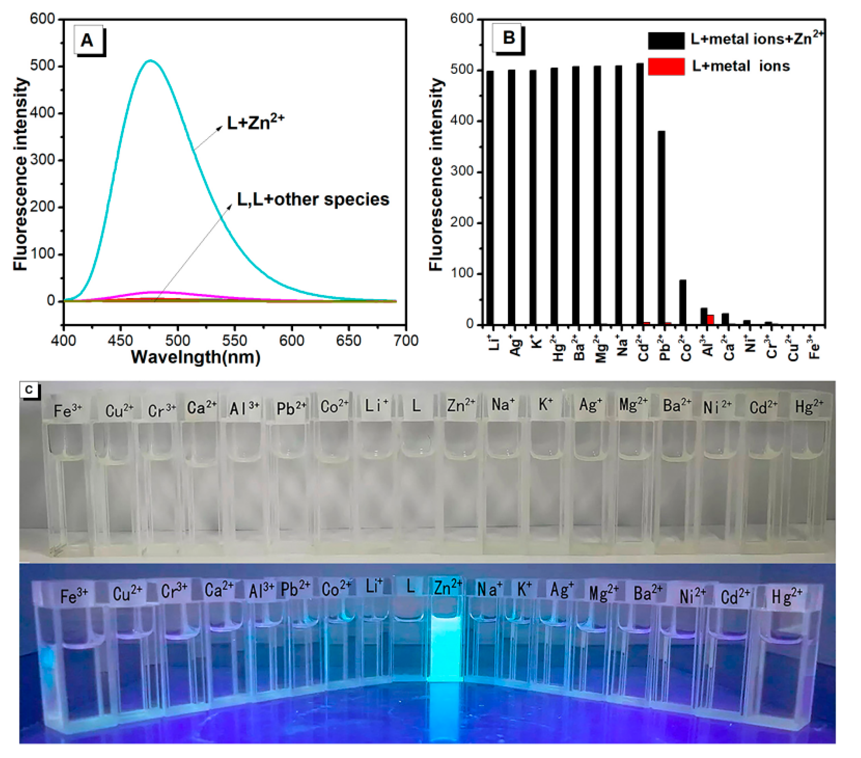
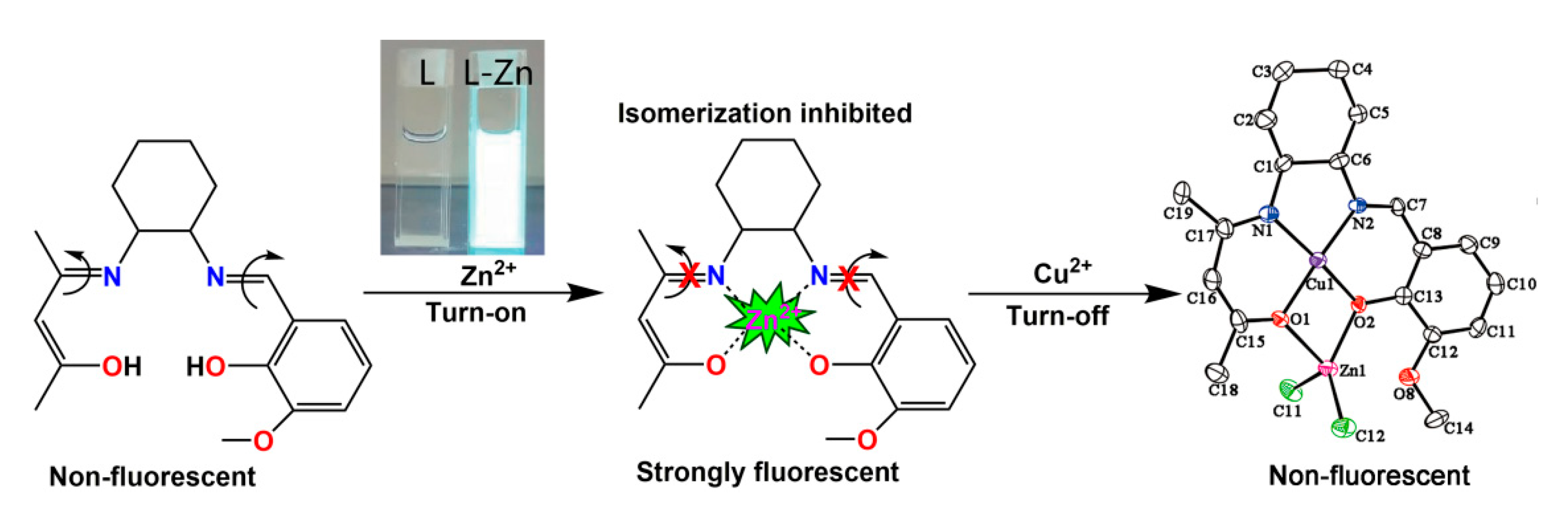
2.1. Selectivity of the Probe L

To determine potential practical applications, the spectroscopic properties of L were measured under simulated physiological conditions (50 μM in ethanol solution). The probe will be deactivated when the water content exceeds 5%, and thus the fluorescence spectral response of L to metal ions was recorded in ethanol solution excited at 354 nm as shown in Figure 1. The fluorescence spectroscopic response of L toward metal ions was evaluated in ethanol solution upon excitation at 354 nm as presented in Figure 1. The fluorescence spectroscopy indicated that the addition of Zn2+ resulted in a significant enhancement of the emission intensity positioned at 475 nm (Figure 1A, blue line). Under the same conditions, next to no responsive changes are observed in the presence of 1 equiv. of various metal cations (solutions of Zn2+, Li+, Na+, K+, Ag+, Mg2+, Ca2+, Sr2+, Ba2+, Al3+, Fe3+, Co2+, Ni2+, Cu2+, Pb2+, Cd2+, and Hg2+ were prepared from their chloride salts) in ethanol. Thus, according to the spectroscopy changes, the Schiff base probe L can detect Zn2+ with good selectivity.

2.2. Competition Experiments

It is necessary for a metal ion fluorescence chemosensor to achieve higher selectivity over other competing metal ions. Therefore, we evaluated the fluorescence behavior of probe L for Zn2+ in the presence of various competing metal ions, in which L was treated with 1 equiv. of Zn2+ in the presence of 1 equiv. of other metal ions. Figure 1B summarizes the results, and reveals that the presence of Li+, Na+, K+, Ag+, Hg2+, Mg2+, Hg2+, Ba2+, and Pb2+ caused only minor interference for the detection of Zn2+, whilst the presence of Co2+, Al3+, Ca2+, Ni2+, and Cr3+ resulted in low fluorescence intensity but were clearly detectable. However, in the case of Fe3+ and Cu2+, quenching of the fluorescence signal was observed, which may be due to the paramagnetic nature of Cu(II) and Fe(III) and their stronger coordinating ability than Zn(II). The fluorescence probe for detecting Zn2+ in the presence of coexisting metal ions, except for Fe3+ and Cu2+, clearly maintains higher emission than observed for the free probe L. Moreover, Cd2+ did not inhibit the emission intensity of Zn2+. Thus, these results indicate that probe L could be used as a selective probe for Zn2+ for distinguishing Zn2+ from Cd2+, which commonly share similar properties.

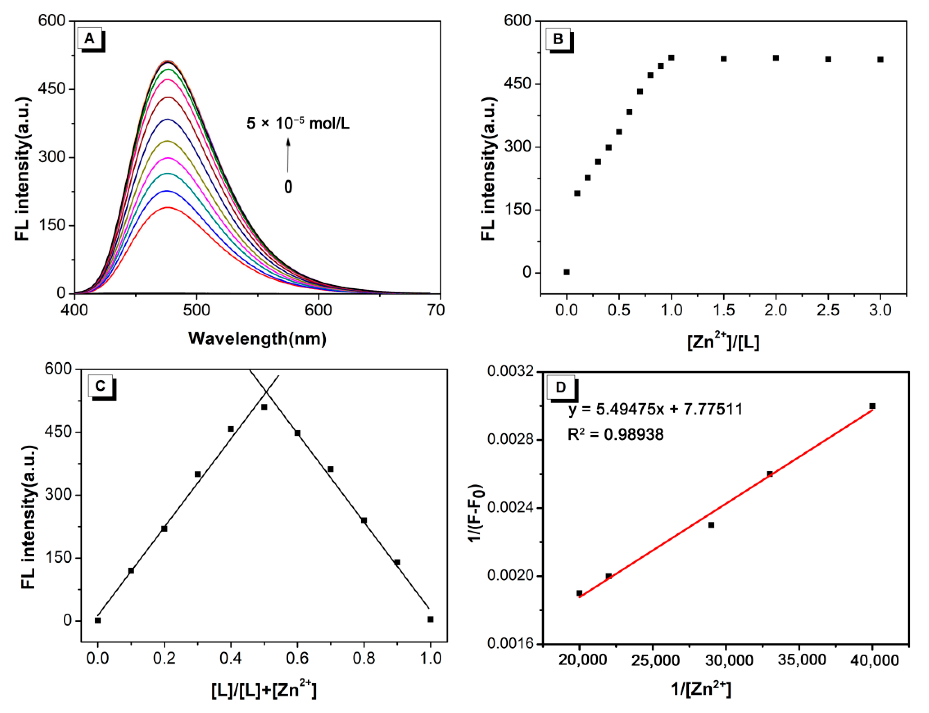
2.3. Fluorescence Spectroscopic Studies of L toward Zn2+

The result of the fluorometric titration of the free probe L and those in the presence of incremental amounts of Zn2+ in ethanol solution is shown in Figure 2A. The fluorescence spectrum showed that the fluorescence intensity increases steadily and smoothly on increasing the Zn2+ concentration. On addition of up to 1 equiv. of Zn2+, the turn-on ratio was observed to increase by over 512-fold. Additionally, the dependence of the emission intensity at 475 nm on the Zn2+ concentration is shown in Figure 2B. The fluorescence intensity remained constant in the presence of more than 1 equiv. of Zn2+, and therefore, the formation of a 1:1 complex between L and the Zn2+ was proposed. According to the fluorescence titration data, the association constant for L–Zn2+ complexation was calculated at 1.42 × 10⁴ mol/L from the Benesi-Hildebrand plot (Figure 2C). For practical purposes, the detection limit of probe L is an important parameter. The detection limit of probe L for Zn2+ was determined to be 9.53 × 10⁻⁸ mol/L according to the IUPAC definition (CDL = 3 Sb/m) from 10 blank solutions [52,53]. The Job’s plot analysis shows that a maximum emission was observed when the molar fraction reached 0.5, suggesting that the complex formation between L and Zn2+ has the stoichiometric ratio of 1:1. A linear relationship (R2 = 0.99173) for the plot of the normalized fluorescence intensity at 475 nm against Zn2+/L is shown in Figure 2D.
For a rough comparison between present complex with other reported similar fluorescent sensors [35,37,38,39,54,55], based on the detection limits of fluorescent sensors for Zn2+, see the results gathered in Table 1. The present compound is clearly more sensitive than most of the other Schiff base fluorescent sensors for the detection of Zn2+ via turn-on fluorescence.

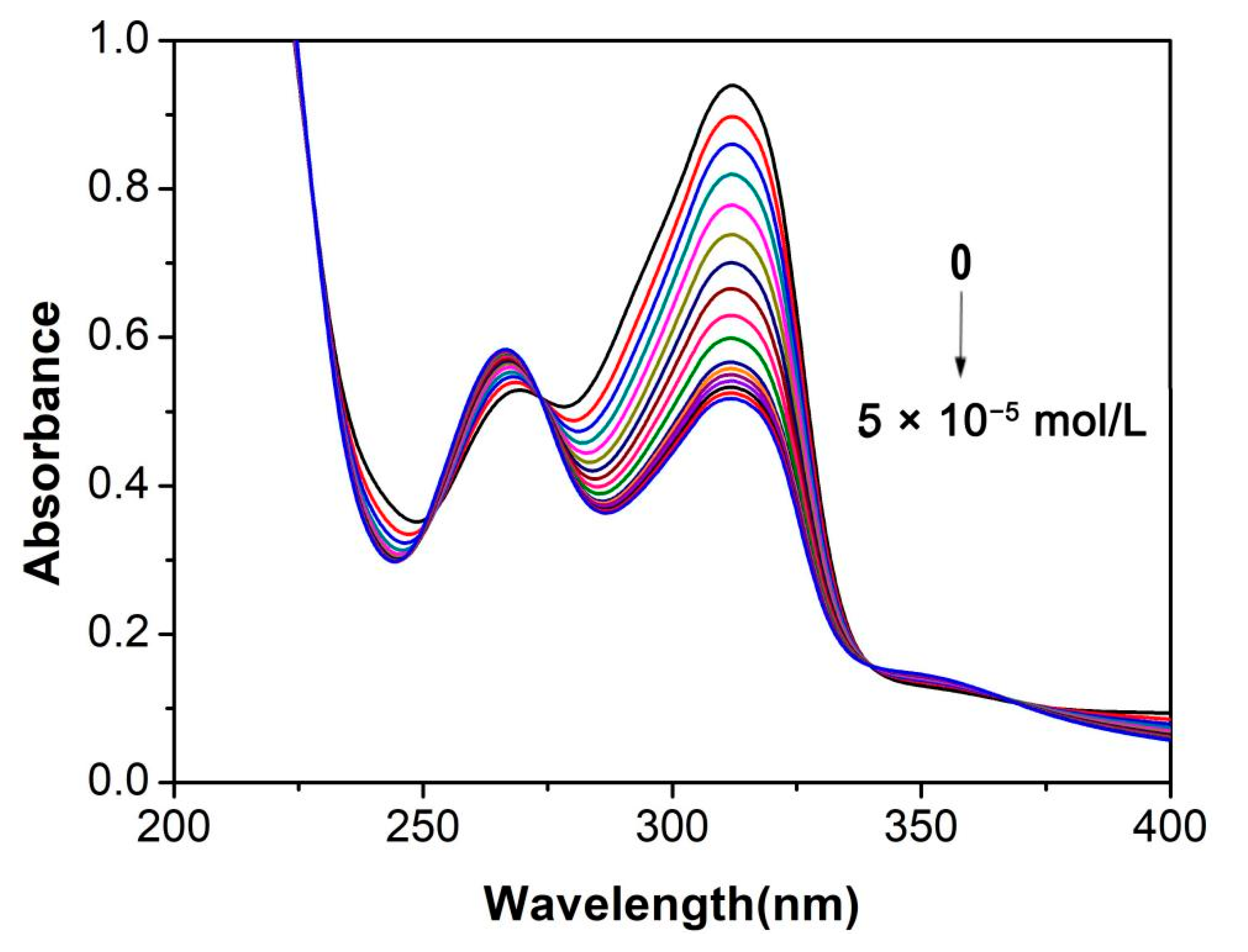
2.4. UV-Vis Absorbance Response of Probe L towards Zn2+

As illustrated in Figure 3, the probe L exhibited a maximal absorption at 313 nm. Upon addition of Zn2+ ions (0−1 equiv.), the absorbance at 313 nm decreased gradually on gradually increasing the Zn2+ concentration (Figure S4, Supplementary Materials). The presence of two clear isosbestic points at 273, 339 nm is consistent with the conversion of the free probe L to the Zn2+ complex. Moreover, the absorbance at 313 nm hardly changes in the presence of more than 1 equiv. of Zn2+ ions, indicating the formation of a 1:1 complex between L and the zinc ion. This is in good agreement with a 1:1 stoichiometry for the Zn2+ complex as determined by the Job’s plot obtained from UV-Vis absorption (Figure S5, Supplementary Materials).

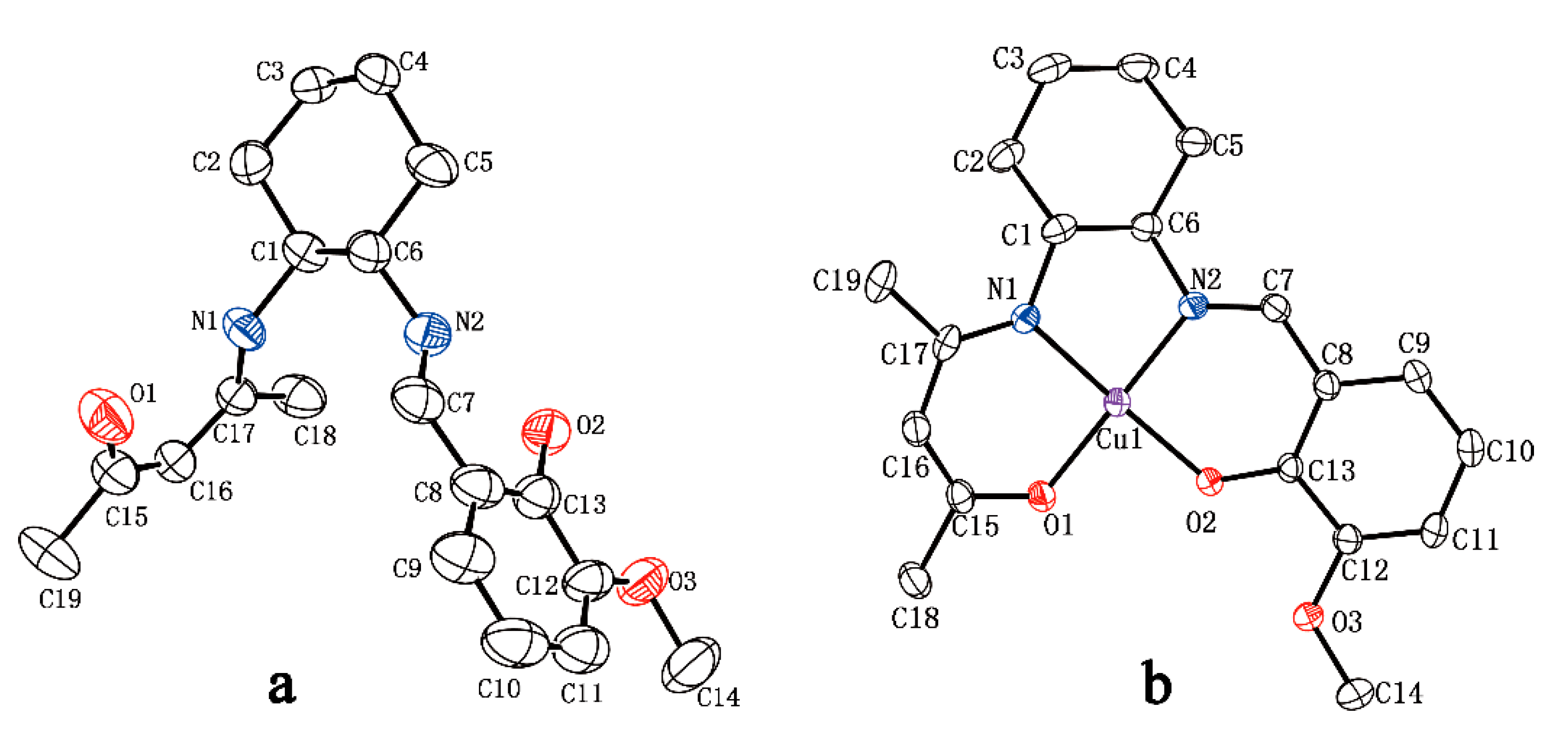
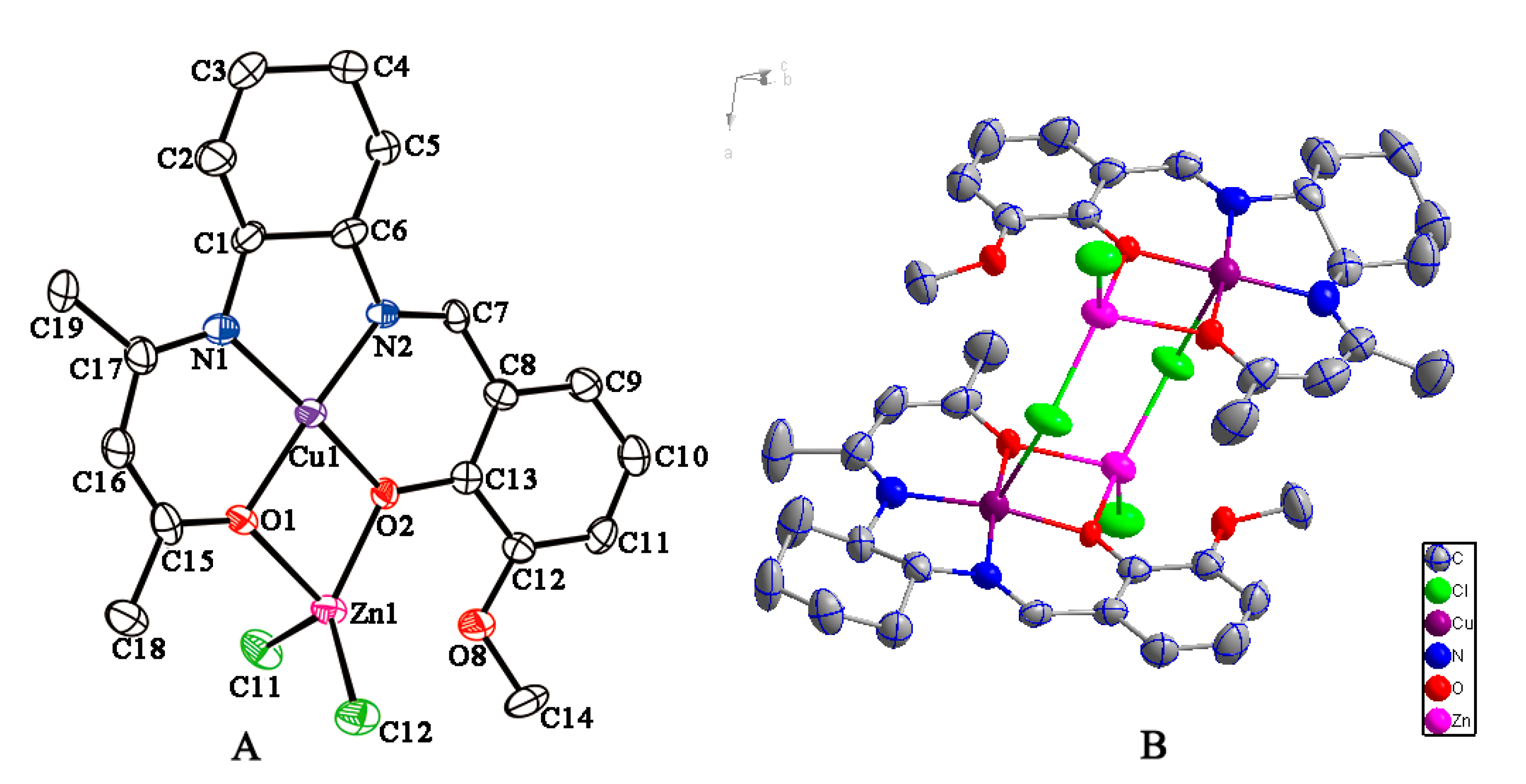
2.5. Crystal Structures of Probe L and Metal Complexes

To further investigate the binding mode of L with other metal ions, probe L and the organic framework of L possess an approximate torsional structure which chelates with one equivalent of Cu2+ ions. Moreover, a heteronuclear bimetallic complex was also obtained. The geometrical parameters of the mononuclear Cu complex, Cu-Zn complex, and L are listed in Table S1, Supplementary Materials. Treatment of 2,4-pentanedione with (1R, 2R)-diaminocyclohexane in refluxing methanol for 15 min, cooling, followed by the addition of 2-hydroxy-3-methoxybenzaldehyde led to the target product L, which was obtained from the filtrate on standing overnight below 0 °C. The reaction generally affords a high yield (84%), and yellow crystals suitable for X-ray determination were obtained (Figure 4a). The probe L is found to be unsymmetrical as shown by the crystal structure and the potential coordination sites in the two side chains point in opposite directions. Additionally, the bond lengths of N1–C17 and N2–C7 are 1.319(10) Å and 1.310(12) Å, respectively, corresponding to a C=N double bond. Both bond lengths of C15–C16 (1.348(15) Å) and C15–O1 (1.293(14) Å) were between single and double as a result of contributing to the conjugation effect. Moreover, all the atoms (except for hydrogen) in each side chain are coplanar and the dihedral angle of the two planes is 61.28°, thereby making the whole molecule appear “V” shaped.
To synthesize the copper complex, ligand L was treated with an equivalent of CuCl2 at room temperature, and following work-up, brown crystals were afforded. X-ray crystallographic analysis (Figure 4b) shows that the Cu2+ complex is a mononuclear complex, where the asymmetric unit consists of one ligand L and one Cu atom, giving rise to a 1:1 ligand to metal coordination. The central Cu2+ is four-coordinate with one phenoxide oxygen atom, and one enolic hydroxyl oxygen atom and two imine nitrogen atoms. Moreover, the four-coordinated atoms are almost coplanar, in which the dihedral angle of N1–Cu1–O1 and N2–Cu2–O2 is only 9.21°. The bond lengths of N1–C17 and N2–C7 are 1.309(5) Å and 1.290(5) Å, respectively, and are shorter than in the neutral ligand. The bond lengths to Cu are comparable with corresponding values observed in similar complexes [56,57]. The Cu complex clearly shows quenched fluorescence, which might be due to the d9 electron configuration of the Cu2+ ions making the transfer of ligand electrons from the excited states to the d-orbital of Cu2+ rather than transferring back to the ground state of the ligand [58].
In order to understand the strong quenching behavior when coexisting Cu2+ and Zn2+ are present, we added CuCl2 to the solution of ZnCl2/L. As expected, the solution shows an obvious quenched fluorescence. Moreover, a heteronuclear bimetallic complex was obtained by concentrating the solution. The solid-state molecular structure is shown in Figure 5, as determined by X-ray crystallography. This revealed that the asymmetric unit is composed of two fragments that are connected by two bridged chlorine atoms to construct a dimeric heteronuclear metal complex. The coordination environment of the copper atom is a slightly distorted square-planar involving two O and N atoms from the L2− ligand, which is similar to that in the above-mentioned mononuclear Cu-complex. Moreover, the zinc has a distorted tetrahedral environment, in which the zinc atom is coordinated by the two O atoms of the L2− ligand and the two Cl atoms. The average angle of Cl–Zn–Cl is 116.96(11) and the Cl–Zn–Cl planes are nearly perpendicular to the CuL units, respectively, which decreases the steric effects between them. The distances between Cu and Zn are 3.106 Å and 3.101 Å in each half of the molecule, respectively. The core feature of the bimetallic structure possesses an average Cu–O bond of 1.919 Å and Zn–O bond of 2.146 Å, accompanied by an average Zn–Cl bond of 2.220 Å and Cu–N bond of 1.939 Å. These values are comparable with corresponding values observed in similar Cu-Zn complexes [59,60]. The formation of the Cu-Zn complex indicates that the Cu2+ can displace the Zn2+ to coordinate with the imine nitrogen atoms and the hydroxyl oxygen atoms of probe L. In this regard, the formation of complex Cu-Zn would be the main cause of the strong quenching behavior when Cu2+ coexists in the presence of Zn2+/L.

2.6. Proposed Recognition Mechanism

To better understand the recognition mechanism in the detection of L for Zn2+, the 1H NMR spectroscopic titration experiment was performed as shown in Figure S6, Supplementary Materials. It shows that the proton peaks at δ = 13.49 and 10.74 ppm of the phenolic hydroxyl and enol hydroxyl, respectively, of probe L gradually decreased on the addition of zinc ions. Especially, the proton peak of the phenolic hydroxyl was almost completely gone when the addition of Zn2+ reached 1.0 equivalent, which indicated that the Zn2+ coordinates to the two O atoms with the stoichiometric ratio 1:1. The enol hydroxyl proton peak remains because the enol and ketone can still interchange. On the other hand, based on the Job’s plot analysis and the structures of similar types of zinc complexes reported in the literature [61,62,63], we propose the structure of a 1:1 complex for L and Zn2+ is as shown in Scheme 2. The remarkable increase of the fluorescence of probe L at 475 nm can be explained as follows: the probe L with a C=N containing structure shows little fluorescence because of C=N isomerization, which leads to the predominant decay in the excited states [64]. In contrast, the nitrogen containing lone pair electrons coordinate with the zinc ions and form a bond to restrain the C=N isomerization so that its fluorescence increases drastically [65]. Moreover, the complexation of Zn2+ with L leads to a more rigid molecule, and produces a large chelation-enhanced fluorescence detection effect, which leads to a large increase in the fluorescence [66]. On the other hand, it has been reported that transition metal cations with closed shell d-orbitals cannot form low-energy metal center excited states or charge-separated excited states to provide the obvious fluorescence enhancement. However, transition metal cations with open shell d-orbitals usually quench fluorescence due to electron or energy transfer between the metal cation and the fluorophore, providing rapid and effective non-radiative decay of the excited state [67]. The formation of Cu-Zn complex implies that Cu2+ with its open shell d-orbitals replaces Zn2+ with closed shell d-orbitals and coordinates with C=N bond of the ligand. Hence, strong quenching of the fluorescence was observed following the addition of CuCl2 to the ZnCl2/L system.

3. Materials and Methods

3.1. Reagents and Equipment

All of the starting materials and solvents were commercially available and used without further purification. Ultrapure water was used throughout the experiments. The solutions of the metal ions were prepared from their chloride salts. The UV-Vis absorption spectra were determined at room temperature on a UV-2600 Milton Ray Spectrofluorometer (Shimadzu, Kyoto, Japan) in a 1 cm quartz cell. Fluorescence spectroscopy measurements were recorded on a Cary Eclipse Hitachi 4500 spectrophotometer (Hitachi, Tokyo, Japan) (Varian). 1H-NMR spectra were measured using an Inova-600 Bruker AV 600 spectrometer (Bruker, Karlsruhe, Germany) at room temperature. DMSO-d6 was used as a solvent and tetramethylsilane (TMS) as an internal standard. Single crystal X-ray diffraction was conducted on a Bruker Smart Apex II single crystal diffractometer (Bruker, Karlsruhe, Germany). The spectroscopic properties of the probe L were investigated in an ethanol solution at 1 mmol/L that was diluted to the required concentrations. All the metal ions and anions were initially prepared as a 1 mmol/L ethanol solution and then diluted to the needed concentrations.

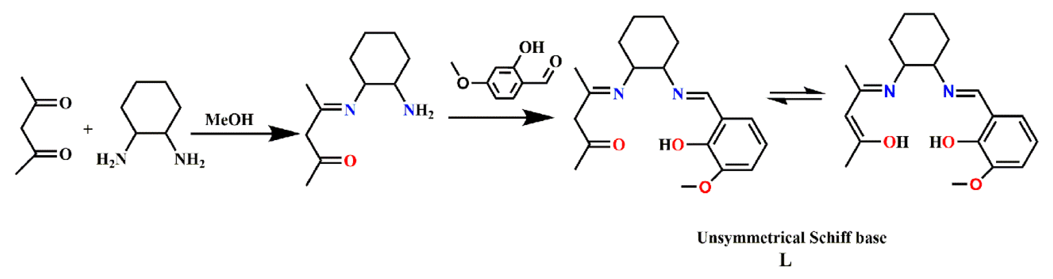
3.2. Synthesis of the Fluorescent Probe L

Here, 2,4-pentanedione (1.00 g, 10 mmol) in methanol (15 mL) was added dropwise to a methanol (20 mL) solution of (1R, 2R)-diaminocyclohexane (1.14 g, 10 mmol). The mixture was heated to reflux for 15 min, and cooled to room temperature. Then, 2-hydroxy-3-methoxybenzaldehyde (1.52 g, 10 mmol) was added and the solution was stirred for an additional 2 h at room temperature. It was filtered and the filtrate was left to stand overnight below 0 °C. The resulting yellow precipitate was collected by filtration to give the Schiff-base ligand L (2.67 g, 84%). The precipitate was recrystallized from methanol. X-ray quality yellow single crystals of ligand L were obtained by slow evaporation of a saturated methanol solution. 1H-NMR (600 MHz, DMSO-TMS): δ/ppm 13.49 (s, 1H, Ar–OH), 10.74 (s, 1H, (C=C)OH), 8.39 (s, 1H, CH=N), 7.03 (d, J = 7.8 Hz, 2H, Ar–H), 6.94 (d, J = 7.8 Hz, 2H, Ar–H), 6.80 (t, J = 7.8 Hz, 1H, Ar–H), 4.74 (s, 1H, CH=C), 3.77 (s, 3H, CH3–O), 3.61 (m, 1H, Cy–H–N), 3.10 (m, 1H, Cy–H–N), 1.81 (s, 3H, CH3(C=N)), 1.78 (s, 3H, CH3(C=C)), 1.95−1.37 (m, 8H, Cy−CH2). 13C-NMR (150.9 MHz, DMSO-TMS): δ/ppm 193.2 (C=C–OH), 165.6 ( CH3–C=N), 162.3 (Ar–C=N), 151.0 (Ar–C–OH), 148.0 (Ar–C–CH3), 123.2 (Ar–C–(C=N)), 118.4 (Ar–C), 118.1 (Ar–C), 114.8 (Ar–C), 94.7 (C=C–OH), 72.5 (Cy–C–N), 55.9 (Cy–C–N), 55.8 (CH3–O), 32.9, 32.6, 28.6, 24.2 (Cy–CH2), 23.5 (CH3(C=C)), 18.8 (CH3(C=N)). ESI-MS m/z: calcd. for [C19H26N2O3 + H]+, 331.2016; found, 331.2011. Elemental analysis calcd. (%) for C19H26N2O3 [330.19 g/mol]: C 68.85, H 8.21, N 8.45. Found (%): C 68.56, H 8.47, N 8.73.

3.3. X-ray Crystallography

Diffraction data for the probe L, Cu-complex, and the Zn-Cu complex were collected on a Bruker SMART APEX II diffractometer at room temperature (298 K) with graphite-monochromated Mo Kα radiation (λ = 0.71073 Å). An empirical absorption correction using SADABS was applied for all data [68]. The structures were solved and refined to convergence on F2 for all independent reflections by the full-matrix least squares method using the SHELXL−2014 programs [69] and OLEX2 1.2 [70]. Hydrogen atoms bonded to carbons were included in idealized geometric positions with thermal parameters equivalent to 1.2 times those of the atom to which they were attached. In compound L, one oxygen atom in the solvent water was disordered and there was a large amount of disorder in the structure. In particular, the disordered side-chains are very dynamic and may be considered as a solvent. Short contacts between disordered fragments are to be expected, which caused the observed level B alerts. Crystallographic data and refinement details for L, Cu-complex, and the Zn-Cu complex are given in Table S1, Supplementary Materials. CCDC: 2067620, L; 2067621, Cu-complex; and 2067622, Zn-Cu complex contain the supplementary crystallographic data for this paper. These data can be obtained free of charge from the Cambridge Crystallographic Data Centre: www.ccdc.cam.ac.uk/data_request/cif.

3.4. General Procedure for Analysis

Before conducting the spectroscopic measurements, the corresponding solutions of probe L and the metal ions were freshly prepared. For fluorescence spectroscopy selective experiments, test solutions were prepared as follows: a stock solution of probe L (1 mM) and the reactive species (1 mM) was prepared in ethanol solution and 0.15 mL of probe L and 0.15 mL of the reactive species were placed into a 3 mL cuvette and then after diluting the solution to 3 mL with ethanol solution; the final concentration was 50 μM. The fluorescence spectra were collected at room temperature.
For fluorescence spectroscopy competitive experiments, test solutions were prepared as follows: a stock solution of probe L (1 mM) and the reactive species (1 mM) was prepared in ethanol solution and 0.15 mL of probe L and 0.15 mL of the reactive species were placed into 3 mL cuvette and mixed. Next, 0.15 mL of the Zn2+ solution was added to the above mixed solution, which was then diluted to 3 mL with ethanol. Fluorescence spectra were recorded at room temperature.
For UV-Vis and fluorescence spectroscopy titrations experiments, a stock solution of the probe L (1 mM) was prepared and 0.15 mL of the probe L solution was placed into 3 mL cuvette. Adding 15–450 µL Zn2+ solution to the above cuvette, and then diluting the solution to 3 mL with different amounts of ethanol solution. UV-Vis and fluorescence spectra were taken at room temperature.
For the Job’s plot measurement, the total concentration of the probe L and Zn2+ was kept at 50 µmol. L and the ratio of the concentration of Zn2+ to the concentration of the probe L was changed to 0:10, 1:9, 2:8, 3:7, 4:6, 5:5, 6:4, 7:3, 8:2, 9:1, and 10:0, with the fluorescence intensity at 475 nm and the ultraviolet absorption intensity at 310 nm as the vertical axis Zn2+ occupies the probe and the molar ratio of Zn2+ is on the horizontal axis. The spectra of these solutions were immediately recorded by means of the UV-Vis method and fluorescence spectroscopy.

4. Conclusions

In summary, a new, low-cost, rapid, and portable Schiff base fluorescent probe L was synthesized and characterized. A “turn-on” fluorescence emission was observed upon sequential addition of Zn2+ and this increases proportionally with increased Zn2+ concentration. Meanwhile, it was found that probe L has great fluorescence selectivity for Zn2+ over many other important metal ions. Additionally, the method of equivalent molarity of fluorescence and UV spectrum indicated a 1:1 binding mode between L and Zn2+. Finally, the formation of the Cu-Zn complex and the strong quenching behavior of coexisting Cu2+ for Zn2+ may lead to the potential application as an on-off chemosensor candidate for Cu2+.

Supplementary Materials

The following are available online: Figure S1: 1H-NMR spectrum of L, Figure S2: 13C-NMR spectrum of L, Figure S3: HR ESI-MS spectrum of probe L, Figure S4: Absorbance of L as a function of [Zn2+]/[L], Figure S5: Job’s plot of probe L with Zn2+. Figure S6: 1H-NMR spectra of probe L with 0–1 equiv. of ZnCl2, Table S1: Crystallographic data and refinement details, Table S2: Selected bond lengths and bond angles for complex L, Cu-complex and Cu-Zn complex.

Author Contributions

L.-Y.S. and X.-L.C. carried out the experiments; H.X., X.Z., and Y.-L.H. analyzed the experiment data; L.-Y.S. and X.-J.Y. analyzed the X-ray structure; C.R. and Q.-L.Z. wrote the paper. All authors have read and agreed to the published version of the manuscript.

Funding

This work was supported by the National Natural Science Foundation of China (22065009, 22066007), the Guizhou Provincial Natural Science Foundation (grant number [2019] 2792], grant number [2018] 5779-14 and 19NSP035) and Guizhou Medical University Startup Project of Doctor Scientific Research (grant number YJ2020-BK024).

Data Availability Statement

No new data were created or analyzed in this study. Data sharing is not applicable to this article.

Acknowledgments

Carl Redshaw thanks the EPSRC for an Overseas Travel Grant (EP/R023816/1).

Conflicts of Interest

The authors declare no conflict of interest.

Sample Availability

Samples of the compounds L, Cu-complex, and Zn-Cu complex are available from the authors.

References

  1. Que, E.L.; Domaille, D.W.; Chang, C.J. Metals in neurobiology: Probing their chemistry and biology with molecular imaging. Chem. Rev. 2008, 108, 1517–1549. [Google Scholar] [PubMed]
  2. Hagimori, M.; Mizuyama, N.; Tominaga, Y.; Mukai, T.; Saji, H. A low-molecular-weight fluorescent sensor with Zn2+ dependent bathochromic shift of emission wavelength and its imaging in living cells. Dyes Pigm. 2015, 113, 205–209. [Google Scholar] [CrossRef]
  3. Na, Y.J.; Hwang, I.H.; Jo, H.Y.; Lee, S.A.; Park, G.J.; Kim, C. Fluorescent chemosensor based-on the combination of julolidine and furan for selective detection of zinc ion. Inorg. Chem. Commun. 2013, 35, 342–345. [Google Scholar] [CrossRef]
  4. Li, X.; Li, J.; Dong, X.; Gao, X.; Zhang, D.; Liu, C. A novel 3-Hydroxychromone fluorescence sensor for intracellular Zn2+ and its application in the recognition of prostate cancer cells. Sens. Actuators B Chem. 2017, 245, 129–136. [Google Scholar] [CrossRef]
  5. Falchuk, K.H. The molecular basis for the role of zinc in developmental biology. Mol. Cell. Biochem. 1998, 188, 41–48. [Google Scholar] [CrossRef]
  6. Outten, E.C.; Ohalloran, T.V. Femtomolar sensitivity of metalloregulatory proteins controlling zinc homeostasis. Science 2001, 292, 2488–2492. [Google Scholar] [CrossRef] [PubMed] [Green Version]
  7. Sirangelo, I.; Iannuzzi, C. The role of metal binding in the amyotrophic lateral sclerosis-related aggregation of copper-zinc superoxide dismutase. Molecules 2017, 22, 1429. [Google Scholar] [CrossRef] [Green Version]
  8. Kim, S.K.; Lee, D.H.; Hong, J.I.; Yoon, J. Chemosensors for Pyrophosphate. Acc. Chem. Res. 2009, 42, 23. [Google Scholar] [CrossRef]
  9. Nikura, K.; Anslyn, E.V. Chemosensor ensemble with selectivity for inositol-trisphosphate. J. Am. Chem. Soc. 1998, 120, 8533. [Google Scholar] [CrossRef]
  10. Mathews, C.P.; van Holde, K.E. Biochemistry; Benjamin/Cummings Publishing Company, Inc.: Redwood City, CA, USA, 1990. [Google Scholar]
  11. Guo, Z.; Park, S.; Yoon, J.; Shin, I. Recent progress in the development of near-infrared fluorescent probes for bioimaging applications. Chem. Soc. Rev. 2014, 43, 16–29. [Google Scholar] [CrossRef]
  12. Li, Y.; Zhao, Q.; Yang, H.; Liu, S.-J.; Liu, X.-M.; Zhang, Y.; Hu, T.; Chen, J.; Chang, Z.; Bu, X. A new ditopic ratiometric receptor for detecting zinc and fluoride ions in living cells. Analyst 2013, 138, 5486–5494. [Google Scholar] [CrossRef]
  13. Nyren, P. Enzymatic method for continuous monitoring of DNA polymerase activity. Anal. Biochem. 1987, 167, 235–238. [Google Scholar] [CrossRef]
  14. Ni, X.l.; Zeng, X.; Redshaw, C.; Yamato, T. Ratiometric fluorescent receptors for both Zn2+ and H2PO4– ions based on a pyrenyl-linked triazole-modified homooxacalix[3]arene: A potential molecular traffic signal with an R-S latch logic circuit. J. Org. Chem. 2011, 76, 5696. [Google Scholar] [CrossRef] [PubMed]
  15. Gupta, V.K.; Mergu, N.; Singh, A.K. Fluorescent chemosensors for Zn2+ ions based on flavonol derivatives. Sens. Actuators B Chem. 2014, 202, 674–682. [Google Scholar] [CrossRef]
  16. Mcquade, L.E.; Lippard, S.J. Cell-trappable quinoline-derivatized fluoresceins for selective and reversible biological Zn(II) detection. Inorg. Chem. 2010, 49, 9535–9545. [Google Scholar] [CrossRef] [PubMed] [Green Version]
  17. Park, S.Y.; Yoon, J.H.; Hong, C.S.; Souane, R.; Kim, J.S.; Matthews, S.E.; Vicens, J. A pyrenyl-appended triazole-based calix[4]arene as a fluorescent sensor for Cd2+ and Zn2+. J. Org. Chem. 2008, 73, 8212–8218. [Google Scholar] [CrossRef] [PubMed]
  18. Tayade, K.; Sahoo, S.K.; Bondhopadhyay, B.; Bhardwaj, V.K.; Singh, N.; Basu, A.; Bendre, R.; Kuwar, A. Highly selective turn-on fluorescent sensor for nanomolar detection of biologically important Zn2+ based on isonicotinohydrazide derivative: Application in cellular imaging. Biosens. Bioelectron. 2014, 61, 429–433. [Google Scholar] [CrossRef]
  19. Gulaboski, R.; Mirceski, V.; Scholz, F. An electrochemical method for determination of the standard Gibbs energy of anion transfer between water and n-octanol. Electrochem. Commun. 2002, 4, 277–283. [Google Scholar] [CrossRef] [Green Version]
  20. Ghaedi, M.; Ahmadi, F.; Shokrollahi, A. Simultaneous preconcentration and determination of copper, nickel, cobalt and lead ions content by flame atomic absorption spectrometry. J. Hazard. Mater. 2007, 142, 272–278. [Google Scholar] [CrossRef]
  21. Khorrami, A.R.; Fakhari, A.R.; Shamsipur, M.; Naeimi, H. Pre-concentration of ultra trace amounts of copper, zinc, cobalt and nickel in environmental water samples using modified C18 extraction disks and determination by inductively coupled plasma–optical emission spectrometry. Int. J. Environ. Anal. Chem. 2009, 89, 319–329. [Google Scholar] [CrossRef]
  22. Wan, J.; Duan, W.; Kai, C.; Tao, Y.; Dang, J.; Zeng, K.; Ge, Y.; Jiang, W.; Dan, L. Selective and sensitive detection of Zn(II) ion using a simple peptide-based sensor. Sens. Actuators 2018, 255, 49–56. [Google Scholar] [CrossRef]
  23. Kim, C.; Kim, B.; Park, K.H.; Kim, K.; Lee, D.; Yang, S.; Joo, N. Effects of short-term chromium supplementation on insulin sensitivity and body composition in overweight children: Randomized, double-blind, placebo-controlled study. J. Nutr. Biochem. 2011, 22, 1030–1034. [Google Scholar] [CrossRef] [PubMed]
  24. Wang, W.; Zhang, Y.; Li, Y.; Zhao, Q. Easily accessible and highly selective “Turn-on” fluorescent sensor for imaging cadmium in living cells. Chem. Res. Chin. Univ. 2013, 29, 632–637. [Google Scholar] [CrossRef]
  25. Zhou, Y.; Jung, J.Y.; Jeon, H.R.; Kim, Y.; Kim, S.-J.; Yoon, J. A novel supermolecular tetrameric vanadate-selective colorimetric and “off–on” sensor with pyrene ligand. Org. Lett. 2011, 13, 2742–2745. [Google Scholar] [CrossRef]
  26. Erdemir, B.S. Tabakci, Selective and sensitive fluorescein-benzothiazole based fluorescent sensor for Zn2+ ion in aqueous media. J. Fluoresc. 2017, 27, 2145–2152. [Google Scholar] [CrossRef] [PubMed]
  27. Tang, X.; Han, J.; Wang, Y.; Bao, X.; Ni, L.; Wang, L.; Li, L. A highly sensitive turn-on fluorescent chemosensor for recognition of Zn2+ and Hg2+ and applications. Spectrochim. Acta A 2017, 184, 177–183. [Google Scholar] [CrossRef]
  28. Santoro, O.; Zhang, X.; Redshaw, C. Synthesis of Biodegradable Polymers: A review on the use of Schiff-base metal complexes as catalysts for the ring opening polymerization (ROP) of cyclic esters. Catalysts 2020, 10, 800. [Google Scholar] [CrossRef]
  29. Epstein, D.M.; Choudhary, S.; Churchill, M.R.; Keil, K.M.; Eliseev, A.V.; Morrow, J.R. Chloroform-soluble Schiff-base Zn(II) or Cd(II) complexes from a dynamic combinatorial library. Inorg. Chem. 2001, 40, 1591–1596. [Google Scholar] [CrossRef]
  30. Raman, N.; Fathima, S.S.A.; Raja, J.D. Designing, synthesis and spectral characterization of Schiff base transition metal complexes: DNA cleavage and antimicrobial activity studies. J. Serb. Chem. Soc. 2008, 73, 1063–1071. [Google Scholar] [CrossRef]
  31. Zhao, Q.; Li, R.F.; Xing, S.K.; Liu, X.M.; Hu, T.L.; Bu, X.H. A highly selective on/off fluorescence sensor for cadmium(II). Inorg. Chem. 2011, 50, 10041–10046. [Google Scholar] [CrossRef]
  32. Aoki, S.; Kagata, D.; Shiro, M.; Takeda, K.; Kimura, E. Metal chelation-controlled twisted intramolecular charge transfer and its application to fluorescent sensing of metal ions and anions. J. Am. Chem. Soc. 2004, 126, 13377–13390. [Google Scholar] [CrossRef] [PubMed]
  33. Lim, N.C.; Schuster, J.V.; Porto, M.C. Coumarin-based chemosensors for Zinc(II):  toward the determination of the design algorithm for CHEF-type and ratiometric probes. Inorg. Chem. 2005, 44, 2018–2030. [Google Scholar] [CrossRef] [PubMed]
  34. Parkesh, R.; Lee, T.C.; Gunnlaugsson, T. Highly selective 4-amino-1,8-naphthalimide based fluorescent photoinduced electron transfer (PET) chemosensors for Zn(ii) under physiological pH conditions. Org. Biomol. Chem. 2007, 5, 310–317. [Google Scholar] [CrossRef]
  35. Dong, Y.; Fan, R.; Chen, W.; Wang, P.; Yang, Y. A simple quinolone Schiff-base containing CHEF based fluorescence ‘turn-on’ chemosensor for distinguishing Zn2+ and Hg2+ with high sensitivity, selectivity and reversibility. Dalton Trans. 2017, 46, 6769–6775. [Google Scholar] [CrossRef]
  36. Chakraborty, S.; Lohar, S.; Dhara, K.; Ghosh, R.; Dam, S.; Zangrando, E.; Chattopadhyay, P. A new half-condensed Schiff base platform: Structures and sensing of Zn2+ and H2PO4− ions in an aqueous medium. Dalton Trans. 2020, 49, 8991–9001. [Google Scholar] [CrossRef]
  37. Dong, W.-K.; Akogun, S.F.; Zhang, Y.; Sun, Y.-X.; Dong, X.-Y. A reversible “turn-on” fluorescent sensor for selective detection of Zn2+. Sens. Actuators B Chem. 2017, 238, 723–734. [Google Scholar] [CrossRef]
  38. Guo, W.T.; Peng, Y.D.; Zhang, Y.; Zhang, H.; Jiang, Y.L. Novel Tridentate Bisoxime Chemosensor for Selective Recognition of Cu2+ and Zn2+ with Different Mechanisms. J. Appl. Spectrosc. 2021, 88, 452–460. [Google Scholar] [CrossRef]
  39. Zhu, J.; Zhang, Y.; Chen, Y.; Sun, T.; Tang, Y.; Huang, Y.; Yang, Q.; Ma, D.; Wang, Y.; Wang, M. A Schiff base fluorescence probe for highly selective turn-on recognition of Zn2+. Tetrahedron Lett. 2017, 58, 365–370. [Google Scholar] [CrossRef]
  40. Lopez, J.; Mintz, E.A.; Hsu, F.-L.; Bu, X.R. Novel unsymmetric chiral Schiff bases possessing two different donor moieties: Unique tetradentate ligands from combination of salicylaldehyde and acetylacetone units. Tetrahedron: Asymmetry 1998, 9, 3741–3744. [Google Scholar] [CrossRef]
  41. Chen, X.Q.; Zhou, Y.; Peng, X.J.; Yoon, J.Y. Fluorescent and colorimetric probes for detection of thiols. Chem. Soc. Rev. 2010, 39, 2120. [Google Scholar] [CrossRef]
  42. An, B.K.; Kwon, S.K.; Jung, S.D.; Park, S.Y. Enhanced Emission and Its Switching in Fluorescent Organic Nanoparticles. J. Am. Chem. Soc. 2002, 124, 14410–14415. [Google Scholar] [CrossRef]
  43. Chung, S.K.; Tseng, Y.R.; Chen, C.Y.; Sun, S.S. A Selective Colorimetric Hg2+ Probe Featuring a Styryl Dithiaazacrown Containing Platinum(II) Terpyridine Complex through Modulation of the Relative Strength of ICT and MLCT Transitions. Inorg. Chem. 2011, 50, 2711–2713. [Google Scholar] [CrossRef]
  44. Ashokkumar, P.; Ramakrishnan, V.T.; Ramamurthy, P. Photoinduced Electron Transfer (PET) Based Zn2+ Fluorescent Probe: Transformation of Turn-On Sensors into Ratiometric Ones with Dual Emission in Acetonitrile. J. Phys. Chem. A 2011, 115, 14292–14299. [Google Scholar] [CrossRef]
  45. Liu, S.L.; Li, D.; Zhang, Z.; Prakash, G.K.S.; Conti, P.S.; Li, Z.B. Efficient synthesis of fluorescent-PET probes based on [18F]BODIPY dye. Chem. Commun. 2014, 50, 7371–7373. [Google Scholar] [CrossRef] [PubMed]
  46. Wu, J.S.; Liu, W.M.; Ge, J.C.; Zhang, H.Y.; Wang, P.F. New sensing mechanisms for design of fluorescent chemosensors emerging in recent years. Chem. Soc. Rev. 2011, 40, 3483–3495. [Google Scholar] [CrossRef] [PubMed]
  47. Wen, X.; Wang, Q.; Fan, Z. Highly selective turn-on fluorogenic chemosensor for Zn(II) detection based on aggregation-induced emission. J. Lumin. 2018, 194, 366–373. [Google Scholar] [CrossRef]
  48. Pasha, S.S.; Yadav, H.R.; Choudhury, A.R.; Laskar, I.R. Synthesis of an aggregation-induced emission (AIE) active salicylaldehyde based Schiff base: Study of mechanoluminescence and sensitive Zn(ii) sensing. J. Mater. Chem. C 2017, 5, 9651–9658. [Google Scholar] [CrossRef]
  49. Sun, Y.Q.; Wang, P.; Liu, J.; Zhang, J.Y.; Guo, W. A fluorescent turn-on probe for bisulfite based on hydrogen bond-inhibited C[double bond, length as m-dash]N isomerization mechanism. Analyst 2012, 137, 3430–3433. [Google Scholar] [CrossRef]
  50. Wang, P.; Liu, J.; Lv, X.; Liu, Y.L.; Zhao, Y.; Guo, W. A Naphthalimide-Based Glyoxal Hydrazone for Selective Fluorescence Turn-On Sensing of Cys and Hcy. Org. Lett. 2012, 14, 520–523. [Google Scholar] [CrossRef] [PubMed]
  51. Jung, H.S.; Ko, K.C.; Lee, J.H.; Kim, S.H.; Bhuniya, S.; Lee, J.Y.; Kim, Y.; Kim, S.J.; Kim, J.S. Rationally Designed Fluorescence Turn-On Sensors: A New Design Strategy Based on Orbital Control. Inorg. Chem. 2010, 49, 8552–8557. [Google Scholar] [CrossRef]
  52. Shiraishi, Y.; Sumiya, S.; Hirai, T. Highly sensitive cyanide anion detection with a coumarin–spiropyran conjugate as a fluorescent receptor. Chem. Commun. 2011, 47, 4953–4955. [Google Scholar] [CrossRef] [PubMed] [Green Version]
  53. Tsui, Y.; Devaraj, S.; Yen, Y.-P. Azo dyes featuring with nitrobenzoxadiazole (NBD) unit: A new selective chromogenic and fluorogenic sensor for cyanide ion. Sens. Actuators B Chem. 2012, 161, 510–519. [Google Scholar] [CrossRef]
  54. Karmakar, M.; Chattopadhyay, S. Synthesis, structure and nitroaromatic sensing ability of a trinuclear zinc complex with a reduced Schiff base ligand: Assessment of the ability of the ligand to sense zinc ion. Polyhedron 2020, 187, 114639. [Google Scholar] [CrossRef]
  55. Das, A.; Jana, S.; Ghosh, A. Modulation of Nuclearity by Zn(II) and Cd(II) in Their Complexes with a Polytopic Mannich Base Ligand:  A Turn-On Luminescence Sensor for Zn(II) and Detection of Nitroaromatic Explosives by Zn(II) Complexes. Cryst. Growth Des. 2018, 18, 2335–2348. [Google Scholar] [CrossRef]
  56. Svoboda, I.; Arici, C.; Nazir, H.; Durmus, Z.; Atakol, O.; Fuess, H. {[μ-N,N′-Bis-(salicyl-idene)-2,2′-di-methyl-1,3-propane-di-amine](piperidine)copper(II)}di-bromo-zinc(II). Acta Cryst. 2001, E57, m584–m586. [Google Scholar]
  57. Tatar, L.; Atakol, O.; Ülkü, D.; Aksu, M. {[μ-Bis(salicyl-idene)-1,3-propanediaminato]-copper(II)}di chlorozinc(II). Acta Cryst. 1999, C55, 923–925. [Google Scholar]
  58. Yu, T.Z.; Ding, X.S.; Zhao, Y.L.; Qian, L.; Fan, D.W. Study on the interaction of transition ions with a open-chain crown ether Schiff base. Imaging Sci. Photochem. 2008, 26, 109–115. [Google Scholar]
  59. You, Z.-L.; Ni, L.-L.; Hou, P.; Zhang, J.-C.; Wang, C. Synthesis, crystal structures, and superoxide dismutase activity of two isostructural copper(II)–zinc(II) complexes derived from N,N′-bis(4-methoxysalicylidene)cyclohexane-1,2-diamine. J. Coord. Chem. 2010, 63, 515–523. [Google Scholar] [CrossRef]
  60. Ülkü, D.; Kaynak, F.B.; Atakol, O.; Aksu, M. Crystal Structure of {[μ-Bis(salicylidene)-1,3-propanediaminato]-copper(II)}dibromozinc(II). Anal. Sci. 2003, 19, 799–800. [Google Scholar] [CrossRef] [Green Version]
  61. Kim, K.B.; Kim, H.; Song, E.J.; Kim, S.; Noh, I.; Kim, C. A cap-type Schiff base acting as a fluorescence sensor for zinc(ii) and a colorimetric sensor for iron(ii), copper(ii), and zinc(ii) in aqueous media. Dalton Trans. 2013, 42, 16569–16577. [Google Scholar] [CrossRef]
  62. Lee, H.G.; Kim, K.B.; Park, G.J.; Na, Y.J.; Jo, H.Y.; Lee, S.A.; Kim, C. An anthracene-based fluorescent sensor for sequential detection of zinc and copper ions. Inorg. Chem. Commun. 2014, 39, 61–65. [Google Scholar] [CrossRef]
  63. Lee, J.J.; Lee, S.A.; Kim, H.; Nguyen, L.; Noh, I.; Kim, C. A highly selective CHEF-type chemosensor for monitoring Zn2+ in aqueous solution and living cells. RSC Adv. 2015, 5, 41905–41913. [Google Scholar] [CrossRef]
  64. Wu, J.; Liu, W.; Zhuang, X.; Wang, F.; Wang, P.; Tao, S.; Zhang, X.; Wu, S.; Lee, S. Fluorescence turn on of coumarin derivatives by metal cations:  A new signaling mechanism based on C=N isomerization. Org. Lett. 2007, 9, 33–36. [Google Scholar] [CrossRef] [PubMed]
  65. Pan, S.; Tang, H.; Song, Z.; Li, J.; Guo, Y. A novel dual channel fluorescent probe for Ca2+ and Zn2+ based on a coumarin Schiff base. Chin. J. Chem. 2017, 35, 1263–1269. [Google Scholar] [CrossRef]
  66. Ma, Y.; Wang, F.; Kambam, S.; Chen, X. A quinoline-based fluorescent chemosensor for distinguishing cadmium from zinc ions using cysteine as an auxiliary reagent. Sens. Actuators B Chem. 2013, 188, 1116–1122. [Google Scholar] [CrossRef]
  67. Li, H.; Gao, S.; Xi, Z. A colorimetric and “turn-on” fluorescent chemosensor for Zn(II) based on coumarin Shiff-base derivative. Inorg. Chem. Commun. 2009, 12, 300–303. [Google Scholar] [CrossRef]
  68. Sheldrick, G.M. Program SADABS: Area-Detector Absorption Correction; University of Göttingen: Göttingen, Germany, 1996. [Google Scholar]
  69. Sheldrick, G.M. Crystal structure of 8-[7,8-bis-(4-chloro-benzo-yl)-7H-cyclo penta-[a]ace-naphthylen-9-yl]naphthalene-1-carb-oxy-lic acid. Acta Crystallogr. Sect. C Cryst. Struct. Commun. 2015, 71, 3–8. [Google Scholar] [CrossRef] [PubMed]
  70. Dolomanov, O.V.; Bourhis, L.J.; Gildea, R.J.; Howard, J.A.K.; Puschmann, H. OLEX2: A complete structure solution, refinement and analysis program. J. Appl. Cryst. 2009, 42, 339–341. [Google Scholar] [CrossRef]
Scheme 1. Synthesis of Schiff base compound L.
Molecules 26 03825 sch001
Figure 1. (A) Fluorescence spectra of L (50 μmol/L) and upon the addition of salts (1 equiv.) of Li+, Na+, K+, Ag+, Mg2+, Ca2+, Ba2+, Al3+, Fe3+, Co2+, Ni2+, Cu2+, Zn2+, Pb2+, Cd2+, Hg2+, and Cr3+ in CH3CH2OH. (B) Black bar: Relative fluorescence of L with 1 equiv. of metal ions stated; Red bar: Relative fluorescence of L and 1 equiv. of Zn2+ with 1 equiv. of metal ions stated (Eex = 354 nm, Eem = 475 nm). (C) Photos of L responding to 17 kinds of other metal ions and Zn2+ under visible light (above) and UV light (below).
Molecules 26 03825 g001
Figure 2. (A) Fluorescent titration spectra of L (50 μmol/L) in the presence of different concentrations of ZnCl2. (B) The fluorescence at 475 nm of L as a function of the Zn2+ concentration. (C) Job’s plot for the determination of the stoichiometry of L and Zn2+ in ethanol, the total concentration of L and Zn2+ was 50 μM. (D) The Benesi-Hildebrand plot of 1 / (F − F0) versus 1 / [Zn2+].
Molecules 26 03825 g002
Figure 3. UV-Vis spectra of L (50 μmol/L) with increasing amounts of ZnCl2 (0–3 equiv.).
Molecules 26 03825 g003
Figure 4. (a) X-ray crystal structure of probe L; (b) X-ray crystal structure of Cu-complex. Thermal ellipsoids are set at the 30% probability level and all hydrogen atoms and solvent molecules were omitted for clarity.
Molecules 26 03825 g004
Figure 5. (A) X-ray crystal structure of the Cu-Zn complex (thermal ellipsoids are set at the 30% probability level); (B) ball-and-stick views of the dimer. All hydrogen atoms and solvent molecules were omitted for clarity.
Molecules 26 03825 g005
Scheme 2. The proposed receptor-metal chelation mechanism.
Molecules 26 03825 sch002
Table 1. Comparison of various fluorescent sensors for Zn2+.
CompoundSolventDetection LimitRef.
Molecules 26 03825 i001CH3OH/H2O2.84 × 10−7 M54
Molecules 26 03825 i002CH3OH7.74 × 10−7 M38
Molecules 26 03825 i003CH3OH/H2O4.80 × 10−7 M37
Molecules 26 03825 i004CH3OH7.69 × 10−9 M55
Molecules 26 03825 i005EtOH/HEPES buffer5.03 × 10−7 M39
Molecules 26 03825 i006DMSO/water~1.00 × 10−8 M35
Molecules 26 03825 i007EtOH9.53 × 10−8 MPresent work
Publisher’s Note: MDPI stays neutral with regard to jurisdictional claims in published maps and institutional affiliations.

