
Modification of Alginates to Modulate Their Physic-Chemical Properties and Obtain Biomaterials with Different Functional Properties


Abstract
:1. Alginates General Information and Their Structure
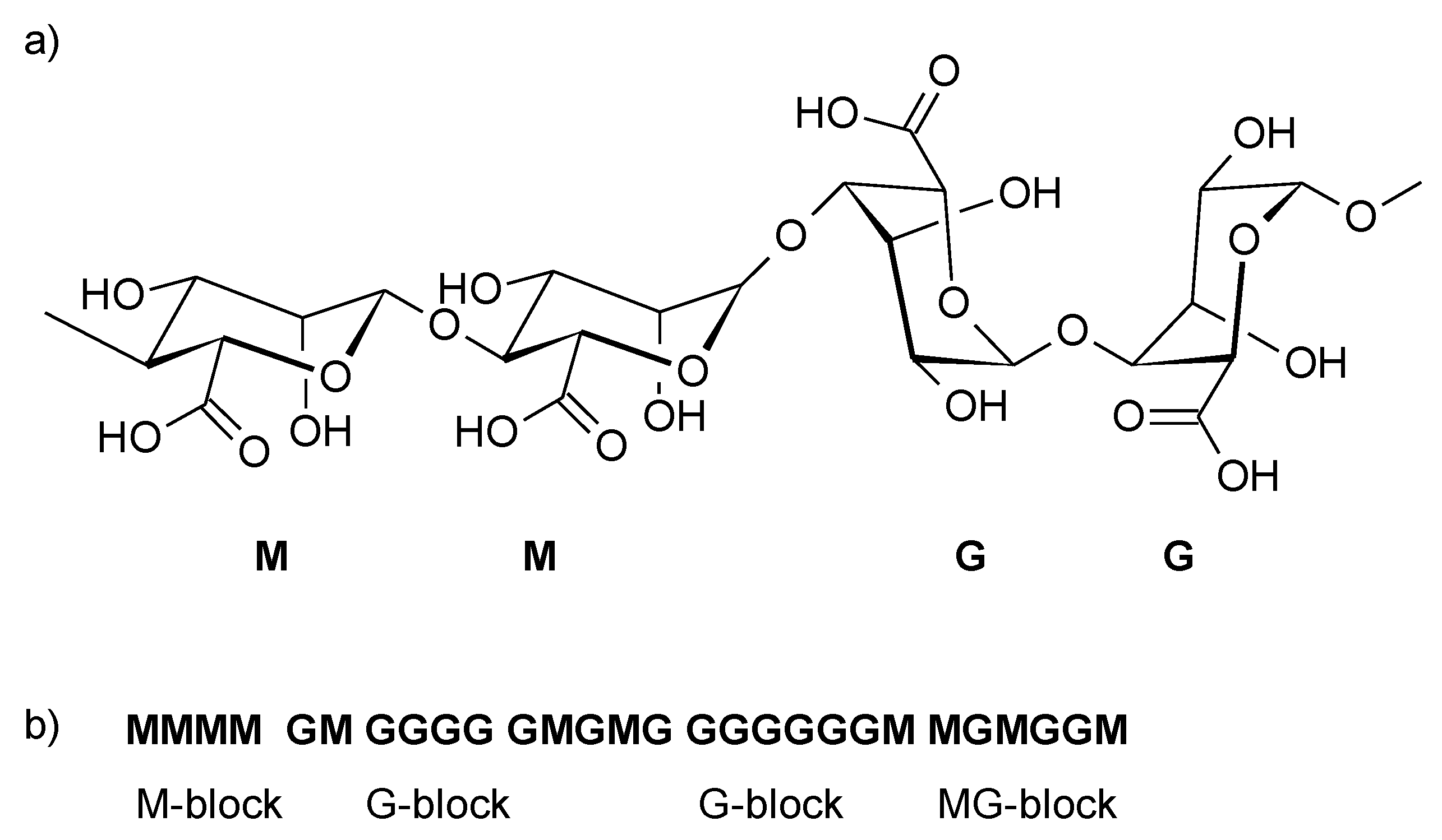
The Structure of Alginates
2. Modulating the Physical Properties of Alginates
3. Chemical Properties of Alginates and Modification Methods
3.1. Reaction of Hydroxyl Groups
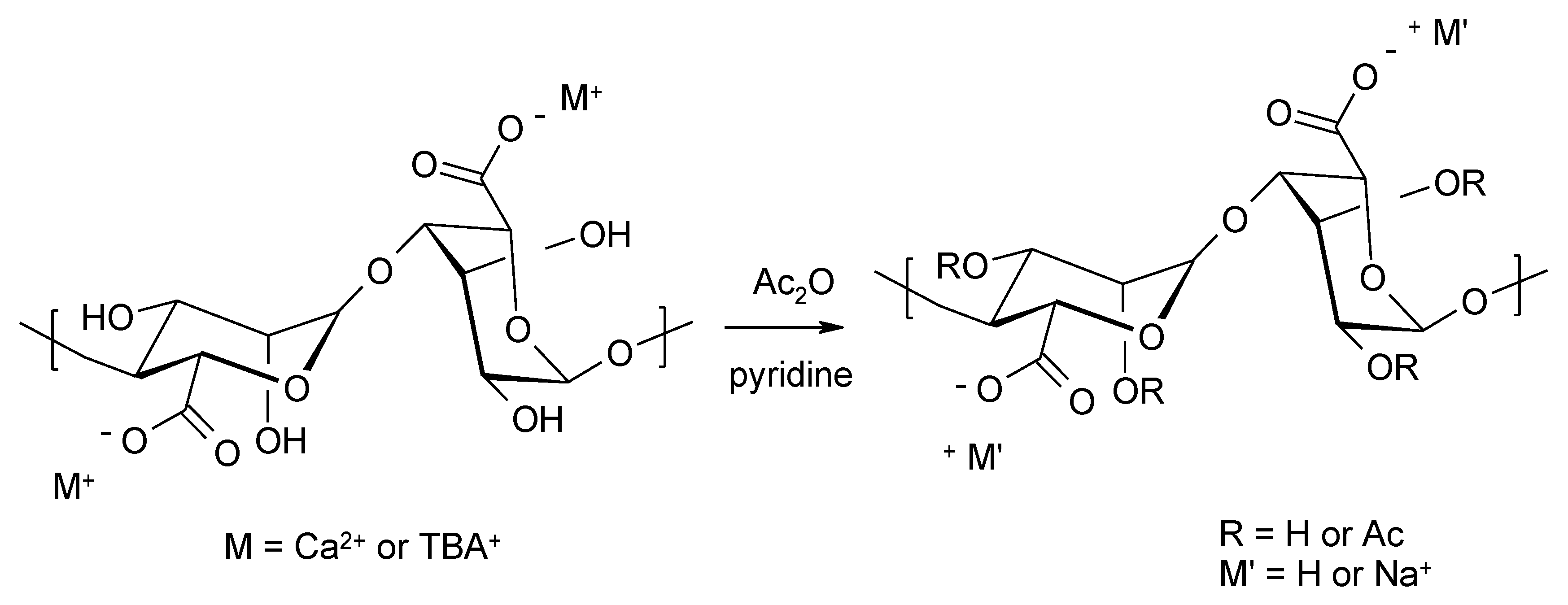
3.1.1. Acylation of Hydroxyl Groups
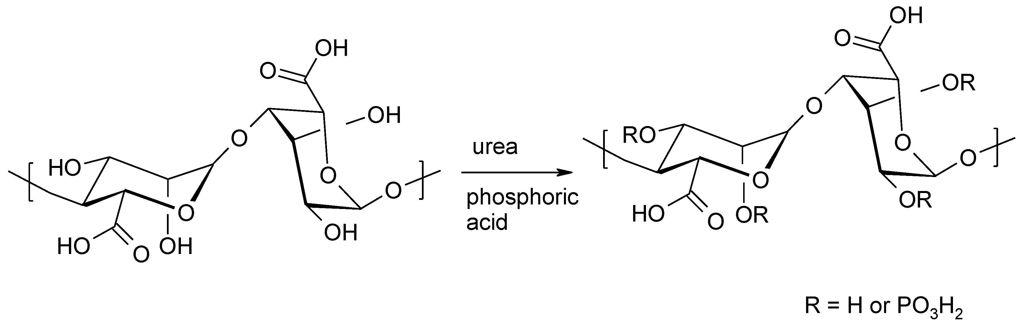
3.1.2. Phosphorylation of Alginates
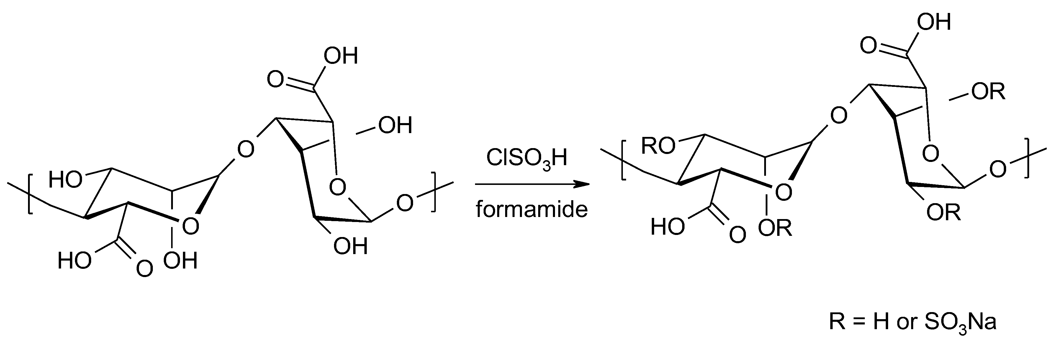
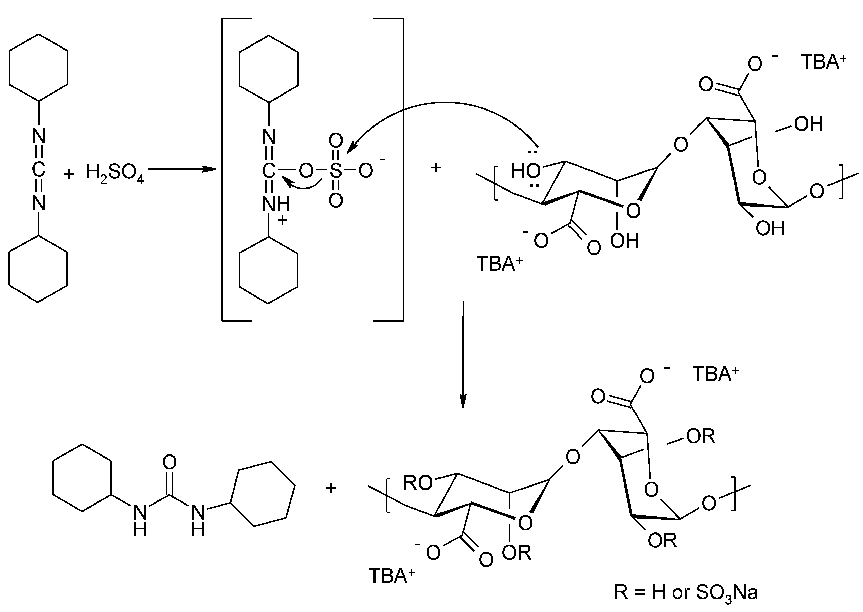
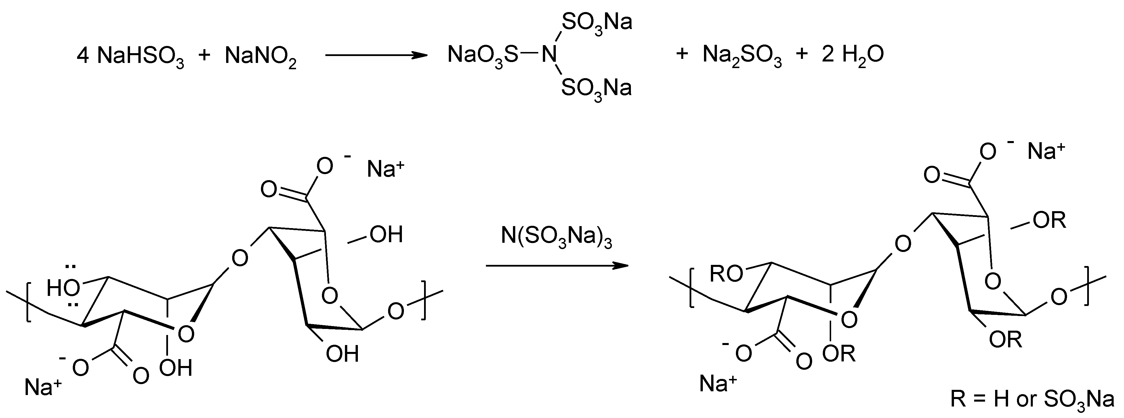
3.1.3. Incorporation of Sulphate Residues into Alginates
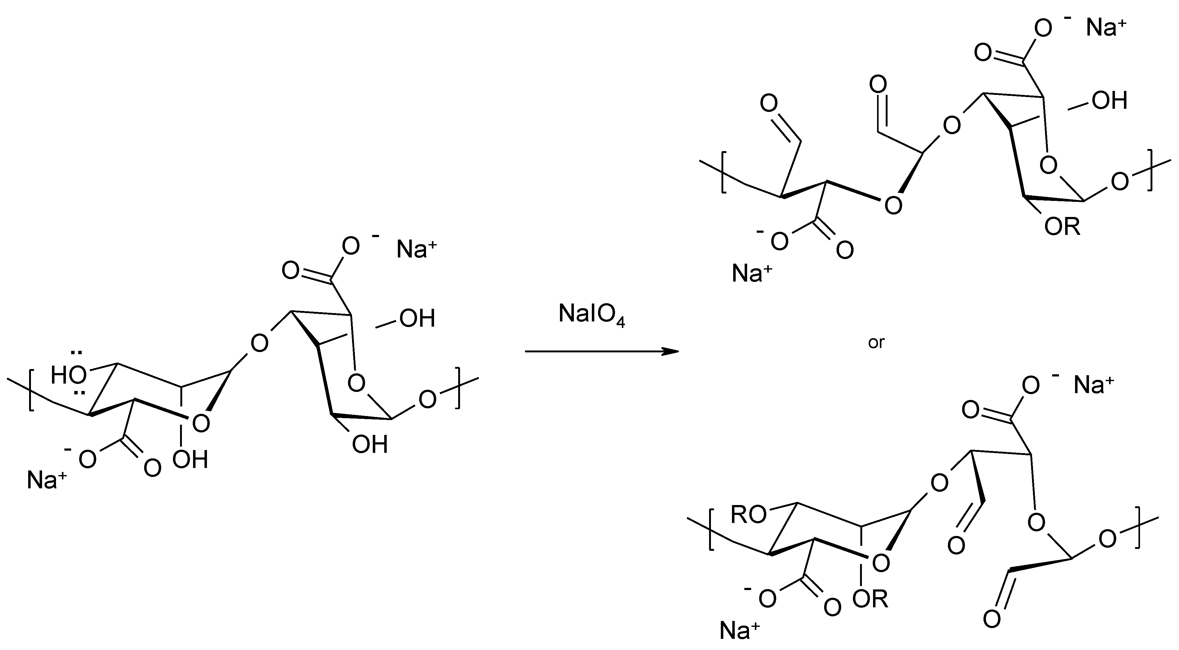
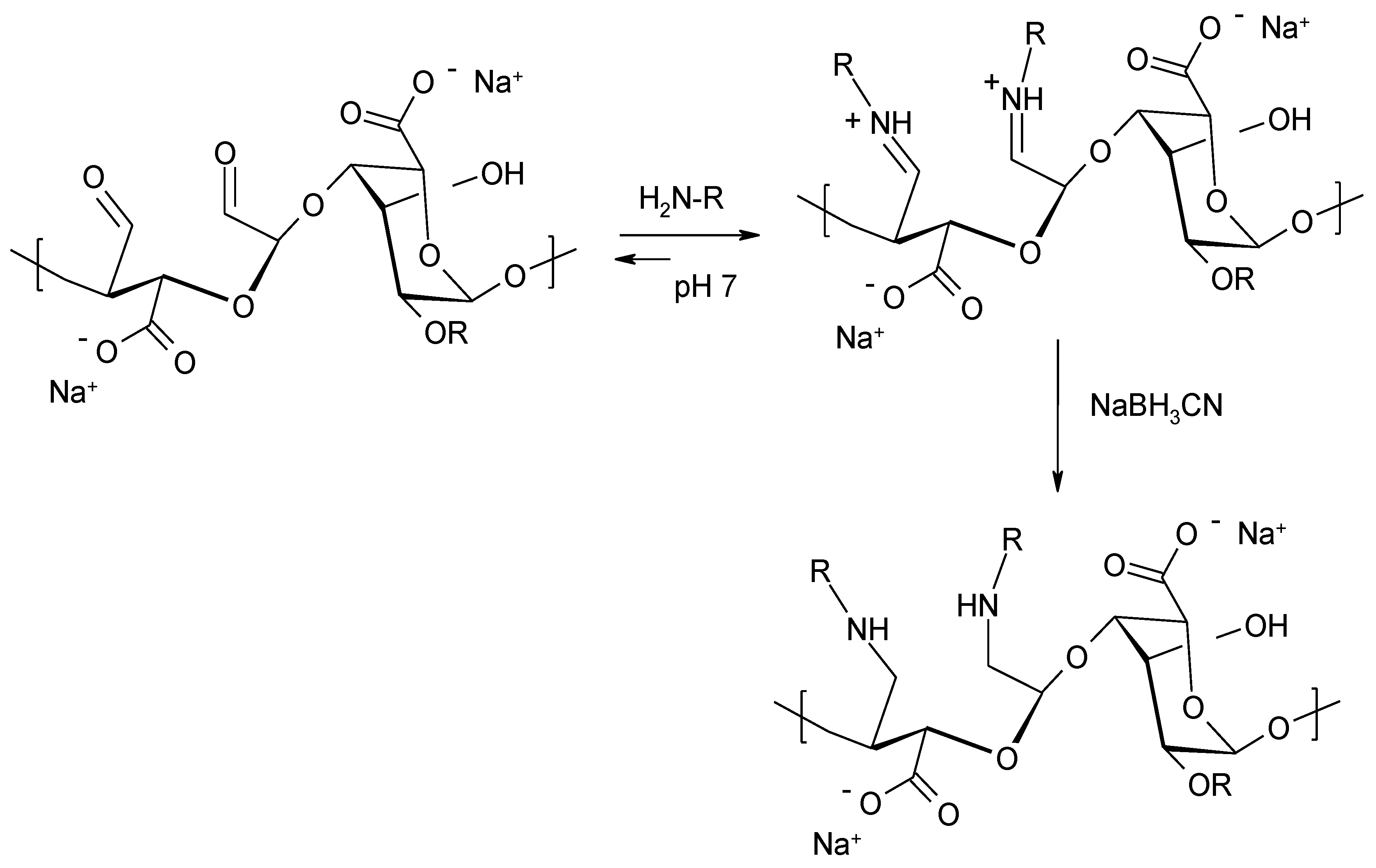
3.2. Oxidation of Alginates and Use of Carbonyl Groups for Further Functionalization
3.3. Chemical Modification of Alginated via Click Chemistry Reactions
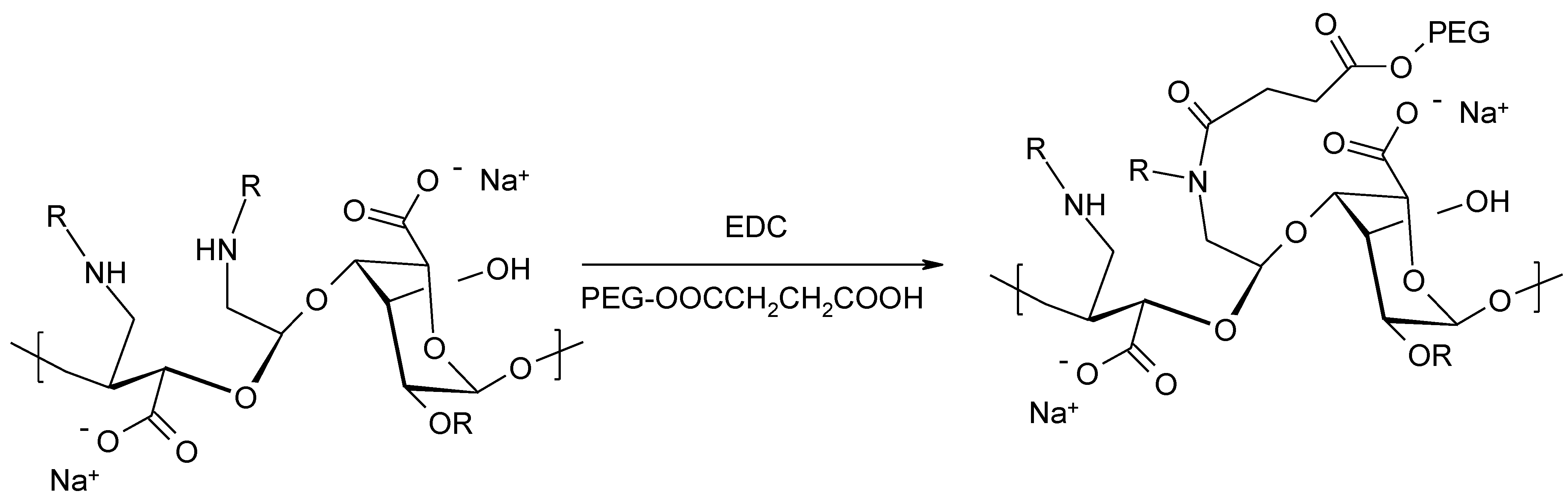
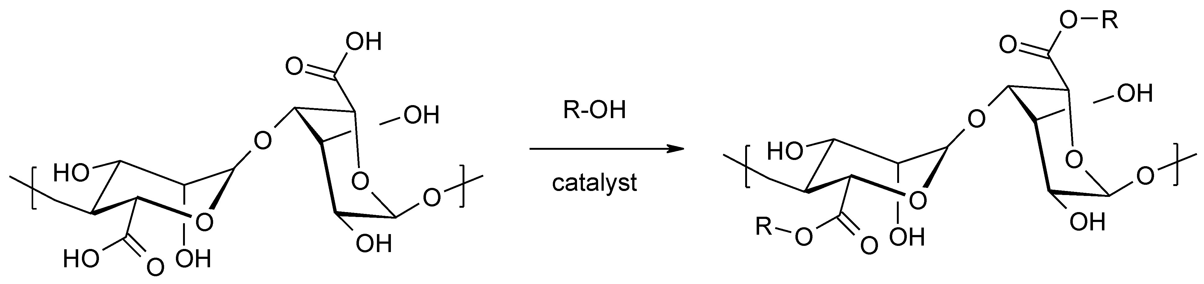
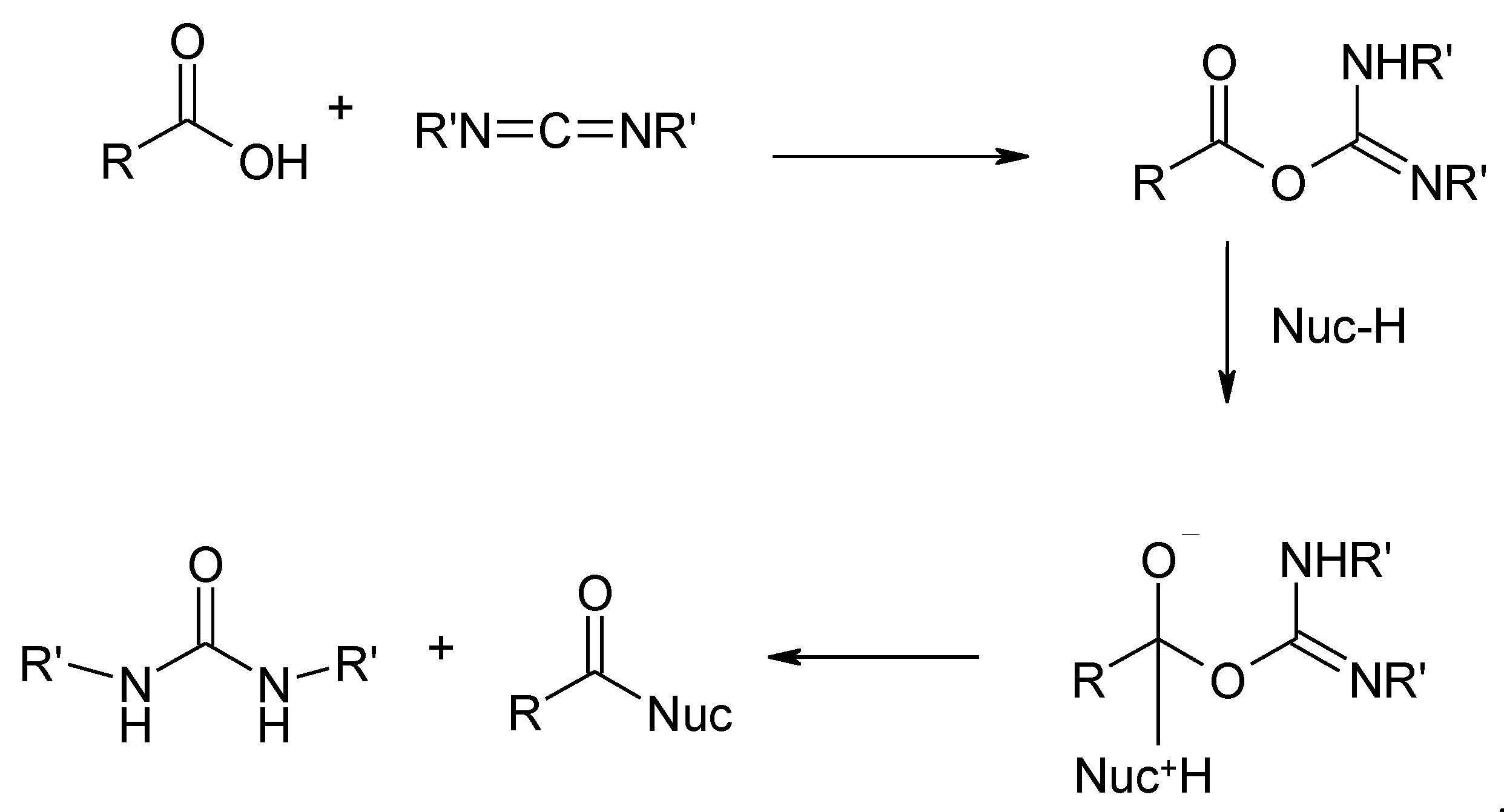
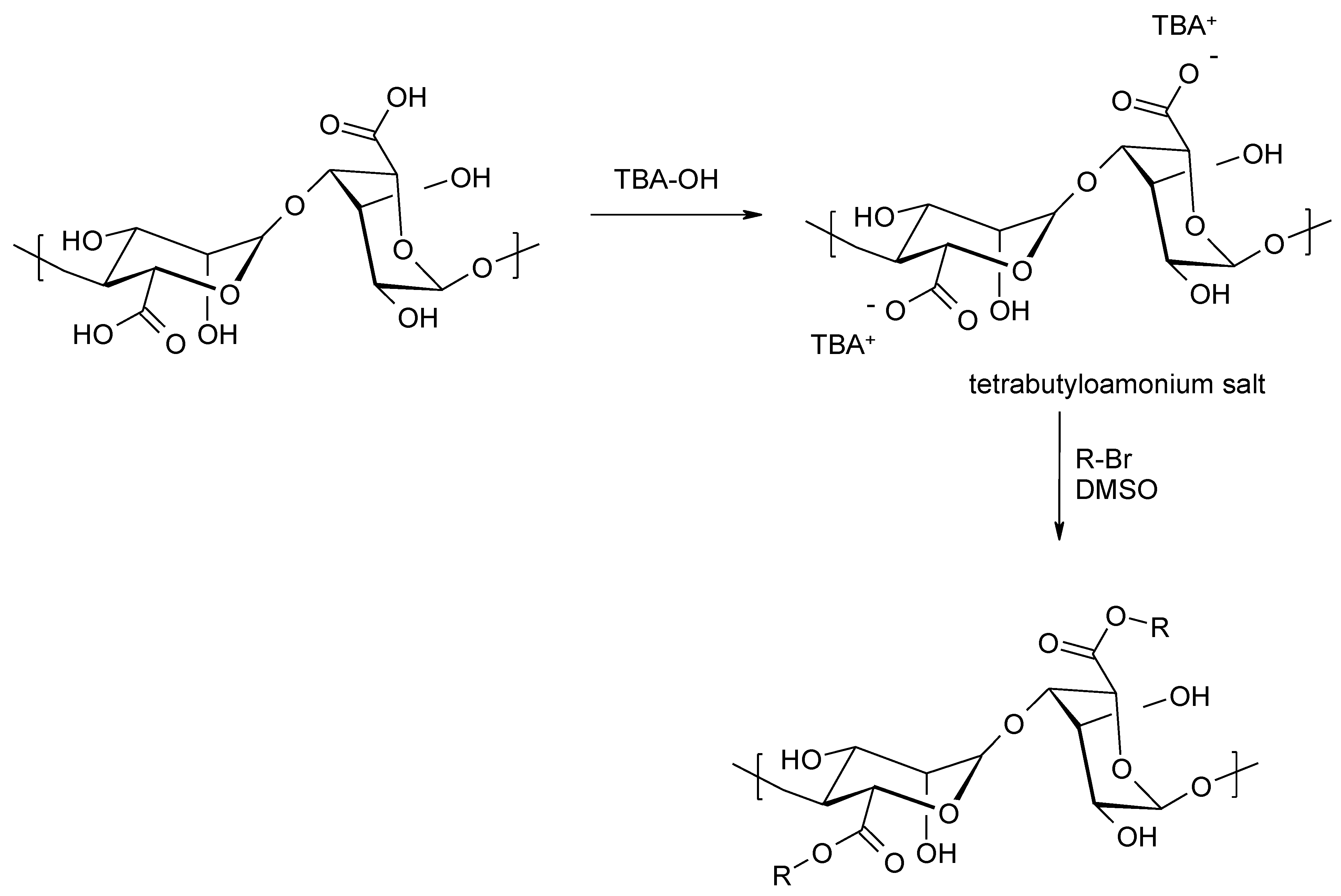
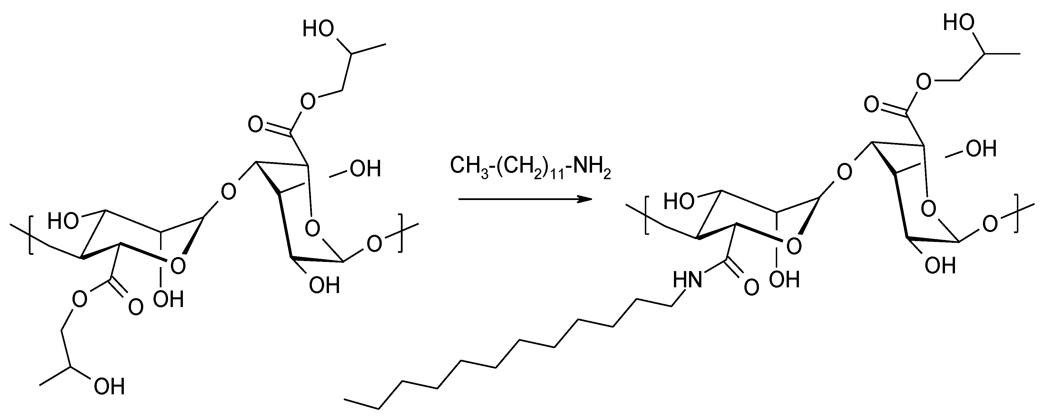
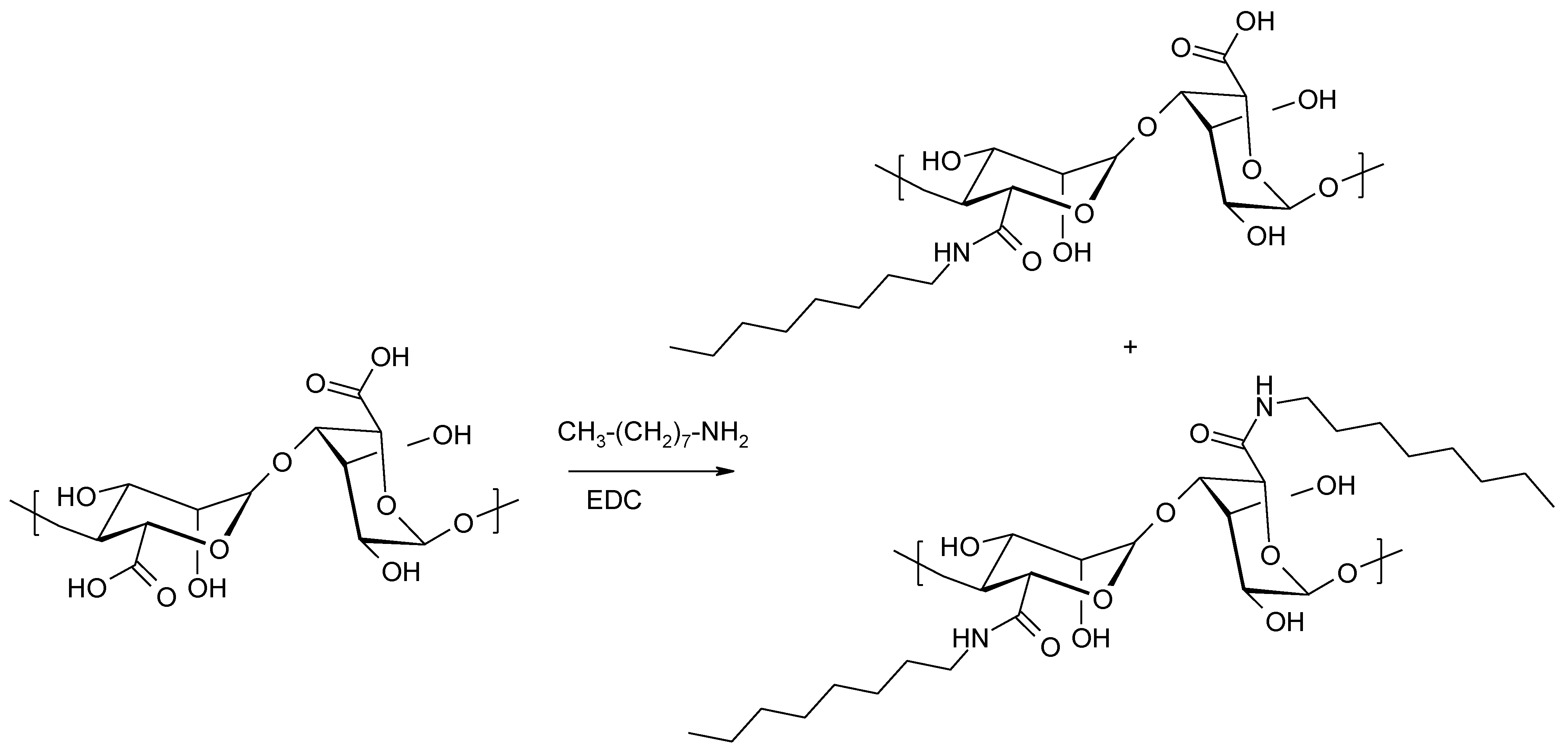
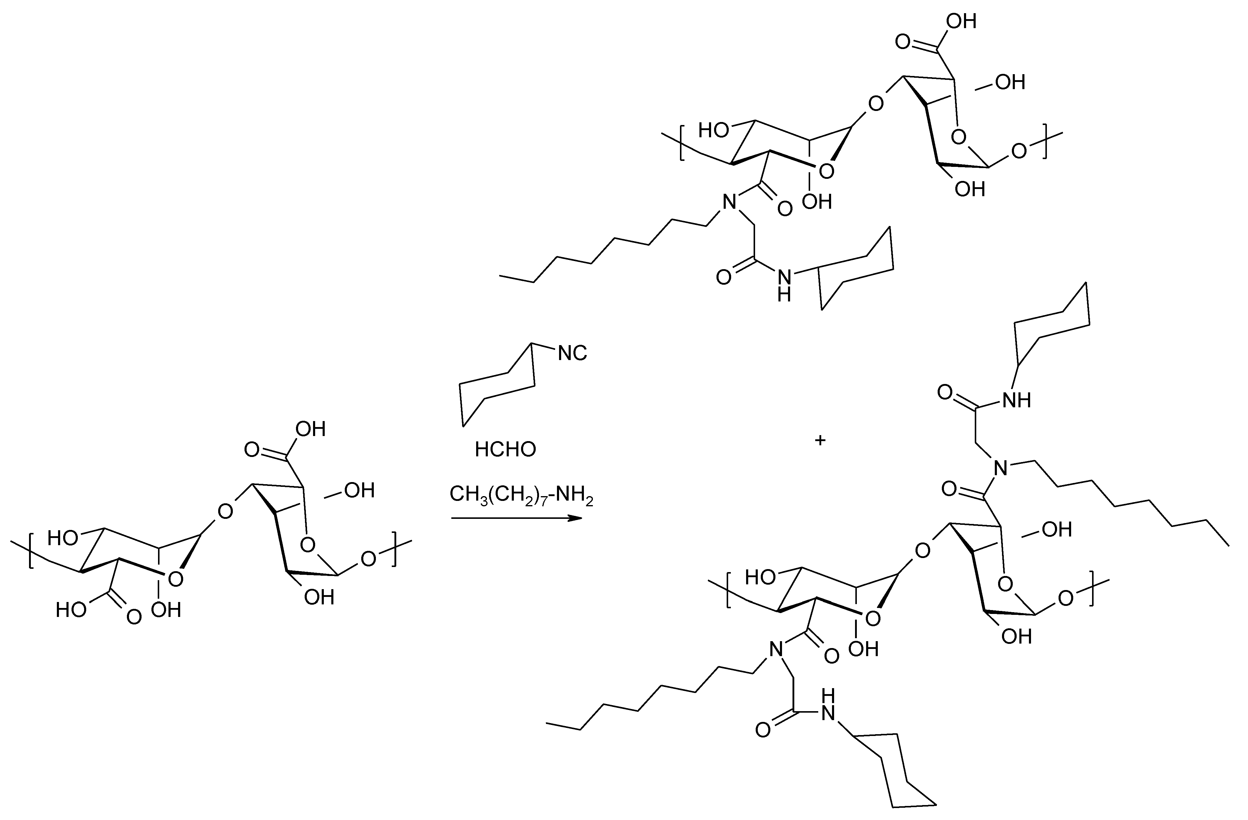
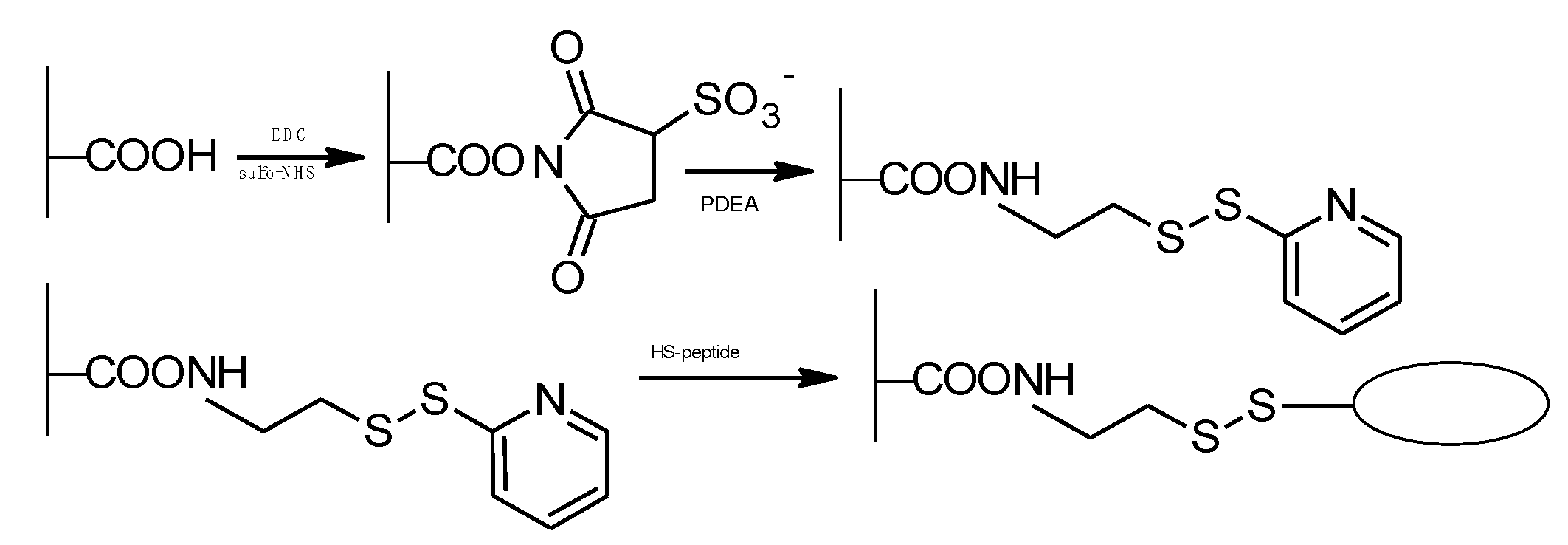
3.4. Chemical Modification of Carboxyl Groups
Reactions Using the Carboxyl Functions of Alginic Acid to Attach Biologically Active Ligands
4. Application of Alginate-Based Materials
- Drugs administered orally (Gastrotuss baby syrup [180], Algicid suspension/tablets [181], Gaviscon Double Action Liquid [182] and Tablets [183]), creating a mechanical barrier between the stomach and esophagus that prevents reflux, choking, dysphagia, heartburn, belching, and irritability, which accelerates the movement of the stomach and regenerates the mucous membranes of the esophagus
- Materials applied to the skin (Flaminal Forte gel [184], Purilon Gel gel [185], Saf-Gel gel [186], Hyalogran dressing [187], SeaSorb dressing [188], Tromboguard dressing [189,190], Fibracol Plus dressing [191], Algivon dressing [192], Guardix-SG [193,194]), which affect dissolution of the dry layer and necrotic tissue, ensure a moist environment at the wound surface, have hemostatic and antibacterial activity, and influence tissue granulation, epithelialization, and healing
- Rectal agents (Natalsid suppositories [195]) used for chronic hemorrhoids, proctitis, and chronic anal fissures after rectal surgery
- Agents applied arthroscopically (ChondroArt 3D injection [199]), used in degenerative diseases of the joints and spine.
5. Summary
Author Contributions
Funding
Acknowledgments
Conflicts of Interest
References
- March, N.H. The Structure and Properties of Liquids. Phys. Bull. 1969, 20, 109. [Google Scholar] [CrossRef]
- McHugh, D.J. A Guide to the Seaweed Industry; Food and Agriculture Organization of the United Nations: Rome, Italy, 2003; pp. 39–50. [Google Scholar]
- Wasikiewicz, J.M.; Yoshii, F.; Nagasawa, N.; Wach, R.A.; Mitomo, H. Degradation of chitosan and sodium alginate by gamma radiation, sonochemical and ultraviolet methods. Radiat. Phys. Chem. 2005, 73, 287–295. [Google Scholar] [CrossRef]
- Draget, K.I. Handbook of Hydrocolloids (incl. Alginates); Woodhead Publishing Limited, Abington Hall, Granta Park, Great Abington: Cambridge, UK, 2009; pp. 807–825. [Google Scholar]
- Ghidoni, I.; Chlapanidas, T.; Bucco, M.; Crovato, F.; Marazzi, M.; Vigo, D.; Torre, M.L.; Faustini, M. Alginate cell encapsulation: New advances in reproduction and cartilage regenerative medicine. Cytotechnology 2008, 58, 49–56. [Google Scholar] [CrossRef] [PubMed] [Green Version]
- Lee, K.Y.; Mooney, D.J. Alginate: Properties and biomedical applications. Prog. Polym. Sci. 2012, 37, 106–126. [Google Scholar] [CrossRef] [PubMed] [Green Version]
- Zimmermann, H.; Shirley, S.G.; Zimmermann, U. Alginate-based encapsulation of cells: Past, present, and future. Curr. Diab. Rep. 2007, 7, 314–320. [Google Scholar] [CrossRef]
- Soon-Shiong, P.; Feldman, E.; Nelson, R.; Heintz, R.; Yao, Q.; Yao, Z.; Zheng, T.; Merideth, N.; Skjak-Braek, G.; Espevik, T. Long-term reversal of diabetes by the injection of immunoprotected islets. Proc. Natl. Acad. Sci. USA 1993, 90, 5843–5847. [Google Scholar] [CrossRef] [Green Version]
- Boateng, J.S.; Matthews, K.H.; Stevens, H.N.E.; Eccleston, G.M. Wound Healing Dressings and Drug Delivery Systems: A Review. J. Pharm. Sci. 2008, 97, 2892–2923. [Google Scholar] [CrossRef] [PubMed]
- Ramsey, D.M.; Wozniak, D.J. Understanding the control of Pseudomonas aeruginosa alginate synthesis and the prospects for management of chronic infections in cystic fibrosis. Mol. Microbiol. 2005, 56, 309–322. [Google Scholar] [CrossRef]
- Haug, A.; Larsen, B.; Smidsrød, O.; Møller, J.; Brunvoll, J.; Bunnenberg, E.; Djerassi, C.; Records, R. A Study of the Constitution of Alginic Acid by Partial Acid Hydrolysis. Acta Chem. Scand. 1966, 20, 183–190. [Google Scholar] [CrossRef]
- Haug, A.; Larsen, B.; Smidsrød, O.; Smidsrød, O.; Eriksson, G.; Blinc, R.; Paušak, S.; Ehrenberg, L.; Dumanović, J. Studies on the Sequence of Uronic Acid Residues in Alginic Acid. Acta Chem. Scand. 1967, 21, 691–704. [Google Scholar] [CrossRef] [Green Version]
- Draget, K.I.; Taylor, C. Chemical, physical and biological properties of alginates and their biomedical implications. Food Hydrocoll. 2011, 25, 251–256. [Google Scholar] [CrossRef]
- Qin, Y. Alginate fibres: An overview of the production processes and applications in wound management. Polym. Int. 2008, 57, 171–180. [Google Scholar] [CrossRef]
- Grasdalen, H.; Larsen, B.; Smidsrød, O. A p.m.r. study of the composition and sequence of uronate residues in alginates. Carbohydr. Res. 1979, 68, 23–31. [Google Scholar] [CrossRef]
- Penman, A.; Sanderson, G.R. A method for the determination of uronic acid sequence in alginates. Carbohydr. Res. 1972, 25, 273–282. [Google Scholar] [CrossRef]
- Smidsrød, O.; Whittington, S.G. Monte Carlo Investigation of Chemical Inhomogeneity in Polymers. Macromolecules 1969, 2, 42–44. [Google Scholar] [CrossRef]
- Painter, T.; Smidsrød, O.; Larsen, B.; Haug, A.; Paasivirta, J. A Computer Study of the Changes in Composition-Distribution Occurring during Random Depolymerization of a Binary Linear Heteropolysaccharide. Acta Chem. Scand. 1968, 22, 1637–1648. [Google Scholar] [CrossRef]
- Larsen, B.; Smidsrød, O.; Painter, T.; Haug, A.; Rasmussen, S.E.; Sunde, E.; Sørensen, N.A. Calculation of the Nearest-neighbour Frequencies in Fragments of Alginate from the Yields of Free Monomers after Partial Hydrolysis. Acta Chem. Scand. 1970, 24, 726–728. [Google Scholar] [CrossRef] [Green Version]
- Pawar, S.N.; Edgar, K.J. Chemical modification of alginates in organic solvent systems. Biomacromolecules 2011, 12, 4095–4103. [Google Scholar] [CrossRef] [PubMed]
- Pawar, S.N.; Edgar, K.J. Alginate derivatization: A review of chemistry, properties and applications. Biomaterials 2012, 33, 3279–3305. [Google Scholar] [CrossRef]
- Sikorski, P.; Mo, F.; Skjåk-Bræk, G.; Stokke, B.T. Evidence for Egg-Box-Compatible Interactions in Calcium−Alginate Gels from Fiber X-ray Diffraction. Biomacromolecules 2007, 8, 2098–2103. [Google Scholar] [CrossRef]
- Donati, I.; Holtan, S.; Mørch, Y.A.; Borgogna, M.; Dentini, M. New Hypothesis on the Role of Alternating Sequences in Calcium−Alginate Gels. Biomacromolecules 2005, 6, 1031–1040. [Google Scholar] [CrossRef] [PubMed]
- Mørch, Ý.A.; Donati, I.; Strand, B.L.; Skjåk-Bræk, G. Effect of Ca2+, Ba2+, and Sr2+ on alginate microbeads. Biomacromolecules 2006, 7, 1471–1480. [Google Scholar] [CrossRef] [PubMed]
- Dalheim, M.Ø.; Vanacker, J.; Najmi, M.A.; Aachmann, F.L.; Strand, B.L.; Christensen, B.E. Efficient functionalization of alginate biomaterials. Biomaterials 2016, 80, 146–156. [Google Scholar] [CrossRef] [PubMed]
- Grant, G.T.; Morris, E.R.; Rees, D.A.; Smith, P.J.C.; Thom, D. Biological interactions between polysaccharides and divalent cations: The egg-box model. FEBS Lett. 1973, 32, 195–198. [Google Scholar] [CrossRef] [Green Version]
- Boguń, M. Nanocomposite alginate fibers and composites with their participation for applications in biomaterial engineering. Sci. J. Lodz Univ. Technol. 2010, Z.389, 3–210. [Google Scholar]
- Skjåk-Bræk, G.; Grasdalen, H.; Smidsrød, O. Inhomogeneous polysaccharide ionic gels. Carbohydr. Polym. 1989, 10, 31–54. [Google Scholar] [CrossRef]
- Draget, K.I.; Skjåk Bræk, G.; Smidsrød, O. Alginic acid gels: The effect of alginate chemical composition and molecular weight. Carbohydr. Polym. 1994, 25, 31–38. [Google Scholar] [CrossRef]
- Aarstad, O.A.; Tøndervik, A.; Sletta, H.; Skjåk-Bræk, G. Alginate Sequencing: An Analysis of Block Distribution in Alginates Using Specific Alginate Degrading Enzymes. Biomacromolecules 2012, 13, 106–116. [Google Scholar] [CrossRef]
- Lang, Y.; Zhao, X.; Liu, L.; Yu, G. Applications of Mass Spectrometry to Structural Analysis of Marine Oligosaccharides. Mar. Drugs 2014, 12, 4005–4030. [Google Scholar] [CrossRef] [Green Version]
- Faidi, A.; Farhat, F.; Boina, D.A.; Touati, M.; Le-Nouen, D.; Stumbé, J.-F. Physico-chemical characterization of alginates isolated from a Tunisian Padina pavonica algae as a sustainable biomaterial. Polym. Int. 2020, 69, 1130–1139. [Google Scholar] [CrossRef]
- Usov, A.I. Alginic acids and alginates: Analytical methods used for their estimation and characterisation of composition and primary structure. Russ. Chem. Rev. 1999, 68, 957–966. [Google Scholar] [CrossRef]
- Cuong, D.X.; Thanh Tuyen, D.T.; Kieu, D.T. Biophysical-chemistry Characterization of Alginate in Brown Algae Species Sargassum dupplicatum. WJFST 2020, 4, 17–22. [Google Scholar] [CrossRef]
- Timell, T.E. The acid hydrolysis of glycosides: I. General conditions and the effect of the nature of aglycone. Can. J. Chem. 1964, 42, 1456–1472. [Google Scholar] [CrossRef]
- Smidsrød, O.; Haug, A.; Larsen, B.; von Hofsten, B.; Williams, D.H.; Bunnenberg, E.; Djerassi, C.; Records, R. The Influence of pH on the Rate of Hydrolysis of Acidic Polysaccharides. Acta Chem. Scand. 1966, 20, 1026–1034. [Google Scholar] [CrossRef] [PubMed] [Green Version]
- Preiss, J.; Ashwell, G. Polygalacturonic acid metabolism in bacteria. II. Formation and metabolism of 3-deoxy-D-glycero-2, 5-hexodiulosonic acid. J. Biol. Chem. 1963, 238, 1577–1583. [Google Scholar] [CrossRef]
- Tsujino, I.; Saito, T. A New Unsaturated Uronide isolated from Alginase Hydrolysate. Nature 1961, 192, 970–971. [Google Scholar] [CrossRef]
- Haug, A.; Larsen, B.; Smidsröd, O.; Munch-Petersen, J.; Munch-Petersen, J. The Degradation of Alginates at Different pH Values. Acta Chem. Scand. 1963, 17, 1466–1468. [Google Scholar] [CrossRef]
- Haug, A.; Larsen, B.; Smidsrød, O.; Haug, A.; Hagen, G. Alkaline Degradation of Alginate. Acta Chem. Scand. 1967, 21, 2859–2870. [Google Scholar] [CrossRef]
- Smidsrød, O.; Haug, A.; Larsen, B.; Alivaara, A.; Trætteberg, M. Degradation of Alginate in the Presence of Reducing Compounds. Acta Chem. Scand. 1963, 17, 2628–2637. [Google Scholar] [CrossRef]
- Smidsrød, O.; Haug, A.; Larsen, B.; Munch-Petersen, J.; Munch-Petersen, J. The Influence of Reducing Substances on the Rate of Degradation of Alginates. Acta Chem. Scand. 1963, 17, 1473–1474. [Google Scholar] [CrossRef]
- Leo, W.J.; McLoughlin, A.J.; Malone, D.M. Effects of sterilization treatments on some properties of alginate solutions and gels. Biotechnol. Prog. 1990, 6, 51–53. [Google Scholar] [CrossRef] [PubMed]
- Yang, J.S.; Xie, Y.J.; He, W. Research progress on chemical modification of alginate: A review. Carbohydr. Polym. 2011, 84, 33–39. [Google Scholar] [CrossRef]
- Chamberlain, N.H.; Cunningham, G.E.; Speakman, J.B. Alginic Acid Diacetate. Nature 1946, 158, 553. [Google Scholar] [CrossRef]
- Wassermann, A. Alginic acid acetate. J. Chem. Soc. 1948, 158, 197. [Google Scholar] [CrossRef]
- Schweiger, R.G. Acetylation of Alginic Acid. II. Reaction of Algin Acetates with Calcium and Other Divalent Ions. J. Org. Chem. 1962, 27, 1789–1791. [Google Scholar] [CrossRef]
- Skjåk-Bræk, G.; Paoletti, S.; Gianferrara, T. Selective acetylation of mannuronic acid residues in calcium alginate gels. Carbohydr. Res. 1989, 185, 119–129. [Google Scholar] [CrossRef]
- Köhler, S.; Heinze, T. New solvents for cellulose: Dimethyl sulfoxide/ammonium fluorides. Macromol. Biosci. 2007, 7, 307–314. [Google Scholar] [CrossRef] [PubMed]
- Chen, X.; Zhu, Q.; Liu, C.; Li, D.; Yan, H.; Lin, Q. 1Esterification of Alginate with Alkyl Bromides of Different Carbon Chain Lengths via the Bimolecular Nucleophilic Substitution Reaction: Synthesis, Characterization, and Controlled Release Performance. Polymers 2021, 13, 3351. [Google Scholar] [CrossRef]
- Yang, J.S.; Zhou, Q.Q.; He, W. Amphipathicity and self-assembly behavior of amphiphilic alginate esters. Carbohydr. Polym. 2013, 92, 223–227. [Google Scholar] [CrossRef]
- Li, Q.; Liu, C.G.; Huang, Z.H.; Xue, F.F. Preparation and characterization of nanoparticles based on hydrophobic alginate derivative as carriers for sustained release of vitamin D3. J. Agric. Food Chem. 2011, 59, 1962–1967. [Google Scholar] [CrossRef]
- Manohara, H.M.; Nayak, S.S.; Franklin, G.; Kotrappanavar Nataraj, S.; Mondal, D. Progress in marine derived renewable functional materials and biochar for sustainable water purification. Green Chem. 2021, 23, 8305–8331. [Google Scholar] [CrossRef]
- Linhardt, R.J.; Claude, S. Hudson Award Address in Carbohydrate Chemistry. Heparin: Structure and Activity. J. Med. Chem. 2003, 46, 2551–2564. [Google Scholar] [CrossRef]
- Kim, H.S.; Song, M.; Lee, E.J.; Shin, U.S. Injectable Hydrogels Derived from Phosphorylated Alginic Acid Calcium Complexes. Mater. Sci. Eng. C 2015, 51, 139–147. [Google Scholar] [CrossRef] [PubMed]
- Alban, S.; Schauerte, A.; Franz, G. Anticoagulant sulfated polysaccharides: Part I. Synthesis and structure-activity relationships of new pullulan sulfates. Carbohydr. Polym. 2002, 47, 267–276. [Google Scholar] [CrossRef]
- Freeman, I.; Kedem, A.; Cohen, S. Preparation and in vitro anticoagulant activities of alginate sulfate and its quaterized derivatives. Biomaterials 2008, 29, 3260–3268. [Google Scholar] [CrossRef] [PubMed]
- Fan, L.; Jiang, L.; Xu, Y.; Zhou, Y.; Shen, Y.; Xie, W.; Long, Z.; Zhou, J. Synthesis and anticoagulant activity of sodium alginate sulfates. Carbohydr. Polym. 2011, 83, 1797–1803. [Google Scholar] [CrossRef]
- Gama, C.I.; Tully, S.E.; Sotogaku, N.; Clark, P.M.; Rawat, M.; Vaidehi, N.; Goddard, W.A.; Nishi, A.; Hsieh-Wilson, L.C. Sulfation patterns of glycosaminoglycans encode molecular recognition and activity. Nat. Chem. Biol. 2006, 2, 467–473. [Google Scholar] [CrossRef] [PubMed] [Green Version]
- Freeman, I.; Kedem, A.; Cohen, S. The effect of sulfation of alginate hydrogels on the specific binding and controlled release of heparin-binding proteins. Biomaterials 2008, 29, 3260–3268. [Google Scholar] [CrossRef]
- Arlov, Ø.; Skjåk-Bræk, G. Sulfated Alginates as Heparin Analogues: A Review of Chemical and Functional Properties. Molecules 2017, 22, 778. [Google Scholar] [CrossRef] [Green Version]
- Zeng, Y.; Yang, D.; Qiu, P.; Han, Z.; Zeng, P.; He, Y.; Guo, Z.; Xu, L.; Cui, Y.; Zhou, Z.; et al. Efficacy of Heparinoid PSS in Treating Cardiovascular Diseases and beyond-A Review of 27 Years Clinical Experiences in China. Clin. Appl. Thromb. 2016, 22, 222–229. [Google Scholar] [CrossRef]
- Mhanna, R.; Kashyap, A.; Palazzolo, G.; Vallmajo-Martin, Q.; Becher, J.; Möller, S.; Schnabelrauch, M.; Zenobi-Wong, M. Chondrocyte Culture in Three Dimensional Alginate Sulfate Hydrogels Promotes Proliferation While Maintaining Expression of Chondrogenic Markers. Tissue Eng. Part A 2014, 20, 1454–1464. [Google Scholar] [CrossRef] [PubMed] [Green Version]
- Öztürk, E.; Arlov, Ø.; Aksel, S.; Ling, L.; Ornitz, D.M.; Skjåk-Bræk, G.; Zenobi-Wong, M. Sulfated Hydrogel Matrices Direct Mitogenicity and Maintenance of Chondrocyte Phenotype through Activation of FGF Signaling. Adv. Funct. Mater. 2016, 26, 3649–3662. [Google Scholar] [CrossRef] [PubMed] [Green Version]
- Ma, H.; Qiu, P.; Xin, M.; Xu, X.; Wang, Z.; Xu, H.; Yu, R.; Xu, X.; Zhao, C.; Wang, X.; et al. Structure-Activity Relationship of Propylene Glycol Alginate Sodium Sulfate Derivatives for Blockade of Selectins Binding to Tumor Cells. Carbohydr. Polym. 2019, 210, 225–233. [Google Scholar] [CrossRef]
- Lee, J.-B.; Takeshita, A.; Hayashi, K.; Hayashi, T. Structures and antiviral activities of polysaccharides from Sargassum trichophyllum. Carbohydr. Polym. 2011, 86, 995–999. [Google Scholar] [CrossRef]
- Ahmadi, A.; Moghadamtousi, S.Z.; Abubakar, S.; Zandi, K. Antiviral potential of algae polysaccharides isolated from marine sources: A review. BioMed Res. Int. 2015, 2015, 825203. [Google Scholar] [CrossRef] [Green Version]
- Wang, S.-X.; Zhang, X.-S.; Guan, H.-S.; Wang, W. Potential anti-HPV and related cancer agents from marine resources: An overview. Mar. Drugs 2014, 12, 2019–2035. [Google Scholar] [CrossRef] [PubMed] [Green Version]
- Xu, X.; Wu, X.; Wang, Q.; Cai, N.; Zhang, H.; Jiang, Z.; Wan, M.; Oda, T. Immunomodulatory effects of alginate oligosaccharides on murine macrophage RAW264.7 cells and their structure-activity relationships. J. Agric. Food Chem. 2014, 62, 3168–3176. [Google Scholar] [CrossRef]
- Meiyu, G.; Fuchuan, L.; Xianliang, X.; Jing, L.; Zuowei, Y.; Huashi, G. The potential molecular targets of marine sulfated polymannuroguluronate interfering with HIV-1 entry. Interaction between SPMG and HIV-1 rgp120 and CD4 molecule. Antivir. Res. 2003, 59, 127–135. [Google Scholar] [CrossRef]
- Son, E.H.; Moon, E.Y.; Rhee, D.K.; Pyo, S. Stimulation of various functions in murine peritoneal macrophages by high mannuronic acid–containing alginate (HMA) exposure in vivo. Int. Immunopharmacol. 2001, 1, 147–154. [Google Scholar] [CrossRef]
- He, S.L.; Zhang, M.; Geng, Z.J.; Yao, K.D. Preparation and Characterization of Partially Oxidized Sodium Alginate. Chin. J. Appl. Chem. 2005, 22, 1007–1011. [Google Scholar]
- Boontheekul, T.; Kong, H.-J.; Mooney, D.J. Controlling alginate gel degradation utilizing partial oxidation and bimodal molecular weight distribution. Biomaterials 2005, 26, 2455–2465. [Google Scholar] [CrossRef] [PubMed]
- Kong, H.J.; Kaigler, D.; Kim, K.; Mooney, D.J. Controlling Rigidity and Degradation of Alginate Hydrogels via Molecular Weight Distribution. Biomacromolecules 2004, 5, 1720–1727. [Google Scholar] [CrossRef] [PubMed]
- Gomez, C.G.; Rinaudo, M.; Villar, M.A. Oxidation of sodium alginate and characterization of the oxidized derivatives. Carbohydr. Polym. 2007, 67, 296–304. [Google Scholar] [CrossRef]
- Andresen, I.L.; Painter, T.; Smidsrød, O. Concerning the effect of periodate oxidation upon the intrinsic viscosity of alginate. Carbohydr. Res. 1977, 59, 563–566. [Google Scholar] [CrossRef]
- Carré, M.-C.; Delestre, C.; Hubert, P.; Dellacherie, E. Covalent coupling of a short polyether on sodium alginate: Synthesis and characterization of the resulting amphiphilic derivative. Carbohydr. Polym. 1991, 16, 367–379. [Google Scholar] [CrossRef]
- Kang, H.-A.; Jeon, G.-J.; Lee, M.-Y.; Yang, J.-W. Effectiveness test of alginate-derived polymeric surfactants. J. Chem. Technol. Biotechnol. 2002, 77, 205–210. [Google Scholar] [CrossRef]
- Kang, H.-A.; Shin, M.S.; Yang, J.-W. Preparation and characterization of hydrophobically modified alginate. Polym. Bull. 2002, 47, 429–435. [Google Scholar] [CrossRef]
- Li, Z.; Ni, C.; Xiong, C.; Li, Q. Preparation and drug release of hydrophobically modified alginate. Chem. Bull./Huaxue Tongbao 2009, 72, 93–96. [Google Scholar]
- Laurienzo, P.; Malinconico, M.; Motta, A.; Vicinanza, A. Synthesis and characterization of a novel alginate-poly(ethylene glycol) graft copolymer. Carbohydr. Polym. 2005, 62, 274–282. [Google Scholar] [CrossRef]
- Reakasame, S.; Boccaccini, A.R. Oxidized Alginate-Based Hydrogels for Tissue Engineering Applications: A Review. Biomacromolecules 2018, 19, 3–21. [Google Scholar] [CrossRef] [PubMed]
- Kumar, A.; Nutan, B.; Jewrajka, S.K. Modulation of Properties through Covalent Bond Induced Formation of Strong Ion Pairing between Polyelectrolytes in Injectable Conetwork Hydrogels. ACS Appl. Bio Mater. 2021, 4, 3374–3387. [Google Scholar] [CrossRef]
- Teng, K.; An, Q.; Chen, Y.; Zhang, Y.; Zhao, Y. Recent Development of Alginate-Based Materials and Their Versatile Functions in Biomedicine, Flexible Electronics, and Environmental Uses. ACS Biomater. Sci. Eng. 2021, 7, 1302–1337. [Google Scholar] [CrossRef]
- Distler, T.; McDonald, K.; Heid, S.; Karakaya, E.; Detsch, R.; Boccaccini, A.R. Ionically and Enzymatically Dual Cross-Linked Oxidized Alginate Gelatin Hydrogels with Tunable Stiffness and Degradation Behavior for Tissue Engineering. ACS Biomater. Sci. Eng. 2020, 6, 3899–3914. [Google Scholar] [CrossRef] [PubMed]
- Wang, S.; Lei, J.; Yi, X.; Yuan, L.; Ge, L.; Li, D.; Mu, C. Fabrication of Polypyrrole-Grafted Gelatin-Based Hydrogel with Conductive, Self-Healing, and Injectable Properties. ACS Appl. Polym. Mater. 2020, 2, 3016–3023. [Google Scholar] [CrossRef]
- Soltan, N.; Ning, L.; Mohabatpour, F.; Papagerakis, P.; Chen, X. Printability and Cell Viability in Bioprinting Alginate Dialdehyde-Gelatin Scaffolds. ACS Biomater. Sci. Eng. 2019, 5, 2976–2987. [Google Scholar] [CrossRef] [PubMed]
- Mousavi, A.; Mashayekhan, S.; Baheiraei, N.; Pourjavadi, A. Biohybrid oxidized alginate/myocardial extracellular matrix injectable hydrogels with improved electromechanical properties for cardiac tissue engineering. Int. J. Biol. Macromol. 2021, 180, 692–708. [Google Scholar] [CrossRef] [PubMed]
- Distler, T.; Kretzschmar, L.; Schneidereit, D.; Girardo, S.; Goswami, R.; Friedrich, O.; Detsch, R.; Guck, J.; Boccaccini, A.R.; Budday, S. Mechanical properties of cell- and microgel bead-laden oxidized alginate-gelatin hydrogels. Biomater. Sci. 2021, 9, 3051–3068. [Google Scholar] [CrossRef] [PubMed]
- Kong, X.; Chen, L.; Li, B.; Quan, C.; Wu, J. Applications of oxidized alginate in regenerative medicine. J. Mater. Chem. B 2021, 9, 2785–2801. [Google Scholar] [CrossRef] [PubMed]
- Distler, T.; Lauria, I.; Detsch, R.; Sauter, C.M.; Bendt, F.; Kapr, J.; Rütten, S.; Boccaccini, A.R.; Fritsche, E. Neuronal Differentiation from Induced Pluripotent Stem Cell-Derived Neurospheres by the Application of Oxidized Alginate-Gelatin-Laminin Hydrogels. Biomedicines 2021, 9, 261. [Google Scholar] [CrossRef]
- Reakasame, S.; Jin, A.; Zheng, K.; Qu, M.; Boccaccini, A.R. Biofabrication and Characterization of Alginate Dialdehyde-Gelatin Microcapsules Incorporating Bioactive Glass for Cell Delivery Application. Macromol. Biosci. 2020, 20, 2000138. [Google Scholar] [CrossRef]
- Distler, T.; Solisito, A.A.; Schneidereit, D.; Friedrich, O.; Detsch, R.; Boccaccini, A.R. 3D printed oxidized alginate-gelatin bioink provides guidance for C2C12 muscle precursor cell orientation and differentiation via shear stress during bioprinting. Biofabrication 2020, 12, 045005. [Google Scholar] [CrossRef]
- Tallawi, M.; Germann, N. Self-crosslinked hydrogel with delivery carrier obtained by incorporation of oxidized alginate microspheres into gelatin matrix. Mater. Lett. 2020, 263, 127211. [Google Scholar] [CrossRef]
- Xiang, G.; Lippens, E.; Hafeez, S.; Duda, G.N.; Geissler, S.; Qazi, T.H. Oxidized alginate beads for tunable release of osteogenically potent mesenchymal stromal cells. Mater. Sci. Eng. C 2019, 104, 109911. [Google Scholar] [CrossRef] [PubMed]
- Deng, Y.; Shavandi, A.; Okoro, O.V.; Nie, L. Alginate modification via click chemistry for biomedical applications. Carbohydr. Polym. 2021, 270, 118360. [Google Scholar] [CrossRef] [PubMed]
- Fischer, A.; Houzelle, M.C.; Hubert, P.; Axelos, M.A.V.; Geoffroy-Chapotot, C.; Carré, M.C.; Viriot, M.L.; Dellacherie, E. Detection of Intramolecular Associations in Hydrophobically Modified Pectin Derivatives Using Fluorescent Probes. Langmuir 1998, 14, 4482–4488. [Google Scholar] [CrossRef]
- Leonard, M.; De Boisseson, M.R.; Hubert, P.; Dalençon, F.; Dellacherie, E. Hydrophobically modified alginate hydrogels as protein carriers with specific controlled release properties. J. Control. Release 2004, 98, 395–405. [Google Scholar] [CrossRef]
- Pelletier, S.; Hubert, P.; Payan, E.; Marchal, P.; Choplin, L.; Dellacherie, E. Amphiphilic derivatives of sodium alginate and hyaluronate for cartilage repair: Rheological properties. J. Biomed. Mater. Res. 2001, 54, 102–108. [Google Scholar] [CrossRef]
- Broderick, E.; Lyons, H.; Pembroke, T.; Byrne, H.; Murray, B.; Hall, M. The characterisation of a novel, covalently modified, amphiphilic alginate derivative, which retains gelling and non-toxic properties. J. Colloid Interface Sci. 2006, 298, 154–161. [Google Scholar] [CrossRef]
- Yang, L.; Zhang, B.; Wen, L.; Liang, Q.; Zhang, L.M. Amphiphilic cholesteryl grafted sodium alginate derivative: Synthesis and self-assembly in aqueous solution. Carbohydr. Polym. 2007, 68, 218–225. [Google Scholar] [CrossRef]
- Pelletier, S.; Hubert, P.; Lapicque, F.; Payan, E.; Dellacherie, E. Amphiphilic derivatives of sodium alginate and hyaluronate: Synthesis and physico-chemical properties of aqueous dilute solutions. Carbohydr. Polym. 2000, 43, 343–349. [Google Scholar] [CrossRef]
- Babak, V.G.; Skotnikova, E.A.; Lukina, I.G.; Pelletier, S.; Hubert, P.; Dellacherie, E. Hydrophobically Associating Alginate Derivatives: Surface Tension Properties of Their Mixed Aqueous Solutions with Oppositely Charged Surfactants. J. Colloid Interface Sci. 2000, 225, 505–510. [Google Scholar] [CrossRef] [PubMed]
- Rastello De Boisseson, M.; Leonard, M.; Hubert, P.; Marchal, P.; Stequert, A.; Castel, C.; Favre, E.; Dellacherie, E. Physical alginate hydrogels based on hydrophobic or dual hydrophobic/ionic interactions: Bead formation, structure, and stability. J. Colloid Interface Sci. 2004, 273, 131–139. [Google Scholar] [CrossRef] [PubMed]
- Pawar, S.N.; Edgar, K.J. Alginate esters via chemoselective carboxyl group modification. Carbohydr. Polym. 2013, 98, 1288–1296. [Google Scholar] [CrossRef] [PubMed]
- Sinquin, A.; Hubert, P.; Dellacherie, E. Amphiphilic derivatives of alginate: Evidence for intra- and intermolecular hydrophobic associations in aqueous solution. Langmuir 1993, 9, 3334–3337. [Google Scholar] [CrossRef]
- Sinquin, A.; Hubert, P.; Marchal, P.; Choplin, L.; Dellacherie, E. Rheological properties of semi-dilute aqueous solutions of hydrophobically modified propylene glycol alginate derivatives. Colloids Surf. A Physicochem. Eng. Asp. 1996, 112, 193–200. [Google Scholar] [CrossRef]
- Sinquin, A.; Houzelle, M.C.; Hubert, P.; Choplin, L.; Viriot, M.L.; Dellacherie, E. Amphiphilic Derivatives of Propylene Glycol Alginate: A Revisit of Their Physicochemical Behavior in Dilute Aqueous Solution. Langmuir 1996, 12, 3779–3782. [Google Scholar] [CrossRef]
- Sinquin, A.; Hubert, P.; Dellacherie, E. Intermolecular associations in hydrophobically modified derivatives of propyleneglycol alginate. Polymer 1994, 35, 3557–3560. [Google Scholar] [CrossRef]
- Maestro, A.; González, C.; Gutiérrez, J.M. Rheological behavior of hydrophobically modified hydroxyethyl cellulose solutions: A linear viscoelastic model. J. Rheol. 2002, 46, 127–143. [Google Scholar] [CrossRef] [Green Version]
- Galant, C.; Kjøniksen, A.-L.; Nguyen, G.T.M.; Knudsen, K.D.; Nyström, B. Altering Associations in Aqueous Solutions of a Hydrophobically Modified Alginate in the Presence of β-Cyclodextrin Monomers. J. Phys. Chem. B 2006, 110, 190–195. [Google Scholar] [CrossRef]
- Gomez, C.G.; Chambat, G.; Heyraud, A.; Villar, M.; Auzély-Velty, R. Synthesis and characterization of a β-CD-alginate conjugate. Polymer 2006, 47, 8509–8516. [Google Scholar] [CrossRef]
- Polyak, B.; Geresh, S.; Marks, R.S. Synthesis and Characterization of a Biotin-Alginate Conjugate and Its Application in a Biosensor Construction. Biomacromolecules 2004, 5, 389–396. [Google Scholar] [CrossRef] [PubMed]
- Abulateefeh, S.R.; Khanfar, M.A.; Al Bakain, R.Z.; Taha, M.O. Synthesis and characterization of new derivatives of alginic acid and evaluation of their iron(III)-crosslinked beads as potential controlled release matrices. Pharm. Dev. Technol. 2014, 19, 856–867. [Google Scholar] [CrossRef]
- Zhu, H.; Ji, J.; Lin, R.; Gao, C.; Feng, L.; Shen, J. Surface engineering of poly(dl-lactic acid) by entrapment of alginate-amino acid derivatives for promotion of chondrogenesis. Biomaterials 2002, 23, 3141–3148. [Google Scholar] [CrossRef]
- Abu-Rabeah, K.; Polyak, B.; Ionescu, R.E.; Cosnier, S.; Marks, R.S. Synthesis and Characterization of a Pyrrole−Alginate Conjugate and Its Application in a Biosensor Construction. Biomacromolecules 2005, 6, 3313–3318. [Google Scholar] [CrossRef]
- Vallée, F.; Müller, C.; Durand, A.; Schimchowitsch, S.; Dellacherie, E.; Kelche, C.; Cassel, J.C.; Leonard, M. Synthesis and rheological properties of hydrogels based on amphiphilic alginate-amide derivatives. Carbohydr. Res. 2009, 344, 223–228. [Google Scholar] [CrossRef] [PubMed]
- Labre, F.; Mathieu, S.; Chaud, P.; Morvan, P.-Y.; Vallée, R.; Helbert, W.; Fort, S. DMTMM-mediated amidation of alginate oligosaccharides aimed at modulating their interaction with proteins. Carbohydr. Polym. 2018, 184, 427–434. [Google Scholar] [CrossRef]
- Wang, G.; Zhu, J.; Chen, X.; Dong, H.; Li, Q.; Zeng, L.; Cao, X. Alginate based antimicrobial hydrogels formed by integrating Diels–Alder “click chemistry” and the thiolene reaction. RSC Adv. 2018, 8, 11036–11042. [Google Scholar] [CrossRef] [Green Version]
- Bu, H.; Nguyen, G.T.M.; Kjøniksen, A.-L. Effects of the Quantity and Structure of Hydrophobes on the Properties of Hydrophobically Modified Alginates in Aqueous Solutions. Polym. Bull. 2006, 57, 563–574. [Google Scholar] [CrossRef]
- Bu, H.; Kjøniksen, A.L.; Knudsen, K.D.; Nyström, B. Rheological and structural properties of aqueous alginate during gelation via the Ugi multicomponent condesation reaction. Biomacromolecules 2004, 5, 1470–1479. [Google Scholar] [CrossRef]
- Bu, H.; Kjøniksen, A.-L.; Knudsen, K.D.; Nyström, B. Effects of Surfactant and Temperature on Rheological and Structural Properties of Semidilute Aqueous Solutions of Unmodified and Hydrophobically Modified Alginate. Langmuir 2005, 21, 10923–10930. [Google Scholar] [CrossRef]
- Augst, A.D.; Kong, H.J.; Mooney, D.J. Alginate hydrogels as biomaterials. Macromol. Biosci. 2006, 6, 623–633. [Google Scholar] [CrossRef] [PubMed]
- Shapiro, L.; Cohen, S. Novel alginate sponges for cell culture and transplantation. Biomaterials 1997, 18, 583–590. [Google Scholar] [CrossRef]
- Zhou, Y.; Petrova, S.P.; Edgar, K.J. Chemical synthesis of polysaccharide–protein and polysaccharide–peptide conjugates: A review. Carbohydr. Polym. 2021, 274, 118662. [Google Scholar] [CrossRef] [PubMed]
- Donati, I.; Vetere, A.; Gamini, A.; Coslovi, A.; Campa, C.; Paoletti, S. Galactose Substituted Alginate: Preliminary Characterization and Study of Gelling Properties. Biomacromolecules 2003, 4, 624–631. [Google Scholar] [CrossRef] [PubMed]
- Yang, J.; Goto, M.; Ise, H.; Cho, C.-S.; Akaike, T. Galactosylated alginate as a scaffold for hepatocytes entrapment. Biomaterials 2002, 23, 471–479. [Google Scholar] [CrossRef]
- Formo, K.; Cho, C.H.H.; Vallier, L.; Strand, B.L. Culture of hESC-derived pancreatic progenitors in alginate-based scaffolds. J. Biomed. Mater. Res. Part. A 2015, 103, 3717–3726. [Google Scholar] [CrossRef] [Green Version]
- Rowley, J.A.; Mooney, D.J. Alginate type and RGD density control myoblast phenotype. J. Biomed. Mater. Res. 2002, 60, 217–223. [Google Scholar] [CrossRef]
- Sandvig, I.; Karstensen, K.; Rokstad, A.M.; Aachmann, F.L.; Formo, K.; Sandvig, A.; Skjåk-Braek, G.; Strand, B.L. RGD-peptide modified alginate by a chemoenzymatic strategy for tissue engineering applications. J. Biomed. Mater. Res. Part A 2015, 103, 896–906. [Google Scholar] [CrossRef]
- Fonseca, K.B.; Bidarra, S.J.; Oliveira, M.J.; Granja, P.L.; Barrias, C.C. Molecularly designed alginate hydrogels susceptible to local proteolysis as three-dimensional cellular microenvironments. Acta Biomater. 2011, 7, 1674–1682. [Google Scholar] [CrossRef]
- Bidarra, S.J.; Barrias, C.C.; Fonseca, K.B.; Barbosa, M.A.; Soares, R.A.; Granja, P.L. Injectable in situ crosslinkable RGD-modified alginate matrix for endothelial cells delivery. Biomaterials 2011, 32, 7897–7904. [Google Scholar] [CrossRef]
- Dhoot, N.O.; Tobias, C.A.; Fischer, I.; Wheatley, M.A. Peptide-modified alginate surfaces as a growth permissive substrate for neurite outgrowth. J. Biomed. Mater. Res. A 2004, 71, 191–200. [Google Scholar] [CrossRef] [PubMed]
- Andersen, T.; Markussen, C.; Dornish, M.; Heier-Baardson, H.; Melvik, J.E.; Alsberg, E.; Christensen, B.E. In Situ Gelation for Cell Immobilization and Culture in Alginate Foam Scaffolds. Tissue Eng. Part A 2013, 20, 131128071850006. [Google Scholar] [CrossRef] [Green Version]
- Rokstad, A.M.; Donati, I.; Borgogna, M.; Oberholzer, J.; Strand, B.L.; Espevik, T.; Skjåk-Bræk, G. Cell-compatible covalently reinforced beads obtained from a chemoenzymatically engineered alginate. Biomaterials 2006, 27, 4726–4737. [Google Scholar] [CrossRef] [PubMed]
- Ertesvåg, H. Alginate-modifying enzymes: Biological roles and biotechnological uses. Front. Microbiol. 2015, 6, 1–10. [Google Scholar] [CrossRef] [Green Version]
- Bubenikova, S.; Stancu, I.C.; Kalinovska, L.; Schacht, E.; Lippens, E.; Declercq, H.; Cornelissen, M.; Santin, M.; Amblard, M.; Martinez, J. Chemoselective cross-linking of alginate with thiol-terminated peptides for tissue engineering applications. Carbohydr. Polym. 2012, 88, 1239–1250. [Google Scholar] [CrossRef]
- Guo, L.; Wang, W.; Chen, Z.; Zhou, R.; Liu, Y.; Yuan, Z. Promotion of microvasculature formation in alginate composite hydrogels by an immobilized peptide GYIGSRG. Sci. China Chem. 2012, 55, 1781–1787. [Google Scholar] [CrossRef]
- Farjah, A.; Owlia, P.; Siadat, S.D.; Mousavi, S.F.; Shafieeardestani, M. Conjugation of alginate to a synthetic peptide containing T- and B-cell epitopes as an induction for protective immunity against Pseudomonas aeruginosa. J. Biotechnol. 2014, 192, 240–247. [Google Scholar] [CrossRef]
- Sarker, B.; Papageorgiou, D.G.; Silva, R.; Zehnder, T.; Gul-E-Noor, F.; Bertmer, M.; Kaschta, J.; Chrissafis, K.; Detsch, R.; Boccaccini, A.R. Fabrication of alginate-gelatin crosslinked hydrogel microcapsules and evaluation of the microstructure and physico-chemical properties. J. Mater. Chem. B 2014, 2, 1470–1482. [Google Scholar] [CrossRef] [Green Version]
- Klontzas, M.E.; Reakasame, S.; Silva, R.; Morais, J.C.F.F.; Vernardis, S.; Macfarlane, R.J.; Heliotis, M.; Tsiridis, E.; Panoskaltsis, N.; Boccaccini, A.R.; et al. Oxidized alginate hydrogels with the GHK peptide enhance cord blood mesenchymal stem cell osteogenesis: A paradigm for metabolomics-based evaluation of biomaterial design. Acta Biomater. 2019, 88, 224–240. [Google Scholar] [CrossRef]
- Sarker, M.D.; Naghieh, S.; McInnes, A.D.; Ning, L.; Schreyer, D.J.; Chen, X. Bio-fabrication of peptide-modified alginate scaffolds: Printability, mechanical stability and neurite outgrowth assessments. Bioprinting 2019, 14, e00045. [Google Scholar] [CrossRef]
- Jeon, O.; Powell, C.; Ahmed, S.M.; Alsberg, E. Biodegradable, photocrosslinked alginate hydrogels with independently tailorable physical properties and cell adhesivity. Tissue Eng. Part A 2010, 16, 2915–2925. [Google Scholar] [CrossRef]
- Re’em, T.; Tsur-Gang, O.; Cohen, S. The effect of immobilized RGD peptide in macroporous alginate scaffolds on TGFβ1-induced chondrogenesis of human mesenchymal stem cells. Biomaterials 2010, 31, 6746–6755. [Google Scholar] [CrossRef] [PubMed]
- Shachar, M.; Tsur-Gang, O.; Dvir, T.; Leor, J.; Cohen, S. The effect of immobilized RGD peptide in alginate scaffolds on cardiac tissue engineering. Acta Biomater. 2011, 7, 152–162. [Google Scholar] [CrossRef]
- Ochbaum, G.; Bitton, R. Effect of peptide self-assembly on the rheological properties of alginate-peptide conjugates solutions. Polymer 2017, 108, 87–96. [Google Scholar] [CrossRef]
- Tsur-Gang, O.; Ruvinov, E.; Landa, N.; Holbova, R.; Feinberg, M.S.; Leor, J.; Cohen, S. The effects of peptide-based modification of alginate on left ventricular remodeling and function after myocardial infarction. Biomaterials 2009, 30, 189–195. [Google Scholar] [CrossRef] [PubMed]
- Grigoletto, A.; Maso, K.; Mero, A.; Rosato, A.; Schiavon, O.; Pasut, G. Drug and protein delivery by polymer conjugation. J. Drug Deliv. Sci. Technol. 2016, 32, 132–141. [Google Scholar] [CrossRef]
- Yamada, Y.; Hozumi, K.; Katagiri, F.; Kikkawa, Y.; Nomizu, M. Biological activity of laminin peptide-conjugated alginate and chitosan matrices. Biopolymers 2010, 94, 711–720. [Google Scholar] [CrossRef]
- Ghosh, M.; Halperin-Sternfeld, M.; Grinberg, I.; Adler-Abramovich, L. Injectable alginate-peptide composite Hydrogel as a scaffold for bone tissue regeneration. Nanomaterials 2019, 9, 497. [Google Scholar] [CrossRef] [PubMed] [Green Version]
- Toppazzini, M.; Coslovi, A.; Boschelle, M.; Marsich, E.; Benincasa, M.; Gennaro, R.; Paoletti, S. Can the interaction between the antimicrobial peptide LL-37 and alginate be exploited for the formulation of new biomaterials with antimicrobial properties? Carbohydr. Polym. 2011, 83, 578–585. [Google Scholar] [CrossRef]
- Lin, Z.; Wu, T.; Wang, W.; Li, B.; Wang, M.; Chen, L.; Xia, H.; Zhang, T. Biofunctions of antimicrobial peptide-conjugated alginate/hyaluronic acid/collagen wound dressings promote wound healing of a mixed-bacteria-infected wound. Int. J. Biol. Macromol. 2019, 140, 330–342. [Google Scholar] [CrossRef]
- Flórez-Castillo, J.M.; Ropero-Vega, J.L.; Perullini, M.; Jobbágy, M. Biopolymeric pellets of polyvinyl alcohol and alginate for the encapsulation of Ib-M6 peptide and its antimicrobial activity against E. coli. Heliyon 2019, 5, e01872. [Google Scholar] [CrossRef] [Green Version]
- Luo, Z.; Yang, Y.; Deng, Y.; Sun, Y.; Yang, H.; Wei, S. Peptide-incorporated 3D porous alginate scaffolds with enhanced osteogenesis for bone tissue engineering. Colloids Surf. B Biointerfaces 2016, 143, 243–251. [Google Scholar] [CrossRef]
- Chen, Z.; Wang, W.; Guo, L.; Yu, Y.; Yuan, Z. Angiogenesis stimulated by adhesion peptide modified alginates, a mechanistic study. J. Control. Release 2013, 172, e141–e142. [Google Scholar] [CrossRef]
- Hashimoto, T.; Suzuki, Y.; Tanihara, M.; Kakimaru, Y.; Suzuki, K. Development of alginate wound dressings linked with hybrid peptides derived from laminin and elastin. Biomaterials 2004, 25, 1407–1414. [Google Scholar] [CrossRef]
- Hurteaux, R.; Edwards-Lévy, F.; Laurent-Maquin, D.; Lévy, M.C. Coating alginate microspheres with a serum albumin-alginate membrane: Application to the encapsulation of a peptide. Eur. J. Pharm. Sci. 2005, 24, 187–197. [Google Scholar] [CrossRef]
- Callewaert, M.; Millot, J.M.; Lesage, J.; Laurent-Maquin, D.; Edwards-Lévy, F. Serum albumin-alginate coated microspheres: Role of the inner gel in binding and release of the KRFK peptide. Int. J. Pharm. 2009, 366, 103–110. [Google Scholar] [CrossRef] [PubMed]
- Wang, B.; Wang, W.; Yu, Y.; Zhang, Y.; Zhang, J.; Yuan, Z. The study of angiogenesis stimulated by multivalent peptide ligand-modified alginate. Colloids Surf. B Biointerfaces 2017, 154, 383–390. [Google Scholar] [CrossRef] [PubMed]
- Fan, L.; Cao, M.; Gao, S.; Wang, T.; Wu, H.; Peng, M.; Zhou, X.; Nie, M. Preparation and characterization of sodium alginate modified with collagen peptides. Carbohydr. Polym. 2013, 93, 380–385. [Google Scholar] [CrossRef] [PubMed]
- Wasko, J.; Fraczyk, J.; Becht, A.; Kaminski, Z.J.; Flincec Grgac, S.; Tarbuk, A.; Kaminska, M.; Dudek, M.; Gliscinska, E.; Draczynski, Z.; et al. Conjugates of Chitosan and Calcium Alginate with Oligoproline and Oligohydroxyproline Derivatives for Potential Use in Regenerative Medicine. Materials 2020, 13, 3079. [Google Scholar] [CrossRef]
- Veernala, I.; Roopmani, P.; Singh, R.; Hasan, U.; Giri, J. Cell encapsulated and microenvironment modulating microbeads containing alginate hydrogel system for bone tissue engineering. Prog. Biomater. 2021, 10, 131–150. [Google Scholar] [CrossRef]
- Ansari, S.; Pouraghaei Sevari, S.; Chen, C.; Sarrion, P.; Moshaverinia, A. RGD-Modified Alginate-GelMA Hydrogel Sheet Containing Gingival Mesenchymal Stem Cells: A Unique Platform for Wound Healing and Soft Tissue Regeneration. ACS Biomater. Sci. Eng. 2021, 7, 3774–3782. [Google Scholar] [CrossRef] [PubMed]
- Wang, W.; Liu, Y.; Liu, Z.; Li, S.; Deng, C.; Yang, X.; Deng, Q.; Sun, Y.; Zhang, Y.; Ma, Z.; et al. Evaluation of Interleukin-4-Loaded Sodium Alginate–Chitosan Microspheres for Their Support of Microvascularization in Engineered Tissues. ACS Biomater. Sci. Eng. 2021, 7, 4946–4958. [Google Scholar] [CrossRef]
- Miao, F.; Liu, T.; Zhang, X.; Wang, X.; Wei, Y.; Hu, Y.; Lian, X.; Zhao, L.; Chen, W.; Huang, D. Engineered bone tissues using biomineralized gelatin methacryloyl/sodium alginate hydrogels. J. Biomater. Sci. Polym. Ed. 2021, 1–18. [Google Scholar] [CrossRef]
- Del Vento, F.; Poels, J.; Vermeulen, M.; Ucakar, B.; Giudice, M.G.; Kanbar, M.; des Rieux, A.; Wyns, C. Accelerated and improved vascular maturity after transplantation of testicular tissue in hydrogels supplemented with VEGF-and PDGF-loaded nanoparticles. Int. J. Mol. Sci. 2021, 22, 5779. [Google Scholar] [CrossRef] [PubMed]
- Kohli, N.; Sharma, V.; Orera, A.; Sawadkar, P.; Owji, N.; Frost, O.G.; Bailey, R.J.; Snow, M.; Knowles, J.C.; Blunn, G.W.; et al. Pro-angiogenic and osteogenic composite scaffolds of fibrin, alginate and calcium phosphate for bone tissue engineering. J. Tissue Eng. 2021, 12, 1–17. [Google Scholar] [CrossRef]
- Liu, C.; Shi, Z.; Sun, H.; Zhao, L.; Wang, X.; Huang, F. Tissue factor-loaded collagen/alginate hydrogel beads as a hemostatic agent. J. Biomed. Mater. Res. Part B Appl. Biomater. 2021, 109, 1116–1123. [Google Scholar] [CrossRef]
- Chen, X.; Zhu, Q.; Li, Z.; Yan, H.; Lin, Q. The Molecular Structure and Self-Assembly Behavior of Reductive Amination of Oxidized Alginate Derivative for Hydrophobic Drug Delivery. Molecules 2021, 26, 5821. [Google Scholar] [CrossRef] [PubMed]
- Ching, S.H.; Bansal, N.; Bhandari, B. Alginate gel particles–A review of production techniques and physical properties. Crit. Rev. Food Sci. Nutr. 2017, 57, 1133–1152. [Google Scholar] [CrossRef]
- Angra, V.; Sehgal, R.; Kaur, M.; Gupta, R. Commercialization of bionanocomposites. Bionanocomposites Tissue Eng. Regen. Med. 2021, 31, 587–610. [Google Scholar] [CrossRef]
- Rehm, B.H.A.; Moradali, M.F. Alginates and Their Biomedical Applications, 1st ed.; Springer Publisher: Singapore, 2018; Volume 11, pp. 1–268. [Google Scholar]
- Rehm, B.H.A. Alginates: Biology and Applications: Biology and Applications, 1st ed.; Springer: Berlin/Heidelberg, Germany, 2015; Volume 13, pp. 1–276. [Google Scholar]
- Venkatesan, J.; Nithya, R.; Sudha, P.N.; Kim, S.K. Role of alginate in bone tissue engineering. Adv. Food Nutr. Res. 2014, 73, 45–57. [Google Scholar]
- Goh, C.H.; Heng, P.W.S.; Chan, L.W. Alginates as a useful natural polymer for microencapsulation and therapeutic applications. Carbohydr. Polym. 2012, 88, 1–12. [Google Scholar] [CrossRef]
- Sachan, K.N.; Pushkar, S.; Jha, A.; Bhattcharya, A. Sodium alginate: The wonder polymer for controlled drug delivery. J. Pharm. Res. 2009, 2, 1191–1199. [Google Scholar]
- Trinh, K.T.L.; Le, N.X.T.; Lee, N.Y. Microfluidic-based fabrication of alginate microparticles for protein delivery and its application in the in vitro chondrogenesis of mesenchymal stem cells. J. Drug Deliv. Sci. Technol. 2021, 66, 102735. [Google Scholar] [CrossRef]
- Tønnesen, H.H.; Karlsen, J. Alginate in drug delivery systems. Drug Dev. Ind. Pharm. 2002, 28, 621–630. [Google Scholar] [CrossRef] [PubMed]
- Szekalska, M.; Pucibowska, A.; Szymanska, E.; Ciosek, P.; Winnicka, K. Alginate: Current Use and Future Perspectives in Pharmaceutical and Biomedical Applications. Int. J. Polym. Sci. 2016, 2016, 7697031. [Google Scholar] [CrossRef] [Green Version]
- Ummarino, D.; Miele, E.; Martinelli, M.; Scarpato, E.; Crocetto, F.; Sciorio, E.; Staiano, A. Efect of magnesium alginate plus simethicone on gastroesophageal refux in infants. J. Pediatr. Gastroenterol. Nutr. 2015, 60, 230–235. [Google Scholar] [CrossRef] [PubMed]
- Jakaria, M.; Zaman, R.; Parvez, M.; Islam, M.; Areeful Haque, M.; Abu Sayeed, M.; Hazrat Ali, M. Comparative study among the different formulation of antacid tablets by using acid-base neutralization reaction. Global J. Pharmacol. 2015, 9, 278–281. [Google Scholar] [CrossRef]
- De Ruigh, A.; Roman, S.; Chen, J.; Pandolfino, J.E.; Kahrilas, P.J. Gaviscon Double Action Liquid (antacid & alginate) is more effective than antacid in controlling post-prandial oesophageal acid exposure in GERD patients: A double-blind crossover study. Aliment. Pharmacol. Ther. 2014, 40, 531–537. [Google Scholar] [PubMed]
- Thomas, E.; Wade, A.; Crawford, G.; Jenner, B.; Levinson, N.; Wilkinson, J. Randomised clinical trial: Relief of upper gastrointestinal symptoms by an acid pocket-targeting alginate-antacid (Gaviscon Double Action)—A double-blind, placebo-controlled, pilot study in gastro-oesophageal reflux disease. Aliment. Pharmacol. Ther. 2014, 39, 595–602. [Google Scholar] [CrossRef]
- Rashaan, Z.M.; Krijnen, P.; van den Akker- van Marle, M.E.; van Baar, M.E.; Vloemans, A.F.P.; Dokter, J.; Tempelman, F.R.H.; van der Vlies, C.H.; Breederveld, R.S. Clinical effectiveness, quality of life and cost-effectiveness of Flaminal versus Flamazine in the treatment of partial thickness burns: Study protocol for a randomized controlled trial. Trials 2016, 17, 122. [Google Scholar] [CrossRef] [Green Version]
- Calo, E.; Khutoryanskiy, V.V. Biomedical applications ’of hydrogels: A review of patents and commercial products. Eur. Polym. J. 2015, 65, 252–267. [Google Scholar] [CrossRef] [Green Version]
- Sussman, C.; Bates-Jensen, B. Management of the wound environment with dressings and topical agents. In Wound Care a Collaborative Practice Manual for Health Professionals, 3rd ed.; Lippincott Williams & Wilkins: Philadelphia, PA, USA; Aspen Publishers Incorporated: Gaihthersburg, MD, USA, 2007; p. 254. [Google Scholar]
- Carella, S.; Maruccia, M.; Fino, P.; Onesti, M.G. An atypical case of Henoch-Shonlein purpura in a young patient: Treatment of the skin lesions with hyaluronic acid-based dressings. In Vivo 2013, 27, 147–151. [Google Scholar]
- Ausili, E.; Paolucci, V.; Triarico, S.; Maestrini, C.; Murolo, D.; Focarelli, B.; Rendeli, C. Treatment of pressure sores in spina bifida patients with calcium alginate and foam dressings. Eur. Rev. Med. Pharmacol. Sci. 2013, 17, 1642–1647. [Google Scholar] [PubMed]
- Witkowski, W.; Surowiecka-Pastewka, A.; Sujka, W.; Matras-Michalska, J.; Bielarska, A.; Stępniak, M. Ocena kliniczna PMCF skuteczności, bezpieczeństwa stosowania oraz uwalniania jonów srebra z opatrunku hemostatycznego do udzielania pierwszej pomocy i opatrywania ran urazowych Tromboguard (badanie uzupełniające fazę III). Lek. Wojsk. 2015, 93, 301–314. [Google Scholar]
- Latańska, I.; Rosiak, P.; Paul, P.; Sujka, W.; Kolesińska, B. Modulating the Physicochemical Properties of Chitin and Chitosan as a Method of Obtaining New Biological Properties of Biodegradable Materials in Chitin and Chitosan-Physicochemical Properties and Industrial Applications; Berrada, M., Ed.; IntechOpen Limited: London, UK, 2021. [Google Scholar] [CrossRef]
- Bale, S.; Baker, N.; Crook, H.; Rayman, A.; Rayman, G.; Harding, K.G. Exploring the use of an alginate dressing for diabetic foot ulcers. J. Wound Care 2001, 10, 81–84. [Google Scholar] [CrossRef]
- Porter, M.; Kelly, J. Pressure ulcer treatment in a patient with spina bifida. Nurs. Stand. 2014, 28, 60–69. [Google Scholar] [CrossRef]
- Sohn, E.J.; Ahn, H.B.; Roh, M.S.; Ryu, W.Y.; Kwon, Y.H. Efficacy of temperature-sensitive guardix-sg for adhesiolysis in experimentally induced eyelid adhesion in rabbits. Ophthalmic Plast. Reconstr. Surg. 2013, 29, 458–463. [Google Scholar] [CrossRef]
- Park, S.O.; Han, J.; Minn, K.W.; Jin, U.S. Prevention of capsular contracture with Guardix-SG after silicone implant insertion. Aesth. Plast. Surg. 2013, 37, 543–548. [Google Scholar] [CrossRef]
- Abramowitz, L.; Weyandt, G.H.; Havlickova, B.; Matsuda, Y.; Didelot, J.-M.; Rothhaar, A.; Sobrado, C.; Szabadi, A.; Vitalyos, T.; Wiesel, P. The diagnosis and management of haemorrhoidal disease from a global perspective. Aliment. Pharmacol. Ther. 2010, 31, 1–58. [Google Scholar] [CrossRef]
- Gruskin, E.; Doll, B.A.; Futrell, F.W.; Schmitz, J.P.; Hollinger, J.O. Demineralized bone matrix in bone repair: History and use. Adv. Drug Deliv. Rev. 2012, 64, 1063–1077. [Google Scholar] [CrossRef]
- Al Machot, E.; Hoffmann, T.; Lorenz, K.; Khalili, I.; Noack, B. Clinical outcomes after treatment of periodontal intrabony defects with nanocrystalline hydroxyapatite (Ostim) or enamel matrix derivatives (Emdogain): A randomized controlled clinical trial. Biomed. Res. Int. 2014, 2014, 786353. [Google Scholar] [CrossRef] [PubMed] [Green Version]
- Yan, X.Z.; Rathe, F.; Gilissen, C.; van der Zande, M.; Veltman, J.; Junker, R.; Yang, F.; Jansen, J.A.; Walboomers, X.F. The effect of enamel matrix derivative (Emdogain) on gene expression profiles of human primary alveolar bone cells. J. Tissue Eng. Regen. Med. 2014, 8, 463–472. [Google Scholar] [CrossRef] [PubMed]
- Barzegari, A.; Saei, A.A. An update to space biomedical research: Tissue engineering in microgravity bioreactors. Bioimpacts 2012, 2, 23–32. [Google Scholar] [CrossRef]
- Usoltseva, R.V.; Belik, A.A.; Kusaykin, M.I.; Malyarenko, O.S.; Zvyagintseva, T.N.; Ermakova, S.P. Laminarans and 1,3-β-D-glucanases. Int. J. Biol. Macromol. 2020, 163, 1010–1025. [Google Scholar] [CrossRef]
- Benavides, S.; Villalobos-Carvajal, R.; Reyes, J.E. Physical, mechanical and antibacterial properties of alginate film: Effect of the crosslinking degree and oregano essential oil concentration. J. Food Eng. 2012, 110, 232–239. [Google Scholar] [CrossRef]
- Roy, S.; Rhim, J.-W. Effect of CuS reinforcement on the mechanical, water vapor barrier, UV-light barrier, and antibacterial properties of alginate-based composite films. Int. J. Biol. Macromol. 2020, 164, 37–44. [Google Scholar] [CrossRef]
- Khan, S.; Tøndervik, A.; Sletta, H.; Klinkenberg, G.; Emanuel, C.; Onsøyen, E.; Myrvold, R.; Howe, R.A.; Walsh, T.R.; Hill, K.E.; et al. Overcoming drug resistance with alginate oligosaccharides able to potentiate the action of selected antibiotics. Antimicrob. Agents Chemother. 2012, 56, 5134–5141. [Google Scholar] [CrossRef] [Green Version]
- Pritchard, M.F.; Powell, L.C.; Menzies, G.E.; Lewis, P.D.; Hawkins, K.; Wright, C.; Doull, I.; Walsh, T.R.; Onsøyen, E.; Dessen, A.; et al. A new class of safe oligosaccharide polymer therapy to modify the mucus barrier of chronic respiratory disease. Mol. Pharm. 2016, 13, 863–872. [Google Scholar] [CrossRef] [Green Version]
- Ngo, D.-H.; Kim, S.-K. Sulfated polysaccharides as bioactive agents from marine algae. Int. J. Biol. Macromol. 2013, 62, 70–75. [Google Scholar] [CrossRef]
- Taşkin, A.K.; Yaşar, M.; Özaydin, I.; Kaya, B.; Bat, O.; Ankarali, S.; Yildirim, Ü.; Aydin, M. The hemostatic effect of calcium alginate in experimental splenic injury model. TJTES 2013, 19, 195–199. [Google Scholar] [CrossRef] [Green Version]
- Hattori, H.; Amano, Y.; Nogami, Y.; Takase, B.; Ishihara, M. Hemostasis for severe hemorrhage with photocrosslinkable chitosan hydrogel and calcium alginate. Ann. Biomed. Eng. 2010, 38, 3724–3732. [Google Scholar] [CrossRef]
- Pavel, T.I.; Chircov, C.; Rădulescu, M.; Grumezescu, A.M. Regenerative Wound Dressings for Skin Cancer. Cancers 2020, 12, 2954. [Google Scholar] [CrossRef]
- Kim, S.; Ward, E.; Dicianno, B.E.; Clayton, G.H.; Sawin, K.J.; .Beierwaltes, P.; Thibadeau, J. Factors Associated With Pressure Ulcers in Individuals with Spina Bifida. Arch. Phys. Med. Rehabil. 2015, 96, 1435–1441.e1. [Google Scholar] [CrossRef] [Green Version]
- Gardner, R.L. Application of alginate gels to the study of Mammalian development. Methods Mol. Biol. 2004, 254, 383–392. [Google Scholar] [CrossRef] [PubMed]
- Terakado, S.; Ueno, M.; Tamura, Y.; Toda, N.; Yoshinaga, M.; Otsuka, K.; Numabe, A.; Kawabata, Y.; Murota, I.; Sato, N.; et al. Sodium alginate oligosaccharides attenuate hypertension and associated kidney damage in Dahl salt-sensitive rats fed a high-salt diet. Clin. Exp. Hypertens. 2012, 34, 99–106. [Google Scholar] [CrossRef] [PubMed]
- Moriya, C.; Shida, Y.; Yamane, Y.; Miyamoto, Y.; Kimura, M.; Huse, N.; Ebisawa, K.; Kameda, Y.; Nishi, A.; Du, D.D.; et al. Subcutaneous administration of sodium alginate oligosaccharides prevents salt induced hypertension in dahl salt-sensitive rats. Clin. Exp. Hypertens. 2013, 35, 607–613. [Google Scholar] [CrossRef]
- Chen, Y.-Y.; Ji, W.; Du, J.-R.; Yu, D.-K.; He, Y.; Yu, C.-X.; Li, D.-S.; Zhao, C.; Qiao, K. Preventive effects of low molecular mass potassium alginate extracted from brown algae on DOCA salt-induced hypertension in rats. Biomed. Pharmacother. 2010, 64, 291–295. [Google Scholar] [CrossRef] [PubMed]
- Zhou, R.; Shi, X.; Gao, Y.; Cai, N.; Jiang, Z.; Xu, X. Antiinflammatory activity of guluronate oligosaccharides obtained by oxidative degradation from alginate in lipopolysaccharideactivated murine macrophage RAW 264.7 cells. J. Agric. Food Chem. 2015, 63, 160–168. [Google Scholar] [CrossRef] [PubMed]
- Namvar, F.; Mohamad, R.; Baharara, J.; Zafar-Balanejad, S.; Fargahi, F.; Rahman, H.S. Antioxidant, antiproliferative, and antiangiogenesis effects of polyphenol-rich seaweed (Sargassum muticum). Biomed. Res. Int. 2013, 2013, 604787. [Google Scholar] [CrossRef] [Green Version]
- Yamamoto, Y.; Kurachi, M.; Yamaguchi, K.; Oda, T. Stimulation of multiple cytokine production in mice by alginate oligosaccharides following intraperitoneal administration. Carbohydr. Res. 2007, 342, 1133–1137. [Google Scholar] [CrossRef]
- Imbs, T.I.; Ermakova, S.P.; Malyarenko Vishchuk, O.S.; Isakov, V.V.; Zvyagintseva, T.N. Structural elucidation of polysaccharide fractions from the brown alga Coccophora langsdorfii and in vitro investigation of their anticancer activity. Carbohydr. Polym. 2016, 135, 162–168. [Google Scholar] [CrossRef]
- Maciel, D.; Figueira, P.; Xiao, S.; Hu, D.; Shi, X.; Rodrigues, J.; Tomás, H.; Li, Y. Redox-responsive alginate nanogels with enhanced anticancer cytotoxicity. Biomacromolecules 2013, 14, 3140–3146. [Google Scholar] [CrossRef] [PubMed]
- Jeong, H.-J.; Lee, S.-A.; Moon, P.-D.; Na, H.-J.; Park, R.-K.; Um, J.-Y.; Kim, H.-M.; Hong, S.-H. Alginic acid has anti-anaphylactic effects and inhibits inflammatory cytokine expression via suppression of nuclear factor-kB activation. Clin. Exp. Allergy 2006, 36, 785–794. [Google Scholar] [CrossRef] [PubMed]
- Uno, T.; Hattori, M.; Yoshida, T. Oral administration of alginic acid oligosaccharide suppresses IgE production and inhibits the induction of oral tolerance. Biosci. Biotechnol. Biochem. 2006, 70, 3054–3057. [Google Scholar] [CrossRef]
- Yavorska, N. Sodium alginate—A potential tool for weight management: Effect on subjective appetite, food intake, and glycemic and insulin regulation. JULS 2012, 6, 66–69. [Google Scholar]
- Wolf, B.W.; Lai, C.-S.; Kipnes, M.S.; Ataya, D.G.; Wheeler, K.B.; Zinker, B.A.; Garleb, K.A.; Firkins, J.L. Glycemic and insulinemic responses of nondiabetic healthy adult subjects to an experimental acid-induced viscosity complex incorporated into a glucose beverage. Nutrition 2002, 18, 621–626. [Google Scholar] [CrossRef]
- Szekalska, M.; Wroblewska, M.; Sosnowska, K.; Winnicka, K. Influence of sodium alginate on hypoglycemic activity of metformin hydrochloride in the microspheres obtained by the spray drying. Int. J. Polym. Sci. 2016, 2016, 8635408. [Google Scholar] [CrossRef] [Green Version]
- Tarling, C.A.; Woods, K.; Zhang, R.; Brastianos, H.C.; Brayer, G.D.; Andersen, R.J.; Withers, S.G. The search for novel human pancreatic-amylase inhibitors: High-throughput screening of terrestrial and marine natural product extracts. ChemBioChem 2008, 9, 433–438. [Google Scholar] [CrossRef] [PubMed]
- Puscaselu, R.G.; Lobiuc, A.; Dimian, M.; Covasa, M. Alginate: From Food Industry to Biomedical Applications and Management of Metabolic Disorders. Polymers 2020, 12, 2417. [Google Scholar] [CrossRef]
- Godugu, C.; Singh, M. AlgiMatrix-based 3D cell culture system as an in vitro tumor model: An important tool in cancer research. In Cancer Chemoprevention. Methods in Molecular Biology; Strano, S., Ed.; Springer: Berlin/Heidelberg, Germany; Humana Press: New York, NY, USA, 2016; Volume 1379, pp. 117–128. [Google Scholar]
- Andersen, T.; Auk-Emblem, P.; Dornish, M. 3D cell culture in alginate hydrogels. Microarray 2015, 4, 133–161. [Google Scholar] [CrossRef]
- Bygd, H.C.; Bratlie, K.M. Investigating the synergistic effects of combined modified alginates on macrophage phenotype. Polymers 2016, 8, 422. [Google Scholar] [CrossRef] [PubMed] [Green Version]
- Bygd, H.C.; Bratlie, K.M. The effect of chemically modified alginates on macrophage phenotype and biomolecule transport. J. Biomed. Mater. Res. A 2016, 104, 1707–1719. [Google Scholar] [CrossRef] [PubMed]
- Lagopati, N.; Pavlatou, E.A. Advanced Applications of Biomaterials Based on Alginic Acid. Am. J. Biomed. Sci. Res. 2020, 9, AJBSR.MS.ID.001350. [Google Scholar] [CrossRef]
- Sahoo, D.R.; Biswal, T. Alginate and its application to tissue engineering. SN Appl. Sci. 2021, 3, 30. [Google Scholar] [CrossRef]
- Wang, Y.; Peng, W.; Liu, X.; Zhu, M.; Sun, T.; Peng, Q.; Zeng, Y.; Feng, B.; Zhi, W.; Weng, J.; et al. Study of bilineage differentiation of human-bone-marrow-derived mesenchymal stem cells in oxidized sodium alginate/n-succinyl chitosan hydrogels and synergistic effects of RGD modification and low-intensity pulsed ultrasound. Acta Biomater. 2014, 10, 2518–2528. [Google Scholar] [CrossRef] [PubMed]
- Huang, S.; Jia, S.; Liu, G.; Fang, D.; Zhang, D. Osteogenic differentiation of muscle satellite cells induced by platelet-rich plasma encapsulated in three-dimensional alginate scaffold. Oral Surg. Oral Med. Oral Pathol. Oral Radiol. 2012, 114, S32–S40. [Google Scholar] [CrossRef]
- Valente, J.F.A.; Valente, T.A.M.; Alves, P.; Ferreira, P.; Silva, A.; Correia, I.J. Alginate based scaffolds for bone tissue engineering. Mater. Sci. Eng. C 2012, 32, 2596–2603. [Google Scholar] [CrossRef]
- Vanacker, J.; Luyckx, V.; Dolmans, M.M.; Des Rieux, A.; Jaeger, J.; Van Langendonckt, A.; Donnez, J.; Amorim, C.A. Transplantation of an alginate-matrigel matrix containing isolated ovarian cells: First step in developing a biodegradable scaffold to transplant isolated preantral follicles and ovarian cells. Biomaterials 2012, 33, 6079–6085. [Google Scholar] [CrossRef]
- Alsberg, E.; Anderson, K.W.; Albeiruti, A.; Franceschi, R.T.; Mooney, D.J. Cell-interactive alginate hydrogels for bone tissue engineering. J. Dent. Res. 2001, 80, 2025–2029. [Google Scholar] [CrossRef]
- Sapir, Y.; Kryukov, O.; Cohen, S. Integration of multiple cell-matrix interactions into alginate scaffolds for promoting cardiac tissue regeneration. Biomaterials 2011, 32, 1838–1847. [Google Scholar] [CrossRef]
- Nakaoka, R.; Hirano, Y.; Mooney, D.J.; Tsuchiya, T.; Matsuoka, A. Study on the potential of RGD- and PHSRN-modified alginates as artificial extracellular matrices for engineering bone. J. Artif. Organs 2013, 16, 284–293. [Google Scholar] [CrossRef]
- Sweeney, I.R.; Miraftab, M.; Collyer, G. Absorbent alginate fibres modified with hydrolysed chitosan for wound care dressings—II. Pilot scale development. Carbohydr. Polym. 2014, 102, 920–927. [Google Scholar] [CrossRef]
- Knill, C.J.; Kennedy, J.F.; Mistry, J.; Miraftab, M.; Smart, G.; Groocock, M.R.; Williams, H.J. Alginate fibres modified with unhydrolysed and hydrolysed chitosans for wound dressings. Carbohydr. Polym. 2004, 55, 65–76. [Google Scholar] [CrossRef]
- Feng, X.; Zhang, X.; Li, S.; Zheng, Y.; Shi, X.; Li, F.; Guo, S.; Yang, J. Preparation of aminated fish scale collagen and oxidized sodium alginate hybrid hydrogel for enhanced full-thickness wound healing. Int. J. Biol. Macromol. 2020, 164, 626–637. [Google Scholar] [CrossRef]
- Fraczyk, J.; Wasko, J.; Walczak, M.; Kaminski, Z.J.; Puchowicz, D.; Kaminska, I.; Bogun, M.; Kolasa, M.; Stodolak-Zych, E.; Scislowska-Czarnecka, A.; et al. Conjugates of Copper Alginate with Arginine-Glycine-Aspartic Acid (RGD) for Potential Use in Regenerative Medicine. Materials 2020, 13, 337. [Google Scholar] [CrossRef] [PubMed] [Green Version]
- Tobias, C.A.; Dhoot, N.O.; Wheatley, M.A.; Tessler, A.; Murray, M.; Fischer, I. Grafting of encapsulated BDNF-producing fibroblasts into the injured spinal cord without immune suppression in adult rats. J. Neurotrauma 2001, 18, 287–301. [Google Scholar] [CrossRef] [PubMed]
- Tobias, C.A.; Han, S.S.W.; Shumsky, J.S.; Kim, D.; Tumolo, M.; Dhoot, N.O.; Wheatley, M.A.; Fischer, I.; Tessler, A.; Murray, M. Alginate encapsulated BDNF-producing fibroblast grafts permit recovery of function after spinal cord injury in the absence of immune suppression. J. Neurotrauma 2005, 22, 138–156. [Google Scholar] [CrossRef] [PubMed] [Green Version]
- Lu, C.; Han, L.; Wang, J.; Wan, J.; Song, G.; Rao, J. Engineering of magnetic nanoparticles as magnetic particle imaging tracers. Chem. Soc. Rev. 2021, 50, 8102–8146. [Google Scholar] [CrossRef]
- Morales, M.A.; Finotelli, P.V.; Coaquira, J.A.H.; Rocha-Leão, M.H.M.; Diaz-Aguila, C.; Baggio-Saitovitch, E.M.; Rossi, A.M. In situ synthesis and magnetic studies of iron oxide nanoparticles in calcium-alginate matrix for biomedical applications. Mater. Sci. Eng. C 2008, 28, 253–257. [Google Scholar] [CrossRef]


| H2O | EG | DMAc | DMF | DMSO | DMAc /LiCl | DMF /TBAF | DMSO /TBAF | DMAc /TBAF | DMI /TBAF | |
|---|---|---|---|---|---|---|---|---|---|---|
| H-Alg | − | − | − | − | − | − | − | − | − | −- |
| Alg-Na | + | − | − | − | − | − | − | − | − | − |
| Alg-TBAF | + | + | − | − | − | - | + | + | + | + |
| Origin of Alginate | DS | Acetylation (%) | |
|---|---|---|---|
| M | G | ||
| L. hyperborea | 0.1 | >90 | <10 |
| L. hyperborea | 0.4 | 80 | 20 |
| A. nodosum | 0.4 | 3 | 36 |
| Substrate | DS (Homogeneous Reaction) | DS (Heterogeneous Reaction) |
|---|---|---|
| 100% M | 0.80 | 0.91 |
| 63% M | 0.80 | 0.48 |
| 29% M | 0.78 | 0.63 |
| 13% M | 0.78 | 0.54 |
| 0% M | 0.82 | 0.72 |
| Click Reaction | Functional Groups Involved in Reaction | Characteristic |
|---|---|---|
| Copper-(I)-Catalyzed Azide-Alkyne Cycloaddition (CuAAC) | Azide–Alkyne | Cu-catalyzed (cytotoxic and difficult to remove from product) Reversible Bioorthogonal No side product |
| Strain-Promoted Alkyne-Azide Cycloaddition (SPAAC) | Azide–cyclic Alkyne | No catalyst needed Hydrophobicity of alkyne ring hindering the reaction |
| Inverse Electron Demand Diels–Alder Cycloaddition (IEDDA) | Dienophile–Diene | No catalyst needed |
| Diels–Alder Reaction | Diene–Alkene | No catalyst needed Thermally reversible |
| Thiol-Ene Addition | Alkene–Thiol | Photoinitiator needed Spatial and temporal control |
| Thiol-Michael Addition Click Reactions | Thiol–α,β-Unsaturated carbonyl compound | No catalyst needed Mild conditions |
| Thiol-Yne Addition | Alkyne–Thiol | Initiator needed |
| Oxime Coupling | Aminooxy compound–Aldehyde/Ketone | No catalyst needed Fast reaction under mild conditions |
| Peptide/Protein | Origin of Peptides | Bonding to Alginate | Material Morphology | Biological Impact | Ref. | |
|---|---|---|---|---|---|---|
| RGD | Fibronectin | Directly by EDC coupling | Printed hydrogel | Neuronal cells, increased neurite growth | [142] | |
| Directly by EDC coupling | Hydrogel layer | Increased adhesion to the matrix | [110] | |||
| 3D beads | Increased viability in a short time, lack of enhanced survival | |||||
| Directly by EDC coupling to methacrylate alginate | Hydrogel | Modified alginate promoted chondrocyte adhesion and spreading on the surface of the hydrogels | [143] | |||
| Directly by EDC coupling on macroporous alginate scaffold | Hydrogel | Increased proliferation of MSCs in chondrogenic medium | [144] | |||
| GGGGRGDY (RGD) | Directly by EDC coupling | Hydrogel | Increased cell attachment to the matrix, increased cell survival and recovery, inducing the organization of cardiac muscle tissue | [145] | ||
| G6KRGDY (RGD) | Directly bonding self-assembling peptides by EDC coupling | Hydrogel | - | [146] | ||
| A6KRGDY (RGD) | ||||||
| V6KRGDY (RGD) | ||||||
| GGGGRGEY | HCV E2 glycoprotein | Directly by EDC coupling | Solution | Less effective at attenuating scar expansion compared to unmodified alginate, similar myofibroblast infiltration into the scar compared to different biomaterial-treated infarcts | Lack of cardiofibroblast attachment to matrix | [147] |
| GGGGRGDY/YIGSRYIGSRY (RGD/YIGSR) | Fibronectin/Laminin | Strong interaction of cardiofibroblast with the matrix | ||||
| YIGSR | Laminin | Directly by EDC coupling | Gel | Allowed adhesion of cells to the peptide-gel conjugate and promoted neurite outgrowth from the attached NB2a cells | [148] | |
| YIGSR | Laminin | Directly by EDC coupling | Printed hydrogel | Neuronal cells, increased neurite growth | [122] | |
| RGD-YIGSR | Fibronectin, Laminin | |||||
| GYIGSRG | Laminin | Directly via tetrabutyloamonium alginate salt, followed by 2-chloro-N-methyl pyridine/TEA coupling | Hydrogel | Induced angiogenesis 14 days after implantation and increased neovascular density almost 1.5 times in comparison to the control | [118] | |
| AGTFALRGDNPQG | Laminin | Directly by EDC coupling | Matrice0073 | Promoted integrin avb3-mediated cell attachment and neurite outgrowth | [149] | |
| RKRLQVQLSIRT | Promoted syndecan-mediated cell attachment, lower amounts of peptide-alginate showed strong neurite outgrowth activity decreasing with increasing amounts of peptide-alginate | |||||
| ATLQLQEGRLHFXFDLGKGR, X: Nle | Promoted strong cell attachment, promoted extensive cell spreading of human dermal fibroblasts | |||||
| ERRANAVRDVLVNEY | Outer membrane proteins of Pseudomonas aeruginosa, Position 294–308 | Adipic acid dihydrazide as linker | - | Immunogenicity, Higher levels of IgG antibodies in comparison to alginate and native peptide 294–308 | [119] | |
| AGLGVGFNFGGSKAA | Outer membrane proteins of P. aeruginosa, Position 176–190 | Immunogenicity, no difference between conjugate and alginate or native peptide 176–190 | ||||
| Fmoc-FF-OH | Self-assembling peptide | Alginate-Fmoc-FF composite | Hydrogel | High biocompatibility confirmed with osteoblasts, properties similar to a natural extracellular matrix | [150] | |
| LL-37 peptide | Native | Mixture | - | Reduced toxicity to mammalian cells while maintaining antibacterial properties | [151] | |
| Tet213 | Modified | Directly by EDC coupling | ALG/HA/ COL-Tet213 3D-porous dressing | Improved antimicrobial activity, tested against MRSA, Staphylococcus aureus, Escherichia coli, confirmed biocompatibility | [152] | |
| EWGRRMMGWGRGRRMMRRWW (Ib-M6) | Ib-AMP4 analogue | Peptide encapsulated in PVA-Alg pellets | Polyvinyl alcohol- alginate (PVA-Alg) matrix | Inhibited growth of microorganisms | [153] | |
| GQGFSYPYKAVFSTQ sequence) | Bone forming peptide-1 | Peptide incorporated in the structure | Porous scaffold with incorporated peptide | Positive impact on cell adhesion, proliferation and aggregation towards MG-63 cells in vitro | [154] | |
| GGGGGHKSP (GHK) | Native peptide analogue | Bonding to aldehyde group at oxidized alginate | Hydrogel | Increased proliferation and viability of the cells, ability to stimulate osteogenic differentiation of MSCs | [121] | |
| Gelatin- GGGGGHKSP (gelatin-GHK) | Native protein-native peptide analogue | |||||
| REDV | Fibronectin | - | Hydrogel | Increased proliferation and enhanced angiogenesis | [155] | |
| Ac-KSIRVAVAPG * | Hybrid peptide laminin/Elastin | Cross-linked by ethylenediamine fibrous calcium alginate with peptide directly bonded by EDC coupling | Fibrous dressing | Induced cell attachment, effective promotion of granulation tissue regeneration and epithelialization | [156] | |
| Ac-KSIRIAIAPG * | Lower activity than other peptides studied in this research | |||||
| Ac-KSIRIAIAPG * | Induced cell attachment, effective promotion of granulation tissue regeneration and epithelialization | |||||
| Ac-KSIRIGIGPG * | Slightly increased proliferation in comparison to control | |||||
| Ac-KSIKVAV (SIKVAV) * | Laminin | No significantly enhanced activity | ||||
| Ac-KVGVAPG (VGVAPG) * | Elastin | |||||
| Human serum albumin | Native | Hydrogel microspheres encapsulating a FITC-KRFK peptide coated with a human serum albumin-alginate membrane (HSA linked to ester groups of PGA by amide bonds) | Microspheres made of Ca2+ cross-linked PGA and alginate (2:1) | Surface membrane survived citrate treatment and lyophilization, no cytotoxicity against osteoblasts, slow release of the bioactive peptide from the core | [157] | |
| Hydrogel microspheres encapsulating KRFK peptide coated with a human serum albumin-alginate membrane (HSA linked to ester groups of PGA by amide bonds) | Gel strength did not influence the amount of the peptide, the size of the microspheres did not influence affinity for the peptide biding sites, unlabeled KRFK peptide appeared to release significantly faster from microspheres, properties such as size, charge, and hydrophilicity impacted the time needed to release the peptide from the microspheres | [158] | ||||
| c(RGD) | Fibronectin | Peptide bound to alginate via amide bond of heterobifunctional linkage, peptide connected via disulphide bond | Hydrogel | No cytotoxicity and increased ability to attach osteoblasts | [117] | |
| FHRRIKA | Bone sialoprotein | No cytotoxicity | ||||
| KRSR | Fibronectin, vitronectin, bone sialoprotein, thrombospondin, osteopontin | |||||
| CGGREDV | Fibronectin | Multivalent surface – thiol modified alginate bonded with gold nanoparticles by Au-S bond, peptide bonded to gold particles on the surface of AuNPs | Hydrogel | Multivalent ligand maintained EC selectivity of the REDV peptide, compared with the monovalent ligand the REDV cluster showed superior EC adhesion capability, the multivalent ligand more efficiently promoted angiogenesis in vivo, the density of a new blood vessel in hydrogel containing the multivalent ligand was approximately 20% higher than that of the monovalent scaffold | [159] | |
| GREDV | Monovalent surface–peptide connected with alginate by amide bond | |||||
| Collagen originated peptides | Collagen | Directly by EDC coupling | - | Showed good hydrogen peroxide scavenging activity and promoted cell growth | [160] | |
| Oligoproline derivatives | PPI/PPII helical structure | Directly by DMT/NMM/TosO- | Calcium alginate nonwoven modified with oligoproline derivatives | No cytotoxicity | [161] | |
| Gelatin | Native | Hydrogel loaded with microbeads of blended gelatin-pectin | Hydrogel loaded with microbeads | Enhanced stem cell function and osteogenic differentiation capability | [162] | |
| Gelatin methacrylate RGD | Gelatin | Alginate-RGD hydrogel with encapsulated gingival mesenchymal stem cells | Hydrogel | Accelerated wound closure and healing without infection | [163] | |
| Interleukin-4 | Native | Hydrogel complexed with alginate-chitosan microspheres loaded with interleukin-4 and endothelial progenitor cells (EPCs) and RAW264.7 | Hydrogel loaded with microbeads | Promotes microvascularization | [164] | |
| Gelatin methacryloyl | Gelatin | Alginate/Gelatin methacryloyl hydrogel loaded with hydroxyapatite | Hydrogel | Enable cell proliferation, spreading and adhesion. Promote the osteogenic differentiation of MC3T3- E1 cells | [165] | |
| Vascular endothelial growth factor (VEGF), Platelet-derived growth factor (PDGF) | Native | VEGF and D+PDGF nanoparticles encapsulated in calcium alginate hydrogel | Hydrogel loaded with | Improvement of vascularization and vascular maturity in testicular tissue grafts | [166] | |
| Fibrin | Native | Fibrin/alginate scaffold with incorporated calcium phosphate deposits | Foam | Biocompatible and proangiogenic | [167] | |
| Collagen | Native | Alginate/collagen hydrogel bead containing lapidated tissue factor | Hydrogel beads | Hemostatic activity, undetectable cytotoxicity | [168] | |
| Laminin Gelatin | Native | Mixed oxidized alginate-gelatin-laminin hydrogel | Hydrogel | Increase in neuronal differentiation in comparison to oxidized alginate-gelatin, with enhanced neuron migration from the neurospheres to the bulk 3D hydrogel matrix | [169] | |
Publisher’s Note: MDPI stays neutral with regard to jurisdictional claims in published maps and institutional affiliations. |
© 2021 by the authors. Licensee MDPI, Basel, Switzerland. This article is an open access article distributed under the terms and conditions of the Creative Commons Attribution (CC BY) license (https://creativecommons.org/licenses/by/4.0/).
Share and Cite
Rosiak, P.; Latanska, I.; Paul, P.; Sujka, W.; Kolesinska, B. Modification of Alginates to Modulate Their Physic-Chemical Properties and Obtain Biomaterials with Different Functional Properties. Molecules 2021, 26, 7264. https://0-doi-org.brum.beds.ac.uk/10.3390/molecules26237264
Rosiak P, Latanska I, Paul P, Sujka W, Kolesinska B. Modification of Alginates to Modulate Their Physic-Chemical Properties and Obtain Biomaterials with Different Functional Properties. Molecules. 2021; 26(23):7264. https://0-doi-org.brum.beds.ac.uk/10.3390/molecules26237264
Chicago/Turabian StyleRosiak, Piotr, Ilona Latanska, Paulina Paul, Witold Sujka, and Beata Kolesinska. 2021. "Modification of Alginates to Modulate Their Physic-Chemical Properties and Obtain Biomaterials with Different Functional Properties" Molecules 26, no. 23: 7264. https://0-doi-org.brum.beds.ac.uk/10.3390/molecules26237264